molecules-19-06671-v2
DESCRIPTION
moleculasTRANSCRIPT

Molecules 2014, 19, 6671-6682; doi:10.3390/molecules19056671
molecules ISSN 1420-3049
www.mdpi.com/journal/molecules
Article
Synthesis and Antiproliferative Effects of Amino-Modified Perillyl Alcohol Derivatives
Zi Hui 1, Meihui Zhang 1, Lin Cong 2, Mingyu Xia 2,* and Jinhua Dong 1,*
1 Key Laboratory of Structure-Based Drug Design and Discovery, Ministry of Education,
Shenyang Pharmaceutical University, Shenyang 110016, China;
E-Mails: [email protected] (Z.H.); [email protected] (M.Z.) 2 School of Life Science and Biopharmaceutics, Shenyang Pharmaceutical University,
Shenyang 110016, China; E-Mail: [email protected]
* Authors to whom correspondence should be addressed; E-Mails: [email protected] (M.X.);
[email protected] (J.D.); Tel.: +86-24-2398-6402 (J.D.); Fax: +86-24-2390-4249 (J.D.).
Received: 18 April 2014; in revised form: 6 May 2014 / Accepted: 16 May 2014 /
Published: 22 May 2014
Abstract: Two series of amino-modified derivatives of (S)-perillyl alcohol were designed
and synthesized using (S)-perillaldehyde as the starting material. These derivatives showed
increased antiproliferative activity in human lung cancer A549 cells, human melanoma
A375-S2 cells and human fibrosarcoma HT-1080 cells comparing with that of (S)-perillyl
alcohol. Among these derivatives, compounds VI5 and VI7 were the most potent agents,
with the IC50s below 100 μM. It was demonstrated that the antiproliferative effect of VI5
was mediated through the induction of apoptosis in A549 cells.
Keywords: perillyl alcohol; perillyl alcohol derivative; amino-modification; antiproliferation;
apoptosis induction
1. Introduction
In recent decades natural products continue to attract intense attention due to their various
bioactivities. Most of the drugs on the clinic market today are inspired by or derived from natural
sources [1]. Perillyl alcohol, a naturally occurring monoterpene found in lavender, cherries and mint,
has been suggested to be an effective agent against a variety of tumors as a farnesyl transferase
(FTase) inhibitor [2–7]. Perillyl alcohol has been put into phase II clinical trials in cancer patients and
OPEN ACCESS

Molecules 2014, 19 6672
the preliminary results indicate that this agent is well tolerated [8,9]. Since the potency of perillyl
alcohol is modest compared to many antitumor agents [10], structural modification of perillyl alcohol
has been carried out in recent years, and several kinds of perillyl alcohol derivatives have been
synthesized. Among these derivatives, the perillyl alcohol carbamates, which were conjugated
compounds of perillyl alcohol with some therapeutic agents, were found to be more active
compounds [11], whereas other perillyl alcohol esters [12,13] and glucosides [14] were proved to be
less potent than perillyl alcohol in vitro.
Amino-modification has been proved to be an efficient approach to increase water solubility and/or
antitumor activity of several natural products, such as that of comptothecin [15,16], β-elemene [17] and
limonene [18]. Thus, introduction of an amino moiety into the skeleton of perillyl alcohol might be
favorable to improving antitumor activity. In this communication, two series of amino-modified
derivatives of perillyl alcohol IV, VI were synthesized. Their activity of inhibiting tumor cell growth
and the potential mechanism were studied in a few of cancer cell lines.
2. Results and Discussion
2.1. Synthesis of (S)-Perillyl Alcohol Derivatives
The synthetic route of the perillyl alcohol derivatives starting with (S)-perillaldehyde is outlined
in Scheme 1, where the substituent groups are listed. (S)-Perillyl alcohol (I) was obtained from
(S)-perillaldehyde via reduction with sodium borohydride.
Scheme 1. The synthetic routes and the substitutes of perillyl alcohol derivatives.
CHO
(S)-Perillaldehyde
a
CH2OH
CH2OAc
Cl
b
f
c
CH2OAc
Cl
d e
CH2OH
NR1R2
g
NR1R2
Compd. NR1R2 Compd NR1R2 Compd. NR1R2
IV1
IV2
IV3
IV4
IV5
IV6
IV7
VI1
VI2
VI3
VI4
VI5
VI6
VI7
NHN
NCH(CH3)2N
NN
N N OCH3
N N
H3CO
NCH2PhN
NHN
NHN
NCH(CH3)2N
NCH3N
N(CH2CH2OH)2
HN
HN
I
II III IV
V VI
Reagents and conditions: (a) NaBH4, EtOH, 0 °C-r.t., 3 h; (b) Ac2O, pyridine, r.t., 4 h; (c) NaClO, AcOH, 0 °C, 0.5 h; (d) R1R2NH, K2CO3, EtOH, reflux, 8–12 h; (e) NaOH, H2O, reflux, 2 h; (f) Ph3P, CCl4, CH2Cl2, r.t.; (g) R1R2NH, K2CO3, CH3CN, reflux, 6–8 h.

Molecules 2014, 19 6673
For a selective chlorination at the terminal allyl group of (S)-perillyl alcohol, acetylation of the
hydroxyl group was carried out. The resulted perillyl acetate (II) was reacted with hypochloric acid,
affording the intermediate III. The nucleophilic substitution of III with a heterocyclic amine or an
aromatic amine and subsequent hydrolysis gave the target compound IV. The selective chlorination
from (S)-perillyl alcohol (I) to the intermediate V was achieved in a mild condition via Appel reaction,
avoiding the undesired chlorination of olefins by other reagents. The substitution reaction of V with an
aliphatic amine or a heterocyclic amine gave the target compound VI.
2.2. Antiproliferative Effects in Tumor Cells
The cell growth inhibitory effect of these amino-modified derivatives was measured in A549,
A375-S2 and HT-1080 cells using MTT assay. As shown in Table 1, the IC50s of (S)-perillyl alcohol
in the three cells were more than 1,000 μM (the highest concentration used in this experiment). All the
synthesized derivatives except IV6 displayed much more potent cytotoxicity than (S)-perillyl alcohol.
Among them, the two secondary aliphatic amines, VI5 and VI7, were the most effective agents with the
IC50s below 100 μM in the three tumor cells. Introduction of a substituted piperazinyl moiety (IV1–IV5,
VI1–VI3) was of benefit to antiproliferative activity to some extent, but it did not show the same
enhanced effect as that in the modification of β-elemene [17] and limonene [18]. It was found that the
replacement of the hydroxyl group of (S)-perillyl alcohol with an amino moiety was more favorable to
improving cytotoxic activity than the introduction of an amino moiety at the terminal allyl group, by
comparing the IC50s of the two kinds of derivatives bearing the same substitutes (VI1 vs. IV1, VI2 vs. IV2,
VI4 vs. IV6).
Table 1. The antiproliferative effects of target compounds in A549, A375-S2 and HT1080 cells.
Compound IC50 (μM)
A549 A375-S2 HT1080
I >1000 >1000 >1000 IV1 437.76 309.61 556.38 IV2 948.35 501.32 >1000 IV3 405.07 287.79 59.91 IV4 403.22 110.07 386.35 IV5 384.63 395.10 340.43 IV6 735.29 >1000 >1000 IV7 417.03 436.77 73.11 VI1 270.11 359.23 426.01 VI2 427.52 463.80 409.16 VI3 393.69 472.75 483.37 VI4 619.11 90.05 761.80 VI5 53.80 53.80 56.17 VI6 432.46 424.74 496.41 VI7 69.50 72.77 69.37
The tumor cells were treated with a variety of concentrations of each compound for 48 h and the concentrations
(IC50s) which inhibited 50% of cell growth were calculated. The data are presented as the mean of the results
from three independent experiments.
Molecules 2014, 19 6674
To further verify the effect of compound VI5, we measured the viability of A549 cells treated
with (S)-perillyl alcohol, amantadine, VI5 or the combination of perillyl alcohol and amantadine
(with the same concentrations) for 24 h. It was shown that the IC50 (152.72 μM) of VI5 was 6-fold less
than the IC50 (920.75 μM) of the combination of perillyl alcohol and amantadine while A549 cells
treated with (S)-perillyl alcohol or amantadine alone didn’t reveal significant viability inhibition at
concentration below 2,000 μM. The result indicated that the inhibitory effect of VI5 was not caused by
an additive effect or synergistic effect, but the specific structure of VI5.
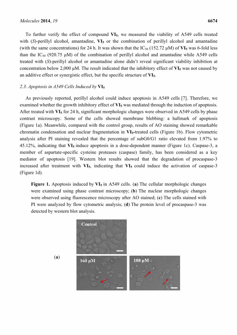
2.3. Apoptosis in A549 Cells Induced by VI5
As previously reported, perillyl alcohol could induce apoptosis in A549 cells [7]. Therefore, we
examined whether the growth inhibitory effect of VI5 was mediated through the induction of apoptosis.
After treated with VI5 for 24 h, significant morphologic changes were observed in A549 cells by phase
contrast microscopy. Some of the cells showed membrane blebbing: a hallmark of apoptosis
(Figure 1a). Meanwhile, compared with the control group, results of AO staining showed remarkable
chromatin condensation and nuclear fragmentation in VI5-treated cells (Figure 1b). Flow cytometric
analysis after PI staining revealed that the percentage of subG0/G1 ratio elevated from 1.97% to
45.12%, indicating that VI5 induce apoptosis in a dose-dependent manner (Figure 1c). Caspase-3, a
member of aspartate-specific cysteine proteases (caspase) family, has been considered as a key
mediator of apoptosis [19]. Western blot results showed that the degradation of procaspase-3
increased after treatment with VI5, indicating that VI5 could induce the activation of caspase-3
(Figure 1d).
Figure 1. Apoptosis induced by VI5 in A549 cells. (a) The cellular morphologic changes
were examined using phase contrast microscopy; (b) The nuclear morphologic changes
were observed using fluorescence microscopy after AO stained; (c) The cells stained with
PI were analyzed by flow cytometric analysis; (d) The protein level of procaspase-3 was
detected by western blot analysis.
(a)

Molecules 2014, 19 6675
Figure 1. Cont.
(b)
(c)
(d)
3. Experimental
3.1. General Information
All reagents and solvents (analytical grade) were commercially available and used without further
purification. Melting points were determined with a Yanaco micro melting point apparatus and were

Molecules 2014, 19 6676
uncorrected. 1H-NMR spectra and 13C-NMR spectrum were recorded in CDCl3 on a Bruker ARX-300
spectrometer. The coupling constants were recorded in hertz (Hz) and chemical shifts were reported in
parts per million (δ, ppm) downfield from tetramethylsilane (TMS). High-resolution mass spectra
(HRMS) were recorded on a high-resonance electrospray time-of-flight mass spectrometer LC/MSD
QTOF 6520 (Agilent). Specific rotation was measured on a Perkin-Elmer 241 MC polarimeter
(path length 1 cm). Column chromatography was performed on silica gel. Analytical TLC was
performed on plates precoated with silica gel and iodine vapor was used to develop color on the plates.
3.2. (S)-(4-(Prop-1-en-2-yl)cyclohex-1-enyl)methanol (I)
To a solution of (S)-perillaldehyde (15.0 g, 0.1 mo1) in ethanol (100 mL) cooled to 0 °C, sodium
borohydride (7.57 g, 0.2 mol) was added in portions. Then, the mixture was stirred at room
temperature for 3 h. After the solvent was evaporated in vacuo, brine (30 mL) was added and the
mixture was extracted with dichloromethane (3 × 30 mL). The combined organic extracts were washed
with brine, dried over anhydrous sodium sulfate, and filtered. The filtrate was concentrated in vacuo.
The residue was purified on a silica gel column with petroleum ether–ethyl acetate (3:1, Rf = 0.51) as
eluent to afford compound I as a colorless liquid (13.2 g, yield 86.8%). [α]20 D : −86° (c = 1, MeOH), in
lit. [20], [α]22 D : −88° (c = 1, MeOH). 1H-NMR δ: 5.70 (1H, br s, CH), 4.73, 4.71 (2H, s, s, CH2),
4.02–3.97 (2H, m, OCH2), 2.18–2.05 (4H, m), 2.00–1.93 (1H, m), 1.88–1.84 (1H, m), 1.74 (3H, s,
CH3), 1.61–1.42 (1H, m). 13C-NMR(75 MHz) δ: 149.8, 137.3, 122.5, 108.7, 67.3, 41.2, 30.4, 27.5,
26.1, 20.8 [21].
3.3. (S)-(4-(Prop-1-en-2-yl)cyclohex-1-enyl)methyl Acetate (II)
To a solution of compound I (12.1 g, 0.08 mol) in pyridine (25 mL), acetic anhydride (25 mL)
was added dropwise. The mixture was stirred at room temperature for 4 h. The reaction was terminated
by addition of methanol (2 mL), followed by addition of ethyl acetate (50 mL). The mixture was
washed with aqueous sodium bicarbonate solution and brine. The organic layer was dried over
anhydrous sodium sulfate, and filtered. The filtrate was concentrated in vacuo. The residue was
purified on a silica gel column with petroleum ether–ethyl acetate (400:1, Rf = 0.41) as eluent to afford
compound II as a colorless liquid (14.8 g, yield 95.4%). 1H-NMR δ: 5.76 (1H, br s, CH), 4.73, 4.71
(2H,s, s, CH2), 4.46 (2H, s, OCH2), 2.24–1.80 (9H, m), 1.74 (3H, s, CH3), 1.52–1.44 (1H, m).
3.4. (S)-(4-(1-Chloroprop-2-en-2-yl)cyclohex-1-enyl)methyl Acetate (III)
To a mixture of compound II (11.6 g, 0.06 mol), acetic acid (5.4 g, 0.09 mol) and dichloromethane
(150 mL) cooled to 0 °C, aqueous sodium hypochlorite solution (containing 10% available chlorine,
72 mL, 0.24 mol) was added dropwise. After the mixture was stirred for 0.5 h, saturated aqueous
sodium sulfite solution (50 mL) was added and the mixture was extracted with dichloromethane
(3 × 50 mL). The combined organic extracts were washed with brine, dried over anhydrous sodium
sulfate, and filtered. The filtrate was concentrated in vacuo. The residue was purified on a silica
gel column with petroleum ether–ethyl acetate (200:1, Rf = 0.57) as eluent to afford compound III
as a pale yellow liquid (11.78 g, yield 85.9%). 1H-NMR δ: 5.76 (1H, br s, CH), 5.19, 5.01 (2H, s, s,

Molecules 2014, 19 6677
CH2), 4.46 (2H, s, OCH2), 4.10 (2H, s, CH2Cl), 2.45–2.42 (1H, m), 2.40–2.25 (1H, m), 2.20–1.80 (7H,
m), 1.52–1.44 (1H, m).
3.5. (S)-1-Chloromethyl-4-(prop-1-en-2-yl)cyclohex-1-ene (V)
To a solution of compound I (9.0 g, 0.06 mo1) and triphenyl phosphine (31.5 g, 0.12 mol) in
dichloromethane (50 mL) cooled to 0 °C, a mixed solution of carbon tetrachloride (12 mL) and
dichloromethane (20 mL) was added dropwise. The mixture was stirred at room temperature
overnight. Then, cyclohexane (200 mL) was added. The white solid precipitated was filtered. The
filtrate was concentrated in vacuo. The residue was purified on a silica gel column with petroleum
ether (Rf = 0.68) as eluent to afford compound V as a pale yellow liquid (7.20 g, yield 70.4%). 1H-NMR δ: 5.83 (1H, br s, CH), 4.73, 4.71 (2H, s, s, CH2), 4.00 (2H, s, CH2Cl), 2.30–2.10 (4H, m),
2.05–1.84 (2H, m), 1.73 (3H, s, CH3), 1.61–1.42 (1H, m).
3.6. General Procedure for the Synthesis of Target Compounds IV1–IV7
To a solution of compound III (0.9 g, 4 mmol) in ethanol (10 mL), potassium carbonate (1.10 g,
8 mmol) and amine (4.4 mmol) were added. The mixture was stirred and refluxed for 8–12 h. Then,
aqueous sodium hydroxide solution (20%, 2 mL) was added and the resulting mixture was refluxed for
another 2 h. The solvent was evaporated in vacuo. Brine (15 mL) was added to the residue and the
mixture was extracted with dichloromethane (3 × 10 mL). The combined organic extracts were washed
with brine, dried over anhydrous sodium sulfate, and filtered. The filtrate was concentrated in vacuo.
The residue was purified on a silica gel column with dichloromethane–methanol (100:1 → 50:1 → 20:1) as
eluent to afford the target product.
(S)-(4-(3-(3,5-cis-Dimethylpiperazin-1-yl)prop-1-en-2-yl)cyclohex-1-enyl)methanol (IV1): Yield: 62.7%;
mp: 106-108 °C; Rf = 0.30 (CH2Cl2/MeOH/Et3N: 200/10/1). 1H-NMR δ: 5.70 (1H, br s, CH), 4.95, 4.89
(2H, s, s, CH2), 4.00 (2H, s, OCH2), 3.05–2.85 (4H, m, NCH2, 2 × NCH), 2.81–2.70 (2H, m),
2.32–2.17 (2H, m), 2.15–1.83 (4H, m), 1.69–1.43 (3H, m), 1.12 (6H, d, J = 6.4, 2 × CH3); HRMS: m/z
calcd. for C16H29N2O [M+H]+ 265.2280, found: 265.2274.
(S)-(4-(3-(4-Isopropylpiperazin-1-yl)prop-1-en-2-yl)cyclohex-1-enyl)methanol (IV2): Yield: 57.5%;
mp: 52–53 °C; Rf = 0.39 (CH2Cl2/MeOH/Et3N: 200/10/1). 1H-NMR δ: 5.71 (1H, br s, CH), 4.97, 4.89
(2H, s, s, CH2), 4.02 (2H, s, OCH2), 2.95 (2H, s, NCH2), 2.76–2.30 (9H, m, 4 × NCH2, NCH),
2.27–2.22 (1H, m), 2.17–2.09 (2H, m), 2.05–1.84 (3H, m), 1.57–1.50 (1H, m), 1.09 (6H, d, J = 6.5,
CH(CH3)2); HRMS: m/z calcd. for C17H31N2O [M+H]+ 279.2436, found: 279.2430.
(S)-(4-(3-(4-(4-Methoxyphenyl)piperazin-1-yl)prop-1-en-2-yl)cyclohex-1-enyl)methanol (IV3): Yield:
54.6%; mp: 58–59 °C; Rf = 0.49 (CH2Cl2/MeOH: 22/5). 1H-NMR δ: 6.93–6.79 (4H, m, Ar-H),
5.70 (1H, br s, CH), 5.00, 4.92 (2H, s, s, CH2), 4.00 (2H, s, OCH2), 3.76 (3H, s, OCH3), 3.18–2.93
(6H, m, 3 × NCH2), 2.65–2.45 (4H, m, 2 × NCH2), 2.37–1.86 (6H, m), 1.60–1.49 (1H, m); HRMS:
m/z calcd. for C21H31N2O2 [M+H]+ 343.2385, found: 343.2381.

Molecules 2014, 19 6678
(S)-(4-(3-(4-(2-Methoxyphenyl)piperazin-1-yl)prop-1-en-2-yl)cyclohex-1-enyl)methanol (IV4): yield:
50.2%; pale yellow oil; Rf = 0.47 (CH2Cl2/MeOH: 22/5). 1H-NMR δ: 7.04–6.79 (4H, m, Ar-H),
5.71 (1H, br s, CH), 5.01, 4.92 (2H, s, s, CH2), 4.01 (2H, s, OCH2), 3.86 (3H, s, OCH3), 3.19–2.89
(6H, m, 3 × NCH2), 2.79–2.40 (4H, m, 2 × NCH2), 2.38-1.84 (6H, m), 1.61-1.48 (1H, m); HRMS:
m/z calcd. for C21H31N2O2 [M+H]+ 343.2385, found: 343.2380.
(S)-(4-(3-(4-Benzylpiperazin-1-yl)prop-1-en-2-yl)cyclohex-1-enyl)methanol (IV5): Yield: 61.3%; pale
yellow oil; Rf = 0.44 (CH2Cl2/MeOH: 22/5). 1H-NMR δ: 7.34–7.21 (5H, m, Ar-H), 5.69 (1H, br s, CH),
4.94, 4.88 (2H, s, s, CH2), 4.00 (2H, s, OCH2), 3.51 (2H, s, NCH2), 2.98–2.88 (2H, m, NCH2),
2.63–2.31 (8H, m, 4 × NCH2), 2.30–2.07 (4H, m), 2.01–1.83 (2H, m), 1.56–1.44 (1H, m); HRMS: m/z
calcd. for C21H31N2O [M+H]+ 327.2436, found: 327.2431.
(S)-(4-(3-(Piperidin-1-yl)prop-1-en-2-yl)cyclohex-1-enyl)methanol (IV6): Yield: 51.3%; pale yellow
oil; Rf = 0.49 (CH2Cl2/MeOH/Et3N: 200/10/1). 1H-NMR δ: 5.70 (1H, br s, CH), 4.96, 4.87 (2H, s, s,
CH2), 4.00 (2H, s, OCH2), 2.90 (2H, s, NCH2), 2.41–2.08 (8H, m), 2.01–1.84 (2H, m), 1.59–1.41 (7H,
m); HRMS: m/z calcd. for C15H26NO [M+H]+ 236.2014, found: 236.2009.
(S)-(4-(3-(Pyridin-2-ylamino)prop-1-en-2-yl)cyclohex-1-enyl)methanol (IV7): Yield: 40.1%; pale
yellow oil; Rf = 0.39 (CH2Cl2/MeOH: 22/5). 1H-NMR δ: 8.07 (1H, d, J = 3.9, Ar-H), 7.46–7.39
(1H, m, Ar-H), 6.65–6.49 (1H, m, Ar-H), 6.36 (1H, d, J = 8.4, Ar-H), 5.71 (1H, br s, CH), 5.03, 4.92
(2H, s, s, CH2), 4.01 (2H, s, OCH2), 3.95–3.85 (2H, m, NCH2), 2.34–1.88 (6H, m), 1.62–1.51 (1H, m);
HRMS: m/z calcd. for C15H21N2O [M+H]+ 245.1654, found: 245.1648.
3.7. General Procedure for the Synthesis of Target Compounds VI1–VI7
To a solution of compound V (0.85 g; 5 mmo1) in acetonitrile (10 mL); potassium carbonate
(1.04 g; 7.5 mmol) and amine (5.5 mmol) were added. The mixture was stirred and refluxed for 6–8 h.
Then the solvent was evaporated in vacuo. Brine (15 mL) was added to the residue and the mixture
was extracted with dichloromethane (3 × 10 mL). The combined organic extracts were washed with
brine; dried over anhydrous sodium sulfate; and filtered. The filtrate was concentrated in vacuo.
The residue was purified on a silica gel column with dichloromethane–methanol (100:1 → 50:1 → 20:1)
as eluent to afford the target product VI.
(S)-3,5-cis-Dimethyl-1-((4-(prop-1-en-2-yl)cyclohex-1-enyl)methyl)piperazine (VI1): Yield: 54.1%;
colorless oil; Rf = 0.48 (CH2Cl2/MeOH/Et3N: 300/10/1). 1H-NMR δ: 5.59 (1H, br s, CH), 4.72, 4.71
(2H, s, s, CH2), 3.04–2.96 (2H, m, 2 × NCH), 2.81–2.73 (2H, m, NCH2), 2.38–2.13 (8H, m), 1.99–1.93
(1H, m), 1.85–1.79 (1H, m), 1.74 (3H, s, CH3), 1.49–1.42 (1H, m), 1.10 (6H, d, J = 6.0, 2 × CH3);
HRMS: m/z calcd. for C16H29N2 [M+H]+ 249.2331, found: 249.2325.
(S)-1-Isopropyl-4-((4-(prop-1-en-2-yl)cyclohex-1-enyl)methyl)piperazine (VI2): Yield: 53.8%; pale
yellow oil; Rf = 0.55 (CH2Cl2/MeOH/Et3N: 300/10/1). 1H-NMR δ: 5.60 (1H, s, CH), 4.77–4.63 (2H, m,
CH2), 2.86–2.76 (3H, m, NCH2, NCH), 2.69–2.42 (8H, m, 4 × NCH2), 2.15–1.91 (5H, m), 1.87–1.78
(1H, m), 1.73 (3H, s, CH3), 1.50–1.38 (1H, m), 1.10 (6H, d, J = 6.5, CH(CH3)2); HRMS: m/z calcd. for
C17H31N2 [M+H]+ 263.2487, found: 263.2482.

Molecules 2014, 19 6679
(S)-1-Methyl-4-((4-(prop-1-en-2-yl)cyclohex-1-enyl)methyl)piperazine (VI3): Yield: 48.2%; pale
yellow oil; Rf = 0.50 (CH2Cl2/MeOH/Et3N: 300/10/1). 1H-NMR δ: 5.59 (1H, br s, CH), 4.70 (2H, s,
CH2), 2.82(2H, s, NCH2), 2.54–2.29 (8H, m, 4 × NCH2), 2.28 (3H, s, NCH3), 2.25–1.79 (6H, m), 1.73
(3H, s, CH3), 1.54–1.33 (1H, m); HRMS: m/z calcd. for C15H27N2 [M+H]+ 235.2174, found: 235.2169.
(S)-1-((4-(Prop-1-en-2-yl)cyclohex-1-enyl)methyl)piperidine (VI4): Yield: 34.2%; pale yellow oil;
Rf = 0.44 (CH2Cl2/MeOH/Et3N: 300/10/1). 1H-NMR δ: 5.59 (1H, br s, CH), 4.72 (2H, s, CH2),
2.86–2.76(2H, m, NCH2), 2.38–2.23 (5H, m, 2 × NCH2, CH), 2.18–1.82 (5H, m), 1.75 (3H, s, CH3),
1.60–1.41 (7H, m) [22]; HRMS: m/z calcd. for C15H26N [M+H]+ 220.2065, found: 220.2060.
(S)-N-((4-(Prop-1-en-2-yl)cyclohex-1-enyl)methyl)amantadine (VI5): Yield: 36.2%; mp: 223–224 °C;
Rf = 0.32 (CH2Cl2/MeOH/Et3N: 300/10/1). 1H-NMR δ: 5.98 (1H, br s, CH), 4.71, 4.67 (2H, s, s, CH2),
3.49–3.40 (2H, m, NCH2), 2.29 (2H, m), 2.17–2.05 (11H, m), 1.96–1.82 (2H, m), 1.73–1.65 (9H, m),
1.51–1.45 (1H, m); HRMS: m/z calcd. for C20H32N [M+H]+ 286.2535, found: 286.2529.
(S)-N-((4-(Prop-1-en-2-yl)cyclohex-1-enyl)methyl)diethanolamine (VI6): Yield: 38.4%; colorless oil;
Rf = 0.34 (CH2Cl2/MeOH/Et3N: 300/10/1). 1H-NMR δ: 5.71–5.55 (1H, br, CH), 4.79–4.64 (2H, m, CH2),
3.63 (4H, t, J = 5.4, 2 × OCH2), 3.03 (2H, s, NCH2), 2.64 (4H, t, J = 5.4, 2 × NCH2), 2.17–1.81
(6H, m), 1.74 (3H, s, CH3), 1.53–1.39 (1H, m); HRMS: m/z calcd. for C14H26NO2 [M+H]+ 240.1963,
found: 240.1957.
(S)-N-((4-(Prop-1-en-2-yl)cyclohex-1-enyl)methyl)cyclohexanamine (VI7): Yield: 35.4%; pale yellow oil;
Rf = 0.39 (CH2Cl2/MeOH/Et3N: 300/10/1). 1H-NMR δ: 5.63 (1H, br s, CH), 4.72, 4.70 (1H, s, s, CH2),
3.20 (2H, s, NCH2), 2.50–2.45 (1H, m, NCH), 2.15–2.06 (3H, m), 1.96–1.81 (4H, m), 1.73 (3H, s,
CH3), 1.70–1.43 (4H, m), 1.28–1.13 (6H, m); HRMS;: m/z calcd. for C16H28N [M+H]+ 234.2222,
found: 234.2216.
3.8. Biological Activity
Cell culture: Human lung cancer A549 cells, human melanoma A375-S2 Cells and human
fibrosarcoma HT-1080 cells were obtained from American Type Culture Collection (ATCC,
Manassas, VA, USA). A549 cells and HT1080 were cultured in DMEM medium with 10% fetal
bovine serums (FBS, Tianjin Haoyang Biological Manufacture Co. LTD, Tianjin, China), 2 mM
L-glutamine, 100 U/mL penicillin and 100 μg/mL streptomycin. A375-S2 cells were cultured in MEM
medium with 10% fetal bovine serums, 2 mM L-glutamine, 100 U/mL penicillin and 100 μg/mL
streptomycin at 37 °C in 5% CO2.
MTT assay [23]: Cells were planted in 96-well flat bottom micro titer plates (Corning, Tewksbury,
MA, USA) with 7 × 10³ cells per well. After 24 h incubation, they were treated with the tested
agents for the indicated times. After washing the plates with PBS, a 20 μL aliquot of
3-(4,5-dimethylthiazol-2-yl)-2,5-diphenyltetrazolium (MTT) solution (5.0 mg/mL) was added to each
well and incubated for 3 h. The resulting crystal was dissolved in dimethyl sulfoxide. Optical
density was detected by ELISA reader (Tecan, Salzburg, Austria). The percentage of cell viability
inhibition was calculated as follows:

Molecules 2014, 19 6680
Inhibitory ratio (%) = (A492, control − A492, sample)/(A492, control − A492, blank) × 100% (1)
Observation of morphologic changes: A549 cells were treated with 0, 120, 160 and 180 μM VI5 for
24 h on 24-well flat bottom plates. Then changes in cellular morphology were examined using phase
contrast microscopy (Olympus, Tokyo, Japan).
Acridine orange (AO) staining [23]: A549 cells were treated with 0, 120, 160 and 180 μM VI5
for 24 h on 24-well flat bottom plates. Then cells were washed with PBS, followed by incubation at
room temperature with PBS containing 20 μg/mL AO for 15 min. The fluorescence of cells was
observed using fluorescence microscopy.
Flow cytometric analysis using propidium iodide (PI) [24]: A549 cells were treated with 0, 120,
160 and 180 μM VI5 for 24 h on 6-well flat bottom plates. The cells were harvested and washed by
PBS and fixed by 70% cold ethanol at 4 °C for more than 18 h. After stained with 50 μg/mL PI and
1 mg/mL DNaseA-free RNaseA on ice in dark for 1 h, cells were analyzed on FACScan flow
cytometer (Becton Dickinson, Franklin Lakes, NJ, USA).
Western blot analysis [24]: After treated with 0, 100, 120, 140, 160 and 180 μM VI5 for 24 h,
both adherent and floating cells were collected and lysed by Ultrasonic Cell Disruptor (Ningbo
Scientz Biotechnology Co., Ltd, Ningbo, China) in whole cell lyse buffer (50 mM HEPES (pH 7.4),
1% Triton-X 100, 2 mM sodium orthovanadate, 100 mM sodiumfluoride, 1 mM edetic acid, 1 mM PMSF,
10 μg/mL aprotinin and 10 μg/mL leupeptin). The protein extracts was separated by 12% SDS-PAGE
and transferred to PVDF membranes (Millipore, Billerica, MA, USA). After blocked with 5% skim
milk, incubated with primary antibodies against procaspase-3 and caspase-3 at 4 °C overnight.
Statistical assay: All the presented data were confirmed at least three independent experiments.
The data were analyzed by ANOVA using Statistics Package for Social Science SPSS software
(version 13.0; SPSS, Chicago, IL, USA).
4. Conclusions
Two series of amino-modified derivatives of (S)-perillyl alcohol were designed and synthesized. The
target compounds showed improved antiproliferative activity against A549, A375-S2 and HT-1080
cells. The structure-activity relationships revealed that the replacement of the hydroxyl group of
(S)-perillyl alcohol with an amino moiety was more favorable to improving cytotoxic activity than the
introduction of an amino moiety at the terminal allyl group. The antiproliferative effect of VI5 was
proved to be mediated through the induction of apoptosis in A549 cells.
Acknowledgments
This work was supported by Foundation of Liaoning Province Education Department (L2011175)
and PhD Research Startup Foundation of Liaoning Province (20111138).
Author Contributions
Conceived and designed the experiments: Jinhua Dong, Mingyu Xia. Performed the experiments:
Zi Hui, Meihui Zhang and Lin Cong. Analyzed the data: Zi Hui, Mingyu Xia and Jinhua Dong. Wrote
the paper: Zi Hui, Mingyu Xia and Jinhua Dong.

Molecules 2014, 19 6681
Conflicts of Interest
The authors declare no conflict of interest.
References
1. Newman, D.J. Natural products as leads to potential drugs: An old process or the new hope for
drug discovery? J. Med. Chem. 2008, 51, 2589–2599.
2. Crowell, P.L.; Chang, R.R.; Ren, Z.; Elson, C.E.; Gould, M.N. Selective inhibition of
isoprenylation of 21–26-kDa proteins by the anticarcinogen d-limonene and its metabolites.
J. Biol. Chem. 1991, 266, 17679–17685.
3. Crowell, P.L.; Ren, Z.; Lin, S.; Vedejs, E.; Gould, M.N. Structure-activity relationships among
monoterpene inhibitors of protein isoprenylation and cell proliferation. Biochem. Pharmacol.
1994, 47, 1405–1415.
4. Gelb, M.H.; Tamanoi, F.; Yokoyama, K.; Ghomashchi, F.; Esson, K.; Gould, M.N. The inhibition
of protein prenyltransferases by oxygenated metabolites of limonene and perillyl alcohol. Cancer
Lett. 1995, 91, 169–175.
5. Ren, Z.; Elson, C.E.; Gould, M.N. Inhibition of type I and type II geranylgeranyl-protein
transferases by the monoterpene perillyl alcohol in NIH3T3 cells. Biochem. Pharmacol. 1997, 54,
113–120.
6. Shi, W.; Gould, M.N. Induction of cytostasis in mammary carcinoma cells treated with the
anticancer agent perillyl alcohol. Carcinogenesis 2002, 23, 131–142.
7. Yeruva, L.; Pierre, K.J.; Elegbede, A.; Wang, R.C.; Carper, S.W. Perillyl alcohol and perillic acid
induced cell cycle arrest and apoptosis in non small cell lung cancer cells. Cancer Lett. 2007, 257,
216–216.
8. Liu, G.; Oettel, K.; Bailey, H.H.; van Ummersen, L.; Tutsch, K.; Staab, M.J.; Horvath, D.;
Alberti, D.; Arzoomanian, R.; Rezazadeh, H.; et al. Phase II trial of perillyl alcohol (NSC
641066) administered daily in patients with metastatic androgen independent prostate cancer.
Investig. New Drugs 2003, 21, 367–372.
9. Bailey, H.H.; Attia, S.; Love, R.R.; Fass, T.; Chappell, R.; Tutsch, K.; Harris, L.; Jumonville, A.;
Hansen, R.; Shapiro, G.R.; et al. Phase II trial of daily oral perillyl alcohol (NSC 641066)
in treatment-refractory metastatic breast cancer. Cancer Chemother. Pharmacol. 2008, 62,
149–157.
10. Leonard, D.M. Ras farnesyltransferase: A new therapeutic target. J. Med. Chem. 1997, 40,
2971–2990.
11. Chen, T.; Levin, D.; Pupalli, S. Pharmaceutical compositions comprising POH derivatives.
WO2012027693, 2012.
12. Eummer, J.T.; Gibbs, B.S.; Zahn, T.J.; Sebolt-Leopold, J.S.; Gibbs, R.A. Novel limonene
phosphonate and farnesyl diphosphate analogues: Design, synthesis, and evaluation as potential
protein-farnesyl transferase inhibitors. Bioorg. Med. Chem. 1999, 7, 241–250.

Molecules 2014, 19 6682
13. Das, B.C.; Mahalingam, S.M.; Panda, L.; Wang, B.; Campbell, P.D.; Evans, T. Design and
synthesis of potential new apoptosis agents: Hybrid compounds containing perillyl alcohol and
new constrained retinoids. Tetrahedron Lett. 2010, 51, 1462–1466.
14. Xanthakis, E.; Magkouta, S.; Loutrari, H.; Stamatis, H.; Roussos, C.; Kolisis, F.N. Enzymatic
synthesis of perillyl alcohol derivatives and investigation of their antiproliferative activity.
Biocatal. Biotransfor. 2009, 27, 170–178.
15. Kollmannsberger, C.; Mross, K.; Jakob, A.; Kanz, L.; Bokemeyer, C. Topotecan—A novel topoisome raseⅠinhibitor: Pharmacology and clinical experience. Oncology 1999, 56, 1–12.
16. Sandier, A.; van Oosterom, A.T. Irinotecan in cancer of the lung and cervix. Anti-Cancer Drugs
1999, 10, S13–S17.
17. Xu, L.; Tao, S.; Wang, X.; Yu, Z.; Wang, M.; Chen, D.; Jing, Y.; Dong, J. The synthesis and
anti-proliferative effects of â-elemene derivatives with mTOR inhibition activity. Bioorg. Med.
Chem. 2006, 14, 5351–5356.
18. Chen, J.; Lu, M.; Jing, Y.; Dong, J. The synthesis of L-carvone and limonene derivatives with
increased antiproliferative effect and activation of ERK pathway in prostate cancer cells.
Bioorg. Med. Chem. 2006, 14, 6539–6547.
19. Mazumder, S.; Plesca, D.; Almasan, A. Caspase-3 activation is a critical determinant of genotoxic
stress-induced apoptosis. Methods Mol. Biol. 2008, 414, 13–21.
20. Karlson, J.; Borg-Karlson, A.-K.; Unelius, R.; Shoshan, M.C.; Wilking, N.; Ringborg, U.; Linder, S.
Inhibition of tumor cell growth by monoterpenes in vitro: Evidence of a Ras-independent
mechanism of action. Anti-Cancer Drugs 1996, 7, 422–429.
21. Constantino, M.G.; Lacerda Júnior, V.; Invernize, P.R.; Carlos da Silva Filho, L.; José da Silva, G.V.
Opening of epoxide rings catalyzed by niobium pentachloride. Synth. Commun. 2007, 37,
3529–3539.
22. Sahli, Z.; Sundararaju, B.; Achard, M.; Bruneau, C. Selective carbon–carbon bond formation:
Terpenylations of amines involving hydrogen transfers. Green Chem. 2013, 15, 775–779.
23. Fan, S.; Li, L.; Chen, S.; Yu, Y.; Qi, M.; Tashiro, S.I.; Onodera, S.; Ikejima, T. Silibinin
induced-autophagic and apoptotic death is associated with an increase in reactive oxygen and
nitrogen species in HeLa cells. Free Radic. Res. 2011, 45, 1307–1324.
24. Qi, M.; Yao, G.; Fan, S.; Cheng, W.; Tashiro, S.I.; Onodera, S.; Ikejima, T. Pseudolaric acid B
induces mitotic catastrophe followed by apoptotic cell death in murine fibrosarcoma L929 cells.
Eur. J. Pharmacol. 2012, 683, 16–26.
Sample Availability: Samples of the compounds are available from the authors.
© 2014 by the authors; licensee MDPI, Basel, Switzerland. This article is an open access article
distributed under the terms and conditions of the Creative Commons Attribution license
(http://creativecommons.org/licenses/by/3.0/).