mollusc-algal chloroplast endosymbiosis. … chloroplast endosymbiosis. photosynthesis, thylakoid...
TRANSCRIPT
Mollusc-Algal Chloroplast Endosymbiosis. Photosynthesis,Thylakoid Protein Maintenance, and Chloroplast GeneExpression Continue for Many Months in theAbsence of the Algal Nucleus1
Brian J. Green, Wei-Ye Li, James R. Manhart, Theodore C. Fox, Elizabeth J. Summer, Robert A. Kennedy2,Sidney K. Pierce3, and Mary E. Rumpho4*
Program in Molecular and Environmental Plant Sciences (B.J.G., W.-Y.L., T.C.F., R.A.K., M.E.R.), Departmentof Horticultural Sciences (B.J.G., W.-Y.L., T.C.F., E.J.S., M.E.R.), and Department of Biology (J.R.M., R.A.K.),Texas A&M University, College Station, Texas 77843; and Department of Biology, University of Maryland,College Park, Maryland 20742 (S.K.P.)
Early in its life cycle, the marine mollusc Elysia chlorotica Gould forms an intracellular endosymbiotic association withchloroplasts of the chromophytic alga Vaucheria litorea C. Agardh. As a result, the dark green sea slug can be sustained inculture solely by photoautotrophic CO2 fixation for at least 9 months if provided with only light and a source of CO2. Herewe demonstrate that the sea slug symbiont chloroplasts maintain photosynthetic oxygen evolution and electron transportactivity through photosystems I and II for several months in the absence of any external algal food supply. This activity iscorrelated to the maintenance of functional levels of chloroplast-encoded photosystem proteins, due in part at least to denovo protein synthesis of chloroplast proteins in the sea slug. Levels of at least one putative algal nuclear encoded protein,a light-harvesting complex protein homolog, were also maintained throughout the 9-month culture period. The chloroplastgenome of V. litorea was found to be 119.1 kb, similar to that of other chromophytic algae. Southern analysis and polymerasechain reaction did not detect an algal nuclear genome in the slug, in agreement with earlier microscopic observations.Therefore, the maintenance of photosynthetic activity in the captured chloroplasts is regulated solely by the algal chloroplastand animal nuclear genomes.
The majority of animal-algal symbioses are cellularassociations with a unicellular alga either residingbetween animal cells or within a vacuole producedby the animal (Douglas, 1994). In contrast, the asco-glossan sea slug Elysia chlorotica Gould establishes anintracellular symbiotic association with chloroplastsfrom the siphonaceous, chromophytic alga Vaucherialitorea C. Agardh (West, 1979; West et al., 1984). Ju-venile sea slugs feed on V. litorea filaments andphagocytotically incorporate the chloroplasts into thecytoplasm of one of two morphologically distinct
epithelial cells that line the tubules of the digestivesystem (Graves et al., 1979; West, 1979). During thisprocess the chloroplast endoplasmic reticulum, astructural characteristic of chromophytic plastids(Lee, 1989), is lost resulting in symbiotic plastids withtheir outer envelope in direct contact with the animalcytoplasm (Graves et al., 1979; Mujer et al., 1996;Rumpho et al., 2000). Heterokont algae (chromo-phytes or autotrophic stramenopiles) such as V. lito-rea do not typically contain nucleomorphs, and elec-tron microscopy studies have not revealed anyunusual nucleomorph-type structures or algal nucleiin the sea slugs (Graves et al., 1979; Mujer et al., 1996;Rumpho et al., 2000). It is important to note that thecaptured chloroplasts are functional, i.e. they are ca-pable of light dependent oxygen evolution (Graves etal., 1979; West, 1979).
When maintained in the laboratory in artificial sea-water (ASW), E. chlorotica sustains itself apart fromany algal food source for at least 9 months whenprovided with only light and a source of CO2 (Mujeret al., 1996; Pierce et al., 1996). Whether in their nativesalt marsh or in culture, the life cycle of the sea slugslasts 8 to 10 months. There is no evidence for plastiddivision in the animals and the plastids are not trans-mitted in the eggs; thus, the endosymbiosis must bere-established with each generation (West, 1979;
1 This work was supported in part by the American ChemicalSociety Herman Frasch Foundation (grant no. 343–HF92 in Agri-cultural Chemistry to M.E.R.), by the National Science Foundation(grant nos. IBN–9505416 to S.K.P. and M.E.R. and IBN–9808904 toM.E.R. and J.R.M.), by the Texas A&M University InterdisciplinaryResearch Initiative (grants to M.E.R. and J.R.M.), and by the TexasAgricultural Experiment Station.
2 Present address: Department of Biology, University of Maine,Orono, ME 04469.
3 Present address: Department of Biology, University of SouthFlorida, Tampa, FL 33620.
4 Present address: Department of Biochemistry, Microbiologyand Molecular Biology, 5735 Hitchner Hall, Room 273, Universityof Maine, Orono, ME 04469 –5735.
* Corresponding author; e-mail [email protected]; fax979 – 845– 0627.
Plant Physiology, September 2000, Vol. 124, pp. 331–342, www.plantphysiol.org © 2000 American Society of Plant Physiologists 331 www.plantphysiol.orgon June 5, 2018 - Published by Downloaded from Copyright © 2000 American Society of Plant Biologists. All rights reserved.
West et al., 1984). Symbiotic associations of this typeoccur in other ascoglossan species, but they are farmore transient (Greene, 1970; Trench, 1975; Clark andBusacca, 1978; Rumpho et al., 2000). The E. chlo-rotica/V. litorea symbiosis represents the longestknown functional association of its kind (West, 1979;Pierce et al., 1996).
The longevity and functional capacity of E. chlo-rotica is surprising considering the complexity ofchloroplast function and regulation evidenced, inpart, by the unsuccessful attempts to culture isolatedchloroplasts on a long-term basis in an artificial sys-tem (Nass, 1969; Ridley and Leech, 1970; Giles andSarafis, 1971). Seventy percent to 90% of all polypep-tides needed for plastid function have a nuclear ori-gin in plants (Reith, 1995; Palmer and Delwiche, 1996;Martin and Herrmann, 1998). Even in chromophyticalgae whose chloroplast genomes tend to have agreater coding capacity than chlorophytes (due inpart to low intron no. and relatively small invertedrepeats), only 120 to 130 gene products are plastidencoded, accounting for only about 13% of all geneproducts required for plastid function (Reith, 1995;Martin and Herrmann, 1998). Furthermore, althoughthe gene products D1, D2, PsaA/B, and several otherpolypeptides that assemble to form the photosyn-thetic complexes are plastid encoded, they depend onnuclear regulation at either or both the transcrip-tional and translational levels (Stern et al., 1997; Mer-chant and Dreyfuss, 1998). In turn this nuclear regu-lation can be influenced by additional environmentaland physiological factors (Aro et al., 1993; Christopherand Mullet, 1994; Russell et al., 1995).
The complex nucleocytosolic/chloroplast interac-tions required for plastid function in plant and algalspecies presents E. chlorotica with what would seemto be insurmountable obstacles for maintaining plas-tid function in a foreign environment for such anextended period of time. Despite this, previous re-sults indicated that several chloroplast encodedpolypeptides (specifically, D1, D2, CP43, the largesubunit of Rubisco [Rubisco LS]), and one probablynuclear encoded protein (related to a fucoxanthinchlorophyll [chl] a/c-binding protein [FCP]) arepresent in the molluscan chloroplasts (Mujer et al.,1996; Pierce et al., 1996). At least two of these pro-teins, D1 and Rubisco LS, are synthesized de novo inthe animal (Mujer et al., 1996; Pierce et al., 1996). Inaddition, levels of D1 transcripts were detected in theendosymbiotic chloroplasts for as long as 7 months(Mujer et al., 1996). Taken together, these data sug-gest that in addition to photosynthesis, the capturedplastids are functionally capable of transcription andtranslation.
To further understand how chloroplast activity canbe maintained in an animal cell, we have extendedthese earlier studies and measured oxygen evolutionand photosynthetic electron transport (PET) capacityof E. chlorotica chloroplasts through 9 months after
removal of the sea slugs from an algal food source.We have found a preliminary correlation between thepresence of photosystem (PS) proteins and activity.We also provide the first molecular evidence sup-porting earlier structural analysis that the photosyn-thetically active animals do not contain an intactalgal nuclear genome.
RESULTS
Rates of Photosynthesis and Respiration inIntact Animals
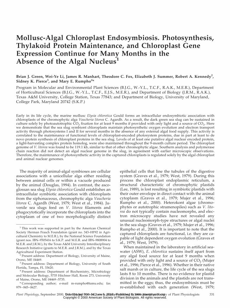
E. chlorotica’s symbiont plastids remained photo-synthetically competent and capable of splitting wa-ter through 7 months in culture in the absence of analgal food source (Fig. 1). Oxygen evolution rateswere fairly low relative to typical values obtained forplant and algal specimens including V. litorea fila-ments, which exhibited rates between 98 and 120mmol O2 evolved mg21 chl h21. The lower sea slugphotosynthetic rates probably reflect an underesti-mate of the true rates due to gaseous diffusion limi-tations to O2 and CO2 through the mucus-covered seaslug body, the rapid respiration and O2 utilization ofthe animals even in the light, and the variability inexposing the animals to the actinic light source (theanimals tend to fold up). Decreasing illuminationbelow the maximal output of the light source (1,500mmol photons m22 s21) resulted in a decrease inapparent photosynthetic rates (data not shown), sug-gesting maximal rates may not have been obtained.
After 5 months of endosymbiotic association, pho-tosynthetic rates dropped significantly in the seaslugs and dark respiration rates exceeded the appar-ent photosynthetic rates. However, respiration ratesalso decreased over time in culture, indicating anoverall decline in metabolic activity as the animals
Figure 1. Changes in photosynthetic and respiratory activity in E.chlorotica symbiotically associated with V. litorea chloroplasts andcultured over a 7-month period in the absence of algae. Apparentphotosynthesis was calculated by measuring the rate of CO2- andlight-dependent O2 evolution. Respiration rates were based on theuptake of O2 in the dark. Gross photosynthesis was estimated bysumming the apparent photosynthesis and respiration rates. Datarepresent means 6 SE of at least three sea slugs for each time point.
Green et al.
332 Plant Physiol. Vol. 124, 2000 www.plantphysiol.orgon June 5, 2018 - Published by Downloaded from Copyright © 2000 American Society of Plant Biologists. All rights reserved.
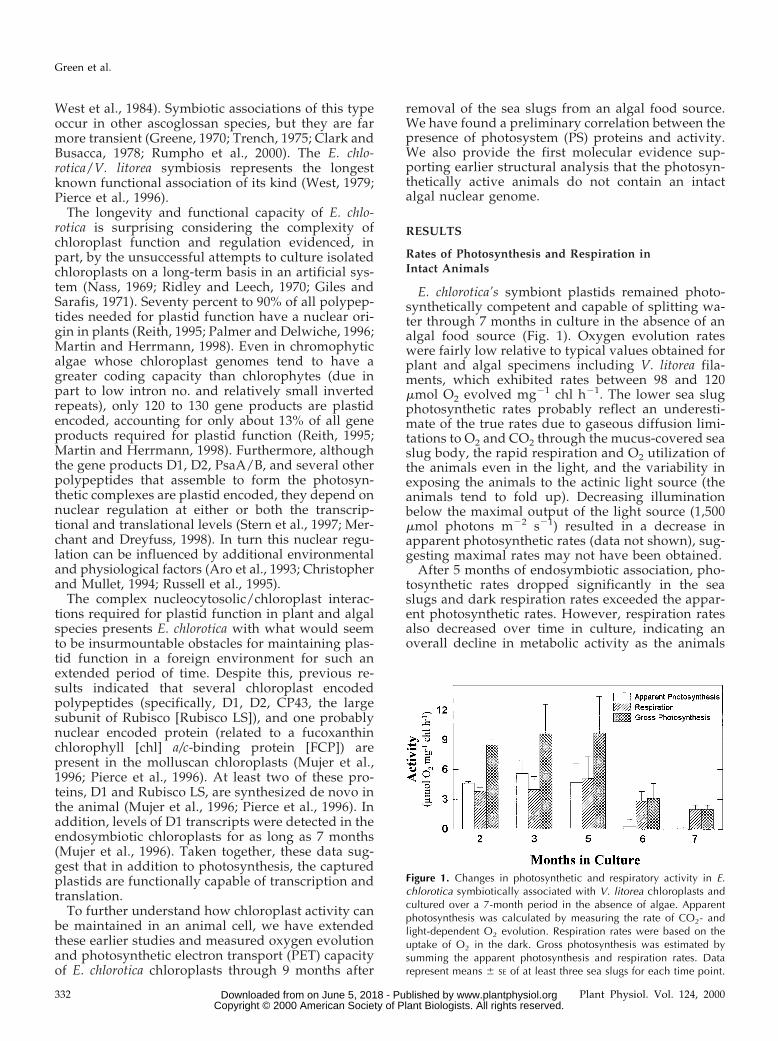
aged. The decline in activity after 5 months preceded,by 1 to 2 months, a measurable decrease in chl con-centration in the sea slugs (Table I). Eighty-five per-cent of the original chl concentration was maintainedthrough 6 months in culture, ultimately decreasing to50% by month 9. An increase in the ratio of chl a tochl c was observed at month 7 as a result of a largerdecrease in the accessory pigment chl c (80% decline),compared with the reaction center pigment chl a(45% decline). Both pigments remained at a steady,but low, level through the final 3 months. As ex-pected, the concentration of chl in the paler chromo-phytic alga (0.27 mg chl mg21 fresh weight) wassignificantly less than in the densely green sea slugs(1.28 mg chl mg21 fresh weight at month 1). However,the algal chl a to chl c ratio (17.1) was approximatelythe same as the sea slug’s through the first 6 monthsin culture, ranging from a low of 14.4 to a high of 17.1(Table I).
PET Activity
Individual PS activities and whole chain PET weremeasured in isolated thylakoids to eliminate any per-
meability problems with the intact animals and todetermine the competency of each PS after severalmonths of symbiotic association and separation fromthe algal nucleus. The ability to isolate chloroplastsfrom E. chlorotica is limited by the copious amount ofmucus produced by the animals (Rumpho et al.,1994); consequently, thylakoid yields were low perextraction despite pooling several sea slugs. Theselow yields limited the extent of analysis per extrac-tion and collection, thereby necessitating analysisfrom multiple collections. In any case reproducibleresults were obtained with subsequent extractions orcollections and a consistent pattern for PSI and PSIIactivity was observed.
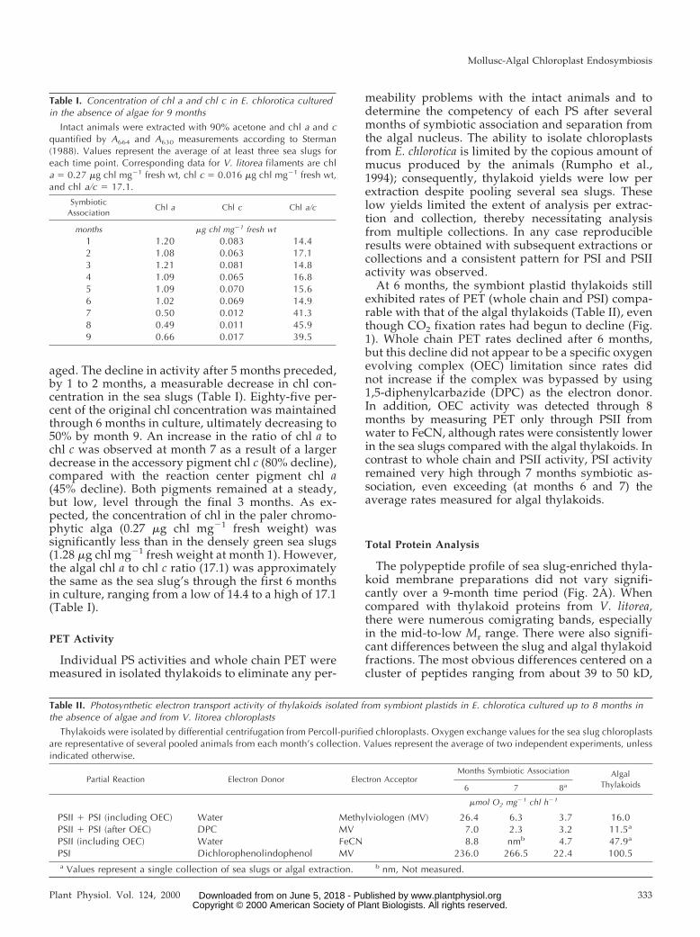
At 6 months, the symbiont plastid thylakoids stillexhibited rates of PET (whole chain and PSI) compa-rable with that of the algal thylakoids (Table II), eventhough CO2 fixation rates had begun to decline (Fig.1). Whole chain PET rates declined after 6 months,but this decline did not appear to be a specific oxygenevolving complex (OEC) limitation since rates didnot increase if the complex was bypassed by using1,5-diphenylcarbazide (DPC) as the electron donor.In addition, OEC activity was detected through 8months by measuring PET only through PSII fromwater to FeCN, although rates were consistently lowerin the sea slugs compared with the algal thylakoids. Incontrast to whole chain and PSII activity, PSI activityremained very high through 7 months symbiotic as-sociation, even exceeding (at months 6 and 7) theaverage rates measured for algal thylakoids.
Total Protein Analysis
The polypeptide profile of sea slug-enriched thyla-koid membrane preparations did not vary signifi-cantly over a 9-month time period (Fig. 2A). Whencompared with thylakoid proteins from V. litorea,there were numerous comigrating bands, especiallyin the mid-to-low Mr range. There were also signifi-cant differences between the slug and algal thylakoidfractions. The most obvious differences centered on acluster of peptides ranging from about 39 to 50 kD,
Table I. Concentration of chl a and chl c in E. chlorotica culturedin the absence of algae for 9 months
Intact animals were extracted with 90% acetone and chl a and cquantified by A664 and A630 measurements according to Sterman(1988). Values represent the average of at least three sea slugs foreach time point. Corresponding data for V. litorea filaments are chla 5 0.27 mg chl mg21 fresh wt, chl c 5 0.016 mg chl mg21 fresh wt,and chl a/c 5 17.1.
SymbioticAssociation
Chl a Chl c Chl a/c
months mg chl mg21 fresh wt1 1.20 0.083 14.42 1.08 0.063 17.13 1.21 0.081 14.84 1.09 0.065 16.85 1.09 0.070 15.66 1.02 0.069 14.97 0.50 0.012 41.38 0.49 0.011 45.99 0.66 0.017 39.5
Table II. Photosynthetic electron transport activity of thylakoids isolated from symbiont plastids in E. chlorotica cultured up to 8 months inthe absence of algae and from V. litorea chloroplasts
Thylakoids were isolated by differential centrifugation from Percoll-purified chloroplasts. Oxygen exchange values for the sea slug chloroplastsare representative of several pooled animals from each month’s collection. Values represent the average of two independent experiments, unlessindicated otherwise.
Partial Reaction Electron Donor Electron AcceptorMonths Symbiotic Association Algal
Thylakoids6 7 8a
mmol O2 mg21 chl h21
PSII 1 PSI (including OEC) Water Methylviologen (MV) 26.4 6.3 3.7 16.0PSII 1 PSI (after OEC) DPC MV 7.0 2.3 3.2 11.5a
PSII (including OEC) Water FeCN 8.8 nmb 4.7 47.9a
PSI Dichlorophenolindophenol MV 236.0 266.5 22.4 100.5a Values represent a single collection of sea slugs or algal extraction. b nm, Not measured.
Mollusc-Algal Chloroplast Endosymbiosis
Plant Physiol. Vol. 124, 2000 333 www.plantphysiol.orgon June 5, 2018 - Published by Downloaded from Copyright © 2000 American Society of Plant Biologists. All rights reserved.
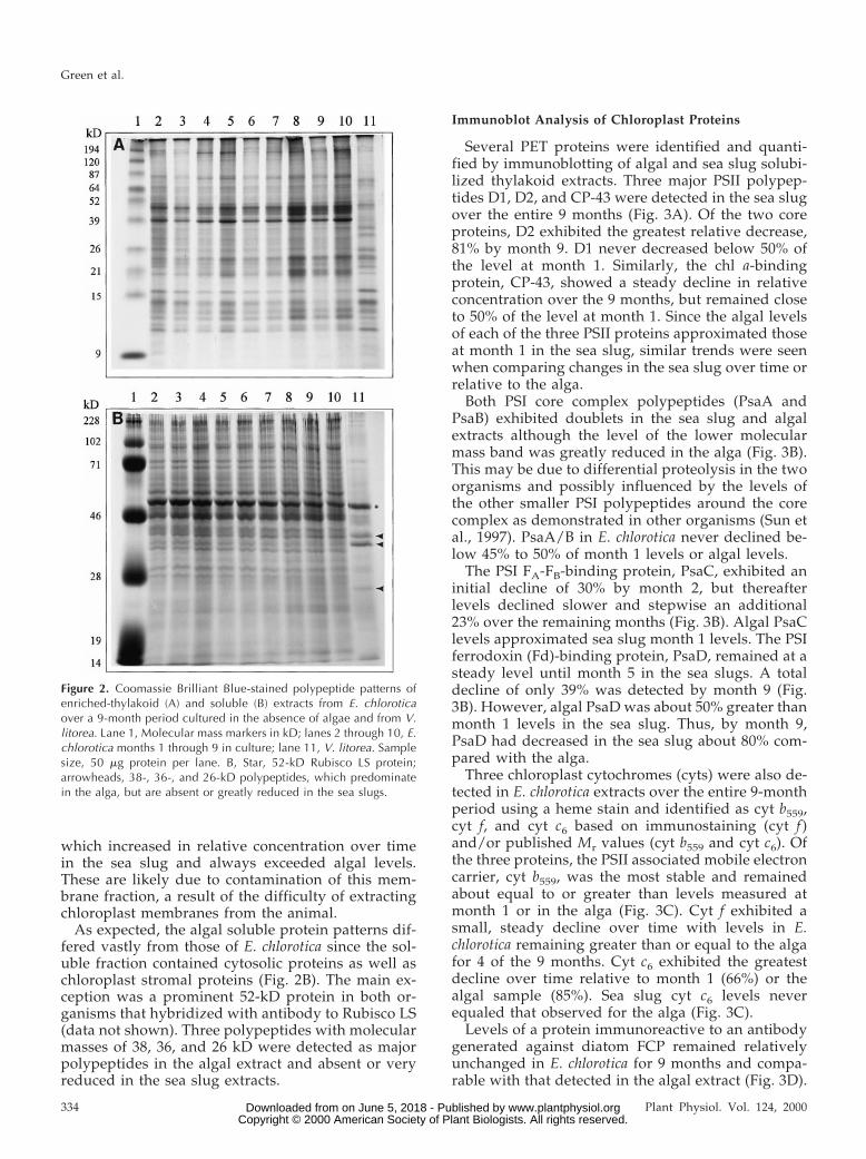
which increased in relative concentration over timein the sea slug and always exceeded algal levels.These are likely due to contamination of this mem-brane fraction, a result of the difficulty of extractingchloroplast membranes from the animal.
As expected, the algal soluble protein patterns dif-fered vastly from those of E. chlorotica since the sol-uble fraction contained cytosolic proteins as well aschloroplast stromal proteins (Fig. 2B). The main ex-ception was a prominent 52-kD protein in both or-ganisms that hybridized with antibody to Rubisco LS(data not shown). Three polypeptides with molecularmasses of 38, 36, and 26 kD were detected as majorpolypeptides in the algal extract and absent or veryreduced in the sea slug extracts.
Immunoblot Analysis of Chloroplast Proteins
Several PET proteins were identified and quanti-fied by immunoblotting of algal and sea slug solubi-lized thylakoid extracts. Three major PSII polypep-tides D1, D2, and CP-43 were detected in the sea slugover the entire 9 months (Fig. 3A). Of the two coreproteins, D2 exhibited the greatest relative decrease,81% by month 9. D1 never decreased below 50% ofthe level at month 1. Similarly, the chl a-bindingprotein, CP-43, showed a steady decline in relativeconcentration over the 9 months, but remained closeto 50% of the level at month 1. Since the algal levelsof each of the three PSII proteins approximated thoseat month 1 in the sea slug, similar trends were seenwhen comparing changes in the sea slug over time orrelative to the alga.
Both PSI core complex polypeptides (PsaA andPsaB) exhibited doublets in the sea slug and algalextracts although the level of the lower molecularmass band was greatly reduced in the alga (Fig. 3B).This may be due to differential proteolysis in the twoorganisms and possibly influenced by the levels ofthe other smaller PSI polypeptides around the corecomplex as demonstrated in other organisms (Sun etal., 1997). PsaA/B in E. chlorotica never declined be-low 45% to 50% of month 1 levels or algal levels.
The PSI FA-FB-binding protein, PsaC, exhibited aninitial decline of 30% by month 2, but thereafterlevels declined slower and stepwise an additional23% over the remaining months (Fig. 3B). Algal PsaClevels approximated sea slug month 1 levels. The PSIferrodoxin (Fd)-binding protein, PsaD, remained at asteady level until month 5 in the sea slugs. A totaldecline of only 39% was detected by month 9 (Fig.3B). However, algal PsaD was about 50% greater thanmonth 1 levels in the sea slug. Thus, by month 9,PsaD had decreased in the sea slug about 80% com-pared with the alga.
Three chloroplast cytochromes (cyts) were also de-tected in E. chlorotica extracts over the entire 9-monthperiod using a heme stain and identified as cyt b559,cyt f, and cyt c6 based on immunostaining (cyt f )and/or published Mr values (cyt b559 and cyt c6). Ofthe three proteins, the PSII associated mobile electroncarrier, cyt b559, was the most stable and remainedabout equal to or greater than levels measured atmonth 1 or in the alga (Fig. 3C). Cyt f exhibited asmall, steady decline over time with levels in E.chlorotica remaining greater than or equal to the algafor 4 of the 9 months. Cyt c6 exhibited the greatestdecline over time relative to month 1 (66%) or thealgal sample (85%). Sea slug cyt c6 levels neverequaled that observed for the alga (Fig. 3C).
Levels of a protein immunoreactive to an antibodygenerated against diatom FCP remained relativelyunchanged in E. chlorotica for 9 months and compa-rable with that detected in the algal extract (Fig. 3D).
Figure 2. Coomassie Brilliant Blue-stained polypeptide patterns ofenriched-thylakoid (A) and soluble (B) extracts from E. chloroticaover a 9-month period cultured in the absence of algae and from V.litorea. Lane 1, Molecular mass markers in kD; lanes 2 through 10, E.chlorotica months 1 through 9 in culture; lane 11, V. litorea. Samplesize, 50 mg protein per lane. B, Star, 52-kD Rubisco LS protein;arrowheads, 38-, 36-, and 26-kD polypeptides, which predominatein the alga, but are absent or greatly reduced in the sea slugs.
Green et al.
334 Plant Physiol. Vol. 124, 2000 www.plantphysiol.orgon June 5, 2018 - Published by Downloaded from Copyright © 2000 American Society of Plant Biologists. All rights reserved.
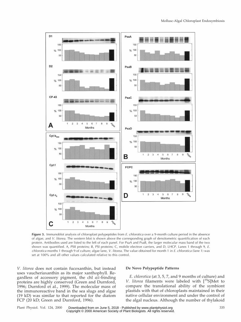
V. litorea does not contain fucoxanthin, but insteaduses vaucheriaxanthin as its major xanthophyll. Re-gardless of accessory pigment, the chl a/c-bindingproteins are highly conserved (Green and Durnford,1996; Durnford et al., 1999). The molecular mass ofthe immunoreactive band in the sea slugs and algae(19 kD) was similar to that reported for the diatomFCP (20 kD; Green and Durnford, 1996).
De Novo Polypeptide Patterns
E. chlorotica (at 3, 5, 7, and 9 months of culture) andV. litorea filaments were labeled with [35S]Met tocompare the translational ability of the symbiontplastids with that of chloroplasts maintained in theirnative cellular environment and under the control ofthe algal nucleus. Although the number of thylakoid
Figure 3. Immunoblot analysis of chloroplast polypeptides from E. chlorotica over a 9-month culture period in the absenceof algae, and V. litorea. The western blot is shown above the corresponding graph of densitometric quantification of eachprotein. Antibodies used are listed to the left of each panel. For PsaA and PsaB, the larger molecular mass band of the twoshown was quantified. A, PSII proteins; B, PSI proteins; C, mobile electron carriers, and D, LHCP. Lanes 1 through 9, E.chlorotica months 1 through 9 of culture; algae lane, V. litorea. The value obtained for month 1 in E. chlorotica (lane 1) wasset at 100% and all other values calculated relative to this control.
Mollusc-Algal Chloroplast Endosymbiosis
Plant Physiol. Vol. 124, 2000 335 www.plantphysiol.orgon June 5, 2018 - Published by Downloaded from Copyright © 2000 American Society of Plant Biologists. All rights reserved.
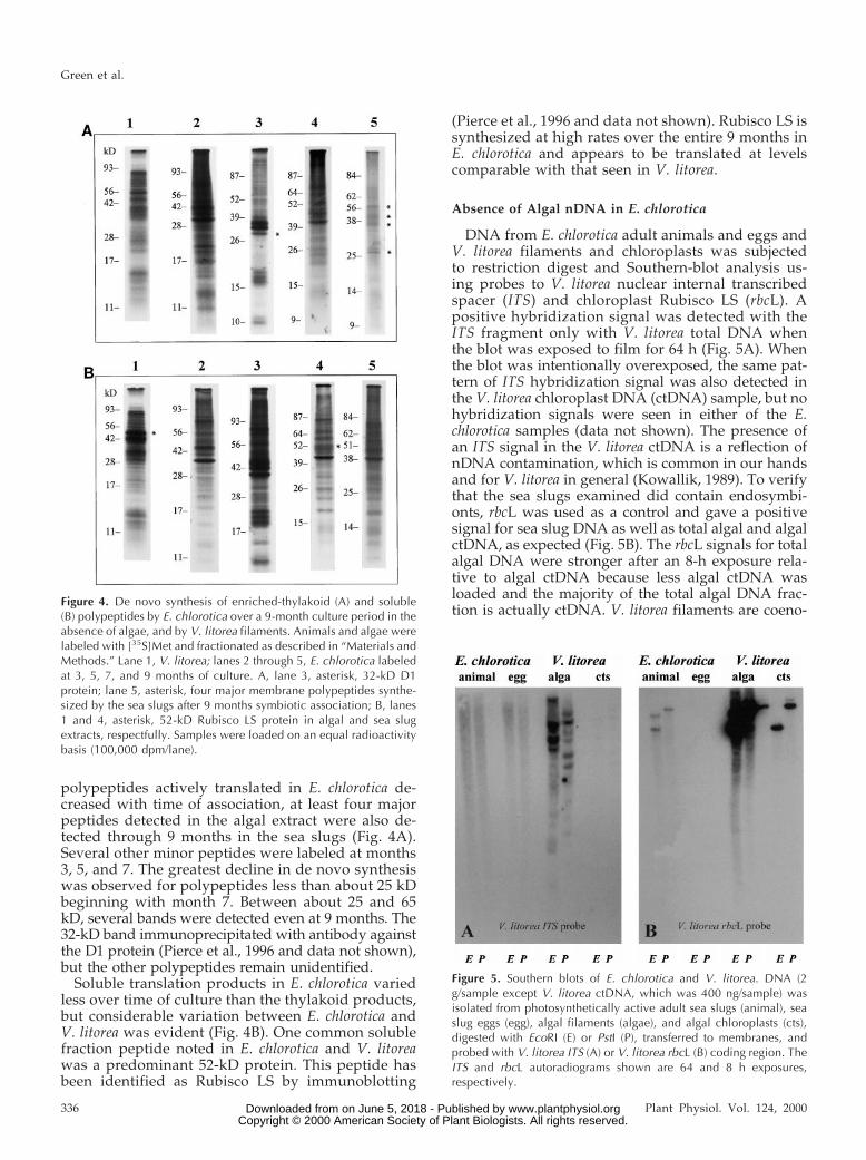
polypeptides actively translated in E. chlorotica de-creased with time of association, at least four majorpeptides detected in the algal extract were also de-tected through 9 months in the sea slugs (Fig. 4A).Several other minor peptides were labeled at months3, 5, and 7. The greatest decline in de novo synthesiswas observed for polypeptides less than about 25 kDbeginning with month 7. Between about 25 and 65kD, several bands were detected even at 9 months. The32-kD band immunoprecipitated with antibody againstthe D1 protein (Pierce et al., 1996 and data not shown),but the other polypeptides remain unidentified.
Soluble translation products in E. chlorotica variedless over time of culture than the thylakoid products,but considerable variation between E. chlorotica andV. litorea was evident (Fig. 4B). One common solublefraction peptide noted in E. chlorotica and V. litoreawas a predominant 52-kD protein. This peptide hasbeen identified as Rubisco LS by immunoblotting
(Pierce et al., 1996 and data not shown). Rubisco LS issynthesized at high rates over the entire 9 months inE. chlorotica and appears to be translated at levelscomparable with that seen in V. litorea.
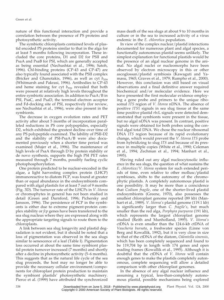
Absence of Algal nDNA in E. chlorotica
DNA from E. chlorotica adult animals and eggs andV. litorea filaments and chloroplasts was subjectedto restriction digest and Southern-blot analysis us-ing probes to V. litorea nuclear internal transcribedspacer (ITS) and chloroplast Rubisco LS (rbcL). Apositive hybridization signal was detected with theITS fragment only with V. litorea total DNA whenthe blot was exposed to film for 64 h (Fig. 5A). Whenthe blot was intentionally overexposed, the same pat-tern of ITS hybridization signal was also detected inthe V. litorea chloroplast DNA (ctDNA) sample, but nohybridization signals were seen in either of the E.chlorotica samples (data not shown). The presence ofan ITS signal in the V. litorea ctDNA is a reflection ofnDNA contamination, which is common in our handsand for V. litorea in general (Kowallik, 1989). To verifythat the sea slugs examined did contain endosymbi-onts, rbcL was used as a control and gave a positivesignal for sea slug DNA as well as total algal and algalctDNA, as expected (Fig. 5B). The rbcL signals for totalalgal DNA were stronger after an 8-h exposure rela-tive to algal ctDNA because less algal ctDNA wasloaded and the majority of the total algal DNA frac-tion is actually ctDNA. V. litorea filaments are coeno-
Figure 4. De novo synthesis of enriched-thylakoid (A) and soluble(B) polypeptides by E. chlorotica over a 9-month culture period in theabsence of algae, and by V. litorea filaments. Animals and algae werelabeled with [35S]Met and fractionated as described in “Materials andMethods.” Lane 1, V. litorea; lanes 2 through 5, E. chlorotica labeledat 3, 5, 7, and 9 months of culture. A, lane 3, asterisk, 32-kD D1protein; lane 5, asterisk, four major membrane polypeptides synthe-sized by the sea slugs after 9 months symbiotic association; B, lanes1 and 4, asterisk, 52-kD Rubisco LS protein in algal and sea slugextracts, respectfully. Samples were loaded on an equal radioactivitybasis (100,000 dpm/lane).
Figure 5. Southern blots of E. chlorotica and V. litorea. DNA (2g/sample except V. litorea ctDNA, which was 400 ng/sample) wasisolated from photosynthetically active adult sea slugs (animal), seaslug eggs (egg), algal filaments (algae), and algal chloroplasts (cts),digested with EcoRI (E) or PstI (P), transferred to membranes, andprobed with V. litorea ITS (A) or V. litorea rbcL (B) coding region. TheITS and rbcL autoradiograms shown are 64 and 8 h exposures,respectively.
Green et al.
336 Plant Physiol. Vol. 124, 2000 www.plantphysiol.orgon June 5, 2018 - Published by Downloaded from Copyright © 2000 American Society of Plant Biologists. All rights reserved.

cytic with numerous ellipsoid chloroplasts found pe-ripheral to a large central vacuole (Lee, 1989). Nochloroplasts are found in sea slug eggs and no rbcLsignal was detected even when the blots were overex-posed (data not shown). The larger rbcL signal in theE. chlorotica “animal” and V. litorea “alga” EcoRI di-gested samples represents incomplete digestion due tothe large number of EcoRI sites in the nDNA relativeto the ctDNA. The stray rbcL hybridization signal tothe right of the V. litorea “alga” lane resulted fromspillover of the sample. This spillover was also evidentin the ITS Southern blot when it was overexposed(data not shown).
PCR reactions were also utilized as an independentand more sensitive means for determining if the seaslugs contain even small amounts of an intact algalnuclear genome. When V. litorea DNA was utilized asa template, primer sets for rbcL and ITS both gavestrong positive signals (approximately 900 and 200bp, respectively; Fig. 6). In contrast, when E. chloroticaDNA was utilized, a positive signal was observedonly for rbcL. Negative controls did not yield positivesignals for either primer set.
Chloroplast Genome Size
In the absence of an algal nuclear genome in the seaslug, it is possible that V. litorea chloroplasts have an
unusual chloroplast genome, possibly with a signif-icantly increased coding capacity compared with thatof other algae and this is the major contributingfactor to the long-term functional activity of the sym-bionts. V. litorea ctDNA was digested to completionwith the restriction endonuclease PstI and the frag-ment sizes summed to reveal a genome size of 119.1kb (Fig. 7). This value is typical for chromophytesand suggests that there is nothing remarkable aboutthe size of the V. litorea chloroplast genome.
DISCUSSION
The E. chlorotica/V. litorea symbiosis is the longest-lived functional mollusc/plastid association known(West, 1979; Pierce et al., 1996). Although it is possi-ble that the chloroplasts provide some other benefitto these shell-less molluscs (such as camouflage), thisand previous studies (Mujer et al., 1996; Pierce et al.,1996) indicate that the plastids are photosyntheticallyactive for at least 5 months and minimally functionalfor at least 9 months, following incorporation by thesea slug. Here we have more closely dissected the
Figure 6. Ethidium bromide stained 1% (w/v) agarose gel showingPCR reactions of photosynthetically active E. chlorotica (E) and V.litorea (V) DNA and negative water control (W) using primers spe-cific for the V. litorea ITS region (lanes 2–4) or rbcL (lanes 5–7). Alsoshown is 0.5 g of size standards (1-kb ladder, Gibco-BRL, no. 15615-016); SS, lane 1.
Figure 7. V. litorea ctDNA digested with PstI and electrophoresedthrough 1% (w/v) agarose. Fragment sizes (in kb) in descending orderare 19.5, 15.6, 11.8, 10.6, 7.9 3 2, 6.8, 6.3, 5.7, 4.8, 4.1, 3.7, 3.6,3.3, 2.9, 2.2, 1.7, and 0.7 (not visible on picture) for a total chloro-plast genome size of 119.1 kb.
Mollusc-Algal Chloroplast Endosymbiosis
Plant Physiol. Vol. 124, 2000 337 www.plantphysiol.orgon June 5, 2018 - Published by Downloaded from Copyright © 2000 American Society of Plant Biologists. All rights reserved.
nature of this functional interaction and provide acorrelation between the presence of PS proteins andphotosynthetic activity.
The symbiotic chloroplasts contained levels of plas-tid encoded PS proteins similar to that in the alga forat least 5 months following incorporation. These in-cluded the core proteins, D1 and D2 for PSII andPsaA and PsaB for PSI, which are generally acceptedas being essential (Nechushtai et al., 1996; Satoh,1996). Chl-binding proteins (CP-43 and CP-47) arealso typically found associated with the PSII complex(Bricker and Ghanotakis, 1996), as well as cyt b559(Whitmarsh and Pakrasi, 1996). Antibodies to CP-43and heme staining for cyt b559 revealed that bothwere present at relatively high levels throughout theentire symbiotic association. In addition to PsaA/B inPSI, PsaC, and PsaD, the terminal electron acceptorand Fd-docking site of PSI, respectively (for review,see Nechushtai et al., 1996), were also present in thesymbionts.
The decrease in oxygen evolution rates and PETactivity after about 5 months of incorporation paral-leled reductions in PS core polypeptides, especiallyD2, which exhibited the greatest decline over time ofany PS polypeptide examined. The lability of PSII-D2and the stability of PSI-PsaA/B were also docu-mented previously when a shorter time period wasexamined (Mujer et al., 1996). The maintenance ofhigh levels of PsaA through D by protein stability orde novo synthesis, supports the high PSI PET ratesmeasured through 7 months, possibly fueling cyclicphotophosphorylation.
One protein predicted to be nuclear-encoded in thealgae, a light harvesting complex protein (LHCP)immunoreactive to diatom FCP, was found at greaterthan or equal abundance in the endosymbionts com-pared with algal plastids for at least 7 out of 9 months(Fig. 3D). The turnover rate of the LHCPs in V. litoreaor chromophytes in general has not been studied indetail (Green and Durnford, 1996; Pichersky andJansson, 1996). The persistence of FCP in the symbi-onts is either due to extreme pigment-protein com-plex stability or fcp genes have been transferred to thesea slug nucleus where they are expressed along withthe appropriate targeting signals to route them to thechloroplasts.
A link between sea slug longevity and plastid deg-radation is not evident, but it should be noted that aloss of pigmentation was observed in the sea slugssimilar to senescence of a leaf (Table I). Pigmentationloss occurred at about the same time symbiont plas-tid protein turnover increased (7 months) and shortlyafter a decline in photosynthetic activity (5–6 months).This suggests that as the natural life cycle of the seaslug proceeds, the host may no longer be able toprovide the necessary cellular environment/compo-nents for chloroplast protein production to maintainthe symbiont plastids’ photosynthetic machinery.Pierce et al. (1999) have attributed the synchronized
mass death of the sea slugs at about 9 to 10 months inculture or in the sea to increased activity of a virusendemic in the E. chlorotica population.
In view of the complex nuclear/plastid interactionsdocumented for numerous plant and algal species, afunctionally autonomous plastid seems unlikely. Thesimplest explanation for functional plastids would bethe presence of an algal nuclear genome in the ani-mal. No algal nuclei or nucleomorphs have beenobserved by electron microscopy in this or otherascoglossan/plastid symbiosis (Kawaguti and Ya-masu, 1965; Graves et al., 1979; Rumpho et al., 2000).However, this conclusion was still based on visualobservations and a final definitive answer requiredbiochemical and/or molecular evidence. Here wehave presented the first molecular evidence employ-ing a gene probe and primers to the unique ribo-somal ITS region of V. litorea nDNA. The absence ofpositive ITS1 signals in sea slug tissue at the sametime positive plastid rbcL signals were detected dem-onstrated that symbionts were present in the tissue,but no algal nDNA was present. In contrast, positivesignals were obtained for both probes using the con-trol algal total DNA. We chose the nuclear ribosomalDNA ITS region because of its rapid evolutionarychange, which would prevent the V. litorea ITS probefrom hybridizing to slug ITS and because of its pres-ence in multiple copies (White et al., 1990; Colemanet al., 1994; Zechman et al., 1994; Manhart et al.,1995).
Having ruled out any algal nucleocytosolic influ-ence in the sea slugs, the question of what sustains theE. chlorotica/V. litorea system for such extended peri-ods of time, even relative to other mollusc/plastidsymbioses, shifts to the autonomy of the chromo-phytic plastids. A completely autonomous plastid isone possibility. It may be more than a coincidencethat Codium fragile, one of the shorter-lived plastidendosymbionts (Greene, 1970), also possesses thesmallest chloroplast genome reported (89 kb) (Man-hart et al., 1989). V. litorea’s plastid genome (119.1 kb)is significantly larger than C. fragile’s, but muchsmaller than the red alga, Porphyra purpurea (191 kb),which represents the largest chloroplast genomestudied (Reith and Munholland, 1995). V. litorea’sctDNA is even smaller than the 124.6-kb ctDNA ofVaucheria bursata, a freshwater species (Linne vonBerg and Kowallik, 1992), but it is very close in sizeto that of the ctDNA of the diatom, Odontella sinensis,which has been completely sequenced and found tobe 119,704 bp in length with 174 genes and openreading frames (Kowallik et al., 1995). Although it isdoubtful that the ctDNA of V. litorea will containenough genes to make the plastids completely auton-omous, complete sequencing will allow a detailedcomparison with other chloroplast genomes.
In the absence of any algal nuclear influence andassuming a typical, less-than-completely autono-mous plastid, possible mechanisms being explored
Green et al.
338 Plant Physiol. Vol. 124, 2000 www.plantphysiol.orgon June 5, 2018 - Published by Downloaded from Copyright © 2000 American Society of Plant Biologists. All rights reserved.
that might contribute to the functional symbiosis in-clude unusual stability of nuclear encoded plastidproteins in the animal, unanticipated activity of tran-scription/translation/PS complexes in the absence ofnormally critical nuclear encoded subunits, targetingof animal cytosolic or mitochondrial proteins withrelated functions to the chloroplast, and lateral trans-fer of algal genes to the animal nucleus (for review,see Rumpho et al., 2000).
By combining information from oxygen exchangeand PET measurements, immunostaining, the highlysimilar trends in peptide banding patterns betweenE. chlorotica and algal de novo and constitutive thy-lakoid preparations, and the previous work demon-strating accumulation of chloroplast transcripts andde novo protein biogenesis (Mujer et al., 1996; Pierceet al., 1996), it appears that a significant number ofprocesses related to gene expression and protein bio-synthesis are all occurring in the symbiotic plastidsfor several months, enabling long-term photosyn-thetic activity and survival. For a plastid within itsusual cellular environment to produce and processtranscripts and synthesize, fold, and processpolypeptides into their active forms, nuclear assis-tance is required in most higher plant systems (Bar-kan et al., 1994; Hayes et al., 1996). Understandinghow this all occurs in a marine mollusc harboringsymbiont algal plastids with a relatively small chlo-roplast genome and no detectable algal nucleocyto-solic influence continues to be investigated.
MATERIALS AND METHODS
Sea Slug and Algal Cultures
Elysia chlorotica Gould specimens originated from collec-tions made in November 1997 and October 1998 from anintertidal marsh on Martha’s Vineyard Island, MA. Theanimals were maintained photoautotrophically in aeratedaquaria containing ASW (925 mosmol kg-1, Instant Ocean,Aquarium Systems, Mentor, OH) at 10°C on a 14-h photo-period. Sea slugs were either sampled live for photosyn-thetic measurements and chloroplast isolation or sacrificedmonthly for 9 months and analyzed later. For the monthlyharvests, 20 to 25 individual animals were blotted dry,pooled, frozen, and powdered immediately in liquid nitro-gen, and stored at 280°C until analyzed. “Months Symbi-otic Association” or “Months in Culture” in the text andfigures refers to the number of months from the time ofcollection from the intertidal marsh in which the sea slugsare no longer associated with any algae. Vaucheria litorea C.Agardh is maintained in culture in enriched one-quarter-strength ASW (250 mosmol kg-1) and a modified f/2 me-dium as described previously (Pierce et al., 1996), exceptthat the cultures are illuminated by natural lighting andaeration is limited to daily manual swirling.
Oxygen Evolution Measurements
Rates of O2 exchange were measured at 15°C with anoxygen electrode (2.5 mL vol, DW1, Hansatech, King’s
Lynn, UK) illuminated with a high-intensity tungsten-halogen light source (1,500 mmol photons m22 s21; LS2,Hansatech). Individual animals were separated from thestirring apparatus by a screen and suspended in 1 mL ofASW supplemented with 10 mm NaHCO3. Samples wereincubated in the dark for 1 min prior to illumination. Rateswere recorded for 5 to 10 min, after which illuminationceased and dark respiration rates (O2 uptake) were re-corded for at least 5 min. Apparent photosynthesis wascalculated by measuring the rate of CO2- and light-dependent O2 evolution, whereas respiration rates werebased on the uptake of O2 in the dark. Gross photosynthe-sis was calculated by summing the apparent photosynthe-sis and dark respiration rates. At least three sea slugs wereanalyzed for each time point. Rates were calculated on atotal chl basis with chl a and c quantified by A664 and A630
measurements following homogenization in 90% (v/v) ac-etone (Sterman, 1988).
Electron Transport Measurements
Chloroplasts were isolated from live sea slugs and algalfilaments and purified on Percoll gradients (Pierce et al.,1996). Intact chloroplasts were lysed by hypertonic freeze-thawing and thylakoid membranes were collected by cen-trifugation at 35,000g for 5 min. The washed thylakoidswere resuspended in reaction buffer (50 mm HEPES [4-(2-hydroxyethyl)-1-piperazineethanesulfonic acid], pH 7.6,100 mm sorbitol, 5 mm NaCl, and 5 mm MgCl2) and usedimmediately at a concentration of about 50 mg chl mL21.All PET measurements were made with an oxygen elec-trode at 15°C and 1,500 mmol photons m22 s21 (Hind,1993).
Whole chain PET including the OEC, but excluding Fdand Fd-NADP reductase, was measured with water as theelectron donor and MV as the electron acceptor. Endoge-nous catalase activity was inhibited by adding 5 mm NaN3.For whole chain PET excluding the OEC, electron flowfrom water was inhibited by incubating the thylakoids at50°C for 2 min. The reaction medium was then supple-mented with 5 mm NH4Cl (uncoupler), 0.5 mm DPC (elec-tron donor), 2 mm NaN3, and 50 mm MV. Electron transportthrough PSI (Mehler reaction) was measured as describedfor whole chain PET except, after 5 min illumination, 10 mmdichloromethylurea was added to inhibit PSII and 2 mmascorbate and 50 mm dichlorophenolindophenol wereadded to provide electrons for PSI. Electron flow fromwater through PSII including the dichloromethylurea-sensitive site was measured by additions to the reactionmedia of 5 mm NH4Cl, 1 mm p-phenylenediamine andexcess FeCN (4 mm).
Soluble and Thylakoid Membrane-Enriched Proteins
Powdered samples from the monthly collections werealiquoted (200 mg fresh weight) and proteins isolated asdescribed by Russell et al. (1995). The powdered tissue washomogenized in a microcentrifuge tube containing 400 mL(1:2, w/v) of isolation buffer (330 mm sorbitol, 25 mm
Mollusc-Algal Chloroplast Endosymbiosis
Plant Physiol. Vol. 124, 2000 339 www.plantphysiol.orgon June 5, 2018 - Published by Downloaded from Copyright © 2000 American Society of Plant Biologists. All rights reserved.
HEPES-KOH, pH 7.0, 5 mm MgCl2, 10 mm NaCl, 100 mmN-acetyl-l-Cys, and 1 mm phenylmethylsulfonyl fluoride).Enriched-thylakoid membranes (hereafter, thylakoid pro-teins) were collected by centrifugation at 10,000g for 5 minat 4°C. The resultant supernatant (containing soluble cyto-solic proteins and the chloroplast stroma) was collectedand used as the soluble fraction. Thylakoid pellets werewashed once in isolation buffer lacking sorbitol andN-acetyl-l-Cys, and resuspended in isolation buffer con-taining 1% (v/v) Triton X-100 followed by continuousshaking for at least 2 h at 4°C. Total protein was deter-mined using the Bio-Rad (Hercules, CA) protein stain withbovine serum albumin as the standard and corrected forTriton X-100, when necessary.
Immunoblot Analysis and Quantification
Thylakoid proteins (50 mg protein lane21) were sepa-rated by SDS-PAGE employing a 9% to 18% (w/v) linearpolyacrylamide gradient (Laemmli, 1970). Gels were eitherstained with Coomassie Brilliant Blue G-250 or the proteinswere transferred to polyvinylidene fluoride membrane(Immobilon-P, Millipore, Bedford, MA) by electroblottingat 15°C and 75 V for 3 h (Towbin et al., 1979). Proteins werevisualized using the alkaline phosphatase system as rec-ommended by the manufacturer (Promega, Madison, WI).Polyclonal, heterologous antibodies directed against D1,D2, and CP43 (all from barley), FCP (diatom), PsaA andPsaB (domain specific Synechocystis sp. PCC 6803 syntheticpeptides [Sun et al., 1997]), PsaC and PsaD (Synechocystissp. PCC 6803), and cyt f (Chlamydomonas reinhardtii), wereraised in rabbits and generously provided to us by a num-ber of sources (please see “Acknowledgments”). Immuno-blots were quantified densitometrically (AlphaImager2000, Alpha Innotech, San Leandro, CA). The quantity ofeach polypeptide at month 1 was set at 100% (control) andall other samples were calculated relative to their respec-tive control.
Super Signal Chemiluminescent Detection System
Cyts were detected using the chemiluminescent sub-strate Luminol (Pierce Chemical, Rockford, IL), which re-acts with proteins containing heme-functional groups. Dueto the hydrophobic nature of these proteins they weretransferred as described above, but in the presence of 0.5%to 1% (w/v) SDS and onto nitrocellulose membrane(Schleicher and Schuell). Once transferred, the blots wereincubated in Luminol for 5 min, exposed to x-ray film, andthe cyts identified by comparisons with known molecularweights, protein fraction from which the samples origi-nated, and immunostaining in the case of cyt f.
De Novo Protein Synthesis
For each time point, four to six sea slugs were placed inglass scintillation vials containing 3 mL of ASW and 50 mCimL21 [35S]Met, and illuminated for 7 h at 18°C. V. litorea(450 mg fresh weight) was treated as above, but labeled for
4 h. Following labeling, thylakoid and soluble proteinswere isolated and separated by SDS-PAGE as describedabove. Equal radioactivity (100,000 dpm) was loaded foreach sample. Gels were fixed and processed for fluorogra-phy according to Mujer et al. (1996).
DNA Isolation, Sizing, and Southern Blotting
DNA was isolated from E. chlorotica adult animals, E.chlorotica eggs, V. litorea filaments, and V. litorea chloro-plasts (Palmer, 1986; Mujer et al., 1996). Aliquots of DNA (2mg sample21 except V. litorea ctDNA, which was 400 ngsample21) were digested with EcoRI or PstI and processedfor Southern analysis as described (Mujer et al., 1996). Forprobes, the entire ITS from V. litorea genomic DNA wasamplified using ITS4 and ITS5 primers of White et al.(1990). Primers to ITS1 only (59-CCAACATATTCATCCTCand 59- ATTGCACCATTGCTGGC) were constructed fromthis sequence and used to produce a 203-bp ITS1 probe byamplification of V. litorea genomic DNA. Primers to rbcL(59-CCTTAATACAACTGCAG and 59-CCTTTATTTACA-GCATAC) were designed using sequence informationobtained from V. litorea ctDNA clones (accession no.AF207527) and used to produce an 892-bp rbcL probe. PCRreactions for ITS and rbcL were performed using the prim-ers listed above with 2 ng of genomic DNA from photo-synthetically active E. chlorotica (3 months in culture), V.litorea, or water control as templates following standardprotocols (35 cycles, 19-19-29 extension times at 94°, 50°, and72°C, respectively) using Taq polymerase (Gibco-BRL,Gaithersburg, MD). For chloroplast genome sizing,V. litorea ctDNA was digested with PstI and analyzed asdescribed in Palmer (1986).
ACKNOWLEDGMENTS
We thank the following for generously providing anti-bodies for these studies: John Mullet (Texas A&M Univer-sity), Steven Theg (University of California, Davis), PaulFalkowski (Brookhaven National Laboratory), John Gol-beck (Penn State University), Beverley Green (University ofBritish Columbia), Parag Chitnis (Iowa State University)and Sabeeha Merchant (University of California, Los An-geles [UCLA]). We would also like to thank Beth Dreyfuss(UCLA) for communicating the cyt staining protocol.
Received December 13, 1999; accepted May 5, 2000.
LITERATURE CITED
Aro E, Virgin I, Andersson B (1993) Photoinhibition ofPhotosystem II: inactivation, protein damage and turn-over. Biochim Biophys Acta 1143: 113–134
Barkan A, Walker M, Nolasco M, Johnson D (1994) Anuclear mutation in maize blocks the processing andtranslation of several chloroplast mRNAs and providesevidence for the differential translation of alternativemRNA forms. EMBO J 13: 3170–3181
Bricker TM, Ghanotakis DF (1996) Introduction to oxygenevolution and the oxygen-evolving complex. In DR Ort,
Green et al.
340 Plant Physiol. Vol. 124, 2000 www.plantphysiol.orgon June 5, 2018 - Published by Downloaded from Copyright © 2000 American Society of Plant Biologists. All rights reserved.
CF Yocum, eds, Oxygenic Photosynthesis: The Light Re-actions, Vol 4. Kluwer Academic Publishers, Boston, pp113–136
Christopher DA, Mullet JE (1994) Separate photosensorypathways co-regulate blue light/ultraviolet-A-activatedpsbD-psbC transcription and light-induced D2 and CP43degradation in barley (Hordeum vulgare) chloroplasts.Plant Physiol 104: 1119–1129
Clark KB, Busacca M (1978) Feeding specificity and chlo-roplast retention in four tropical Ascoglossa, with a dis-cussion of the extent of chloroplast symbiosis and theevolution of the order. J Mollusc Stud 44: 272–282
Coleman AW, Suarez A, Goff LJ (1994) Molecular delin-eation of species and syngens in volvocacean green algae(chlorophyta). J Phycol 30: 80–90
Douglas AE (1994) Symbiotic Interactions. Oxford Univer-sity Press, New York
Durnford DG, Deane JA, Tan S, McFadden GI, Gantt E,Green BR (1999) A phylogenetic assessment of the eu-karyotic light-harvesting antenna proteins, with implica-tions for plastid evolution. J Mol Evol 48: 59–68
Giles KL, Sarafis V (1971) On the survival and reproduc-tion of chloroplasts outside the cell. Cytobios 4: 61–74
Graves DA, Gibson MA, Bleakney JS (1979) The digestivediverticula of Alderia modesta and Elysia chlorotica. Veliger21: 415–422
Green BR, Durnford DG (1996) The chlorophyll-carotenoidproteins of oxygenic photosynthesis. Annu Rev PlantPhysiol Plant Mol Biol 47: 685–714
Greene RW (1970) Symbiosis in sacoglossan opistho-branchs: functional capacity of symbiotic chloroplasts.Marine Biol 7: 138–142
Hayes R, Kudla J, Schuster G, Gabay L, Maliga P, Gruis-sem W (1996) Chloroplast mRNA 39-end processing by ahigh molecular weight protein complex is regulated bynuclear encoded RNA binding proteins. EMBO J 15:1132–1141
Hind G (1993) Thylakoid components and processes. InDO Hall, JMO Scurlock, HR Bolhar-Nordenkampf, RCLeegood, SP Long, eds, Photosynthesis and Productionin a Changing Environment: A Field and LaboratoryManual. Chapman & Hall, New York, pp 283–298
Kawaguti S, Yamasu T (1965) Electron microscopy on thesymbiosis between an elysioid gastropod and chloro-plasts of a green alga. Biol J Okayama Univ 11: 57–65
Kowallik K (1989) Molecular aspects and phylogeneticimplications of plastid genomes of certain chromo-phytes. In JC Green, BSC Leadbeater, WL Diver, eds, TheChromophyte Algae: Problems and Perspectives, Vol 38.Clarendon Press, Oxford, pp 101–124
Kowallik K, Stroebe B, Schaffran I, Freier U (1995) Thechloroplast genome of a chlorophyll a 1 c containingalga, Odontella sinensis. Plant Mol Biol Rep 13: 336–342
Laemmli UK (1970) Cleavage of structural proteins duringthe assembly of the head of bacteriophage T4. Nature 22:680–685
Lee RE (1989) Phycology. Cambridge University Press,New York
Linne von Berg K-H, Kowallik KV (1992) Structural orga-nization of the chloroplast genome of the chromophyticalga Vaucheria bursata. Plant Mol Biol 18: 83–95
Manhart JR, Fryxell GA, Celia Villac M, Segura LY (1995)Psuedo-nitzschia pungens and P. multiseries (bacillari-ophyceae): nuclear ribosomal DNAs and species differ-ences. J Phycol 31: 421–427
Manhart JR, Kelly K, Dudock BS, Palmer JD (1989) Un-usual characteristics of Codium fragile chloroplast DNArevealed by physical and gene mapping. Mol Gen Genet216: 417–421
Martin W, Herrmann RG (1998) Gene transfer from or-ganelles to the nucleus: how much, what happens, andwhy? Plant Physiol 118: 9–17
Merchant S, Dreyfuss BW (1998) Posttranslational assem-bly of photosynthetic metalloproteins. Annu Rev PlantPhysiol Plant Mol Biol 49: 25–51
Mujer CV, Andrews DL, Manhart JR, Pierce SK, RumphoME (1996) Chloroplast genes are expressed during intra-cellular symbiotic association of Vaucheria litorea plastidswith the sea slug Elysia chlorotica. Proc Natl Acad SciUSA 93: 12333–12338
Nass MM (1969) Uptake of isolated chloroplasts by mam-malian cells. Science 165: 1128–1131
Nechushtai R, Eden A, Cohen Y, Klein J (1996) Introduc-tion to photosystem I: reaction center function, compo-sition and structure. In DR Ort, CF Yocum, eds, OxygenicPhotosynthesis: The Light Reactions, Vol 4. Kluwer Ac-ademic Publishers, Boston, pp 289–311
Palmer JD (1986) Isolation and structural analysis of chlo-roplast DNA. Methods Enzymol 118: 167–186
Palmer JD, Delwiche CF (1996) Second-hand chloroplastsand the case of the disappearing nucleus. Proc Natl AcadSci USA 93: 7432–7435
Pichersky E, Jansson S (1996) The light-harvesting chloro-phyll a/b-binding polypeptides and their genes in angio-sperm and gymnosperm species. In DR Ort, CF Yocum,eds, Oxygenic Photosynthesis: The Light Reactions, Vol4. Kluwer Academic Publishers, Boston, pp 507–521
Pierce SK, Biron RW, Rumpho ME (1996) Endosymbioticchloroplasts in molluscan cells contain proteins synthe-sized after plastid capture. J Exp Biol 199: 2323–2330
Pierce SK, Maugel TK, Rumpho ME, Hanten JJ, MondyWL (1999) Annual viral expression in a sea slug popula-tion: life cycle control and symbiotic chloroplast mainte-nance. Biol Bull 197: 1–6
Reith M (1995) Molecular biology of rhodophyte and chro-mophyte plastids. Annu Rev Plant Physiol Plant Mol Biol46: 549–575
Reith M, Munholland J (1995) Complete nucleotide se-quence of the Porphyra purpurea chloroplast genome.Plant Mol Biol Rep 13: 333–335
Ridley SM, Leech RM (1970) Division of chloroplasts in anartificial environment. Nature 227: 463–465
Rumpho ME, Mujer CV, Andrews DL, Manhart JR, PierceSK (1994) Extraction of DNA from mucilaginous tissuesof a sea slug (Elysia chlorotica). BioTech 17: 1097–1101
Rumpho ME, Summer EJ, Manhart JR (2000) Solar-
Mollusc-Algal Chloroplast Endosymbiosis
Plant Physiol. Vol. 124, 2000 341 www.plantphysiol.orgon June 5, 2018 - Published by Downloaded from Copyright © 2000 American Society of Plant Biologists. All rights reserved.
powered sea slugs: mollusc/algal chloroplast symbiosis.Plant Physiol 123: 1–10
Russell AW, Critchley C, Robinson SA, Franklin LA,Seaton GGR, Chow WS, Anderson JM, Osmond CB(1995) Photosystem II regulation and dynamics of the chlo-roplast D1 protein in Arabidopsis leaves during photosyn-thesis and photoinhibition. Plant Physiol 107: 943–952
Satoh K (1996) Introduction to the photosystem II reactioncenter: isolation and biochemical and biophysical char-acterization. In DR Ort, CF Yocum, eds, Oxygenic Pho-tosynthesis: The Light Reactions, Vol 4. Kluwer Aca-demic Publishers, Boston, pp 193–211
Sterman NT (1988) Spectrophotometric and fluorometricchlorophyll analysis. In CS Lobban, DJ Chapman, BPKramer, eds, Experimental Phycology. Cambridge Uni-versity Press, Cambridge, pp 35–46
Stern DB, Higgs DC, Yang J (1997) Transcription andtranslation in chloroplasts. Trends Plant Sci 2: 308–315
Sun J, Xu Q, Chitnis VP, Jin P, Chitnis PR (1997) Topog-raphy of the photosystem I core proteins of the cyanobac-terium Synechocystis sp. PCC 6803. J Biol Chem 272:21793–21802
Towbin H, Staehelin T, Gordon J (1979) Electrophoretictransfer of proteins from polyacrylamide gels to nitrocel-
lulose sheets: procedure and some applications. ProcNatl Acad Sci USA 76: 4350–4354
Trench RK (1975) Of “leaves that crawl”: functional chlo-roplasts in animal cells. Soc Exp Biol Cambridge Symp29: 229–266
West HH (1979) Chloroplast symbiosis and developmentof the ascoglossan opisthobranch Elysia chlorotica. PhDthesis, Northeastern University, Boston
West HH, Harrigan J, Pierce SK (1984) Hybridization oftwo populations of a marine opisthobranch with differ-ent developmental patterns. Veliger 26: 199–206
White TJ, Bruns T, Lee S, Taylor J (1990) Amplificationand direct sequencing of fungal RNA genes for phyloge-netics. In M Innis, D Gelfand, J Sninsky, T White, eds,PCR Protocols: A Guide to Methods and Applications.Academic Press, San Diego, pp 315–322
Whitmarsh J, Pakrasi HB (1996) Form and function ofcytochrome b-559. In DR Ort, CF Yocum, eds, OxygenicPhotosynthesis: The Light Reactions, Vol 4. Kluwer Ac-ademic Publishers, Boston, pp 249–264
Zechman FW, Zimmer EA, Theriot EC (1994) Use of ribo-somal DNA internal transcribed spacers for phylogeneticstudies in diatoms. 30: 507–512
Green et al.
342 Plant Physiol. Vol. 124, 2000 www.plantphysiol.orgon June 5, 2018 - Published by Downloaded from Copyright © 2000 American Society of Plant Biologists. All rights reserved.