monitoring of in vitro - university of belgrade
TRANSCRIPT
UNIVERSITY OF BELGRADE
FACULTY OF CHEMISTRY
Maja V. Krstić Ristivojević
MONITORING OF IN VITRO
BIOAVAILABILITY AND UPTAKE OF
GLYCOSYLATED FOOD ALLERGENS
USING CELL-BASED MODELS
Doctoral Dissertation
Belgrade, 2020

UNIVERZITET U BEOGRADU
HEMIJSKI FAKULTET
Maja V. Krstić Ristivojević
PRAĆENjE IN VITRO BIOUSVOJIVOSTI I
PREUZIMANjA GLIKOZILOVANIH
ALERGENA HRANE UPOTREBOM
ĆELIJSKIH MODELA
doktorska disertacija
Beograd, 2020

Supervisor
__________________________________
Ph.D. Tanja Ćirković Veličković, Full Professor,
University of Belgrade-Faculty of Chemistry
Committee members
__________________________________
M.D., Ph.D. Marianne van Hage, Full Professor,
Karolinska Institutet, Department of Medicine Solna, Division of Immunology and
Allergy
__________________________________
Ph.D. Marija Stojadinović, Assistant professor,
University of Belgrade-Faculty of Chemistry
__________________________________
Ph.D. Vesna Jovanović, Senior scientific associate,
University of Belgrade-Faculty of Chemistry
__________________________________
Ph.D. Lidija Burazer, Senior scientific associate,
Institute of Virology, Vaccines and Sera ”Torlak”
Date ___________________________

Monitoring of in vitro bioavailability and uptake of glycosylated food allergens
using cell-based models
SUMMARY
The increasing problem of food allergies in the population worldwide requires
the extensive engagement of researchers in the elucidation of the mechanisms
underlying processes of food digestion, allergen transport, and uptake by the immune
cells and its effector immune responses. The development of the in vitro assays and
cell-based models has allowed bridging the problem of food allergy research and the
respect of the ethical norms in used research procedures.
The red meat allergy is a novel type of food allergy characterized by the
production of an IgE antibody against the carbohydrate galactose-α-1,3-galactose (α-
Gal). Glycoproteins from non-primate mammals are rich with an α-Gal as post-
translational modification. Also, red meat allergy is characterized by the delayed onset
of symptoms which may be related to the mechanism and the fate of α-Gal carrying
proteins in the human gastrointestinal tract. Furthermore, the uptake, processing, and
mechanisms of presentation of α-Gal by the immune cells are still unknown. Therefore
this doctoral dissertation aimed to investigate how protein glycosylation by α-Gal
affects their susceptibility to gastric digestion, does α-Gal conjugated to proteins affects
their transport through the Caco-2 cell monolayer, which mimics the gastrointestinal
layer, and to examine the influence of α-Gal epitopes on the protein surface on their
uptake and processing by immature monocyte-derived dendritic cells (iMDDCs). The
study revealed that the presence of the α-Gal glycosylation on protein surface had an
impact on their susceptibility to gastric digestion and the digestion pattern of the
obtained protein fragments upon pepsinolysis. Prolonged survival, up to 2h of digestion,
was characteristic of the large proteins fragments bearing the α-Gal epitope.
Importantly, transport through the Caco-2 monolayer of proteins conjugated to α-Gal
was hampered in comparison to unconjugated proteins. Furthermore, differential
centrifugation of Caco-2 cell lysates upon transport experiments revealed that α-Gal
could be detected on the intact protein in the endosomal fraction of the cells. Also, the
level of galectin-3, which is abundantly expressed by intestinal epithelial cells and is

possibly associated with pro-inflammatory properties, was not altered by the presence of
α-Gal glycosylated BSA (bovine serum albumin) (BSA-α-Gal) in comparison to BSA.
To monitor the influence of α-Gal epitopes on the protein surface on their uptake and
processing iMDDCs were prepared from healthy blood donors and red meat allergic
patients. Overtime increased internalization of α-Gal carrying proteins in iMDDCs from
healthy individuals and red meat allergic patients was noted. The flow cytometric
analysis also revealed that the uptake of α-Gal carrying proteins was significantly higher
than the uptake of non-α-Gal carrying proteins. For analysis of the spatial distribution of
α-Gal carrying proteins inside iMDDCs confocal microscopy was employed and
interestingly α-Gal carrying proteins were scattered around the cytoplasm while
detection of proteins not carrying α-Gal was negligible. Upon uptake experiments, the
iMMDCs lysates were prepared and resolved on sodium dodecyl sulfate-polyacrylamide
gel electrophoresis which showed that α-Gal carrying proteins were processed to a
lesser extent compared to non-α-Gal carrying proteins.
As a general conclusion, it can be underlined that glycosylation of food proteins
has effects on their digestibility and transport through the intestinal monolayer and on
their successive uptake and processing by cells of the immune system. This thesis has
established new in vitro protocols as well as protocols for setting up cell-based models
that, in addition to examining the effects of glycosylated food allergens, may be used in
some future studies.
Keywords: α-Gal; bioavailability; transcytosis; glycoprotein; Caco-2 cells; mammalian
meat allergy; iMDDCs; uptake; cell-based models.
Scientific field: Chemistry
Scientific subfield: Biochemistry

Praćenje in vitro biousvojivosti i preuzimanja glikozilovanih alergena hrane
upotrebom ćelijskih modela
REZIME
Alergije na hranu su rastući problem u ljudskoj populaciji širom sveta i
rešavanje ovog problema zahteva opsežno angažovanje istraživača u rasvetljavanju
mehanizama uključenih u procese varenja hrane, transporta alergena i njihovog unosa
od strane imunih ćelija odgovornih za efektorske mehanizme imunoloških odgovora.
Ovo je nezamislivo bez razvoja in vitro testova i ćelijskih modela koji premošćuju
problem istraživanja alergija na hranu uz poštovanja etičkih normi u korišćenim
istraživačkim metodama.
Novu vrstu alergije na hranu, alergiju na crveno meso, karakteriše sinteza
imunoglobulina E kao odgovor na prisustvo šećera galaktoza-α-1,3-galaktoza (α-Gal),
koji je prisutan na površini glikoproteina primata. Takođe, alergiju na crveno meso
karakteriše odložena pojava simptoma što može biti rezultat promena u mehanizmu
obrade proteina koji nose α-Gal u gastrointestinalnom traktu čoveka. Dalje, unos,
obrada i mehanizmi prezentacije α-Gal šećera od strane imunih ćelija još uvek nisu
poznati. Stoga ciljevi ove doktorske disertacije su ispitivanje kako α-Gal glikozilacija
proteina utiče na njihovu digestiju od strane pepsina, da li α-Gal glikozilacija proteina
utiče na njihov transport kroz monosloj Caco-2 ćelija, koji oponaša gastrointestinalni
epitel, kao i ispitivanje uticaja α-Gal glikozilacije na površini proteina na njihov unos i
obradu od strane nezrelih dendritičnih ćelijama kultivisanih iz monocita (iMDDC). Iz
dobijenih rezultata moze se zaključiti da prisustvo α-Gal glikozilacije na površini
proteina utiče na njihovu podložnost na digestiju a najviše na obrazac dobijenih
fragmenata proteina nakon pepsinolize. Veliki fragmenti proteina koji nose α-Gal
prisutni su čak i do 2 sata digestije. Takođe, važno je istaći da je transport α-Gal
glikozilovanih proteina kroz Caco-2 monosloj otežan u poređenju sa neglikozilovanim
proteinima. Dalje, diferencijalnim centrifugiranjem lizata aco-2 ćelija nakon
transcitoze pokazalo je da je α-Gal prisutan na intaktnim proteinima u endozomalnim
frakcijama ćelija. Takođe, nivo galektina-3, koji je obilno eksprimiran na intestinalnim
epitelnim ćelijama i za koji se veruje da je u sprezi sa mehanizmima alergijskih reakcija,

ne menja se u prisustu BSA (goveđi serum albumin) glikozilovanog α-Gal-om (BSA-α-
Gal) u poređenju sa neglikozilovanim BSA. Da bi se ispratio uticaj α-Gal glikozilacije
proteina na njihov unos i obradu od strane ćelija imunog sistema, korišćeni su iMDDC
uzeti od zdravih davalaca krvi i pacijenata sa alergijom na crveno meso. rimećena je
povećana internalizacija α-Gal glikozilovanih proteina od strane iMDDC i zdravih
osoba i pacijenata sa alergijom na crveno meso. Korišćenjem metoda protočne
citometrije došlo se do rezultata da je unos proteina, od strane iMDDCs, koji nose α-Gal
bio znatno veći od unosa proteina koji nemaju α-Gal. Za analizu prostorne distribucije
proteina koji nose α-Gal unutar iMDD s korišćena je konfokalna mikroskopija i
zanimljivo je da su proteini koji nose α-Gal rasuti po citoplazmi, dok je detekcija
proteina koji ne nose α-Gal zanemarljiva. Nakon preuzimanja proteina od strane
iMMDCs, pripremljeni su lizati ovih ćelija čiji su proteini razdvojeni u gelu
elektroforetskom tehnikom, ovo je pokazalo da su proteini koji nose α-Gal procesovani
u manjoj meri u poređenju sa proteinima koji ne nose α-Gal.
Kao opšti zaključak, može se navesti da glikozilacija proteina hrane utiče na
njihovu digestibilnost i transport kroz intestinalni monosloj kao i na njihovo dalje
preuzimanje i procesovanje od strane ćelija imunog sistema. Takođe, kao jedno od
postignuća ove doktorske disertacije treba istaći i uspostavljanje novih in vitro
protokola kao i protokola za uspostavljanje ćelijskih modela, koji se, pored ispitivanja
efekata glikozilacije alergena hrane, mogu koristiti u nekim budućim studijama.
Ključne reči: α-Gal; biousvojivost transcitoza glikoprotein aco-2 ćelije alergija na
crveno meso; iMDDCs; usvajanje; ćelijski modeli.
Naučna oblast: Hemija
Naučna disciplina: Biohemija

List of abbreviations
AF488 – Alexa Fluor 488;
APCs – antigen-presenting cells;
BCA - bicinchoninic acid;
BCIP - 5-bromo-4-chloro-3-indolyl phosphate;
BSA – bovine serum albumin;
BSL - biosafety level risk;
bTG – bovine thyroglobulin;
CBB R-250 - Coomassie Brilliant Blue R-250;
CD – cluster of differentiation;
CLEC9A - C-type lectin domain family 9 member A;
CLSM – confocal laser scanning microscopy;
CX3CR1 - C-X3-C motif chemokine receptor 1;
CytD - cytochalasin D;
DCs – dendritic cells;
DMEM – Dulbecco's Modified Eagle's medium;
DMSO - dimethyl sulfoxide;
FBS – fetal bovine serum;
HSA – human serum albumin;
Ig – immunoglobulin;
IL – interleukin;
iMMDCs – immature monocyte-derived dendritic cells;
MDC – monodansylcadaverine;
MHC - major histocompatibility complex;
MTT - 3-(4,5-dimethylthiazol-2-yl)-2,5-diphenyltetrazolium bromide;
NAl – N-Acetyllactosamine;
NBT - nitro blue tetrazolium;
PAA – polyacrylamide;
PBS - phosphate buffered saline;
PBS-T - phosphate buffered saline - Tween 20;
PVDF - polyvinylidene difluoride;
RPMI - Roswell Park Memorial Institute;
RT - room temperature;
ScFV - single-chain variable fragment;
SDS PAGE – sodium dodecyl sulfate polyacrylamide gel electrophoresis;
SGF - simulated gastric fluid;
TBS-T - Tris-buffered saline – Tween 20;
TEER - transepithelial electrical resistance;
Treg – T regulatory cells;
α-Gal - galactosyl-α-1,3-galactose;

Contents
1. Introduction ............................................................................................................... 1
2. Theoretical part ......................................................................................................... 2
2.1. Immune system .................................................................................................. 2
2.1.1. The immune system underlying the gastrointestinal barrier ...................... 6
2.2. Organization of the gastrointestinal tract ........................................................... 9
2.2.1. The intestinal barrier ................................................................................. 11
2.3. Food digestion .................................................................................................. 14
2.3.1. Enzymes involved in food digestion ........................................................ 16
2.4. Food Allergies .................................................................................................. 20
2.4.1. Red meat allergy ....................................................................................... 23
2.5. Development and use of in vitro systems for studies of physiological processes
such as bioavailability and uptake by immune cells ................................................... 27
3. Aims ........................................................................................................................ 31
4. epsin digestion of α-Gal glycosylated bovine serum albumin .............................. 32
4.1. Introduction ...................................................................................................... 32
4.2. Methodology .................................................................................................... 33
4.2.1. Reagents ................................................................................................... 33
4.2.2. Protein sample preparation ....................................................................... 33
4.2.3. In vitro gastric digestion ........................................................................... 33
4.2.4. SDS PAGE analysis ................................................................................. 34
4.2.5. Immunoblot for detection of the BSA and α-Gal epitope ........................ 35
4.3. Results .............................................................................................................. 35
4.3.1. Pepsin digestion pattern of BSA, BSA-α-Gal, and BSA-NAl.................. 35
4.4. Discussion ........................................................................................................ 41
5. Alpha-Gal on the protein surface hampers transcytosis through the Caco-2
monolayer ....................................................................................................................... 42
5.1. Introduction ...................................................................................................... 42
5.2. Methodology .................................................................................................... 43
5.2.1. Reagents ................................................................................................... 43
5.2.2. Protein sample preparation ....................................................................... 43
5.2.4. Transport through the Caco-2 monolayer ................................................. 44
5.2.5. Intracellular visualization of proteins and isolation of the endosomal
fraction of the Caco-2 cell monolayer ..................................................................... 45
5.2.6. Statistical analysis .................................................................................... 45
5.3. Results .............................................................................................................. 46
5.3.1. Parameters which indicate the quality of the Caco-2 monolayer ............. 46
5.3.2. Effect of glycosylation of proteins on their transport through the Caco-2
monolayer ................................................................................................................ 48
5.3.3. The α-Gal carrying proteins can be detected in endosomes ..................... 51
5.3.4. The α-Gal carrying proteins do not influence the level of galectin-3 in
Caco-2 cells ............................................................................................................. 54

5.4. Discussion ........................................................................................................ 54
6. Alpha-Gal on the protein surface affects uptake and degradation in immature
monocyte-derived dendritic cells.................................................................................... 57
6.1. Introduction ...................................................................................................... 57
6.2. Methodology .................................................................................................... 58
6.2.1. Reagents ................................................................................................... 58
6.2.2. Fluorescent labeling of proteins ............................................................... 58
6.2.3. Donors ...................................................................................................... 58
6.2.4. Ethics Statement ....................................................................................... 58
6.2.5. Cell isolation and culturing ...................................................................... 59
6.2.6. Flow cytometry ......................................................................................... 59
6.2.7. SDS-PAGE and Immunoblot analysis of α-Gal proteins ......................... 60
6.2.8. Confocal Laser Scanning Microscopy ...................................................... 61
6.2.9. Statistical analysis .................................................................................... 61
6.3. Results .............................................................................................................. 61
6.3.1. The α-Gal glycosylation of protein influences protein uptake in
iMDDCs .................................................................................................................. 61
6.3.2. Influence of protein size and the type of carbohydrate modification carried
by the protein on the uptake of protein by iMDDC ............................................... 63
6.3.3. Investigation of the internalization routes of proteins by iMDDCs ......... 64
6.3.4. The processing of proteins with and without α-Gal epitope by iMDDC . 66
6.3.5. The α-Gal containing proteins are scattered in the cytoplasm of
iMDDCs .................................................................................................................. 67
6.4. Discussion ........................................................................................................ 70
7. Conclusions ............................................................................................................. 74
8. Appendix ................................................................................................................. 75
9. Literature ................................................................................................................. 81
AUTHOR'S BIOGRAPHY ............................................................................................ 95
Прилог 1. ....................................................................................................................... 97
Прилог 2. ....................................................................................................................... 98
Прилог 3. ....................................................................................................................... 99

Doctoral Dissertation – Maja V. Krstić Ristivojević 2020
1
1. Introduction
Nowadays with the emerging problem of increase of food allergies in the
population worldwide, research on the mechanism underlying processes of food
digestion, allergen transport, and uptake by the immune cells and its effector immune
responses is necessary. The research community, as well as our modern society, agrees
that the establishment and improvement of in vitro models are required. Therefore
efforts of many researchers are directed toward the development and improvement of in
vitro models such as cell-based models to monitor and examine the physiological and
pathological processes which occur in the human gut and are responsible for the
development of food allergies.
To better understand and explain processes related to food processing in the
human gut it is necessary to define the frequently used term bioavailability. In general,
bioavailability refers to gastrointestinal digestion, absorption, tissue distribution,
metabolism, and bioactivity. Bioavailability is usually defined as “the fraction of
ingested nutrient or compound that reaches the systemic circulation and is utilized”.
Furthermore, the term bioavailability also means that the compound of interest is
efficiently digested and absorbed, and as such exhibits effects on human health.
However, already mentioned ethical restrictions as well as practical difficulties in the
assessment of effects of food compounds on human health narrow the term
bioavailability and define it as “the fraction of a given compound or its metabolite that
reaches the systemic circulation without considering bioactivity” (Wood 2005, Holst
and Williamson 2008).

Doctoral Dissertation – Maja V. Krstić Ristivojević 2020
2
2. Theoretical part
The theoretical part of this doctoral dissertation provides background and theoretical
knowledge essential for understanding the rationale, relevance, and significance of the
conducted research.
2.1. Immune system
The immune system represents the defense system enrolled in the protection from the
invading pathogens. The origin of the term immunity is Latin, derived from the word
immunitas which means “exempt from public service” and referred to as a legal
exemption from military service in ancient Rome. A science that deals with the study of
the immune system is immunology and Louis Pasteur is considered to be the father of
modern immunology due to its efforts to firmly establish the germ theory of disease
during the 1860s. Interestingly, the practice of variolation was spread throughout
England in the 1740s, but vaccination is not introduced before 1798 by Edward Jenner
who realized that individuals who had cow’s pox do not get smallpox. Although
Jenner’s observation was correct, it was not supported by an understanding of how
immunity develops (Smith 2012). Besides infectious, non-infectious agents can also
provoke responses of the immune system. Therefore one of the definitions of the
immune response is: “reactions to the components of microbes as well as to
macromolecules such as proteins and polysaccharides and low molecular weight
chemicals that are recognized as foreign, regardless of the physiologic or pathologic
consequence of such a reaction” (Abbas, Lichtman et al. 2007).
The host immunity can be divided into innate and adaptive immunity (Figure 1). The
innate immunity or native is characterized by early reactions, during several hours after
exposure of an organism to pathogens, and provides an early line of defense against
microbes. The principal components of innate immunity are epithelial cells and their
antimicrobial substances, phagocytic and natural killer cells, blood proteins (members
of the complement system), and cytokines. The adaptive immunity is stimulated by the
successive exposure to the “invaders” e.g. microbes and increases in extent and
defensive potential with each exposure. There are two types of adaptive immune
responses, called humoral immunity in which macromolecules like antibodies produced

Doctoral Dissertation – Maja V. Krstić Ristivojević 2020
3
in B lymphocytes are involved and cell-mediated immunity mediated by T lymphocytes
(Abbas, Lichtman et al. 2007). The immune system fulfills its protective role through
several important functions such as detection of the threat, which is referred to as
immunological recognition; elimination of the threat by various immune’s effector
functions, prevention and decrease of damage to the host by immune regulation and
development of immunological memory to respond quickly to future threats.
Figure 1. The host immunity – innate and adaptive immunity.1 NK cells – natural
killer cells
Failure and errors in the immune regulation lead to disease states such as allergy and
autoimmune disease. The mechanism of allergic reactions can be divided into two
phases (a) sensitization and memory and (b) effector phase. During the sensitization
phase (Figure 2) allergens that reach and/or pass through the epithelial layer of gut, skin,
or airways are being encountered by the resident dendritic cells (DCs), captured,
processed, and presented within the major histocompatibility complex (MHC) class II
molecules on the cell surface. In the lymph node antigen-presenting, DCs establish
communication with naïve CD4+
T cells which further differentiate into allergen-
specific CD4+
Th2 cells. Furthermore, naïve B cells under the stimuli of Th2 cell, which
produce high levels of interleukins (ILs) IL-4 and IL-13, and by recognition of
1 Reprinted from Abbas, A. K., A. H. Lichtman and S. Pillai (2007). Cellular and molecular immunology,
Philadelphia: Saunders Elsevier.

Doctoral Dissertation – Maja V. Krstić Ristivojević 2020
4
endogenous antigens via IgM B cell receptor activate IgE isotype class-switching and
differentiate into IgE memory B cells which can mature and differentiate into plasma
cells. Plasma cells produce allergen-specific IgE antibodies which bind to the high-
affinity IgE receptor (FcεRI) on the surface of basophils and mast cell. During the
sensitization phase, there are no symptoms of allergy. Interestingly several studies have
demonstrated that epithelial cells from resident epithelia are involved in this phase of
sensitization, e.g. epithelial cells can produce cytokines such as IL-25, IL-33, IL-31, or
thymic stromal lymphopoietin (TSLP or IL-7) upon stimulation contributing to the
generation of allergen-specific CD4+ Th2 cells and allergic sensitization. Moreover,
epithelial cells are key players in establishing immune tolerance to allergens
(Palomares, Akdis et al. 2017).
Figure 2. Mechanism of allergic sensitization.2 TSLP - thymic stromal lymphopoietin,
DC - dendritic cell, NKT cell - natural killer T cell, Ig - immunoglobulin, MHC - major
histocompatibility complex, TCR – T cell receptor, Th – T helper.
2 Reprinted from Palomares, O., M. Akdis, M. Martín-Fontecha and C. Akdis (2017). "Mechanisms of
immune regulation in allergic diseases: the role of regulatory T and B cells." Immunological Reviews 278(1): 219-236.

Doctoral Dissertation – Maja V. Krstić Ristivojević 2020
5
The effector phase of the allergic reactions occurs when re-encountering the allergen
leads to crosslinking of the IgE bound to FcεRI on the sensitized basophils and mast
cells (Figure 3). This leads to the release of mediators such as histamine, heparin,
proteases, prostaglandins, leukotrienes, and cytokines from activated basophils and mast
cells referred to as the early stage of the IgE mediated hypersensitivity reaction. Besides
the early stage of hypersensitivity, the allergic reaction can be also characterized by a
late phase which starts several hours after exposure to the allergen. The release of
mediators by basophils and mast cells triggers long-term inflammatory processes that
involve attracting various immune cells to allergen sites, including Th2 lymphocytes,
eosinophils, basophils, and monocytes. (Palomares, Akdis et al. 2017). During the
effector phase, individuals can experience various symptoms ranging from mild to
severe anaphylactic reactions.
Figure 3. Effector phase of an allergic reaction.3IL – interleukin, Ig – immunoglobulin,
IFN-γ – interferon γ, ILC2 – innate lymphoid cell type 2, Th – T helper, NKT cell -
natural killer T cell.
3 Reprinted from Palomares, O., M. Akdis, M. Martín-Fontecha and C. Akdis (2017). "Mechanisms of
immune regulation in allergic diseases: the role of regulatory T and B cells." Immunological Reviews 278(1): 219-236.
Doctoral Dissertation – Maja V. Krstić Ristivojević 2020
6
2.1.1. The immune system underlying the gastrointestinal barrier
The immune system underlying the gastrointestinal tract (GIT) is a part of the
common mucosal immune system (Figure 4). The widely used term “common mucosal
immune system” should be replaced by the term “integrated mucosal immune system”
since there is more and more evidence that immunization in one part of the mucosal
immune system does not protect all other mucosal organs (Pabst and Rothkötter 2006).
Although originally belonging to the cells of the intestinal epithelia, the cells specialized
in sampling and presentation of antigens on the luminal side of GIT are microfold cells
(M cells). M cells are located in intestinal lymphoid structures such as ayer’s patches
and isolated lymphoid follicles. The most abundant immune cell type underlying
intestine are mononuclear phagocytes or macrophages and DCs. The main role of these
cells is a sampling of the content in GIT and to orchestrate both innate and adaptive
immune responses.
The immune cells are characterized and differ by the expression of some surface
markers, macrophages with surface expression of CD11c and CX3CR1 are recognized
as the intestinal macrophages involved in the antigen sampling which do not migrate to
the mesenteric lymph nodes in physiological conditions (Schulz, Jaensson et al. 2009).
Besides macrophages and DCs, lymphocytes are present in the intestinal mucosa. Naïve
lymphocytes are primed in ayer’s patches and mesenteric lymph nodes and through the
expression of the gut-homing chemokine receptor 9 and integrin α4β7 can migrate
within the gut (Mora, Bono et al. 2003). Plasma cells that produce IgA antibodies and
CD4+
T cells are mainly distributed in the lamina propria while CD8+
T cells are in the
epithelium (Mowat 2003). Different subsets of CD4+
T cells are present such as T helper
(Th) cells like Th17, which produce IL-17, Th1, and T regulatory (Treg) cells.
Furthermore, Treg cells can be divided into two subsets of cells forkhead box protein P3
(Foxp3) and Type 1 regulatory T (Tr1) cells which are both involved in the tolerogenic
immune response. Innate lymphoid cells (ILCs) are recently discovered cells in the gut
which also belong to the immune system. The main characteristic of ILCs is that they do
not possess receptors and upon stimulation do not undergo clonal selection. Within this
cell population three cell subsets, ILC1, ILC2, and ILC3, can be recognized. ILC1
resemble the most the natural killer (NK) cells but unlike them lack cytolytic activity

Doctoral Dissertation – Maja V. Krstić Ristivojević 2020
7
and have a different developmental pathway. It is believed that ILC2 are typically
associated with the Th2-response, while ILC3 produce high amounts of IL-17A, IL-22
and granulocyte-macrophage colony-stimulating factor (GM-CSF) (Walker, Barlow et
al. 2013, Parigi, Eldh et al. 2015, Geremia and Arancibia-Cárcamo 2017). The all stated
above demonstrates the complexity of the gut immune system and the complex interplay
ongoing among the different subsets of the immune cells which are present in the layers
under the intestinal barrier.
Figure 4. The immune cells underlying the gastrointestinal barrier.4 IEL -
Intraepithelial lymphocyte, M cell - Microfold cell, TREG – T regulatory cell, TH – T
helper, ILC - Innate lymphoid cells, Ig – Immunoglobulin, Ly6hi
- lymphocyte antigen 6
“high” monocyte.
4 Modified and reprinted from Parigi, S. M., M. Eldh, P. Larssen, S. Gabrielsson and E. J. Villablanca
(2015). "Breast Milk and Solid Food Shaping Intestinal Immunity." Frontiers in Immunology 6(415).

Doctoral Dissertation – Maja V. Krstić Ristivojević 2020
8
2.1.1.1. Dendritic cells
Dendritic cells were discovered in the 1970s by two scientists Ralph Steinman and
Zanvil Cohn (Steinman and Cohn 1973), but their role in the immune system was
discovered 40 years later. Dendritic cells efficiently stimulate B and T lymphocytes.
While B cells can directly recognize antigen through their receptors, T cells need
antigen to be processed and presented to them by the antigen-presenting cells to get
activated. Antigen-presenting cells possess on their surface the MHC molecules within
which they present fragments of the processed antigens. On the other side, T cells
possess T cell antigen receptors (TCRs) which recognize antigen fragments presented
by the MHCs. There are two types of MHC molecules, MHC class I which stimulate
cytotoxic T cells, and MHC class II which stimulates T helper cells. APCs process and
present intracellular antigens within MHC class I molecules to cytotoxic T cells which
upon activation directly kill a target cell. On the other hand, extracellular antigens
uptaken and processed by the APCs are presented within MHC class II to the Th cells
which are involved in the immune-regulation (Abbas, Lichtman et al. 2007).
Dendritic cells can be classified as plasmacytoid DCs (pDCs) and classical DCs
(cDCs). The plasmacytoid DCs unlike cDCs express low levels of MHC class II,
costimulatory molecules, and CD11c integrin as well as pattern-recognition receptors
(PRRs) such as Toll-like receptors (TLRs). Upon recognition and uptake of foreign
nucleic acid molecules, pDCs produce high amounts of type I interferon and acquire the
ability to present foreign antigens (Merad, Sathe et al. 2013). The classical DCs have an
important role in capturing foreign antigens and their subsequent processing and
presentation to T lymphocytes, and also in sensing tissue injuries. Also, cDCs are
involved in the development of tolerance to self-antigens which are cellular proteins,
peptides, enzyme complexes, ribonucleoprotein complexes, DNA, and post-
translationally modified antigens. In the human body, it is estimated that cDCs
contribute from 1 to 5 % to the total cell number of a specific organ, and that depending
on the classification of DCs. The estimation of the number of cDCs in some organs can
be quite complex, especially in the human intestine.
The cDCs in the gut are organized within lymphoid tissue such as eyer’s patches,
draining lymph nodes as well as in the lamina propria of the small intestine and colon.

Doctoral Dissertation – Maja V. Krstić Ristivojević 2020
9
The cDCs have been divided into subsets according to the expression of specific
markers or according to the cellular origin. The surface markers according to which
DCs can be classified are: D8α, D11b (also known as integrin αM), D24, D64
(also known as FcγRI), CD103 (also known as integrin αE), CD172a (also known as
SIR α and SH S1), CX3C-chemokine receptor 1 (CX3CR1), F4/80, XC- chemokine
receptor 1 (XCR1), CLEC9A (also known as DNGR1) and E-cadherin (also known as
cadherin 1). The cDCs in the lamina propria can be divided into cDC1 which express
XCR1 and cDC2 which express signal regulatory protein α (SIR α/ D172a). The cDC2
can further be divided to CD103–CD11b
+ cells and a gut-specific CD103
+CD11b
+
population although under the influence of transforming growth factor β (TGFβ)
CD103–CD11b
+ give rise to CD103
+CD11b
+. According to the cellular origin cDCs in
the lamina propria originate from pre-cDC progenitor under the stimuli of cytokine
Fms-like tyrosine kinase 3 ligands (FLT3L). The main characteristic of the cDCs in the
lamina propria is their ability to migrate to the draining lymph nodes in a CCR7-
dependent manner and more importantly to interact with recirculating T cells
(Banchereau and Steinman 1998, Guilliams, Ginhoux et al. 2014, Stagg 2018).
2.2. Organization of the gastrointestinal tract
The definition of the GIT also called the alimentary canal, is “an open-ended or
hollow-like tube, organized into regions and layers with each having peculiar features in
structure and functions” (Welcome 2018). The components of the GIT are mouth,
pharynx, esophagus, stomach, small intestine, large intestine, rectum, and anus (Figure
5). The components of the upper GIT tract are the mouth, pharynx, esophagus, and
stomach, while the lower GIT includes the small and large intestine, rectum, and anus.
The important feature of the digestive system is that it connects other systems such as
the endocrine, lymphatic, muscular, nervous, circulatory, respiratory, urinary,
reproductive, skeletal, and integumentary. The major tissue types which form the GIT
and associated organs (liver and pancreas) are epithelial, muscle, nervous, and
connective tissue which are formed from different cells associated and with a specific
function (Welcome 2018).

Doctoral Dissertation – Maja V. Krstić Ristivojević 2020
10
Figure 5. The organization of the gastrointestinal tract in the adult human.
5
The small intestine (average length 5 – 6 m) consists of the duodenum, jejunum, and
ileum and it is the major site for nutrients and drug absorption which is enabled by the
great surface area. The average length of the duodenum is 25 cm, the jejunum is around
2.5 m and the ileum is 3 m. The major absorptive cells of the small intestine are
enterocytes which on the luminal surface contain microvilli. The great surface area of
the small intestine is achieved by the tightly wrinkled interior cell wall which forms the
so-called intestinal crypts built from enterocytes organized in the finger-like structures
known as the villi which additionally increase the surface area from 0.33 m2 to 120 m
2
with the villi and microvilli extensions (Figure 6) (Kararli 1995).
5Reprinted from Welcome, M. O. (2018). Gastrointestinal physiology: Development, principles and
mechanisms of regulation.

Doctoral Dissertation – Maja V. Krstić Ristivojević 2020
11
Figure 6. Organization of the small intestine.
6
2.2.1. The intestinal barrier
The intestinal barrier is composed of the physical, biochemical, and immune
elements but the main component is the intestinal epithelial layer which represents the
physical barrier between the lumen of the GIT and the body. The lumen of the intestine
which is in direct contact with the outer environment is coated with a mucus layer.
Besides as a physical barrier, the secreted mucus facilitates the movement of food
through the intestine. The rigid structure of the mucus-building proteins and its high
cohesion give mucus the sticky gel-forming appearance. The major components of the
mucus layer are mucins secreted by the goblet cells (GCs) located in the intestinal
epithelial layer among enterocytes (Figure 7). Mucins are high molecular weight
glycoproteins characterized by a high content of sugar moieties that are attached to the
amino acids side-chains of serine or threonine by O-glycosidic bonds (Johansson and
Hansson 2013). Changes in the patterns of mucin secretion are probably the primary
event in rohn’s disease or secondary event during inflammation of GIT (Niv 2016).
6 Image taken from the site http://www.Mtchs.Org/bio/text/chapter29/concept29.2.html

Doctoral Dissertation – Maja V. Krstić Ristivojević 2020
12
Figure 7. Organization and belonging cells of the intestinal epithelial layer.7 M cell -
Microfold cell, CBC stem cell - crypt base columnar stem cells.
The intestinal epithelial layer of villus and crypts consists of enterocytes as major
absorptive cells, goblet cells (GCs), enteroendocrine cells, Paneth cells, stem cells, and
tuft cells (Figure 7) (Okumura and Takeda 2017). The GCs are specialized in mucus
secretion and are scattered among enterocytes in the epithelium of the small intestine
and colon. Interestingly, the number of GCs along GIT ranging from relatively few in
the duodenum up to many cells in the colon. Recent findings suggest that except for
their role in the barrier maintenance GCs are involved in innate immunity. These cells
beside mucins can secrete anti-microbial proteins, chemokines, and cytokines, but
furthermore, they are capable of forming the goblet cell-associated antigen passages and
induction of the adaptive immune responses (Knoop and Newberry 2018).
Enteroendocrine cells are localized individually among the enterocytes and other
exocrine cells of the villus. The main function of these cells is the production of gut
7 Modified and reprinted from Gerbe, F. and P. Jay (2016). "Intestinal tuft cells: epithelial sentinels
linking luminal cues to the immune system." Mucosal Immunology 9(6): 1353-1359.

Doctoral Dissertation – Maja V. Krstić Ristivojević 2020
13
hormones which have key roles in the coordination of processes such as food digestion
and absorption, secretion of insulin, and appetite regulation (Gribble and Reimann
2019). Paneth cells are located at the ends of intestinal crypts and their role is to secrete
antibacterial proteins such as defensins to protect adjacent stem cells inside of the wall
of the crypt (Ganz 2000). The intestinal stem cells are undifferentiated cells capable of
continuous cell division to replenish the intestinal epithelium. Stem cells and
approximately once every four days completely replace all the enterocytes and GCs
(Umar 2010). The tuft cells located in the villus have an important role in promoting
immunity against parasitic helminths and protozoa. Studies on the mice showed that tuft
cells are capable of producing IL-25 which further promotes the rapid expansion of
ILC2. Interestingly, tuft cells possess chemosensory receptors for sweet, bitter, and
umami taste and they are involved in the perception of compounds with these tastes in
food (Harris 2016).
2.2.1.1. Enterocytes
The enterocytes are the main building blocks of the intestinal epithelial layer of the
villus and they have the main role in the digestion process so they are well-studied.
Enterocytes are the main absorptive cells responsible for the uptake of ions, water,
nutrients, vitamins, and unconjugated bile salts. Besides this role, recently it was
discovered that they have an important role in the induction of oral tolerance to food
proteins. The establishment and maintenance of oral tolerance is a very complex and
demanding process that requires the participation of enterocytes in the numerous
mechanisms. Those mechanisms include the constant communication between
enterocytes and the intestinal mucosa-associated lymphoid tissue (MALT) to maintain
the tolerance to food and microbial antigens (Miron and Cristea 2012). It is
demonstrated that enterocytes possess the so-called pattern-recognition receptors
(PRRs), including Toll-like receptors (TLRs), by which they can sense the presence of
microbes (Hornef and Bogdan 2005).
The transport of the allergens from the lumen of the intestine to the underlying
immune system occurs by several routes: by paracellular diffusion, via M cells in
Peyer’s patches, via goblet cell-associated antigen passage, and by enterocyte
transcytosis. Uptake of allergens by enterocytes can occur as an active process under

Doctoral Dissertation – Maja V. Krstić Ristivojević 2020
14
stimuli of the pre-existing allergen-specific IgA and IgE antibodies because enterocytes
possess low-affinity IgE receptor designed as CD23 (Tu, Salim et al. 2005). Enterocytes
constitutively express a low level of MHC class II molecules and in conditions of
mucosal inflammation such as inflammatory bowel disease the expression of the MHC
class II molecules increases (Mayer, Eisenhardt et al. 1991). Cathepsin proteases
involved in the MHC class II-mediated antigen processing are also constitutively
expressed. Furthermore in the enterocytes two distinct pathways of MHC class II
antigen processing are noted, one in the presence of inflammation and another in its
absence (Hershberg, Framson et al. 1997). Enterocytes are very dynamic cells and in
constant contact with adjacent cells and tissues, actively communicating with T-cells,
dendritic cells, granulocytes, monocytes, macrophages, and mast cells (Shaykhiev and
Bals 2007).
2.3. Food digestion
The process of food digestion involves mechanical, chemical, and enzymatic
food breakdown. Food digestion starts in the mouth where food is chewed and mixed
with saliva. The saliva contains the digestive enzyme α-amylase which is secreted by
the parotid glands. Alpha-amylase catalyzes the reaction of starch hydrolysis into
maltose and other small glucose polymers with three to nine glucose units (Munegumi,
Inutsuka et al. 2016). But less than 5 % of the starch is hydrolyzed in the mouth because
the food remains there for less than 5 min (Woolnough, Bird et al. 2010). Next, the
slippery broken-down food mass named bolus moves to the lower parts of the digestive
tract. Through the pharynx, the bolus arrives at the esophagus where peristaltic
contractions and the pressure of the bolus stimulate the lower esophageal sphincter to
open and food moves into the stomach. The digestion process continues in the stomach
which strong muscular contractions additionally mix and mash the food. The lumen of
stomach wall which is in the direct contact with outer environment is coated with
different types of cells. These cells secrete mucus, hydrochloride acid and enzyme
pepsin which additionally contributed to the digestion of bolus, which after mechanical,
chemical and enzymatic digestion becomes semiliquid mass named chyme. Importantly,
a thick layer of mucus coats the stomach wall in order to protect it from digesting itself.
The time which food spends in the stomach depends on the food composition, meals

Doctoral Dissertation – Maja V. Krstić Ristivojević 2020
15
which have high fat and protein content take longer to break down compared to meals
with high carbohydrate content. Usually it takes a few hours to completely empty the
stomach content. From the stomach, chyme enters into the duodenum, and digestion is
aided by the juices secreted by the liver, pancreas, and gallbladder. An interesting fact is
that pancreas secrets up to 1.5 liters of pancreatic juice per day which is composed of
water and contains bicarbonate ions which are very important in the neutralization of
the acidic chyme and enzymes which are important for a further breakdown of the
proteins, lipids, and carbohydrates. Furthermore, bile is excreted in the intestine which
is required for lipid digestion. Bile is synthesized in the liver, stored in the gallbladder,
and excreted via a duct. In the intestine, bile surrounds the fats and acts as an emulsifier
e.g. detergent which emulsifies fats. This process is very important for the digestion of
fats because allows fat to move in the intestinal water environment and its digestion by
lipase. Because of the peristaltic contraction of a muscle in the intestinal wall, chyme
moves in this part of GIT into both directions back and forth this enables further chyme
mixing. In the intestine, a big portion of the meal components are completely broken
down and chyme is now a mixture of amino acids, emulsified fatty acids, and
monosaccharides. The absorbing surface area in the intestine is enormous (120 m2) and
covered with a thick layer of mucus which enables that nutrients such as amino acids,
monosaccharides, water-soluble vitamins, and other components reach to enterocytes,
the main absorptive cells. These compounds are transported through epithelia by trans-
cellular transport and reach blood circulation. On the other hand, the major products of
lipid digestion like fatty acids and 2-monoglycerides, as well as, fat-soluble vitamins
and other lipids enter the enterocyte by simple diffusion across the plasma membrane.
Inside enterocyte fatty acids and 2-monoglycerides are used for the synthesis of
triglycerides and packaged with cholesterol, lipoproteins, and other lipids into particles
called chylomicrons. They are transported first into lymphatic vessels and after that into
blood vessels (Kong and Singh 2008, Boland, Golding et al. 2014).
The process of food digestion is very efficient as reflected in the fact that less than
10 % of food remains undigested. Among the undigested food, the portion is
indigestible fibers which together with undigested food move from the small intestine to
the large intestine. The main process in the large intestine is water reabsorption because
water is present in the food but also in the gastric and the pancreatic juice which in total

Doctoral Dissertation – Maja V. Krstić Ristivojević 2020
16
leads to a few hundred milliliters of liquid in the intestine. This would be tremendous
water lost if we know that in the human body water is highly conserved. Besides water,
also, the absorption of minerals such as sodium and potassium occurs. In this part GIT,
the number of commensal bacteria is huge (greater than 1014
) and this number is bigger
than the total cell number in the human body (1013
) this insight or fact can be
frightening from the aspect that bacteria are disease-causing (Sender, Fuchs et al. 2016).
However, the majority of bacteria in the large intestine are not harmful and some are
even required and beneficial for human health. Some essential nutrients like short-chain
fatty acids, vitamin K and B12 are synthesized by the commensal bacteria in the large
intestine from the undigested food and fibers (Koh, De Vadder et al. 2016).
2.3.1. Enzymes involved in food digestion
The food digestive process is impossible without the different enzymes which are
spatially distributed. Different parts of the human digestive system are characterized by
their specific cocktail of enzymes which operate at different optimal pH range. In the
saliva (pH 7.2) the main digestive enzyme is α-amylase whose systematic name is 1,4-
alpha-D-glucan glucohydrolase. It catalyzes the rapid hydrolysis of 1,4-alpha-D-
glucosidic bonds when the next bond in the sequence is 1,6. There are six isoforms of
this enzyme, which the optimal pH range is around 7. Starch is a natural substrate of this
enzyme that hydrolyzes into maltose, maltotriose, maltotetrose, and some higher
oligosaccharides by the action of this enzyme. (Zakowski and Bruns 1985) Another
digestive enzyme found in the saliva is lingual lipase, present in very low levels and
secreted by the glands of von Ebner located in the human tongue. Lingual lipase was
first detected in the rat and it seems that in humans its role in fat digestion is not so
important as in rat (Dawes, Pedersen et al. 2015).
Besides the lingual, a more potent gastric lipase is secreted in the gut. The main
enzymes secreted in the stomach are pepsin, which will be discussed later and already
mentioned gastric lipase. The main feature of gastric enzymes is the retention of their
activity in the acidic environment at pH 2 which classifies them as an extremophile.
Gastric lipase is involved in the gastrointestinal lipolysis of dietary fat, catalyzes the
hydrolysis of triacylglycerols to free fatty acids, diacylglycerol, monoacylglycerol, and
glycerol, and shows preferential hydrolysis at the sn-3 position of triacylglycerol. It is

Doctoral Dissertation – Maja V. Krstić Ristivojević 2020
17
secreted by the gastric chief cells in the mucosa of the stomach. For its activity, it does
not require cofactors like the pancreatic lipase (Aloulou and Carrière 2008, Nomura and
Casida 2016).
Pancreatic juice is rich with digestive enzymes like trypsin, chymotrypsin,
carboxypeptidase, elastase, pancreatic lipase, sterol esterase, phospholipase, several
nucleases, and pancreatic amylase. Trypsin is a digestive enzyme secreted in the
pancreas in an inactive form (zymogen) as trypsinogen to prevent enzyme activation in
the pancreas. In the small intestine, trypsinogen is cleaved by the enteropeptidase from
the intestinal mucosa into the active form trypsin. Furthermore, trypsin itself can cleave
more trypsinogen and this process is the so-called autoactivation. Trypsin is a serine
protease responsible for the cleavage of the protein peptide bonds at the carboxyl end of
the amino acids lysine and arginine. This process is crucial for the absorption of dietary
proteins because large proteins are broken down into smaller peptides which can further
be degraded to amino acids by the action of other proteases and peptidases (Voet and
Voet 2011). Chymotrypsin is also a serine protease active in the small intestine
produced in the pancreas as a zymogen chymotrypsinogen. In the small intestine,
trypsin cleaves chymotrypsinogen into its active form chymotrypsin. Chymotrypsin
preferentially cleaves peptide amide bonds in which the N-terminal amino acid is
tryptophan, tyrosine, phenylalanine, or leucine. Carboxypeptidase unlike trypsin and
chymotrypsin catalyzes the hydrolysis of the peptide bonds at the carboxy-terminal (C-
terminal) end of the proteins or peptides. There are several types of carboxypeptidases
classified according to their active site mechanism and substrate preference.
Carboxypeptidase A (A-aromatic, aliphatic amino acids) which cleaves peptide bonds
of amino acids containing aromatic or branched hydrocarbon chains is present in the
small intestine. It is also produced in the pancreas in the zymogen form,
procarboxypeptidase, and it is activated in the small intestine by trypsin cleavage. There
are two isoforms of carboxypeptidase, A1 and A2. Another pancreatic carboxypeptidase
is carboxypeptidase B (B-basic) that cleaves peptide bonds of positively charged amino
acids (arginine, lysine). (Laethem, Blumenkopf et al. 1996) Elastase is another serine
protease that breaks elastin a fiber which together with collagen makes the connective
tissue. Also, elastase cleaves the specific peptide bonds on the carboxyl side of small,
hydrophobic amino acids such as glycine, alanine, and valine. Pancreatic lipase

Doctoral Dissertation – Maja V. Krstić Ristivojević 2020
18
(pancreatic triacylglycerol lipase or steapsin) hydrolyzes dietary fats in the human
digestive system. It converts triglyceride substrates from the ingested fats and oils to
monoglycerides and free fatty acids. Together with bile acids, pancreatic lipase is the
main digestive enzyme responsible for lipid digestion, lipids are first emulsified by bile
salts and then broken down by the lipase. Interestingly, unlike pancreatic proteases,
lipase is secreted in its already active form but for its activity requires the presence of
another enzyme colipase. The resulting free fatty acids and monoglycerides are moved
along the small intestine and absorbed from enterocytes into the lymphatic system by a
specialized vessel called the lacteal (Pandol 2010). The initial metabolic transformation
of dietary cholesterol and its esters is catalyzed by sterol esterases. The cholesterol
esterase from pancreatic juice catalyzes the hydrolysis of cholesterol esters to free
sterol, which is the form required for absorption in the intestinal lumen. Further, the
sterol is re-esterified by mucosal sterol esterase before absorption into the lymphatic
system. (Hyun, Kothari et al. 1969) Phospholipases are enzymes that catalyze the
hydrolysis of the phospholipids from the dietary fats into free fatty acids and hydro-,
lipophilic substances. According to the position of bond in phospholipid which catalyze
they are divided into four classes A (subclasses A1 and A2), B, C, and D. Pancreatic
phospholipase A2 is secreted as a zymogen, pro-phospholipase A2, and it is activated in
the small intestine by trypsin cleavage (Gudgeon, Patel et al. 1991). Nucleases catalyze
the cleavage of the phosphodiester bonds between the nucleotides of nucleic acids.
According to the regions of the nucleic acids which they cleave they, are divided into
endonucleases, which act on the middle regions, and exonucleases, which act on the end
of the target molecule (Whitcomb and Lowe 2007).
2.3.1.1. Pepsin
Pepsin is a proteinase secreted by the chief cells of the human gastric mucosa. Inside
the chief cells, the enzyme is stored in granules as inactive zymogens. In the stomach,
zymogen-pepsinogen is activated by low pH below 4.5 but it is easily inactivated by pH
above 7.0. Unlike pepsin, pepsinogen contains 44 additional amino acid residues which
“cover” the enzyme’s active site and prevent premature enzyme activation. The low pH
value (around pH 2) in the stomach causes removal of the additional amino acids which
leads to alteration of the protein structure, so the enzyme’s active site becomes

Doctoral Dissertation – Maja V. Krstić Ristivojević 2020
19
uncovered and available to bind substrate. Besides this, for the protein to be active, one
of the two aspartate residues in the catalytic site has to be protonated (Asp 215), and the
other deprotonated (Asp32). This occurs between pH 1 and 5, so this is the optimal pH
range of pepsin activity. Because pepsin has two aspartate residues in the active site it
belongs to the family of aspartic protease. Pepsin is a monomer composed of two
domains. It has 326 amino acid residues with a high percentage of acidic residues and
has a molecular weight of 34.6 kDa. The secondary structure predominates beta-sheet
but also has a limited number of alpha-helices. There are four reported pepsin proteins:
pepsin A (the predominant gastric protease) and minor forms, pepsin B (parapepsin I),
C (gastricsin), and D (an unphosphorylated version of pepsin A) (Lee and Ryle 1967).
Pepsins B and C share a higher degree of homology (Narita, Oda et al. 2002). The
reaction mechanism of pepsin catalysis is shown in Figure 8. In the first step of the
reaction oxygen from the water, the molecule performs the nucleophilic attack on the
carbonyl carbon on the substrate. This attack of a water molecule on the peptide bond is
facilitated by the attack of deprotonated Asp32 on the water proton whereby oxygen
becomes a stronger nucleophile. At the same time, the oxygen of the carbonyl group
attacks the proton Asp215, which is protonated, which leads to the formation of a
transition state. In the transition Asp32 is protonated, Asp215 deprotonated and the
substrate is in the amid dihydrate form. In the next step, there is proton transfer from the
Asp residue to the nitrogen of amide bond and from the hydroxyl group of carbonyl
carbon to the Asp residue as well as rearrangement of electrons in the peptide bond
which leads to its cleavage (Garrett and Grisham 1999).
Figure 8. Pepsin reaction mechanism.8
8 Reprinted from Garrett, R. H. and C. M. Grisham (1999). Biochemistry. Fort Worth, Orlando, Saunders
College Publ.

Doctoral Dissertation – Maja V. Krstić Ristivojević 2020
20
2.4. Food Allergies
The adverse reactions to food (Figure 9) can be immune-mediated, non-immune
mediated, and toxic. Immune-mediated reactions can be divided into food allergy and
coeliac disease (Turnbull, Adams et al. 2015). Food allergies represent an emerging
health problem especially in the developing countries and within the young children
population. There is no precise information about its prevalence but it could be stated
that ‘‘food allergy affects more than 1 to 2% but less than 10% of the population’’. The
most common allergens in children are cow’s milk (2.2%), peanut (1.8%), and tree nuts
(1.7%), while the most common allergens in adults are shellfish (1.9%), fruits (1.6%),
and vegetables (1.3%) (Sicherer and Sampson 2014).
Figure 9. Adverse reactions to foods.
9
In food allergies, the immune response can be IgE-mediated, non-IgE-mediated, or a
mixture of both. During IgE-mediated food allergies the first step is the phase of
allergen sensitization when specific IgE antibody to food allergen secrets into the
serum. In the second phase or the effector phase, symptoms and signs of food allergy
occur after the exposure to the food allergen. Non-IgE-mediated food allergies are
9 Reprinted from Turnbull, J. L., H. N. Adams and D. A. Gorard (2015). "Review article: the diagnosis and
management of food allergy and food intolerances." Alimentary Pharmacology & Therapeutics 41(1): 3-25.

Doctoral Dissertation – Maja V. Krstić Ristivojević 2020
21
characterized mostly by T cell-mediated processes. The third type of food allergies is
mixed IgE and non-IgE-mediated food allergy such as eosinophilic inflammation of
GIT (Turnbull, Adams et al. 2015).
Sensitization to food allergens occurs in the GIT and oral cavity and those are well-
known routes which include class 1 or oral allergens such as milk, egg, or peanut.
Another sensitization route is via respiratory tract which includes class 2 food allergens
or aeroallergens such as the major birch pollen allergen Bet v 1. The immune responses
to class 2 food allergens cross-react with homologous food allergens, such as the major
apple allergen Mal d 1, and cause oral allergy syndrome. (Valenta, Hochwallner et al.
2015) Furthermore, it is proposed that the third route of sensitization to food allergens
(e.g. peanut) is via skin contact, but still, there is an ongoing research of this process
(Asero and Antonicelli 2011). During food ingestion in the stomach and intestine
proteins from food are digested to peptides by digestive enzymes at appropriate pH
levels. However, the remaining intact food proteins and large peptides are capable of
reaching the intestinal cells and as such transported from the lumen of the gut to the
mucosa by the epithelial cells, M cells localized above the Peyer’s patches or by
mucosal DCs which extend their dendrites into the gut lumen (Sampson, O'Mahony et
al. 2018).
It is well known that the largest secondary lymphoid organ in the human body is gut-
associated intestinal lymphoid tissue. This is understandable because, from the one side,
ingested antigens are constantly sampled and recognized as harmful or non-harmful by
the immune cells of GIT but from the other side some antigens like commensal bacteria
must be actively ignored by these cells. All this explains the complexity of intestinal
lymphoid tissue as well as the regulatory processes occurring within this tissue
(Nowak-Wegrzyn, Szajewska et al. 2017).
To establish and maintain a physiological immune response, it is important to
establish tolerance to food proteins transported through the epithelium in the absence of
other dangerous signals. Currently, scientists show great interest in the understanding
mechanism of establishing tolerance. The study on mice showed that the population of
CD103+ DCs after the uptake of food antigens in the gut migrates to the lymph nodes to
“educate” the naïve T cells (Figure 10a). The characteristic of CD103+ DCs is the

Doctoral Dissertation – Maja V. Krstić Ristivojević 2020
22
induction of Treg cells (Iwata, Hirakiyama et al. 2004, Coombes, Siddiqui et al. 2007).
Also, CD103+ DCs express aldehyde dehydrogenase which enables the metabolism of
vitamin A to retinoic acid, which is important in the regulation of T cells. T-cells
possess the retinoic acid receptor and its interaction with retinoic acid induces the
expression of the gut-homing receptor α4β7 and FOXP3, the master transcription factor
responsible for inducing and maintaining a Treg phenotype (Elias, Laurence et al.
2008). It was shown that ανβ8 integrin expressed on DCs generate the signal which
causes the release of immunosuppressive TGFβ which is involved in the further Treg
cell induction (Worthington, Czajkowska et al. 2011). According to all above mention,
in the GIT there is a very complex interplay between DCs and T-cells.
The food antigens which reach the bloodstream enter the portal circulation and pass
through the liver where again encounters the tolerogenic populations of APCs, the
resident macrophages (Kupffer cells), and liver sinusoidal endothelial cells. This
process is very important for the explanation of an unusually high rate of new-onset
food allergy in the individuals with liver-transplantation because there is a mismatch
between MHC on their T cells and MHC on the liver allograft (Boyle, Hardikar et al.
2005, Brown, Haringman et al. 2012, Renz, Allen et al. 2018).
Figure 10. Establishing of immune tolerance to ingested antigens and tolerance
breakdown.10
ILC - Innate lymphoid cells, Treg – T regulatory cell, TH – T helper, IL –
Interleukin, PAMPs - Pathogen-associated molecular patterns.
10
Reprinted from Renz, H., K. Allen, S. Sicherer, H. Sampson, G. Lack, K. Beyer and H. Oettgen (2018). "Food allergy." Nature Reviews Disease Primers 4.

Doctoral Dissertation – Maja V. Krstić Ristivojević 2020
23
The established tolerance to food antigens can be broken and induction of Treg cells
by CD103+ DCs can be shifted to the generation of proallergic Th2 effector cells
(Figure 10b). The factors which induce the brake of tolerance are various and poorly
understood. It is considered that pathogen-associated molecular patterns (PAMPs),
products of commensal and pathogen microorganisms, induce injury of the intestinal
epithelium but also antigens exposure at other sites, like skin, are the main culprits. The
key proallergic cytokine is IL-4, it is produced by the Th2 cell population upon
exposure to food allergens. IL-4 further drives IgE switching in B cells and influences
mast cell survival and tissue sensitivity to mast cell mediators. (Renz, Allen et al. 2018).
Also, the gut epithelial cytokines induce expansion of Th2 innate cells which produce
IL-4 and IL-13 cytokines that block Treg cell function (Blázquez and Berin 2008).
Another important impact arises from the allergen-specific IgE antibodies which
activate mast cells via FcεRI and activated mast cells to generate IL-4 which further
promotes Th2 cell induction and suppresses Treg cell induction, this is known as the
positive feedback loop (Burton, Noval Rivas et al. 2014). Interestingly, Treg cells can
be reprogramed towards the Th2 phenotype to produce IL-4 while maintaining
expression of FOXP3+ (Noval Rivas, Burton et al. 2015). Beside the intestine, there are
other routes and causes of tolerance breakdown. For example, children who have food
allergy often have atopic dermatitis and food allergens may enter through scratched and
inflamed skin and induce the breakdown of tolerance in the intestine (Brough, Liu et al.
2015). Another example of tolerance breakdown is the oral allergy syndrome in which
IgE antibodies are generated in response to some inhaled aeroallergens cross-react with
food allergens. Hypersensitivity to latex can also induce sensitivity to food such as
banana, avocado, and kiwifruit (Webber and England 2010).
2.4.1. Red meat allergy
The intriguing story of mammalian red meat allergy begun recently in 2007, when
Van Nunen (2009) reported that individuals who had been bitten by ticks experienced
urticarial or anaphylactic reactions after ingestion of red meat. A few years later
Commins (2009) reported anaphylactic and urticarial reactions in individuals who
consumed red meat and previously reported large local reactions to a tick bite. (Levin,
Apostolovic et al. 2019) The most important in elucidating the causative agent of red

Doctoral Dissertation – Maja V. Krstić Ristivojević 2020
24
meat allergy was cetuximab, a chimeric mouse-human monoclonal antibody against the
epidermal growth factor receptor, used for the treatment of patients with colorectal
cancer. It was discovered that these patients have a high titer of IgE antibodies reactive
toward the galactosyl-α-1,3-galactose (α-Gal). In cetuximab, α-Gal is present on 2 N-
linked oligosaccharide domains on the Fab portion of the cetuximab heavy chain (Qian,
Liu et al. 2007, Chung, Mirakhur et al. 2008). Allergic reactions to red meat have been
reported in many other countries besides Australia and the USA, such as Sweden,
Germany, France, Spain, Japan, and Africa (Grönlund, Adédoyin et al. 2009, Jacquenet,
Moneret-Vautrin et al. 2009, Commins, Kelly et al. 2012, Jappe 2012, Sekiya, Fukutomi
et al. 2012). The symptoms of red meat allergy are various, ranging from urticaria,
angioedema, and the most severe allergic reaction, anaphylaxis (Kiewiet, Apostolovic et
al. 2020). Recent findings suggest that abdominal pain is a frequent but often
underreported symptom (Commins, Satinover et al. 2009, Mabelane, Botha et al. 2018).
One more interesting fact about the red meat allergy is the delayed onset of symptoms,
usually 3 to 6 hours after the red meat consumption, but according to some reports,
symptoms could also appear within 2 hours (Commins and Platts-Mills 2009, Mabelane,
Botha et al. 2018). There seems to be some link between the reaction to cetuximab and
high prevalence of bite of star tick, Amblyomma americanum, in red meat allergic
individuals. It is believed that tick bites are an important cause of sensitization to α-Gal.
This epitope is found in the content of gastrointestinal tract and/or saliva of different
species of tick such as Ixodus ricinus, Amblyomma sculptum, Heamaphysalis
longicornis, and Ixodes scapularis (Hamsten, Starkhammar et al. 2013, Araujo, Franco
et al. 2016, Chinuki, Ishiwata et al. 2016, Cabezas-Cruz, Espinosa et al. 2018).
Moreover, intradermal injection of tick salivary gland extract from A. americanum
induced sensitization to α-Gal and allergic responses upon ingestion of pork sausage in
the murine model of α-Gal allergy (Commins and Karim 2017).
The concentration and the total amount of α-Gal present in different foods varies but
it is the most abundant in the food derived from the animal's internal organs (Morisset,
Richard et al. 2012). Interestingly some of the identified α‐Gal‐containing IgE‐binding
proteins, such as creatine kinase M‐type, aspartate aminotransferase, alpha- and beta-
enolase were stable to heat treatment (Apostolovic, Tran et al. 2014). Also, it seems that
lipids present in meat influence the severity of symptoms as consumption of meat with

Doctoral Dissertation – Maja V. Krstić Ristivojević 2020
25
higher lipid content induced more severe symptoms in allergic individuals (Steinke,
Pochan et al. 2016). Moreover, some cofactors can influence and induce more severe
symptoms even after the ingestion of food with a very low content of α-Gal. The
identified cofactors are exercise, nonsteroidal analgesics, and alcohol (Fischer, Eberlein
et al. 2017). The cause of the delayed onset of symptoms and the cofactor mechanism of
action is still unknown and bioavailability and presentation of cofactors and allergen to
immune cells can be one of the explanations.
Also, the prevalence of red meat allergy is higher in middle-aged individuals
compare to young people, probably because they spend more time in nature and are
more exposed to tick bites. Interestingly, in the earliest reports, a higher prevalence of
red meat allergy was noted in the male population than in the female population. This
observation was attributed to the higher environmental exposure of males regarding
their profession, but with the emerging data, a clear line between the frequency of red
meat allergy between men and women cannot be drawn (Commins, James et al. 2011,
Commins, James et al. 2014, Kiewiet, Apostolovic et al. 2020).
2.4.1.1. Alpha-Gal epitope structure and recognition
Alpha-Gal consists of two molecules of galactose linked by an α-1,3-glycosidic
bond and it is the main recognition domain of α-Gal epitope which contains three
carbohydrate residues (Galα1-3Galβ1-4GlcNAc-R) (Figure 11). The alpha-Gal epitope
is found on proteins and lipids of non-primate mammals. The synthesis of the α-Gal
epitope is catalyzed by different enzymes, but in the last step, the enzyme α-1,3-
galactosyltransferase (α1,3GT) catalyzes the formation of the α-1,3-glycosidic bond
between two Gal residues. It is found that humans, apes, and Old World monkeys have
α1,3GT pseudogene on chromosome 9 instead of the normal gene present in non-
primate mammals. It is thought that inactivation of this enzyme has occurred 28 million
years ago when two point mutations (deletion) happened in the exon that codes the main
catalytic domain of the enzyme which causes a frameshift and a premature stop codon
(Larsen, Rivera-Marrero et al. 1990). The lower mammals possess α-Gal as a post-
translational modification while humans do not, so this is a reason why they produce α-
Gal antibodies (Commins 2015).

Doctoral Dissertation – Maja V. Krstić Ristivojević 2020
26
Figure 11. The structural formula of the α-Gal epitope and α-Gal residue.
There are obvious structural similarities among α-Gal and the blood group B-
antigen, with B-antigen possessing an extra fucose residue on the glycan core. Because
of this, in the individuals with B or AB blood group, the levels of IgE and IgG against
α-Gal are measured. The obtained results showed that healthy individuals have almost
the non-existing level of IgE and low level of IgG antibodies to α-Gal. The connection
between the levels of IgG and IgE antibodies was found, thus in patients with higher
IgE levels proportionally higher level of IgG1 was found when compared to the
background level of IgG2 specific for α-Gal. These findings suggest that in the normal
physiological conditions the low level of IgG2 present probably an immune response
against the α-Gal epitope expressed on normal gut bacterial flora. During the immune
response to the α-Gal sensitization by Th2 cells elevated levels of IgE, and IgG1 are
generated in addition to the existing IgG2 (Oostingh, Davies et al. 2003, Rispens,
Derksen et al. 2013). Importantly, the immune response to α-Gal and B-antigen differs
among red meat allergic patients and healthy individuals. The immune response to α-
Gal in red meat allergic patients is characterized by increased levels of IgE, IgG1, and
Doctoral Dissertation – Maja V. Krstić Ristivojević 2020
27
IgG4, while the response to the B-antigen is characterized by increased levels of IgG2
(Apostolovic, Rodrigues et al. 2018).
Production of anti-α-Gal antibodies is a response to continuous antigenic stimulation
by carbohydrate antigens on GI bacteria of the normal flora (Galili, Mandrell et al.
1988) similar to the production of antibodies to the blood group A and B carbohydrate
antigens (Springer and Horton 1969). It was shown that anti-α-Gal antibodies are
capable to differentiate structures with variations in the linkage positions of the terminal
galactose residues but have the same carbohydrate sequence and anomerity (Galili,
Macher et al. 1985). The hydrogen bonds, hydrophobic interactions, and van der Waals’
forces are responsible for the binding of anti-α-Gal antibodies to the α-Gal epitopes.
These forces are of short distance and require closer interaction and more precise
orientation such as the case for the hydrogen bonds which are the main interaction, in
this case, compare to ionic bonds between charged amino acids on the binding surface
of the antibodies and protein allergens (Commins 2015).
2.5. Development and use of in vitro systems for studies of physiological
processes such as bioavailability and uptake by immune cells
The use of in vitro models is required during the experimental study of complex
processes such as food digestion, the release of food nutrients, and their bioavailability.
In the past two decades, a lot of effort has been put into developing these models but
still, there is not a complete, model that describes in detail all these processes in the
organism. Hence, some models mimic conditions in the mouth, stomach, and the small
intestine, the so-called digestion models, and those which serve for monitoring the
transport of the nutrients and allergens across the intestinal barrier or for monitoring the
uptake of allergens by the GIT immune cells. Existing in vitro digestion models can be
firstly classified as the static or dynamic models and secondly as a monocompartmental,
bicompartmental, or multicompartmental model (Guerra, Etienne-Mesmin et al. 2012).
In the literature different in vitro, static models are applied depending on the starting
material such as whole meal, proteins, carbohydrates, etc. For example one of the static
digestion models is developed by Moreno and coworkers for analysis of allergen 2S
albumin (Ber e 1) (Moreno, Mellon et al. 2005). The same protocol with slight
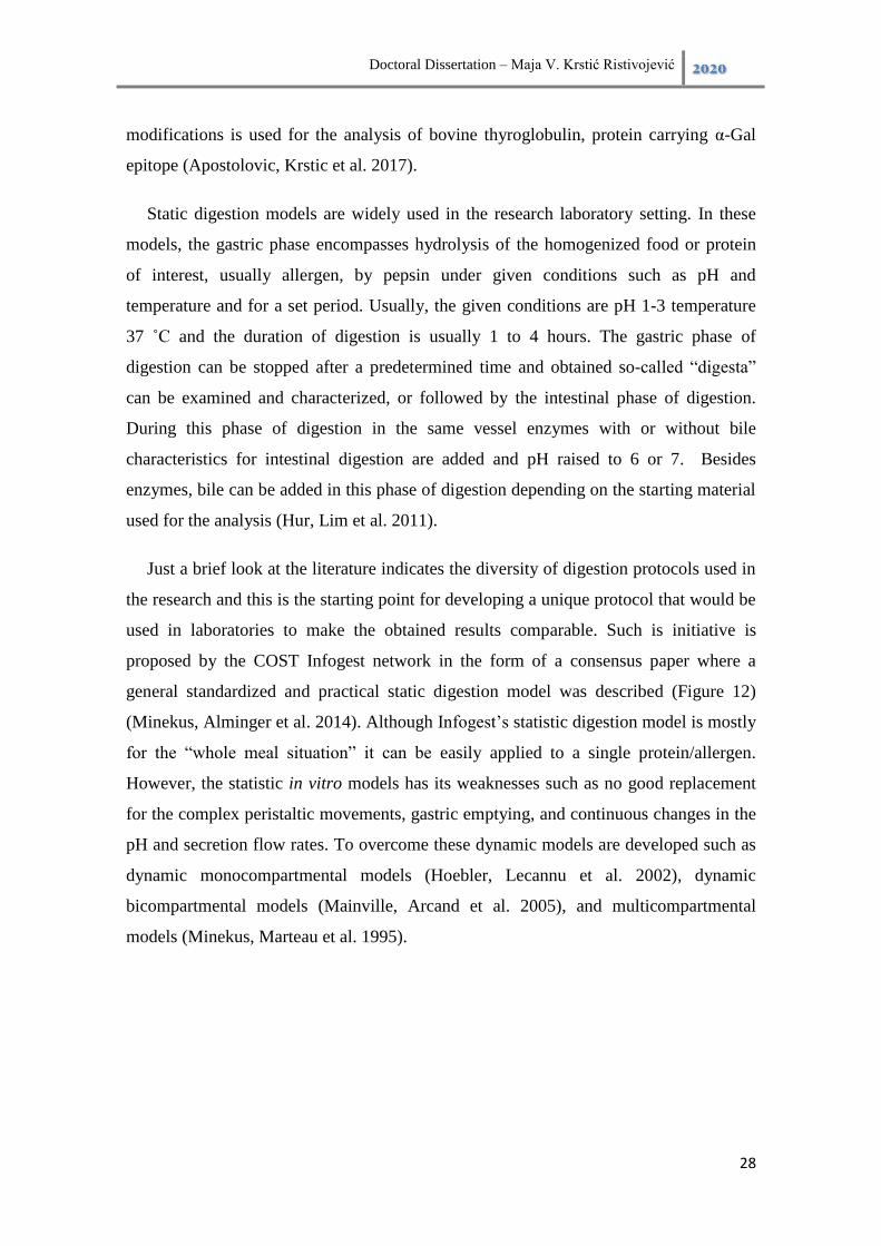
Doctoral Dissertation – Maja V. Krstić Ristivojević 2020
28
modifications is used for the analysis of bovine thyroglobulin, protein carrying α-Gal
epitope (Apostolovic, Krstic et al. 2017).
Static digestion models are widely used in the research laboratory setting. In these
models, the gastric phase encompasses hydrolysis of the homogenized food or protein
of interest, usually allergen, by pepsin under given conditions such as pH and
temperature and for a set period. Usually, the given conditions are pH 1-3 temperature
37 ˚ and the duration of digestion is usually 1 to 4 hours. The gastric phase of
digestion can be stopped after a predetermined time and obtained so-called “digesta”
can be examined and characterized, or followed by the intestinal phase of digestion.
During this phase of digestion in the same vessel enzymes with or without bile
characteristics for intestinal digestion are added and pH raised to 6 or 7. Besides
enzymes, bile can be added in this phase of digestion depending on the starting material
used for the analysis (Hur, Lim et al. 2011).
Just a brief look at the literature indicates the diversity of digestion protocols used in
the research and this is the starting point for developing a unique protocol that would be
used in laboratories to make the obtained results comparable. Such is initiative is
proposed by the COST Infogest network in the form of a consensus paper where a
general standardized and practical static digestion model was described (Figure 12)
(Minekus, Alminger et al. 2014). Although Infogest’s statistic digestion model is mostly
for the “whole meal situation” it can be easily applied to a single protein/allergen.
However, the statistic in vitro models has its weaknesses such as no good replacement
for the complex peristaltic movements, gastric emptying, and continuous changes in the
pH and secretion flow rates. To overcome these dynamic models are developed such as
dynamic monocompartmental models (Hoebler, Lecannu et al. 2002), dynamic
bicompartmental models (Mainville, Arcand et al. 2005), and multicompartmental
models (Minekus, Marteau et al. 1995).

Doctoral Dissertation – Maja V. Krstić Ristivojević 2020
29
Figure 12. Flow diagram of a simulated in vitro digestion method by Minekus and
coworkers. SSF-Simulated Salivary Fluid, SGF-Simulated Gastric Fluid, and SIF-
Simulated Intestinal. Enzyme activities are in units per mL of the final digestion mixture
at each corresponding digestion phase.11
11
Modified and reprinted from Minekus, M., M. Alminger, P. Alvito, S. Ballance, T. Bohn, C. Bourlieu, F. Carrière, R. Boutrou, M. Corredig, D. Dupont, C. Dufour, L. Egger, M. Golding, S. Karakaya, B. Kirkhus, S. Le Feunteun, U. Lesmes, A. Macierzanka, A. Mackie, S. Marze, D. J. McClements, O. Ménard, I. Recio, C. N. Santos, R. P. Singh, G. E. Vegarud, M. S. J. Wickham, W. Weitschies and A. Brodkorb (2014). "A standardised static in vitro digestion method suitable for food – an international consensus." Food & Function 5(6): 1113-1124.
Doctoral Dissertation – Maja V. Krstić Ristivojević 2020
30
The cell-based models for monitoring of transport of food proteins and/or
allergens as well as other nutrients, and very important drugs are widely used. These
experimental models are crucial in the initial phase of the clinical trials and to help to
reduce animal experimentation. Caco-2 cells (cancer cell line), which mimic the
intestinal epithelial cells and forming the intestinal-like barrier under certain growing
conditions, are a gold standard model for evaluation of transport through the intestinal
epithelium (Hubatsch, Ragnarsson et al. 2007). Their phenotype is well characterized
and they are widely used for the analyses of drug transport (Sun, Chow et al. 2008) or
transport of food allergens (Moreno, Rubio et al. 2006, Thierry, Bernasconi et al. 2009,
Price, Ackland et al. 2014). Another cancer cell line widely used in “gut-mimicking”
experiments is T-84 (Raffatellu, Wilson et al. 2005, Tran, Gomez et al. 2009). The main
characteristic of these two cancer cell lines is forming polarized monolayer and
exhibition of microvilli formation. Also for transport experiments these cells are
commonly grown on semi-permeable Transwell® inserts. Beside tremendous
advantages in the first place easy maintenance, cultivation, and reproducibility, these
cell-based model system have some limitations. It is primarily a lack of mucus-secreting
goblet cells in the intestinal-like barrier as well as the expression of different
transporters and metabolic proteins depending on the applied culture conditions. To
overcome some of these obstacles recently the primary human intestinal epithelial cells
(hInEpCs) have become recently commercially available. As these cells are more
difficult to handle, there is only a limited amount of data based on these cells
(Kauffman, Gyurdieva et al. 2013). Also, the hInEpCs line is the basis for the
development of novel, in vivo-like organoid molecules. The organoids represent the
respective native organs in their structure and function features. For this purpose adult
intestinal stem cells are directly isolated from human biopsies and this system can be
maintained even for several months without significant phenotype changes (Sato, Vries
et al. 2009, Sato, Stange et al. 2011, Schweinlin, Wilhelm et al. 2016).

Doctoral Dissertation – Maja V. Krstić Ristivojević 2020
31
3. Aims
Within this doctoral dissertation, several aims have been formulated:
a) To examine the effect of the α-Gal epitope on the protein surface on its susceptibility
to gastric digestion in the pepsin in vitro digestion model.
b) To examine the effect of the α-Gal epitope on protein transport through the
monolayer of Caco-2 cells, a model system of the intestinal epithelium.
c) To examine the effect of the α-Gal epitope on protein uptake and processing in the
immature monocyte-derived dendritic cells.

Doctoral Dissertation – Maja V. Krstić Ristivojević 2020
32
4. Pepsin digestion of α-Gal glycosylated bovine serum albumin
4.1. Introduction
Although in recent years resistance to gastrointestinal digestion of food allergens as
one of the markers of allergenicity was questioned it is still an inevitable tool in
assessing the allergenicity, especially for novel food allergens (Bøgh and Madsen
2016). According to our knowledge, there is a lack of data in the literature on the
digestibility of proteins carrying the α-Gal epitope. Also, it is very hard to compare
results obtained in different studies dealing with resistance to gastrointestinal digestion
since different in vitro digestion protocols have been applied. To avoid these obstacles
Minekus and coworkers (Minekus, Alminger et al. 2014) proposed a consensus model
for simulated in vitro gastrointestinal digestion which was primarily intended for whole
meal digestion simulation but can also be applied to individual proteins/allergens.
The α-Gal-containing proteins from different beef preparations (raw, medium rare,
fried, and boiled beef) have been identified and their potential allergenicity was
investigated by monitoring the binding of IgE antibodies from meat-allergic patients
(Apostolovic, Tran et al. 2014). Although various α-Gal carrying proteins have been
identified little is known about their bioaccessibility during gastrointestinal digestion.
This is important especially in terms of the food matrix which can have a tremendous
impact on the bioaccessibility of proteins from food (Vissers Y.M., Wichers H.J. et al.
2012). Besides α-Gal caring protein, protein without this epitope or deglycosylated
protein is required for an in-depth investigation of the influence of α-Gal epitope on
protein resistance to digestion and pattern of the released digestion peptides.
Bovine thyroglobulin (bTG) is a naturally occurring protein that possess α-Gal
epitope as a posttranslational modification and it is abundantly present in red meat
(Rawitch, Pollock et al. 1993). Apostolovic, Krstic et al. (2017) performed the
simulated in vitro gastrointestinal digestion of bTG using protocols recommended by
Moreno, Mellon et al. (2005) and Koppelman, Hefle et al. (2010) and found that α-Gal
peptides were present during the whole gastric phase of protein digestion. It was also
confirmed that there was not IgE binding to digested peptides if they were
deglycosylated. Besides this, another important finding was that at the acidic pH

Doctoral Dissertation – Maja V. Krstić Ristivojević 2020
33
conditions deglycosylated bTG was insoluble which makes gastric digestion impossible
and thus limiting the use of the deglycosylated bTG as the α-Gal negative control
(Apostolovic, Krstic et al. 2017).
All previously mentioned point out the need for the studies dealing with the
gastrointestinal digestion of red meat as well as individual α-Gal-containing proteins.
Also, it is very important in the research that some consensus models be adopted and
followed in conducting in vitro studies of bioaccessibility of food proteins and
simulated gastrointestinal digestion of individual proteins/allergens. Taking into account
previously mentioned one of the aims of this doctoral dissertation was to check the
usability of the simulated in vitro pepsin digestion for the monitoring digestion of α-Gal
containing food allergens using α-Gal glycosylated bovine serum albumin (BSA-α-Gal),
as a model protein.
4.2. Methodology
4.2.1. Reagents
Used proteins were commercially available. BSA and pepsin were obtained from
Sigma-Aldrich (Merck, Steinheim, Germany). α-Gal glycosylated BSA (BSA-α-Gal)
and N-Acetyllactosamine conjugated BSA (BSA-NAl) were obtained from Dextra
Laboratories (Reading, UK). Other chemicals used in the experiments were p.a. grade
and obtained from Sigma-Aldrich unless stated otherwise.
4.2.2. Protein sample preparation
Used proteins were fluorescently labeled by conjugating AlexaFluor488 (AF488)
fluorescent dye using an AF488 protein labeling kit (Invitrogen, Molecular Probes, Inc.,
Eugene, Oregon, USA) according to the manufacturer’s instructions. For determination
of protein concentration commercial kit Pierce Bicinchoninic acid (BCA) Protein Assay
Kit (Pierce Biotechnology, Rockford, Illinois, USA) was used.
4.2.3. In vitro gastric digestion
“For in vitro gastric digestion the statistic digestion model proposed by Minekus,
Alminger et al. (2014) was used with slight modification. The carbohydrate conjugated

Doctoral Dissertation – Maja V. Krstić Ristivojević 2020
34
proteins BSA-α-Gal, BSA-NAl, and unconjugated BSA fluorescently labeled with
AF488 were pre-incubated at 37 °C for 15 min before pepsin in simulated gastric fluid
(SGF) was added in a 1:3 (volume protein/volume SGF with pepsin) ratio. In vitro
digestion protocol was adjusted to single protein digestion so the final enzyme activity
was 8 U pepsin per mg of protein. The recipe and calculation for the preparation of 4 ×
SGF are presented in Table 1. The pH of the digestion mixture was adjusted to 3 by the
addition of 1 M HCl. Simulated gastric digestion was monitored for up to 4 h at 37 °C,
and aliquots were withdrawn from a single digestion mixture at different time points for
further analysis. The digestion was stopped by raising the pH of the digestion mixture to
7.5 by adding 1 M NaHCO3.” (Krstic Ristivojevic M., Grundström J. et al. 2020)
Table 1. Preparation of stock solution of simulated gastric fluid (SGF). The masses
of substances are calculated to prepare 200 mL 4 × concentrate SGF solution.
SGF pH 3
Constituent 4 × SGF* Final conc. in
SGF
g mmol/L
KCl 0.41 6.90
KH2PO4 0.10 0.90
NaHCO3 1.68 25.00
NaCl 2.21 47.20
MgCl2(H2O)6 0.02 0.10
(NH4)2CO3 0.08 0.50
CaCl2(H2O)2 0.02 0.15
For pH adjustment
mL (12Ma) mmol/L
HCl 1.04 15.6
*to prepare 200 mL of 4 × SGF solution a molarity of a stock solution
4.2.4. SDS PAGE analysis
For analysis of digestion products of BSA with or without glycosylation sodium
dodecyl sulfate-polyacrylamide gel electrophoresis (SDS PAGE) according to Laemmli
(1970) under reducing conditions was performed. The digestion products of BSA with
or without glycosylation were resolved on the 14% polyacrylamide (PAA) hand-cast

Doctoral Dissertation – Maja V. Krstić Ristivojević 2020
35
gels and analysis were carried out using a Hoefer Scientific Instruments apparatus
(Holliston, Massachusetts, USA) with a discontinuous buffer system. The resolved PAA
gels were stained using Coomassie Brilliant Blue R-250 and scanned using the Typhoon
FLA 7000 laser scanner (GE Healthcare, Freiburg, Germany).
4.2.5. Immunoblot for detection of the BSA and α-Gal epitope
“Bovine serum albumin and α-Gal in BSA-α-Gal digests were detected by
immunoblot analysis. Previously resolved proteins by SDS PAGE were transferred to
polyvinylidene difluoride (PVDF) membranes (0.2 µm pore size) using an EBU-4000
Semi-Dry Blotting System (Fisher Scientific Company L.L.C., Pittsburgh,
Pennsylvania, USA). For the immunodetection of in vitro pepsin-digests, peptides of
BSA-α-Gal, BSA-NAl, and BSA the membranes were probed with the rabbit polyclonal
anti-BSA IgG antibody (Invitrogen, A11133) diluted 1:1000 in 0.1% human serum
albumin (HSA) for 1.5 h at room temperature (RT) followed by goat anti-rabbit IgG
conjugated to alkaline phosphatase (AP) (ab97072, Abcam, Cambridge, Massachusetts,
USAab97072) diluted 1:5000 in 0.1% HSA for 1 h at RT. Immunoreactive bands were
visualized using nitro blue tetrazolium (NBT) and 5-bromo-4-chloro-3-indolyl
phosphate (BCIP) substrates. For the immunodetection of α-Gal on peptides of BSA-α-
Gal obtained upon simulated in vitro digestion the membranes were probed with
monoclonal mouse anti-α-Gal antibody M86 (Enzo Life Sciences, Farmingdale, New
York, USA) diluted 1:5 in 0.1% HSA in Tris-buffered saline – Tween 20 (TBS-T) for 3
h followed by AP-labeled goat anti-mouse IgM antibody (Southern Biotech,
Birmingham, Alabama, USA) diluted 1:3000 in TBS-T for 1 h at room RT. Reactive
bands were visualized as stated above.” (Krstic Ristivojevic M., Grundström J. et al.
2020)
4.3. Results
4.3.1. Pepsin digestion pattern of BSA, BSA-α-Gal, and BSA-NAl
The assessment of the stability of a protein or its proteolytic fragments during
pepsin digestion is an important parameter in sensitization in food allergy. Hence, it is
of importance to examine whether the α-Gal glycosylation of BSA has any impact on its
resistance to pepsin digestion and profile of the proteolytic fragments. In the first

Doctoral Dissertation – Maja V. Krstić Ristivojević 2020
36
experiment of this thesis, the difference in molecular weight between molecules of BSA
before and after glycosylation was checked. The pure proteins, BSA (control) and with
α-Gal glycosylation (BSA-α-Gal) were separated onto 12 % PAA gel using SDS-PAGE
(Figure 13).
Figure 13. Fluorescent detection of AF488 labeled BSA and BSA-α-Gal resolved on
the 12 % PAA gel after SDS-PAGE.
To examine the influence of glycosylation of BSA on its resistance to pepsin
digestion, the pepsin digestion profiles of BSA-α-Gal, BSA-NAl and BSA were
compared using SDS PAGE under reducing conditions. Obtained gels were analyzed by
staining with CBB R250 or by immunoblot using a polyclonal anti-BSA antibody
(Figure 14 and 17). Besides this, to investigate the content of the α-Gal epitope on the
released peptides during digestion of BSA-α-Gal by pepsin, the obtained gel was
additionally analyzed by immunoblot using mouse monoclonal anti-α-Gal antibody
M86 (Figure 16). To eliminate the additional bands in the digestion profiles of analyzed
BSA which can be originated from pepsin during its auto-digestion or the presence of
impurities, a sample of pure pepsin was also analyzed at 0 and 4 h from the beginning of
digestion (Figure 15).
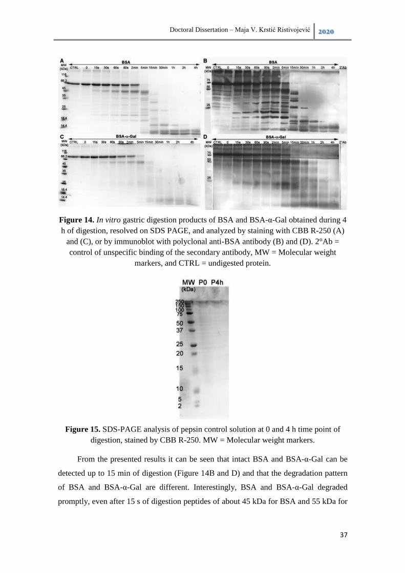
Doctoral Dissertation – Maja V. Krstić Ristivojević 2020
37
Figure 14. In vitro gastric digestion products of BSA and BSA-α-Gal obtained during 4
h of digestion, resolved on SDS PAGE, and analyzed by staining with CBB R-250 (A)
and (C), or by immunoblot with polyclonal anti-BSA antibody (B) and (D). 2°Ab =
control of unspecific binding of the secondary antibody, MW = Molecular weight
markers, and CTRL = undigested protein.
Figure 15. SDS-PAGE analysis of pepsin control solution at 0 and 4 h time point of
digestion, stained by CBB R-250. MW = Molecular weight markers.
From the presented results it can be seen that intact BSA and BSA-α-Gal can be
detected up to 15 min of digestion (Figure 14B and D) and that the degradation pattern
of BSA and BSA-α-Gal are different. Interestingly, BSA and BSA-α-Gal degraded
promptly, even after 15 s of digestion peptides of about 45 kDa for BSA and 55 kDa for
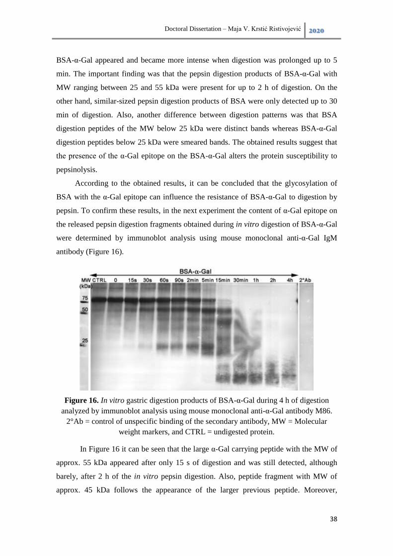
Doctoral Dissertation – Maja V. Krstić Ristivojević 2020
38
BSA-α-Gal appeared and became more intense when digestion was prolonged up to 5
min. The important finding was that the pepsin digestion products of BSA-α-Gal with
MW ranging between 25 and 55 kDa were present for up to 2 h of digestion. On the
other hand, similar-sized pepsin digestion products of BSA were only detected up to 30
min of digestion. Also, another difference between digestion patterns was that BSA
digestion peptides of the MW below 25 kDa were distinct bands whereas BSA-α-Gal
digestion peptides below 25 kDa were smeared bands. The obtained results suggest that
the presence of the α-Gal epitope on the BSA-α-Gal alters the protein susceptibility to
pepsinolysis.
According to the obtained results, it can be concluded that the glycosylation of
BSA with the α-Gal epitope can influence the resistance of BSA-α-Gal to digestion by
pepsin. To confirm these results, in the next experiment the content of α-Gal epitope on
the released pepsin digestion fragments obtained during in vitro digestion of BSA-α-Gal
were determined by immunoblot analysis using mouse monoclonal anti-α-Gal IgM
antibody (Figure 16).
Figure 16. In vitro gastric digestion products of BSA-α-Gal during 4 h of digestion
analyzed by immunoblot analysis using mouse monoclonal anti-α-Gal antibody M86.
2°Ab = control of unspecific binding of the secondary antibody, MW = Molecular
weight markers, and CTRL = undigested protein.
In Figure 16 it can be seen that the large α-Gal carrying peptide with the MW of
approx. 55 kDa appeared after only 15 s of digestion and was still detected, although
barely, after 2 h of the in vitro pepsin digestion. Also, peptide fragment with MW of
approx. 45 kDa follows the appearance of the larger previous peptide. Moreover,

Doctoral Dissertation – Maja V. Krstić Ristivojević 2020
39
peptide fragments with MW of approx. 37, 32, and 25 kDa carrying α-Gal were detected
after 1 h.
To gain a better insight into the effect of the α-Gal epitope on the pepsin
digestion of the α-Gal containing proteins we performed in vitro pepsin digestion of
BSA-NAl, which possesses a carbohydrate modification similar in size to α-Gal (Figure
17). According to the obtained results presented in Figure 17 this carbohydrate
modification of BSA has a significant impact on its resistance to pepsinolysis. Intact
BSA-NAl was still detectable after 15 min of digestion compared to BSA which was,
although barely, detectable up to 5 min of digestion (Figure 14). On the other hand,
immunoblot detection using a polyclonal anti-BSA antibody revealed that the intact
protein was present for up to 2 h of in vitro digestion (Figure 17B). Moreover peptide
fragments with MW of approx. 55, 45, 37, 32, and 25 kDa were detected even after 4 h
of in vitro pepsin digestion.
Figure 17. In vitro gastric digestion products of BSA-NAl obtained during 4 hours of
digestion, resolved on SDS PAGE, and analyzed by staining with CBB R-250 (A), or by
immunoblot with polyclonal anti-BSA antibody (B). 2°Ab = control of unspecific
binding of the secondary antibody, MW = Molecular weight markers, and CTRL =
undigested protein.
Different systems for detection which are more or less sensitive were used for
the analysis of obtained peptide fragments during digestion of different BSA molecules
by pepsin. In Figure 18 the obtained digestion patterns of the AF488 labeled BSA,
BSA-α-Gal, and BSA-NAl using fluorescent detection are shown. Obtained digestion
patterns of proteins are in correlation with the results obtained with the above-
mentioned detection systems such as CBB R-250 and immunoblot analysis using a
chromogenic NBT/BCIP substrate. The intact BSA was visible up to 5 min of
pepsinolysis while BSA-α-Gal and BSA-NAl were visible up to 15 min of pepsinolysis.

Doctoral Dissertation – Maja V. Krstić Ristivojević 2020
40
Also, the presence of the α-Gal epitope on the BSA-α-Gal peptides and NAl on the
BSA-NAl peptides is the probable reason for the obtained smeared bands whereas BSA
peptides were distinct on the resolved PAA gels.
Figure 18. In vitro gastric digestion products of (A) BSA, (B) BSA-α-Gal, and (C)
BSA-NAl obtained during 4 hours of digestion, resolved on SDS PAGE and analyzed
by fluorescent detection using AF488 fluorescent dye. MW = Molecular weight
markers, and CTRL = undigested protein.

Doctoral Dissertation – Maja V. Krstić Ristivojević 2020
41
4.4. Discussion
Glycosylation a posttranslational modification of proteins and lipids contributes to
the complexity of the biological structures available in living organisms. This
modification is especially important for the proteins which are the key players in a
broad spectrum of biological processes (Maureen E. Taylor and Drickamer 2011).
Although carbohydrates as such are mainly not considered as potent inducers of allergic
reactions, α-Gal carbohydrate proves the opposite. The alpha-Gal epitope is present in
beef proteins and even food processing such as cooking does not influence its
allergenicity (Apostolovic, Tran et al. 2014).
The obtained results demonstrated that the digestion stability of proteins and the
pattern of the resulting peptides was influenced by protein glycosylation, one of the
most common posttranslational modifications (Krstic Ristivojevic M., Grundström J. et
al. 2020). One of the findings is that the type of glycosylation influence the protein
susceptibility to pepsinolysis, α-Gal glycosylation increased the resistance of BSA to
pepsinolysis, but NAl glycosylation hampered pepsinolysis even more. “Intact BSA-α-
Gal, similar to BSA, could be detected up to 15 min of in vitro pepsin digestion,
whereas intact BSA-NAl could be detected for up to 2 h. These findings are following a
previous study that demonstrated that glycosylation protects the egg white allergen
ovomucoid from pepsinolysis (Benede, Lopez-Fandino et al. 2013). The major impact
of protein glycosylation was observed in the digestion patterns of BSA-α-Gal compared
to BSA, large digestion products of BSA-α-Gal ≥ 25 kDa were present for up to 2 h,
which was not seen for BSA. Moreover, in the lower molecular weight region where
BSA-α-Gal gave rise to weak smeared peptide bands, BSA generated well-defined
peptide bands. Immunoblot analysis of α-Gal containing proteins upon gastric digestion
resulted in a rich spectrum of peptides abundant in α-Gal epitopes, where α-Gal epitope
was detected on the large peptides even after 2 h of in vitro pepsin digestion. Although
faintly stained, the major peptides carrying the α-Gal epitope, detected at molecular
weights between 25 and 55 kDa provide a prolonged survival of proteins during gastric
digestion and a prolonged availability for uptake by intestinal cells.” (Krstic
Ristivojevic M., Grundström J. et al. 2020) This is of great importance since it was
shown that α-Gal containing peptides resulting from pepsinolysis are allergenic
(Apostolovic, Krstic et al. 2017).
Doctoral Dissertation – Maja V. Krstić Ristivojević 2020
42
5. Alpha-Gal on the protein surface hampers transcytosis through
the Caco-2 monolayer
5.1. Introduction
“There is an evident lack of literature dealing with the fate of the α-Gal carrying
proteins in the gastrointestinal tract, including processing, uptake, and transport of α-
Gal carrying proteins by the intestinal epithelium”. (Krstic Ristivojevic M., Grundström
J. et al. 2020) For consequential examination of bioaccessibility of food proteins and
allergens and their peptides except in vitro simulated gastrointestinal digestion, there are
established cell-based in vitro models for monitoring protein transport through the
intestine. “Enterocytes of the intestinal epithelium are the most important cells for
absorbing nutrients from food and at the same time excluding antigenic macromolecules
and microbes from crossing the barrier. The human colon carcinoma cell-line Caco-2 is
used as a model system of the intestinal epithelium. When cultured on permeable
supports, the cells differentiate into a monolayer that is similar to the small intestinal
villus epithelium and this monolayer expresses brush border proteins, tight junctions
between cells, and is highly polarized (Hubatsch, Ragnarsson et al. 2007, Sun, Chow et
al. 2008). The Caco-2 cell monolayer has been used for investigation of transport of
food allergens (Moreno, Rubio et al. 2006, Thierry, Bernasconi et al. 2009, Tordesillas,
Gomez-Casado et al. 2013, Price, Ackland et al. 2014). Also, the Caco-2 in vitro model
system has been applied to show that allergens can be resistant to digestion and
maintain their allergenicity after passing the epithelial barrier (Tordesillas, Gomez-
Casado et al. 2013).” (Krstic Ristivojevic M., Grundström J. et al. 2020) Importantly,
intestinal cells form one of the most important lines of defense which are exposed to
microbes daily. Hence, in recent years more and more studies revealed that enterocytes
are in constant communication with the surrounding cells such as cells of the immune
system which are underlying the intestinal barrier. “Pathogen-derived glycoconjugates
can be sensed by mammalian lectins, which are of importance in both innate and
acquired immune responses. Galectin-3, the chimera type galectin, can bind to α-Gal
(Peterson, Jin et al. 2005, Jin, Greenwald et al. 2006) and is abundantly expressed by
intestinal epithelial cells (Dumic, Dabelic et al. 2006). Furthermore, galectin-3 is
upregulated in myeloid and epithelial cells upon microbial stimulation and has mainly

Doctoral Dissertation – Maja V. Krstić Ristivojević 2020
43
pro-inflammatory properties (Diaz-Alvarez and Ortega 2017)”.(Krstic Ristivojevic M.,
Grundström J. et al. 2020)
The main goal of this part of the thesis was to investigate the impact of the α-Gal
epitope on the protein surface and for the transport rate through the monolayer of
enterocytes using the Caco-2 monolayer model system. BSA and HSA without and with
bound α-Gal epitope (BSA-α-Gal and HSA-α-Gal, respectively) were used as the model
protein for this study.
5.2. Methodology
5.2.1. Reagents
Proteins were commercially available. BSA and HSA were obtained from Sigma-
Aldrich (Merck, Steinheim, Germany). α-Gal glycosylated BSA (BSA-α-Gal), HSA
(HSA-α-Gal), and N-Acetyllactosamine conjugated BSA (BSA-NAl) were obtained
from Dextra Laboratories (Reading, UK). Other chemicals used in experiments were
p.a. grade and obtained from Sigma-Aldrich unless is stated otherwise.
5.2.2. Protein sample preparation
Used proteins were fluorescently labeled by conjugating AlexaFluor488 (AF488)
fluorescent dye using an AF488 protein labeling kit (Invitrogen, Molecular Probes, Inc.,
Eugene, Oregon, USA) according to the manufacturer’s instructions. For determination
of protein concentration commercial kit Pierce Bicinchoninic acid (BCA) Protein Assay
Kit (Pierce Biotechnology, Rockford, Illinois, USA) was used.
5.2.3. Cell culture and viability assay
Caco-2 (HTB37) cell line was obtained from The American Type Culture Collection
(ATCC, Manassas, Virginia, USA). Caco-2 cells were cultured in complete Minimum
Essential medium (cMEM) supplemented with 10% of fetal bovine serum (FBS), 2 mM
glutamine, 1% of nonessential amino acids (NEAA), 100 IU/mL penicillin, 0.1 mg/mL
streptomycin, and 50 µg/mL gentamicin. Cells were incubated at 37 °C in a humidified
atmosphere with 5% CO2. Caco-2 cells viability in serum-free media was evaluated by
MTT assay 1, 4, and 24 h after the media exchange, according to a slightly modified

Doctoral Dissertation – Maja V. Krstić Ristivojević 2020
44
procedure of Mosmann (1983). Briefly, Caco-2 cells were seeded in 96-well plates at a
density of 10 000 cells per well and cultured for the next 21 days to allow the forming
of the cell monolayer. On the 21st day of culturing, cells were thoroughly washed with
PBS and incubated with MEM medium supplemented with 2 mM glutamine, 1% of
NEAA, 100 IU/mL penicillin, 0.1 mg/mL streptomycin, and 50 µg/mL gentamicin but
without FBS for the next 1, 4 and 24 h. MEM with 10% FBS and mentioned
supplements were added to the cells in the control wells. The cell culture medium was
added to blank wells. After the indicated period, 20 µL of MTT solution (5 mg/mL in
phosphate-buffered saline, PBS) were added to each well and incubated for the next 4 h
at 37°C. The medium was carefully removed and 200 μL of dimethyl sulfoxide
(DMSO) was added to dissolve the formed formazan complexes; absorbance was read
at 492 nm. Data are expressed as the percentage of viability with untreated cells taken as
100%.
5.2.4. Transport through the Caco-2 monolayer
“For transport studies, the Caco-2 cells at passages of 23–68 were seeded at 6 × 10⁴
cells/cm2 (Behrens and Kissel 2003) on cell culture inserts for 12-well plates (ThinCert,
Greiner Bio-One GmbH, Frickenhausen, Germany) and cultured for 21 days before the
start of the experiments. The medium was replaced three times per week. Before the
transport studies and at the end of the experiment, transepithelial electrical resistance
(TEER) was measured using a Millicell-ERS VoltOhmmeter (Millipore, Amsterdam,
Netherlands). Only cell monolayers with TEER > 300 Ωcm2 were used and none of the
added proteins had any effect on the TEER. Transport studies were started by adding
400 µg of AF488-labeled proteins in 500 µL (0.8 mg/mL) complete MEM (cMEM) to
the apical compartment. Aliquots were withdrawn from the basolateral compartment
after 1, 2, and 4 h of incubation, and the fluorescence was measured using a FluoroMax-
4 spectrofluorometer (Horiba Scientific, Kyoto, Japan). Protein concentration was
calculated from standard curves of the corresponding protein, with the limit of detection
(LOD) of 0.011 µg/mL (LOD = 3Sa/b, where Sa is the standard deviation of the
response and b is the slope of the calibration curve).”(Krstic Ristivojevic M.,
Grundström J. et al. 2020)

Doctoral Dissertation – Maja V. Krstić Ristivojević 2020
45
5.2.5. Intracellular visualization of proteins and isolation of the endosomal
fraction of the Caco-2 cell monolayer
“To visualize the internalization of AF488-labeled proteins, confocal laser scanning
microscopy (CLSM) was employed (Vukojevic, Heidkamp et al. 2008). Isolation of the
endosomal fraction of Caco-2 cell lysates was performed as previously described (Mao,
Ou et al. 2016) with slight modification. Briefly, Caco-2 cells were cultured in the same
conditions as for transport studies in 6-well plates. Before the addition of treatments, the
monolayers were washed 3 times with PBS, and the cultivating medium was replaced
with a medium without FBS. Caco-2 cell monolayers were incubated with 100 µg of
AF488-labeled BSA or BSA-α-Gal for 4 and 24 h, unstimulated cells were used as
control. After incubation, the cells were washed 3 times with PBS and detached from
the culture inserts using an ice-cold PBS-EDTA solution. The cell pellets were washed
3 times with PBS and lysed with lysis buffer (250 mM sucrose, 50 mM Tris-Cl (pH
7.4), 5 mM MgCl2, 1 mM EDTA, 1 mM egtazic acid (EGTA)) with a protease inhibitor
cocktail (Sigma-Aldrich). The suspended cell lysates were left overnight at −80 ° . The
next day, endosomes were harvested in the third pellet after three centrifugation steps of
the supernatant: 1st (1000× g, 10 min, performed twice and the supernatants were
pooled), 2nd (16 000× g, 20 min), and 3rd (100 000× g, 1 h) for immunoblot analysis.
All centrifugation steps were performed at 4 °C, and for the last ultracentrifugation step
a Sorvall WX80 ultracentrifuge (Thermo Electron LED GmbH, Langenselbold,
Germany) with T-880 rotor was used.”(Krstic Ristivojevic M., Grundström J. et al.
2020)
5.2.6. Statistical analysis
“Data are presented as mean ± standard deviation (SD) and were analyzed using
GraphPad Prism software version 6.04 software for Windows (La Jolla, California,
www.graphpad.com). Each sample was analyzed in duplicate with two biological
replicates. The differences were analyzed by two-way ANOVA. The transport rates
were calculated by linear regression. Differences were considered significant if p <
0.05.” (Krstic Ristivojevic M., Grundström J. et al. 2020)

Doctoral Dissertation – Maja V. Krstić Ristivojević 2020
46
5.3. Results
5.3.1. Parameters which indicate the quality of the Caco-2 monolayer
Till today FBS supplementation is practically inevitable in culture media used for
maintenance of almost all cell culture lines. FBS with its very complex composition
provides optimal conditions for cell culture growth, BSA is one of the most represented
components of FBS. Because BSA present in the cell culture media can interfere with
the transport of BSA-α-Gal through the Caco-2 monolayer, it was necessary to reduce
its presence in media, but this step can cause the disturbance of optimal Caco-2 cell
viability. Hence, the testing of the viability of cells used in in vitro Caco-2 cell-based
model in conditions of the reduced quantity of FBS in culture media was the first
experiment in this part of the thesis. The results of this experiment are presented in
Figure 19.
Figure 19. The vitality of Caco-2 cells was determined after 21 days of
differentiation and subsequently, Caco-2 cells were maintained for 1, 4, and 24 h in the
medium without FBS. CTRL-Cells in the control wells were maintained in the medium
with FBS.
From Figure 19 it can be concluded that the maintenance of Caco-2 monolayer in
the medium without FBS does not influence the vitality of the established Caco-2
monolayer. This was preferably for the study of the transport of model proteins since
any disruption of the monolayer could lead to leakage of the test protein to the
basolateral compartment of the transwell insert.

Doctoral Dissertation – Maja V. Krstić Ristivojević 2020
47
Another important parameter that indicates the quality of established cell monolayer
is the value of TEER. It was adopted that the value of TEER bigger than 300 Ω/cm2
indicates that the Caco-2 monolayer is of satisfactory quality. Also, TEER must be
measured during the period of differentiation cell, before and after transport
experiments.
Figure 20. TEER values of Caco-2 monolayer obtained on the 21st day of cell
culturing before transport experiment (21DayBE) and after transport experiment
(21DayAE), CTRL – untreated Caco-2 monolayer.
Figure 20 presents the TEER values of Caco-2 monolayer obtained before and after
transport experiments. The variations in obtained values between 300 and 500 Ω/cm2
among used monolayers were expected due to the dynamics and complexity of the used
system. Interestingly in some transwell inserts the measured TEER values were higher
after the transport experiment, while in others were lower than values obtained before
the experiment. In all cases obtained values of TEER after transport experiments were
higher than 300 Ω/cm2 which indicates that transport of proteins did not disturb the
established Caco-2 cell monolayer.

Doctoral Dissertation – Maja V. Krstić Ristivojević 2020
48
5.3.2. Effect of glycosylation of proteins on their transport through the Caco-2
monolayer
In recent years it becomes evident that protein and allergen transport through
intestinal epithelia is also an important parameter in sensitization and food allergy
induction. For this purpose as one of the aims of this thesis, in vitro model system based
on Caco-2 cells was established which was used for the monitoring and comparison of
the transport of different proteins (BSA and HSA) which bearing α-Gal epitope (BSA-
α-Gal and HSA-α-Gal) through the monolayer of these cells during 4 h of the
observation (Figure 21). Their transport rates were compared with the transport rates of
appropriate controls (BSA and HSA), and with the BSA-NAl which carbohydrates'
residues are similar to α-Gal (Table 2).
Table 2. The average transport rates (ng/h) of the different proteins through the
Caco-2 monolayer.
Protein Transport rate
(ng/h) 95 % CI r
2† p-value
‡
BSA 142.8 103.9 – 181.7 0.87 ˂ 0.001
BSA-α-Gal 77.2 53.1 – 101.3 0.84 ˂ 0.001
HSA 158 117.2 – 198.7 0.88 ˂ 0.001
HSA-α-Gal 78.8 61.0 – 96.6 0.91 ˂ 0.001
BSA-NAl 135.0 83.4 – 186.6 0.77 ˂ 0.001
† Goodness of fit, ‡ if the slope is different from zero

Doctoral Dissertation – Maja V. Krstić Ristivojević 2020
49
Figure 21. Transport (ng) of proteins carrying α-Gal through the intestinal Caco-2
monolayer. (A) Time-dependent transcytosis of BSA-α-Gal and BSA, n = 4, and (B)
human serum albumin (HSA) carrying α-Gal (HSA-α-Gal) and HSA, n = 4. * p < 0.05,
** p < 0.01 and *** p < 0.001 analyzed by two-way ANOVA with Bonferroni’s post
hoc test; (C) fluorescent detection of BSA-α-Gal and BSA in Caco-2 lysates resolved on
sodium dodecyl sulfate-polyacrylamide gel electrophoresis (SDS PAGE) gel; (D)
immunoblot detection of the α-Gal epitope in lysates from Caco-2. MW = Molecular
weight markers and CTRL = untreated Caco-2 cells.
Fluorescently labeled BSA-α-Gal in the amount of 400 µg was applied on the
apical side of Caco-2 monolayer and after 1 h of incubation 70 ± 19 ng was transported
to the basolateral compartment and after 4 h, the amount of transported protein had
increased to 299 ± 75 ng (Figure 21A). In comparison to glycosylated protein
unconjugated BSA was transported approximately two times more, administrated in the
same amount in the apical compartment of Caco-2 monolayer, 136 ± 39 ng had been
transported after 1 h and 565 ± 113 ng after 4 h of incubation (Figure 21A). Also, the
transport of HSA-α-Gal through Caco-2 cell monolayer was monitored and compared
with the transport of unconjugated HSA (Figure 21B). When 400 µg of HSA-α-Gal was
applied in the apical compartment 72 ± 3 ng was transported in the basolateral
compartment after 1h of incubation and 306 ± 57 ng of protein was transported in the

Doctoral Dissertation – Maja V. Krstić Ristivojević 2020
50
basolateral compartment after 4 h of incubation. The transport of HSA-α-Gal was
similar to that of BSA-α-Gal but significantly lower compared to the transport of HSA.
After 1 h of incubation 132 ± 13 ng of HSA had been transported in the basolateral
compartment when 400 µg of HSA was applied in the apical compartment and 603 ±
141 ng of HSA was transported in the basolateral compartment after 4 h of incubation.
The obtained results showed that the amount of transported proteins was constant over
time which enables the calculation of the average transport rates for investigated
proteins (Table 2). Unconjugated HSA and BSA have the transport rate almost two
times higher (158 and 142.8 ng/h) than HSA-α-Gal and BSA-α-Gal (78.8 and 77.2
ng/h).
The important question was whether transport through the Caco-2 monolayer
depends on the type of glycosylation present on the protein. To answer that question we
compared the transcytosis of BSA-NAl at different time points (Figure 22). When 400
µg of BSA-NAl was applied in the apical compartment of the transwell inserts, 105 ±
11 ng of protein was transported in the basolateral compartment after 1 h of incubation
and 499 ± 167 ng of protein after 4 h of incubation.
Figure 22. Comparison of transcytosis (expressed as an amount of transported protein)
of BSA-α-Gal, BSA, acetyllactosamine (NAl) conjugated to BSA (BSA-NAl) through
the Caco-2 monolayer, n = 4, * p < 0.05, ** p < 0.01 and *** p < 0.001 analyzed by
two-way ANOVA with Bonferroni’s post hoc test.
According to the result from Table 2, it can be seen that the calculated average
transport rate of BSA-NAl (135.0 ng/h) is almost the same as the transport of BSA
(142.8 ng/h). This result indicates that glycosylation alone is not the cause of why BSA-
α-Gal and HSA-α-Gal have two times smaller transport rates than BSA and HSA, but
also the type of glycosylation.

Doctoral Dissertation – Maja V. Krstić Ristivojević 2020
51
To confirm the presence of the intact BSA proteins with and without α-Gal
epitope in the Caco-2 cells, after transport experiments Caco-2 cell monolayers were
collected, cells were lysed and cell lysates were resolved on SDS PAGE. The used PAA
gels were illuminated and fluorescently labeled BSA and BSA-α-Gal were visible
(Figure 21C). The difference in the MW of the resolved fluorescently labeled proteins
suggests that BSA-α-Gal was intact in the Caco-2 cell lysates. For the additional
confirmation of the detection of the α-Gal epitope western blot analysis with a chicken
single-chain variable fragment (ScFV) anti-α-Gal was performed (Figure 21D). In
Figure 21D is a visible band which corresponds to the MW of approx. 75 kDa and it is a
strong confirmation that the α-Gal epitope is still present on the intact protein.
According to the results of this experiment, it can be concluded that the in vitro
developed model system based on Caco-2 cells monolayer can be used for the
investigation of the transport rate of different glycosylated food allergens, as well as
how specific glycosylation can affect this process.
5.3.3. The α-Gal carrying proteins can be detected in endosomes
In the previous experiment, the presence of intact BSA and BSA-α-Gal was
confirmed inside of the Caco-2 cell. In the next experiment, the confocal laser scanning
microscopy (CLSM) was applied for visual confirmation of the presence of the
fluorescently labeled BSA and BSA-α-Gal inside of Caco-2 cells. This analysis also
enables us to get an impression of the spatial distribution of these proteins inside of
cells.
Figure 23. Spatial distribution of fluorescently labeled BSA-α-Gal and BSA with green
fluorescent dye AF488 in Caco-2 cells after 4 h of incubation at 37 ˚C analyzed by
confocal laser scanning microscopy. CTRL- untreated cells with fluorescently labeled
protein.

Doctoral Dissertation – Maja V. Krstić Ristivojević 2020
52
After 4 h of incubation, CLSM analysis reveals the presence of green fluorescent
dye only in the Caco-2 cells which were incubated with BSA-α-Gal. The lack of the
green signal in the control cells as well as in the cells incubated with BSA suggests that
there was no accumulation of the BSA inside cells. Scattered green in the cytosol of
Caco-2 cell, incubated with BSA-α-Gal implicate that protein is still was present in
some small subcellular structures such as endosomes after 4 h of incubation.
Further, for the confirmation of the spatial distribution of proteins inside the
cells, differential centrifugation was used to isolate endosomes from Caco-2 cells.
Again, using fluorescence detection the presence of intact proteins was confirmed in the
cell lysates and subsequently in the endosomal fraction of cell lysates after 4 and 24 h of
incubation of Caco-2 cells with BSA and BSA-α-Gal (Figure 24A and 24B). The
resolved protein content of the endosomal fraction of Caco-2 cell lysates via SDS
PAGE was transferred on the PVDF membrane and probed with a polyclonal anti-BSA
antibody (Figure 25). In the corresponding endosomal fraction of Caco-2 cell lysates,
both BSA and BSA-α-Gal were detected after 4 and 24 h of incubation. Interestingly
BSA was found in both unstimulated Caco-2 cells (CTRL) and BSA-α-Gal stimulated
cells, probably due to its presence in FBS supplement in the culturing medium used
before the transport experiment.
Figure 24. Fluorescent detection of BSA and BSA-α-Gal in (A) Caco-2 lysates resolved
on sodium dodecyl sulfate-polyacrylamide gel electrophoresis (SDS PAGE) gel and (B)
endosomal fraction of cell lysates after 4 and 24 h of incubation of Caco-2 cells with
BSA and BSA-α-Gal. CTRL-untreated Caco-2cells, MW-Molecular weight markers.

Doctoral Dissertation – Maja V. Krstić Ristivojević 2020
53
Figure 25. Immunoblot analysis of an endosomal fraction of cell lysates after 4 and 24
h of incubation of Caco-2 cells with BSA and BSA-α-Gal using a polyclonal anti-BSA
antibody. CTRL=unstimulated Caco-2cells, MW = molecular weight markers.
The detection of the α-Gal epitope in the endosomal fraction of Caco-2 cell
lysates was performed using a monoclonal anti-α-Gal M86 antibody (Figure 26). After 4
h of incubation of Caco-2 cells with BSA-α-Gal, the α-Gal epitope was detected at the
MW of approx. 75 kDa which corresponds to the intact protein. Moreover, except intact
BSA-α-Gal, an additional band containing the α-Gal epitope was detected at the approx.
MW of 150 kDa in the endosomal fraction of the Caco-2 cell lysates. This additional
band is probably a dimer of the BSA molecule.
Figure 26. Immunoblot detection of BSA and BSA-α-Gal using monoclonal anti-α-Gal
M86 antibody in the endosomal fraction obtained by differential centrifugation of Caco-
2 cell lysates after 4 h of incubation, 2°Ab = control of unspecific binding of the
secondary antibody, MW = Molecular weight markers, and CTRL = unstimulated Caco-
2 cells.

Doctoral Dissertation – Maja V. Krstić Ristivojević 2020
54
5.3.4. The α-Gal carrying proteins do not influence the level of galectin-3 in
Caco-2 cells
Recently the role of galectin-3 is featured in both innate and acquired immune
responses. In that sense, it was of importance to examine if there are potential changes
in the levels of the galectin-3 in differentiated Caco-2 cells upon incubation with α-Gal
conjugated and unconjugated BSA at different time points (Figure 27). According to the
results present in this figure, it can be seen that the intensities of galectin-3 bands in
stimulated Caco-2 cells are the same as the band in the unstimulated cells, which
indicates that the galectin-3 level in Caco-2 cells did not change during transport and
uptake of BSA-α-Gal by Caco-2 monolayers.
Figure 27. Western blot detection of galectin-3 using the polyclonal anti-human
galectin-3 antibody in Caco-2 cell lysates after 4 and 24 h of incubation Caco-2 cells
with BSA and BSA-α-Gal. MW = Molecular weight markers, and CTRL = unstimulated
Caco-2 cells.
5.4. Discussion
Investigation of complex processes of food digestion, nutrients bioaccessibility, and
bioavailability is quite challenging for researchers from the different aspects of research.
To solve this puzzle it is necessary to start whit simpler models and aspects and
accumulate knowledge to obtain a broader and precise picture of ongoing processes.
Since food allergens are transported through gut epithelial cells, it is of importance to
investigate the transcytosis of novel allergenic foods.

Doctoral Dissertation – Maja V. Krstić Ristivojević 2020
55
The results obtained for cell vitality confirmed that culturing conditions did not
interfere with Caco-2 cell viability and monolayer integrity. Consequently obtained
values of TEER measurements before and after transport experiments confirmed that
the Caco-2 monolayer was intact and maintained its integrity after transcytosis.
Regarding the transport “it was demonstrated that the presence of the α-Gal epitope on
BSA hampers transport through the intestinal Caco-2 monolayer compared to
unconjugated BSA, which could be a possible reason why the allergic reaction to α-Gal
is delayed. Interestingly, the transport of BSA-NAl was higher than that of BSA-α-Gal,
even though the carbohydrates α-Gal and NAl are of similar size, suggesting that the
slower transport of BSA-α-Gal is glycosylation specific. In contrast to our results,
Román-Carrasco, Lieder et al. (2019) showed that only α-Gal bound to lipids, not to
proteins, could be detected in the basolateral medium after transport through the Caco-2
monolayer. However, in both studies different methods for the detection of transported
proteins and peptides as well as the amount of α-Gal containing proteins added was
different. In this study Krstic Ristivojevic M., Grundström J. et al. (2020) 800 µg/mL of
pure α-Gal containing protein was applied compared to 1 mg/mL of digested red meat
extract in the study of Román-Carrasco, Lieder et al. (2019), suggesting that the lower
amount of α-Gal containing proteins in the extract could not be detected”.(Krstic
Ristivojevic M., Grundström J. et al. 2020)
Intact proteins with and without α-Gal epitope were detected in Caco-2 cell lysates
upon transport studies. Moreover, CLSM revealed that BSA-α-Gal was scattered inside
the living cells and its apparent accumulation was notable. On the other hand lack of the
green signal arising from BSA suggests that intact protein could not be detected inside
of the Caco-2 cells although its presence in Caco-2 cell lysates was confirmed. One
explanation for the obtained results is that the CLSM fluorescent signal is collected
from max 40 to 50 cells in one confocal plane while for the lysate preparation a
minimum of 20 000 cells was used. Also in immunoblots, the signal is amplified by
using a primary and secondary antibody. “It has been previously shown that
mannosylated BSA is transported by endosomes and lysosomes in alveolar
macrophages (Wileman, Boshans et al. 1985). In this study, BSA-α-Gal and the α-Gal
epitope on the intact protein were detected in the endosomal fraction of Caco-2 cell
lysates, and intact protein was detected even after 24 h of incubation. One of the

Doctoral Dissertation – Maja V. Krstić Ristivojević 2020
56
possible explanations of this delayed processing is that the presence of α-Gal epitopes
on the protein surface leads to steric interference that impedes access to cellular and
endosomal proteases involved in protein degradation (Krstic Ristivojevic M.,
Grundström J. et al. 2020). It has been already demonstrated that protein modifications,
such as lactosylation, can influence protein hydrolysis catalyzed by cathepsin D, a
protease present in endolysosomal cell compartments (Kastrup Dalsgaard, Holm
Nielsen et al. 2007). In the used model of the intestinal epithelium, any effect on the
expression of galectin-3 in the Caco-2 cells after stimulation with BSA-α-Gal is not
seen. BSA-α-Gal is a harmless protein and since healthy gut epithelium should be
tolerant to commensal bacteria of the gut flora, which expresses α-Gal, an inflammatory
response cannot be expected.” (Krstic Ristivojevic M., Grundström J. et al. 2020) In the
shaping of the biological process of immune tolerance to food and microbial antigens
besides immune cells also all the cells in the human intestine are participated (Miron
and Cristea 2012).
“The main results of this study are that α-Gal carrying proteins are transported
through the intestinal Caco-2 monolayer in significantly lower amounts compared to
non-α-Gal carrying proteins and that BSA-α-Gal is notably accumulated and retained in
endosomes of the intestinal cells. The results, contribute significantly to the
understanding of the mechanisms underlying the delayed onset of mammalian meat
allergic symptoms. However, further research is needed to elucidate the kinetics of the
processing of α-Gal containing proteins.” (Krstic Ristivojevic M., Grundström J. et al.
2020)

Doctoral Dissertation – Maja V. Krstić Ristivojević 2020
57
6. Alpha-Gal on the protein surface affects uptake and degradation
in immature monocyte-derived dendritic cells
6.1. Introduction
The cascade of processes leading to an allergic reaction to food starts with the
digestion process and bioaccessibility of allergens through their transport across the
intestinal barrier and encounter with immune cells. One unusual characteristic of red
meat allergy is delayed symptoms, which usually appear from 3 to 6 hours after meat
consumption (Nunez, Carballada et al. 2011). It is assumed that the main reason for this
is the delayed appearance of the allergen in the bloodstream as well as disturbances in
its processing by the immune cells such as dendritic cells and T-cells. “The DCs are
professional antigen-presenting cells and the role of immature DCs is sampling the
environment to capture antigens and initiate an adaptive immune response by activating
and priming naive CD4+ T-cells (Banchereau, Briere et al. 2000). The subsequent
outcome of this interaction can be tolerance promotion or induction of active immunity
(Steinman 2003). Internalization and degradation of antigens by DCs are important
steps in determining whether an antigen will be treated as dangerous or innocuous
(Lambrecht and Hammad 2010). Antigens can be actively internalized in several
different ways: receptor-mediated endocytosis, macropinocytosis, and phagocytosis
(Roche and Furuta 2015). After uptake, the vesicle formed fuses with endosomal
compartments where the antigens will be processed into peptides that can be further
presented on the cell surface.”(Krstic Ristivojevic, Grundstrom et al. 2018) It is very
interesting how glycans on glycoprotein were processed by DCs and how the position of
the glycan group within MHC on the DCs influences the recognition of the T-cell and
antigen-presenting cells. There are two possible scenarios, in the first scenario, glycan is
linked to an amino acid serves to anchors the glycopeptide into the MHC and does not
participate in MHC binding and as such is nonimmunogenic. In the second scenario
when glycan is linked to an amino acid that is pointing away from the MHC molecule
and thus participates in MHC in the binding between MHC and T cell, moreover it
makes dominant part of recognition by T cell. In that case, the glycopeptide is
designated as immunogenic (Werdelin, Meldal et al. 2002). Thus it is very important to
investigate the uptake and the routes of internalization of α-Gal containing proteins by

Doctoral Dissertation – Maja V. Krstić Ristivojević 2020
58
the DCs. Because of this, the last aim of this thesis was to investigate how α-Gal glyco-
component on the protein surface influence their uptake and processing by iMDDCs.
6.2. Methodology
6.2.1. Reagents
Used proteins are commercially available. BSA, HSA, bTG, monodansylcadaverine
(MDC), and cytochalasin D (CytD) were obtained from Sigma-Aldrich (Saint Louis,
MO, USA). α-Gal glycosylated BSA (BSA-α-Gal), HSA (HSA-α-Gal), and N-
Acetyllactosamine conjugated BSA (BSA-NAl) were obtained from Dextra
Laboratories (Reading, UK). Other chemicals used in experiments were p.a. grade and
obtained from Sigma-Aldrich unless is other stated.
6.2.2. Fluorescent labeling of proteins
Used proteins (BSA, BSA-α-Gal, BSA-NAl, HSA, HSA-α-Gal, and bTG) were
fluorescently labeled by conjugating of AlexaFluor488 (AF488) fluorescent dye using
an AF488 protein labeling kit (Invitrogen, Molecular Probes, Inc., Eugene, Oregon,
USA) according to the manufacturer’s instructions. For determination of protein
concentration commercial kit Pierce Bicinchoninic acid (BCA) Protein Assay Kit
(Pierce Biotechnology, Rockford, Illinois, USA) was used.
6.2.3. Donors
In this study, buffy coats from 18 healthy blood donors and four patients diagnosed
with red meat allergy were used. Diagnose of red meat allergy was made by a physician
experienced in allergic diseases.
6.2.4. Ethics Statement
In this study, all experiments were following relevant guidelines and regulations and
approved by the local ethics committee (2014/847-32 and 2016/1447-32). The study
participants gave written informed consent for the sample collection.

Doctoral Dissertation – Maja V. Krstić Ristivojević 2020
59
6.2.5. Cell isolation and culturing
Peripheral blood mononuclear cells (PBMCs) were isolated from buffy coats by
density gradient centrifugation using Ficoll–Paque (GE Healthcare, Uppsala, Sweden).
CD14 MicroBeads (Miltenyi Biotec, Bergisch Gladbach, Germany) were used for
CD14+ monocytes isolation following the manufacturer’s instructions. The isolated
CD14+ monocytes were then cultured for 6 days. The medium for cultivation was
‘complete’ R MI (cRPMI) consisting of R MI‐1640 (Gibco) supplemented with 25
µg/mL gentamicin (Gibco), 10% (v/v) heat‐inactivated fetal calf serum (FCS; Hyclone,
Logan, UT), 2 mM L‐glutamine, 100 IU/mL penicillin (Gibco), 100 µg/mL
streptomycin (Gibco) and 50 µM β‐mercaptoethanol (KEBO‐lab, Spånga, Sweden) in
the additional presence of 800 U recombinant interleukin‐4 (rIL‐4 Nordic BioSite,
Täby, Sweden) and 10 ng/mL granulocyte-macrophage colony‐stimulating factor (GM‐
CSF; Biosource International, Camarillo, A). Fresh rIL‐4 (800 U/mL) and GM‐ SF
(10 ng/mL) were added after 3 days of culture. After 6 days when the MDDCs exhibited
a typical immature phenotype and uptake analysis were performed. “For uptake
analysis, immature MDDCs (iMDDCs) were seeded in multiwell flat-bottom tissue
culture plates and incubated at 37 ˚ with 10 µg/106 cells of fluorescently labeled
proteins for 1, 2, and 4 h. Internalization was inhibited by incubating the iMDDCs at
4°C, or by adding MDC (200µM) or CytD (2µM) 30 min before adding the proteins for
each time point. In all experiments, untreated iMDDCs were used as a negative
control.” (Krstic Ristivojevic, Grundstrom et al. 2018)
6.2.6. Flow cytometry
“Cells were harvested after incubation with AF 488 labeled proteins and stained
with LIVE/DEAD Aqua stain (Molecular probes, Thermo Fisher) or fixable viability
stain 780 (BD Bioscience, San Jose, Calif., US). After this, the samples were fixed with
2% PFA and surface markers (CD11c-PE-Cy7 (clone B-ly6), CD14-APC (clone
M5E2), both from BD Bioscience) were stained. At least 10 000 single cell events were
collected on a FACSCanto II flow cytometer using the BD FACS Diva software (BD
Bioscience). Data analysis was performed using FlowJo version 10 software (FlowJo
LLC, Oregon, US)” (Krstic Ristivojevic, Grundstrom et al. 2018) and used gating
strategy is shown in Figure 28.

Doctoral Dissertation – Maja V. Krstić Ristivojević 2020
60
Figure 28. Gating strategy for assessment of protein uptake by immature monocyte-
derived dendritic cells after 4h of incubation iMDDCs with BSA and BSA-α-Gal.
CTRL= untreated cells. Data analysis was performed using FlowJo version 10 software.
6.2.7. SDS-PAGE and Immunoblot analysis of α-Gal proteins
“After incubation with AF 488 labeled BSA and BSA-α-Gal for 1, 2, 4, 6, 8 and 16
h, iMDDCs were pelleted and the cell pellets were treated with
radioimmunoprecipitation lysis buffer. The lysates were clarified by centrifugation and
the soluble fractions were separated by SDS-PAGE under reducing conditions.
Intracellular AF 488 labeled proteins were visualized on a ChemiDoc system (Bio-Rad,
Hercules, CA, US). Following visualization, resolved proteins were transferred to
PVDF (0.2 µm pore size) using a Bio-Rad turbo system. The membranes were probed
with a previously validated 24 monoclonal anti-α-Gal antibody (M86, Enzo Life
Sciences, Farmingdale, NY, USA) followed by alkaline phosphatase labeled goat anti-
mouse IgM antibody (Southern Biotech, Birmingham, AL, USA). Immunoreactive
bands were visualized using NBT and BCIP substrates (Bio-Rad).” (Krstic Ristivojevic,
Grundstrom et al. 2018)
Doctoral Dissertation – Maja V. Krstić Ristivojević 2020
61
6.2.8. Confocal Laser Scanning Microscopy
“The iMDDCs were cultured for 1 h at 37°C on coverslips coated with poly-L-
lysine (Neuvitro Corporation, WA, USA) to allow cell attachment. After 1 and 4 h of
incubation with AF 488 labeled proteins (10µg/106 cells), the coverslips were washed
with PBS and stained with purified anti-human HLA-DR antibody (clone L243,
BioLegend, San Diego, CA, USA), followed by a secondary goat anti-mouse IgG
antibody conjugated with Alexa Fluor 555 (AF 555) (clone Poly4053, BioLegend). The
coverslips were mounted using Prolong Gold Antifade reagent with DAPI (Molecular
probes). The CLSM was performed using the LSM 510 Meta system (Carl Zeiss, Jena,
Germany) individually modified to allow imaging with improved detection efficiency
using avalanche photodiodes 25. The alpha Plan-Fluar 100x/1.45 oil immersion
objective (Zeiss MicroImaging GmbH, Jena, Germany) was used throughout. Images
were acquired at 1024×1024 pixel resolution, scanning speed 12.6 µs/pixel, without
averaging, and prepared for publication using the Zeiss LSM Image Browser software.”
(Krstic Ristivojevic, Grundstrom et al. 2018)
6.2.9. Statistical analysis
“Data are presented as mean value ± SD. Differences in protein uptake at the same
time point were analyzed by two-way ANOVA using Bonferroni post hoc test.
Differences in protein uptake at different time points were analyzed by two-way
ANOVA using Tukey post hoc test. The effect of inhibition and uptake for red meat
allergic patients was analyzed by paired t-test. Differences were considered significant
if P < 0.05. For statistical analysis, GraphPad Prism version 6.00 software for Windows
(GraphPad Software, La Jolla, CA, USA) was used.” (Krstic Ristivojevic, Grundstrom
et al. 2018)
6.3. Results
6.3.1. The α-Gal glycosylation of protein influences protein uptake in iMDDCs
As one of the cells of the immune system which underlines the layer of the
intestinal cells and one of the most important cells in the initiating of immune
responses, DCs were inevitable in the examination of the uptake of the proteins

Doctoral Dissertation – Maja V. Krstić Ristivojević 2020
62
conjugated to α-Gal epitope and unconjugated ones. The iMDDCs differentiated from
healthy individuals were analyzed by flow cytometry after 1, 2, and 4 h of the
incubation with fluorescently labeled α-Gal and non-α-Gal carrying BSA or HSA
(Figure 29).
Figure 29. Time-dependent uptake of (A) BSA-α-Gal and BSA, n = 10, and (B) HSA-
α-Gal and HSA, n = 8, by iMDDCs generated from healthy donors. *p < 0.05, and ***p
< 0.001 analyzed by two-way ANOVA with Bonferroni’s post hoc test. ( ) Uptake of
BSA-α-Gal and BSA by iMDDCs generated from red meat allergic patients after 4 h of
incubation, n = 4. **p < 0.001 analyzed by paired t-test.

Doctoral Dissertation – Maja V. Krstić Ristivojević 2020
63
From the obtained results it is evident that the internalization of BSA-α-Gal by
iMDDCs was significantly higher compared to internalization of BSA at all three time
points (p ˂ 0.0001, Figure 29A). Also, the accumulation of BSA-α-Gal, inside cells, has
been increasing along with the period of incubation (p = 0.0107, from 1 h to 4 h).
Furthermore, similar results were obtained for the uptake of the HSA-α-Gal and
unconjugated HSA (Figure 29B). Uptake and internalization of both proteins increased
over time (HSA-α-Gal: p < 0.0001 from 1 to 4 h and p = 0.0002 from 2 to 4 h, HSA: p =
0.0441 from 1 to 2 h, p < 0.0001 from 1 to 4 h and 0.0127 from 2 to 4 h). Also, the
uptake of HSA-α-Gal was significantly higher compared to the uptake of HSA (p <
0.0001 at all three time points).
In the next experiment iMDDCs differentiated from four red meat allergic
individuals were used for the monitoring of the uptake and internalization of the α-Gal
conjugated and unconjugated BSA after 4 h of incubation with iMDDCs (Figure 29C).
Obtained results show that BSA-α-Gal was internalized by these cells a statistically
significant in a higher degree than BSA (p = 0.0013). This was similar to the result
obtained for healthy individuals.
6.3.2. Influence of protein size and the type of carbohydrate modification
carried by the protein on the uptake of protein by iMDDC
In the previous experiment, it was proved that iMDDCs very efficiently uptake the
α-Gal-proteins. In the next experiment, iMDDCs obtained from the healthy individuals
were used for the investigation of whether the molecular size of proteins or the type of
carbohydrate modification carried by the protein affect uptake of proteins by iMDDC.
For this experiment, besides BSA and BSA-α-Gal as the controls, bTG and BSA-NAl
were used. These two proteins were chosen because bTG is naturally carrying α-Gal
epitope and has 10 times bigger molecular weight than BSA-α-Gal, while BSA-NAl has
a carbohydrate modification N-acetyllactosamine similar size as α-Gal. The uptake of
above mentioned all proteins labeled by fluorescence dye AF 488 by iMDDCs after 1
and 4 h of incubation was monitored by flow cytometry.
The obtained results showed that the uptake of bTG was significantly higher
compared to BSA after 1 and 4 h of incubation (p < 0.0001). The uptake of bTG was

Doctoral Dissertation – Maja V. Krstić Ristivojević 2020
64
comparable to the uptake of BSA-α-Gal (Figure 30A). The uptake and internalization of
BSA-NAl after 1 and 4 h of incubation of iMDDCs was significantly lower compared to
BSA-α-Gal (p < 0.0001), and it was comparable to the uptake of BSA (Figure 30B).
Figure 30. The impact of protein size and conjugation on protein uptake. The uptake of
bTG compared to the uptake of BSA-α-Gal and BSA. (B) The uptake of BSA-NAl
compared to the uptake of BSA-α-Gal and BSA. iMDDCs were generated from healthy
blood donors, n = 4. ***p < 0.001 analyzed by two-way ANOVA with Bonferroni’s
post hoc test.
According to the obtained results, it can be concluded that: the size of protein did
not have any effect on the uptake of protein by iMDDC; the type of glycosylation has a
big influence even when the size of glycans similar.
6.3.3. Investigation of the internalization routes of proteins by iMDDCs
The uptake and internalization of different proteins (BSA, HSA, bTG) carrying to
α-Gal epitope by iMDDCs obtained from healthy blood donors and red meat allergic
patients were demonstrated in the previous experiments. For the understanding of these
processes, it is very important to examine a potential route of the entrance of those
proteins into the cells as well as whether the uptake of proteins into iMDDCs is an
active process. In this experiment, the uptake of BSA and BSA-α-Gal in the presence of
two inhibitors CytD and MDC and at 4°C by iMDDCs was investigated.
According to the obtained results in Figure 31, it can be seen that the uptake of α-
Gal carrying proteins is an active process since internalization of BSA and BSA- α-Gal
was significantly lower (p = 0.0165; p = 0.0017, respectively) during 4h of cells
incubation at 4 °C compare to the incubation at 37 °C.
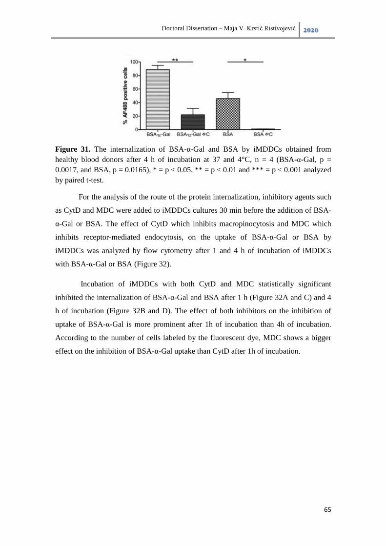
Doctoral Dissertation – Maja V. Krstić Ristivojević 2020
65
Figure 31. The internalization of BSA-α-Gal and BSA by iMDDCs obtained from
healthy blood donors after 4 h of incubation at 37 and 4°C, n = 4 (BSA-α-Gal, p =
0.0017, and BSA, p = 0.0165), * = p < 0.05, ** = p < 0.01 and *** = p < 0.001 analyzed
by paired t-test.
For the analysis of the route of the protein internalization, inhibitory agents such
as CytD and MDC were added to iMDDCs cultures 30 min before the addition of BSA-
α-Gal or BSA. The effect of CytD which inhibits macropinocytosis and MDC which
inhibits receptor-mediated endocytosis, on the uptake of BSA-α-Gal or BSA by
iMDDCs was analyzed by flow cytometry after 1 and 4 h of incubation of iMDDCs
with BSA-α-Gal or BSA (Figure 32).
Incubation of iMDDCs with both CytD and MDC statistically significant
inhibited the internalization of BSA-α-Gal and BSA after 1 h (Figure 32A and C) and 4
h of incubation (Figure 32B and D). The effect of both inhibitors on the inhibition of
uptake of BSA-α-Gal is more prominent after 1h of incubation than 4h of incubation.
According to the number of cells labeled by the fluorescent dye, MDC shows a bigger
effect on the inhibition of BSA-α-Gal uptake than CytD after 1h of incubation.

Doctoral Dissertation – Maja V. Krstić Ristivojević 2020
66
Figure 32. The internalization of BSA-α-Gal and BSA by iMDDCs obtained from
healthy blood donors after 1 h (A and C) and 4 (B and D) of incubation of iMDDCs and
proteins in the presence of (A) and (B) Cytochalasin D,( n = 6; BSA-α-Gal and n = 5;
BSA), and (C) and (D) Monodansylcadaverine (n = 7). *p < 0.05, ***p < 0.001
analyzed by paired t-test.
6.3.4. The processing of proteins with and without α-Gal epitope by iMDDC
Although all previous experiments indicate that proteins, which carrying the α-Gal
epitope are internalized by iMDDCs nothing is known about their further fate in these
cells. One important question is: in which way proteins that carrying α-Gal epitope are
processed by iMDDCs. Are any differences in the rate of processing of proteins with
and without the α-Gal epitope? In an attempt to get answers to these questions, in the
next experiment, the presence of BSA and BSA-α-Gal labeled by fluorescent dye AF
488, in the lysates of iMDDCs obtained at different time points was analyzed by SDS
PAGE. The bands of BSA and BSA-α-Gal in the gel were visualized by fluorescent
detection (Figure 33A), while the presence of α-Gal epitope on the intact proteins or
their digested fragments was analyzed by immunodetection using monoclonal anti-α-
Gal M86 antibody (Figure 33B).
The bands of BSA-α-Gal and BSA were detected at MW which corresponds to
approx. MW of their intact form. Interestingly in the case of BSA-α-Gal, a band above
75 kDa, which corresponds to the intact protein was present for up to 16 h of uptake of

Doctoral Dissertation – Maja V. Krstić Ristivojević 2020
67
BSA-α-Gal. The protein band lower than the intact protein was detected after 4, 6 and 8
h of BSA-α-Gal incubation (Figure 33A). The detected band at lower MW than intact
BSA-α-Gal probably represents degraded products of the protein. Regarding the uptake
of BSA, a strong protein band, which corresponds to the intact protein was detected for
up to 6 h of uptake while after 8 and 16 h of uptake very weak protein band could be
noted at a slightly higher MW.
When the presence of the α-Gal epitope is analyzed in iMDDCs lysates, it can be
seen from Figure 33B that the α-Gal epitope was present for up to 16 h at MW of the
band which corresponds to the intact protein. Besides this band, in all time points of the
protein uptake weak protein bands at approx. MW of 150 kDa was detected.
Figure 33. (A) Fluorescent detection of BSA-α-Gal and BSA labeled by fluorescent dye
AF 488 and (B) Western blot detection of the α-Gal epitope using monoclonal anti-α-
Gal M86 antibody in iMDDCs lysates prepared from iMDDCs after 1 to 16 h of
incubation with BSA-α-Gal and BSA, and resolved using SDS polyacrylamide gel
electrophoresis. iMDDCs were generated from healthy blood donors,
CTRL=unstimulated cells.
6.3.5. The α-Gal containing proteins are scattered in the cytoplasm of
iMDDCs
For a better understanding of the processes behind the processing of proteins that
carry α-Gal epitopes such as BSA-α-Gal and bTG inside of DCs, CLSM analysis was
used. The iMDDCs were incubated with BSA-α-Gal and BSA AF488 labeled for 1 and
4 h at 37 ˚ , after the end of this period iMDDCs were prepared for CLMS to monitor

Doctoral Dissertation – Maja V. Krstić Ristivojević 2020
68
the uptake of proteins because this analysis enables very accurate prediction of the
spatial distribution and localization of proteins inside the cells (Figure 34). Besides
these proteins, CLSM was used for the monitoring of the uptake of bTG and BSA-NAl,
by iMDDCs. The bTG, which naturally possesses α-Gal epitope as posttranslational
modification and has the MW 10 times higher than the MW of BSA-α-Gal and BSA-
NAl, which carrying a glycan similar size as α-Gal were used to check how iMDDCs
uptake and process these proteins (Figure 35).
The obtained images using CLSM are presented in Figure 34 and fluorescent green
signals arising from BSA-α-Gal were detected in almost 50% of the iMDDCs
represented in that confocal plane. Furthermore, after 4 h of incubation green
fluorescent signal was dominantly localized in the perinuclear region of almost all
iMDDCs represented in that confocal plane. On the other hand, there was no detectable
green signal arising from fluorescently labeled BSA neither after 1 or 4 h of the
monitored uptake. Importantly CLSM imaging was following the results obtained using
flow cytometric analysis.

Doctoral Dissertation – Maja V. Krstić Ristivojević 2020
69
Figure 34. The uptake of BSA-α-Gal and BSA by iMDDCs after 1 h and 4 h of
incubation iMDDC with AF488 labeled proteins at 37 °C was analyzed by confocal
laser scanning microscopy. CTRL = unstimulated iMDDCs, green = BSA-α-Gal or
BSA, red = HLA-DR and blue = DAPI stained nuclei.
It is evident the lack of green fluorescent signals arising from BSA-NAl after 1
and 4 h of incubation of iMDDCs with protein, while strong green signal arising from
bTG was detected scattered in cell cytosol of almost all iMDDCs after 1 and 4 h of the
uptake. Overall results demonstrated that proteins with α-Gal epitope as its sugar
modification were retained and accumulated inside of iMDDCs.

Doctoral Dissertation – Maja V. Krstić Ristivojević 2020
70
Figure 35. The uptake of BSA-NAl and bTG by iMDDCs after 1 h and 4 h of
incubation iMDDC with AF488 labeled proteins at 37 °C was analyzed by confocal
laser scanning microscopy. Green = BSA-NAl or bTG, red = HLA-DR and blue =
DAPI stained nuclei.
6.4. Discussion
“The α-Gal rich foods elicit delayed allergic symptoms in red meat-allergic patients,
which could be due to uptake and processing by DCs. The present study (Krstic
Ristivojevic, Grundstrom et al. 2018), shown efficient uptake of the α-Gal model
antigens like BSA-α-Gal and HSA-α-Gal by in vitro cultured human iMDDCs. Similar
results were obtained for iMDDCs generated from healthy individuals and red meat
allergic patients, indicating that altered uptake of α-Gal in DCs per se is not a factor
contributing to red meat allergy. However, DCs are likely involved in the sensitization
phase of red meat allergy where α-Gal containing proteins may be taken up in the close
vicinity of a tick bite. In atopic individuals, the DCs may then initiate allergic

Doctoral Dissertation – Maja V. Krstić Ristivojević 2020
71
sensitization through skewing of CD4+ T cells towards a Th2 response. A recent report
showed that the context of allergen uptake is important for the maturation of iMDDCs
and that the maturation effect differed between healthy and atopic donors (Aglas, Gilles
et al. 2018).
Interestingly, the uptake of BSA and HSA alone was not as efficient as for their α-
Gal carrying counterparts. Thus, the presence of α-Gal on the surface seems to enhance
the internalization of the protein, suggesting an α-Gal dependent uptake mechanism.
The results are in line with previous studies on glycosylated protein uptake, where a
100-fold higher uptake of mannosylated BSA compared with non-mannosylated BSA
was demonstrated (Engering, Cella et al. 1997). Furthermore, advanced glycation end
products of ovalbumin were taken up more efficiently by iMDDCs compared with
ovalbumin (Hilmenyuk, Bellinghausen et al. 2010).
It also investigated if the size and type of conjugation of the proteins have an impact
on the uptake. The iMDDCs from healthy individuals showed no difference in the
uptake of BSA-α-Gal and bTG even though the MW of bTG is approx. 10 times higher
than the MW of BSA-α-Gal. Interestingly, the uptake of BSA-NAl was several times
lower compared to BSA-α-Gal, even though the α-Gal and NAl carbohydrates are of
similar size. Previous studies have shown that MDDCs internalize BSA with different
glycosylation patterns to very varying extent (Avraméas, McIlroy et al. 1996), and for
other glycosylated allergens, the mannose receptor plays a major role in glycoallergen
recognition and uptake (Royer, Emara et al. 2010). The results of this study strongly
suggest that the specific glycosylation of protein antigens predominantly influences the
uptake, while the size of the protein is not a decisive factor. This is in line with previous
studies which have shown that human DCs, in addition to protein antigens, can
internalize large size particles (Foged, Brodin et al. 2005) while the key factor that
influences protein uptake is the surface architecture (Engering, Cella et al. 1997).
What was next elucidated in this study, whether α-Gal could be detected in the
cytoplasm of iMDDCs. Fluorescence detection confirmed the presence of BSA-α-Gal in
iMDDC cytoplasm. Moreover, SDS PAGE analysis showed that BSA-α-Gal was
detected after up to 16 h of incubation, while BSA was only detected up to 6 h. Western
blot with monoclonal anti-α-Gal antibody confirmed that the BSA-α-Gal still carries the

Doctoral Dissertation – Maja V. Krstić Ristivojević 2020
72
epitope. These results suggest that the degradation pathway of α-Gal carrying proteins is
slower than for non-carrying proteins and that they are stored intact inside of cells. Most
likely the α-Gal glycosylation protects the protein from degradation and as such may
increase the processing time and exposure on the surface of the cell. Other studies have
indeed shown that reduced degradation increases antigen presentation on MHCII
(Delamarre, Pack et al. 2005) and that the allergenic Bet v 1a was degraded slower than
its hypoallergenic isoform Bet v 1d (Freier, Dall et al. 2015), suggesting that processing
time affects allergenicity. This is also in line with the delayed appearance of symptoms
in red meat allergic patients.
The route of internalization of proteins was analyzed by adding CytD and MDC to
the iMDDC cultures which resulted in partial inhibition of the internalization of BSA,
regardless of the presence of α-Gal. The inhibition was stronger after 1 h of incubation
than after 4 h, which probably reflects a temporary effect by both inhibitory agents.
MDC blocks receptor-mediated endocytosis whereas CytD affects the invagination of
the cell membrane by blocking actin polymerization, which inhibits macropinocytosis
and phagocytosis (Ivanov 2008). However, the blocking of actin polymerization may
also inhibit receptor-mediated endocytosis (Ivanov 2008). Fluorescently labeled BSA
can be used for measuring uptake via macropinocytosis, but as with chemical inhibition
of antigen-uptake, it is difficult to unambiguously distinguish between
macropinocytosis and receptor-mediated endocytosis as the uptake mechanism in DCs
(Liu and Roche 2015). These results thus indicate that both receptor-mediated
endocytosis and macropinocytosis are involved in the uptake of α-Gal containing
proteins.
The confocal analysis revealed accumulated BSA-α-Gal and bTG after 1 and 4 h of
incubation, whereas no internalization of BSA and BSA-NAl could be detected at any
time point. Thus, for antigen uptake, the conjugated sugar seems to be more important
than the protein size, in line with the flow cytometric analysis. After 1 h the proteins
were found in clusters scattered around the cytoplasm while after 4 h, the proteins were
found closer to the nucleus, suggesting that BSA-α-Gal is taken up and processed in
endosomes.
Doctoral Dissertation – Maja V. Krstić Ristivojević 2020
73
In conclusion, α-Gal containing proteins are actively and promptly internalized by
iMDDCs in a time-dependent manner, and the mechanism of internalization seems to be
α-Gal dependent. Also, the differences in degradation kinetics of proteins carrying α-
Gal and those that do not reflect the delayed onset of symptoms that red meat allergic
patients experience. Taken together, it is shown that α-Gal is important for antigen
processing and these results add new knowledge of α-Gal as a clinically relevant food
allergen.” (Krstic Ristivojevic, Grundstrom et al. 2018)
Doctoral Dissertation – Maja V. Krstić Ristivojević 2020
74
7. Conclusions
According to the aims set out in section 3, the following conclusions are drawn:
a) The α-Gal glycosylation hampers the resistance of BSA to pepsinolysis and alters the
digestion patterns of BSA-α-Gal compared to BSA, whereas large digestion products
of BSA-α-Gal bearing α-Gal were prolonged time present in the digestion mixture.
b) The type of glycosylation influence the protein susceptibility to pepsinolysis, α-Gal
glycosylation hampers the resistance of BSA to pepsinolysis, but NAl glycosylation
hampers pepsinolysis even more.
c) The presence of the α-Gal epitope on BSA influences their transport through the
intestinal Caco-2 monolayer compared to unconjugated BSA and the slower
transport of BSA-α-Gal is glycosylation specific.
d) The intact proteins and the α-Gal epitope on the intact protein can be detected in the
endosomal fraction of Caco-2 cell lysates which implicates the delayed processing of
α-Gal carrying proteins in the intestinal cells.
e) The level of pro-inflammatory factors (galectin-3) in the intestinal cells is not
affected by the presence of BSA-α-Gal in comparison to BSA.
f) The presence of α-Gal on the protein surface enhances their internalization by
iMDDCs.
g) The types of protein glycosylation of antigens influence their uptake by iMDDCs,
while the size of the protein is not a decisive factor.
h) The α-Gal glycosylation protects the protein from degradation in the iMDDCs and as
such may increase their processing time and exposure on the surface of the cell.
i) The receptor-mediated endocytosis and macropinocytosis are involved in the uptake
of α-Gal containing proteins by iMDDCs.
j) The BSA-α-Gal is taken up and processed the most probably in endosomes in
iMDDCs.

Doctoral Dissertation – Maja V. Krstić Ristivojević 2020
75
8. Appendix
Biosafety level risk
Biological agents of plant, animal, or human origin are classified according to
biosafety level risk (BSL). This means that work with such agents requires the strict
following of guidelines for laboratory facilities, safety equipment, and laboratory
practices and techniques. There are four levels of biological containment (BSL 1 - 4)
and experiments performed in this doctoral dissertation were performed in laboratories
that apply BSL-2 containment. This means that agents used in performed experiments
pose a moderate risk to personnel and the environment (BSL-2 risk) and BSL-2
containment required lab coats, gloves and face protection, biological safety cabinets
(Class 2, type A, BSC), decontamination of waste materials, restricted access to
laboratory and laboratory work supervised by a trained scientist who understands the
risk associated with used biological agents.
General cell-culturing protocols
The work with cell cultures and cell-based models follows some specific assay
protocols, also some general protocols which are routines in cell-cultures laboratories
worldwide.
Thawing cells
To ensure the maximum survival of cells upon thawing the next general protocol
should be followed:
1. The culturing medium should be preheated to 37 °C.
2. The cryovials containing frozen cells should be inserted into the water bath adjusted
to 37 °C immediately after removal from liquid nitrogen containers or deep-freezer to
prevent cell lysis.
3. Upon defrosting the content, the cryovials should be transferred to the biological
safety cabinet and the cell suspension should be carefully passed on appropriate volume
prewarmed culturing, usually 5 mL of culturing medium in a 25 mL flask (T-25).

Doctoral Dissertation – Maja V. Krstić Ristivojević 2020
76
4. The cells suspension in the medium should be gently mixed and culturing flask
should be placed in the incubator for maintaining the optimal conditions for cell culture
growth.
5. The following day culturing medium should be changed with a fresh medium to
increase the survival of still fragile cells and to allow cell adhesion for adherent cell
cultures also the viability of the cells and eventual presence of contaminants such as
bacteria should be checked under a microscope.
Maintaining cell culture
The cell cultures and cell-based models are usually grown in an air atmosphere
in incubators which provides them a constant temperature of 37 °C and the optimal
percentage of CO2. The cell suspension or monolayers are kept in appropriate sterile
dishes such as flasks, Petri dishes, transwell inserts, etc. in an optimal culturing medium
usually supplemented with FBS and antibiotics. For optimal cell growth culturing
medium should be regularly replaced with a fresh one, usually, mediums contain pH
indicators such as phenol red whose color change signals the level of metabolic activity
of the cells which results in a change in pH of the culturing medium. The cells grown in
laboratory conditions follow the cell growth curve (Figure 36) which consist of lag
phase which reflects the cells recover from sub-cultivation, attach to the surface and
start to spread, log phase which reflects exponential cells growth and characteristic
doubling rate of cells which defines the cell line’s doubling time, plateau phase in which
the culture is confluent and cell growth slows or even stops and eventually death phase
during which cells start dying and in case of adherent cultures detach from the surface.
Figure 36. Phases of cell cultures growth.
12
12
Available at https://www.leica-microsystems.com/science-lab/how-to-do-a-proper-cell-culture-quick-check/

Doctoral Dissertation – Maja V. Krstić Ristivojević 2020
77
It is crucial to ensure viability and stabile proliferation rate, as well as genetic stability
of the cultured cells for that purpose cells should be kept in their exponential phase of
growth i.e. log phase. To ensure the mentioned characteristics the cell cultures should be
sub-cultured before reach the stationary phase of growth. For each cell line, the growth
curves should be determined since the optimal splitting ratio is evaluated based on it.
The number of sub-cultures is designated as passage number which must be regularly
recorded and kept as low as possible.
Sub-culturing protocol
For the cell cultures grown in suspension, the sub-culturing protocol is quite
simple when the growth curve and subsequently splitting ratio was determined. The cell
suspension should be collected from the culturing vessel, transferred to the appropriate
tube, and centrifuged to spin down the cells to the bottom of the tube. The residual
medium should be carefully discarded and optionally the cells can be washed with PBS
which requires second round centrifugation and the remaining PBS should be discarded.
The cells should be resuspended in an appropriate volume of fresh medium suitable for
use in subsequent cell counting and optimal cell splitting.
For the adherent cells, when cultured cells usually rich 80 – 90% of confluence,
which can be determined by simple inspection of the cell culture under the microscope,
sub-culturing is recommended. First cell monolayer should be washed with PBS to
remove the remaining FBS from the culturing medium, next the cells should be
detached from the bottom of the vessel by the addition of trypsin or in case of semi-
adherent cells prolonged incubation with PBS and simply tapping the bottom of the
vessel is enough. The detached cells should be resuspended in a fresh medium
containing FBS to ensure the inactivation of the trypsin since FBS contains trypsin
inhibitors. Further, the cell suspension should be centrifuged, a medium should be
discarded and the cells should be resuspended in an appropriate volume of fresh
medium suitable for use in subsequent cell counting and optimal cell splitting.
Cell freezing
For the long-term storage and preservation of the cell lines, the cells should
freeze. After passage and cell counting, a definite number of viable cells usually up to 6
x 106
cells in 1 mL of the freezing solution should be left for storage. The used freezing

Doctoral Dissertation – Maja V. Krstić Ristivojević 2020
78
solution must contain a cryoprotective agent such as DMSO or glycerol to reduce the
detrimental effects which freezing can have on cell viability. The cryoprotective agents
reduce the freezing point and increase the permeability of the cell membrane to water.
The cell suspension in freezing solution should be stored at – 80 °C for up to 6 months
and subsequently transferred in liquid nitrogen for long term storage.
Cell counting
Cell counting is performed on regular basis in cell laboratories, for that purpose
commercial devices so-called cell-counters can be used but cell counting using cell-
counting chambers i.e. hemocytometers is unsurpassed (Figure 37).
Figure 37. Hemocytometer and dimensions of counting grid chambers.13
Before counting to discriminate live and dead cells, cell suspension should be mixed
with a 0.1 % Trypan blue solution. Trypan blue is an azo dye which penetrates in death
cell due to impaired membrane permeability while live cells with intact membrane are
non-colored. The cell suspension mixed with Trypan blue should place on top of
13
Available at https://biocyclopedia.com/index/biotechnology_methods/microbiology/cell_count_by_hemocytometer_or_measuring_volume.php

Doctoral Dissertation – Maja V. Krstić Ristivojević 2020
79
hemocytometer and covered with cover glass. For example if in a square with
dimensions 1 mm x 1 mm (depth 0.1 mm) the next calculation should be applied:
⁄
Measurement of transepithelial electrical resistance (TEER)
The measurement of transepithelial/transendothelial electrical resistance is a
sensitive and reliable method for confirming the integrity and permeability of the
monolayer. It is the measurement of electrical resistance across a cellular monolayer.
For TEER measurement commercial devices are in use (Figure 38).
Figure 38. (A) Schematic diagram of TEER measurement, (B) TEER measurement
device.14
TEER should be monitored during the differentiation and growth of cells on the
transwell inserts as well as before and after transport experiments. As blank or control
empty transwell insert without seeded cells only with medium should be used. Before
measurement electrodes should be sterilized by dipping the electrodes in 70 % ethanol
for 15 min, afterward electrodes should be dried on air and dipped in a prewarmed
culturing medium before measurement. TEER measurement should be performed in a
14
Available at https://ebrary.net/24380/health/measurement_transepithelial_electrical_resistance_teer

Doctoral Dissertation – Maja V. Krstić Ristivojević 2020
80
biosafety cabinet to ensure sterile conditions. The average value of three consecutive
measurements is taken as the TEER value. Usually, TEER is expressed as measured
TEER value divided by the surface of the transwell insert in Ω / cm2.

Doctoral Dissertation – Maja V. Krstić Ristivojević 2020
81
9. Literature
Abbas, A. K., A. H. Lichtman and S. Pillai (2007). Cellular and molecular
immunology, Philadelphia: Saunders Elsevier.
Aglas, L., S. Gilles, R. Bauer, S. Huber, G. R. Araujo, G. Mueller, S.
Scheiblhofer, M. Amisi, H. H. Dang, P. Briza, B. Bohle, J. Horejs-Hoeck, C. Traidl-
Hoffmann and F. Ferreira (2018). "Context matters: TH2 polarization resulting from
pollen composition and not from protein-intrinsic allergenicity." J Allergy Clin
Immunol 142(3): 984-987 e986.
Aloulou, A. and F. Carrière (2008). "Gastric lipase: an extremophilic interfacial
enzyme with medical applications." Cellular and Molecular Life Sciences 65(6): 851-
854.
Apostolovic, D., M. Krstic, J. Mihailovic, M. Starkhammar, T. Cirkovic
Velickovic, C. Hamsten and M. van Hage (2017). "Peptidomics of an in vitro digested
α-Gal carrying protein revealed IgE-reactive peptides." Scientific Reports 7(1): 5201.
Apostolovic, D., R. Rodrigues, P. Thomas, M. Starkhammar, C. Hamsten and M.
van Hage (2018). "Immunoprofile of α-Gal- and B-antigen-specific responses
differentiates red meat-allergic patients from healthy individuals." Allergy 73(7): 1525-
1531.
Apostolovic, D., T. A. Tran, C. Hamsten, M. Starkhammar, T. Cirkovic
Velickovic and M. van Hage (2014). "Immunoproteomics of processed beef proteins
reveal novel galactose-alpha-1,3-galactose-containing allergens." Allergy 69(10): 1308-
1315.
Araujo, R. N., P. F. Franco, H. Rodrigues, L. C. B. Santos, C. S. McKay, C. A.
Sanhueza, C. R. N. Brito, M. A. Azevedo, A. P. Venuto, P. J. Cowan, I. C. Almeida, M.
G. Finn and A. F. Marques (2016). "Amblyomma sculptum tick saliva: α-Gal
identification, antibody response and possible association with red meat allergy in
Brazil." International Journal for Parasitology 46(3): 213-220.
Asero, R. and L. Antonicelli (2011). "Does Sensitization to Foods in Adults Occur
Always in the Gut?" International Archives of Allergy and Immunology 154(1): 6-14.
Avraméas, A., D. McIlroy, A. Hosmalin, B. Autran, P. Debré, M. Monsigny, A.
C. Roche and P. Midoux (1996). "Expression of a mannose/fucose membrane lectin on
human dendritic cells." European Journal of Immunology 26(2): 394-400.
Banchereau, J., F. Briere, C. Caux, J. Davoust, S. Lebecque, Y. J. Liu, B.
Pulendran and K. Palucka (2000). "Immunobiology of dendritic cells." Annu Rev
Immunol 18: 767-811.
Banchereau, J. and R. M. Steinman (1998). "Dendritic cells and the control of
immunity." Nature 392(6673): 245-252.

Doctoral Dissertation – Maja V. Krstić Ristivojević 2020
82
Behrens, I. and T. Kissel (2003). "Do cell culture conditions influence the carrier-
mediated transport of peptides in Caco-2 cell monolayers?" European Journal of
Pharmaceutical Sciences 19(5): 433-442.
Benede, S., R. Lopez-Fandino, M. Reche, E. Molina and I. Lopez-Exposito
(2013). "Influence of the carbohydrate moieties on the immunoreactivity and
digestibility of the egg allergen ovomucoid." PLoS One 8(11): e80810.
Blázquez, A. B. and M. C. Berin (2008). "Gastrointestinal Dendritic Cells
Promote Th2 Skewing via OX40L." The Journal of Immunology 180(7): 4441-4450.
Bøgh, K. L. and C. B. Madsen (2016). "Food Allergens: Is There a Correlation
between Stability to Digestion and Allergenicity?" Critical Reviews in Food Science
and Nutrition 56(9): 1545-1567.
Boland, M., M. Golding and H. Singh (2014). Food Structures, Digestion and
Health, Academic Press.
Boyle, R. J., W. Hardikar and M. L. K. Tang (2005). "The development of food
allergy after liver transplantation." Liver Transplantation 11(3): 326-330.
Brough, H. A., A. H. Liu, S. Sicherer, K. Makinson, A. Douiri, S. J. Brown, A. C.
Stephens, W. H. Irwin McLean, V. Turcanu, R. A. Wood, S. M. Jones, W. Burks, P.
Dawson, D. Stablein, H. Sampson and G. Lack (2015). "Atopic dermatitis increases the
effect of exposure to peanut antigen in dust on peanut sensitization and likely peanut
allergy." The Journal of allergy and clinical immunology 135(1): 164-170.
Brown, C., N. Haringman, C. Davies, C. Gore, M. Hussain, G. Mieli-Vergani, D.
Vergani, J. O. Warner, S. D. Marks and R. J. Boyle (2012). "High prevalence of food
sensitisation in young children with liver disease: a clue to food allergy pathogenesis?"
Pediatric Allergy and Immunology 23(8): 770-777.
Burton, O. T., M. Noval Rivas, J. S. Zhou, S. L. Logsdon, A. R. Darling, K. J.
Koleoglou, A. Roers, H. Houshyar, M. A. Crackower, T. A. Chatila and H. C. Oettgen
(2014). "Immunoglobulin E signal inhibition during allergen ingestion leads to reversal
of established food allergy and induction of regulatory T cells." Immunity 41(1): 141-
151.
Cabezas- ruz, A., . J. Espinosa, . Alberdi, L. Šimo, J. J. Valdés, L. Mateos-
Hernández, M. Contreras, M. V. Rayo and J. de la Fuente (2018). "Tick
galactosyltransferases are involved in α-Gal synthesis and play a role during Anaplasma
phagocytophilum infection and Ixodes scapularis tick vector development." Scientific
Reports 8(1): 14224.
Chinuki, Y., K. Ishiwata, K. Yamaji, H. Takahashi and E. Morita (2016).
"Haemaphysalis longicornis tick bites are a possible cause of red meat allergy in Japan."
Allergy 71(3): 421-425.
Chung, C. H., B. Mirakhur, E. Chan, Q. T. Le, J. Berlin, M. Morse, B. A. Murphy,
S. M. Satinover, J. Hosen, D. Mauro, R. J. Slebos, Q. Zhou, D. Gold, T. Hatley, D. J.

Doctoral Dissertation – Maja V. Krstić Ristivojević 2020
83
Hicklin and T. A. Platts-Mills (2008). "Cetuximab-induced anaphylaxis and IgE specific
for galactose-alpha-1,3-galactose." N Engl J Med 358(11): 1109-1117.
Commins, S. P. (2015). "Carbohydrates as Allergens." Current Allergy and
Asthma Reports 15(1): 1-6.
Commins, S. P., H. R. James, L. A. Kelly, S. L. Pochan, L. J. Workman, M. S.
Perzanowski, K. M. Kocan, J. V. Fahy, L. W. Nganga, E. Ronmark, P. J. Cooper and T.
A. Platts-Mills (2011). "The relevance of tick bites to the production of IgE antibodies
to the mammalian oligosaccharide galactose-alpha-1,3-galactose." J Allergy Clin
Immunol 127(5): 1286-1293 e1286.
Commins, S. P., H. R. James, W. Stevens, S. L. Pochan, M. H. Land, C. King, S.
Mozzicato and T. A. Platts-Mills (2014). "Delayed clinical and ex vivo response to
mammalian meat in patients with IgE to galactose-alpha-1,3-galactose." J Allergy Clin
Immunol 134(1): 108-115.
Commins, S. P. and S. Karim (2017). "Development of a novel murine model of
alpha-gal meat allergy." Journal of Allergy and Clinical Immunology 139(2,
Supplement): AB193.
Commins, S. P., L. A. Kelly, E. Rönmark, H. R. James, S. L. Pochan, E. J. Peters,
B. Lundbäck, L. W. Nganga, P. J. Cooper, J. M. Hoskins, S. S. Eapen, L. A. Matos, D.
C. McBride, P. W. Heymann, J. A. Woodfolk, M. S. Perzanowski and T. A. E. Platts-
Mills (2012). "Galactose-α-1,3-Galactose–Specific IgE Is Associated with Anaphylaxis
but Not Asthma." American Journal of Respiratory and Critical Care Medicine 185(7):
723-730.
Commins, S. P. and T. A. Platts-Mills (2009). "Anaphylaxis syndromes related to
a new mammalian cross-reactive carbohydrate determinant." J Allergy Clin Immunol
124(4): 652-657.
Commins, S. P., S. M. Satinover, J. Hosen, J. Mozena, L. Borish, B. D. Lewis, J.
A. Woodfolk and T. A. Platts-Mills (2009). "Delayed anaphylaxis, angioedema, or
urticaria after consumption of red meat in patients with IgE antibodies specific for
galactose-alpha-1,3-galactose." J Allergy Clin Immunol 123(2): 426-433.
Coombes, J. L., K. R. Siddiqui, C. V. Arancibia-Cárcamo, J. Hall, C. M. Sun, Y.
Belkaid and F. Powrie (2007). "A functionally specialized population of mucosal
CD103+ DCs induces Foxp3+ regulatory T cells via a TGF-beta and retinoic acid-
dependent mechanism." J. Exp Med 204: 1757–1764.
Dawes, C., A. M. L. Pedersen, A. Villa, J. Ekström, G. B. Proctor, A. Vissink, D.
Aframian, R. McGowan, A. Aliko, N. Narayana, Y. W. Sia, R. K. Joshi, S. B. Jensen,
A. R. Kerr and A. Wolff (2015). "The functions of human saliva: A review sponsored
by the World Workshop on Oral Medicine VI." Archives of Oral Biology 60(6): 863-
874.

Doctoral Dissertation – Maja V. Krstić Ristivojević 2020
84
Delamarre, L., M. Pack, H. Chang, I. Mellman and E. S. Trombetta (2005).
"Differential lysosomal proteolysis in antigen-presenting cells determines antigen fate."
Science 307.
Diaz-Alvarez, L. and E. Ortega (2017). "The Many Roles of Galectin-3, a
Multifaceted Molecule, in Innate Immune Responses against Pathogens." Mediators
Inflamm 2017: 9247574.
Dumic, J., S. Dabelic and M. Flogel (2006). "Galectin-3: an open-ended story."
Biochim Biophys Acta 1760(4): 616-635.
Elias, K. M., A. Laurence, T. S. Davidson, G. Stephens, Kanno, Y., , E. M.
Shevach and J. J. O'Shea (2008). "Retinoic acid inhibits Th17 polarization and enhances
FoxP3 expression through a Stat-3/Stat-5 independent signaling pathway." Blood
111(3): 1013-1020.
Engering, A. J., M. Cella, D. Fluitsma, M. Brockhaus, E. C. Hoefsmit, A.
Lanzavecchia and J. Pieters (1997). "The mannose receptor functions as a high capacity
and broad specificity antigen receptor in human dendritic cells." Eur J Immunol 27(9):
2417-2425.
Fischer, J., B. Eberlein, C. Hilger, F. Eyer, S. Eyerich, M. Ollert and T.
Biedermann (2017). "Alpha-gal is a possible target of IgE-mediated reactivity to
antivenom." Allergy 72(5): 764-771.
Foged, C., B. Brodin, S. Frokjaer and A. Sundblad (2005). "Particle size and
surface charge affect particle uptake by human dendritic cells in an in vitro model." Int J
Pharm 298(2): 315-322.
Freier, R., E. Dall and H. Brandstetter (2015). "Protease recognition sites in Bet v
1a are cryptic, explaining its slow processing relevant to its allergenicity." Scientific
Reports 5(1): 12707.
Galili, U., B. A. Macher, J. Buehler and S. B. Shohet (1985). "Human natural anti-
α-galactosyl IgG: II. The specific recognition of a(1 → 3)-linked galactose residues."
Journal of Experimental Medicine 162(2): 573-582.
Galili, U., R. E. Mandrell, R. M. Hamadeh, S. B. Shohet and J. M. Griffiss (1988).
"Interaction between human natural anti-alpha-galactosyl immunoglobulin G and
bacteria of the human flora." Infection and immunity 56(7): 1730-1737.
Ganz, T. (2000). "Paneth cells—guardians of the gut cell hatchery." Nature
Immunology 1(2): 99-100.
Garrett, R. H. and C. M. Grisham (1999). Biochemistry. Fort Worth, Orlando,
Saunders College Publ.
Gerbe, F. and P. Jay (2016). "Intestinal tuft cells: epithelial sentinels linking
luminal cues to the immune system." Mucosal Immunology 9(6): 1353-1359.

Doctoral Dissertation – Maja V. Krstić Ristivojević 2020
85
Geremia, A. and C. V. Arancibia-Cárcamo (2017). "Innate Lymphoid Cells in
Intestinal Inflammation." Frontiers in Immunology 8: 1296.
Gribble, F. M. and F. Reimann (2019). "Function and mechanisms of
enteroendocrine cells and gut hormones in metabolism." Nature Reviews Endocrinology
15(4): 226-237.
Grönlund, H., J. Adédoyin, S. P. Commins, T. A. E. Platts-Mills and M. van Hage
(2009). "The carbohydrate galactose-alpha-1,3-galactose is a major IgE-binding epitope
on cat IgA." The Journal of allergy and clinical immunology 123(5): 1189-1191.
Gudgeon, A. M., G. Patel, J. Hermon-Taylor, P. Hurley, R. C. Bowyer and A. M.
T. Jehanli (1991). "Detection of Human Pancreatic Pro-Phospholipase A2 Activation
Using an Immunoassay for the Free Activation Peptide DSGISPR." Annals of Clinical
Biochemistry 28(5): 497-503.
Guerra, A., L. Etienne-Mesmin, V. Livrelli, S. Denis, S. Blanquet-Diot and M.
Alric (2012). "Relevance and challenges in modeling human gastric and small intestinal
digestion." Trends in Biotechnology 30(11): 591-600.
Guilliams, M., F. Ginhoux, C. Jakubzick, S. H. Naik, N. Onai, B. U. Schraml, E.
Segura, R. Tussiwand and S. Yona (2014). "Dendritic cells, monocytes and
macrophages: a unified nomenclature based on ontogeny." Nature Reviews
Immunology 14(8): 571-578.
Hamsten, C., M. Starkhammar, T. A. T. Tran, M. Johansson, U. Bengtsson, G.
Ahlén, M. Sällberg, H. Grönlund and M. van Hage (2013). "Identification of galactose-
α-1,3-galactose in the gastrointestinal tract of the tick Ixodes ricinus; possible
relationship with red meat allergy." Allergy 68(4): 549-552.
Harris, N. (2016). "The enigmatic tuft cell in immunity." Science 351(6279):
1264-1265.
Hershberg, R. M., P. E. Framson, D. H. Cho, L. Y. Lee, S. Kovats, J. Beitz, J. S.
Blum and G. T. Nepom (1997). "Intestinal epithelial cells use two distinct pathways for
HLA class II antigen processing." The Journal of clinical investigation 100(1): 204-215.
Hilmenyuk, T., I. Bellinghausen, B. Heydenreich, A. Ilchmann, M. Toda, S.
Grabbe and J. Saloga (2010). "Effects of glycation of the model food allergen
ovalbumin on antigen uptake and presentation by human dendritic cells." Immunology
129(3): 437-445.
Hoebler, C., G. Lecannu, C. Belleville, M. F. Devaux, Y. Popineau and J. L. Barry
(2002). "Development of an in vitro system simulating bucco-gastric digestion to assess
the physical and chemical changes of food." International journal of food sciences and
nutrition 53(5): 389-402.
Holst, B. and G. Williamson (2008). "Nutrients and phytochemicals: from
bioavailability to bioefficacy beyond antioxidants." Current Opinion in Biotechnology
19(2): 73-82.
Doctoral Dissertation – Maja V. Krstić Ristivojević 2020
86
Hornef, M. W. and C. Bogdan (2005). "The role of epithelial Toll-like receptor
expression in host defense and microbial tolerance." Journal of Endotoxin Research
11(2): 124-128.
Hubatsch, I., E. G. E. Ragnarsson and P. Artursson (2007). "Determination of
drug permeability and prediction of drug absorption in Caco-2 monolayers." Nature
Protocols 2: 2111.
Hur, S. J., B. O. Lim, E. A. Decker and D. J. McClements (2011). "In vitro human
digestion models for food applications." Food Chemistry 125(1): 1-12.
Hyun, J., H. Kothari, E. Herm, J. Mortenson, C. R. Treadwell and G. V. Vahouny
(1969). "Purification and Properties of Pancreatic Juice Cholesterol Esterase." Journal
of Biological Chemistry 244(7): 1937-1945.
Ivanov, A. I. (2008). "Pharmacological inhibition of endocytic pathways: is it
specific enough to be useful?" Methods Mol Biol 440: 15-33.
Iwata, M., A. Hirakiyama, Y. Eshima, H. Kagechika, C. Kato and S. Y. Song
(2004). "Retinoic acid imprints gut-homing specificity on T cells." Immunity 21(4):
527-538.
Jacquenet, S., D.-A. Moneret-Vautrin and B. E. Bihain (2009). "Mammalian
meat-induced anaphylaxis: Clinical relevance of anti-galactose-[alpha]-1,3-galactose
IgE confirmed by means of skin tests to cetuximab." Journal of Allergy and Clinical
Immunology 124(3): 603-605.
Jappe, U. (2012). "[Update on meat allergy. α-Gal: a new epitope, a new entity?]."
Der Hautarzt; Zeitschrift fur Dermatologie, Venerologie, und verwandte Gebiete 63(4):
299-306.
Jin, R., A. Greenwald, M. D. Peterson and T. K. Waddell (2006). "Human
Monocytes Recognize orcine Endothelium via the Interaction of Galectin 3 and α-
GAL." The Journal of Immunology 177(2): 1289.
Johansson, M. E. V. and G. C. Hansson (2013). "Mucus and the goblet cell."
Digestive diseases (Basel, Switzerland) 31(3-4): 305-309.
Kararli, T. T. (1995). "Comparison of the gastrointestinal anatomy, physiology,
and biochemistry of humans and commonly used laboratory animals."
Biopharmaceutics & drug disposition 16(5): 351-380.
Kastrup Dalsgaard, T., J. Holm Nielsen and L. Bach Larsen (2007). "Proteolysis
of milk proteins lactosylated in model systems." Molecular Nutrition & Food Research
51(4): 404-414.
Kauffman, A., A. Gyurdieva, J. Mabus, C. Ferguson, Z. Yan and P. Hornby
(2013). "Alternative functional in vitro models of human intestinal epithelia." Frontiers
in Pharmacology 4: 79.

Doctoral Dissertation – Maja V. Krstić Ristivojević 2020
87
Kiewiet, M. B. G., D. Apostolovic, M. Starkhammar, J. Grundström, C. Hamsten
and M. van Hage (2020). "Clinical and Serological haracterization of the α-Gal
Syndrome—Importance of Atopy for Symptom Severity in a European Cohort." The
Journal of Allergy and Clinical Immunology: In Practice 8(6): 2027-2034.e2022.
Knoop, K. A. and R. D. Newberry (2018). "Goblet cells: multifaceted players in
immunity at mucosal surfaces." Mucosal Immunology 11(6): 1551-1557.
Koh, A., F. De Vadder, P. Kovatcheva-Datchary and F. Bäckhed (2016). "From
Dietary Fiber to Host Physiology: Short-Chain Fatty Acids as Key Bacterial
Metabolites." Cell 165(6): 1332-1345.
Kong, F. and R. P. Singh (2008). "Disintegration of solid foods in human
stomach." J. Food Sci 73(5): R67-R80.
Koppelman, S. J., S. L. Hefle, S. L. Taylor and G. A. H. de Jong (2010).
"Digestion of peanut allergens Ara h 1, Ara h 2, Ara h 3, and Ara h 6: A comparative in
vitro study and partial characterization of digestion-resistant peptides." Molecular
Nutrition & Food Research 54(12): 1711-1721.
Krstic Ristivojevic, M., J. Grundstrom, T. A. T. Tran, D. Apostolovic, V. Radoi,
M. Starkhammar, V. Vukojevic, T. Cirkovic Velickovic, C. Hamsten and M. van Hage
(2018). "alpha-Gal on the protein surface affects uptake and degradation in immature
monocyte derived dendritic cells." Sci Rep 8(1): 12684.
Krstic Ristivojevic M., Grundström J., Apostolovic D., Radomirovic M.,
Jovanovic V., Radoi V., Kiewiet M. B. Gea, Vukojevic V., Cirkovic Velickovic T. and
v. H. M. (2020). "Alpha-Gal on the Protein Surface Hampers Transcytosis through the
Caco-2 Monolayer." International Journal of Molecular Sciences 21(16).
Laemmli, U. K. (1970). "Cleavage of structural proteins during the assembly of
the head of bacteriophage T4." Nature 227(5259): 680-685.
Laethem, R. M., T. A. Blumenkopf, M. Cory, L. Elwell, C. P. Moxham, P. H.
Ray, L. M. Walton and G. K. Smith (1996). "Expression and characterization of human
pancreatic preprocarboxypeptidase A1 and preprocarboxypeptidase A2." Archives of
biochemistry and biophysics 332(1): 8-18.
Lambrecht, B. N. and H. Hammad (2010). "The role of dendritic and epithelial
cells as master regulators of allergic airway inflammation." Lancet 376(9743): 835-843.
Larsen, R. D., C. A. Rivera-Marrero, L. K. Ernst, R. D. Cummings and J. B. Lowe
(1990). "Frameshift and nonsense mutations in a human genomic sequence homologous
to a murine UDP-Gal:β-D-Gal(1,4)-D-GlcNAc α(1,3)-galactosyltransferase cDNA."
Journal of Biological Chemistry 265(12): 7055-7061.
Lee, D. and A. Ryle (1967). "Pepsin D. A minor component of commercial pepsin
preparations." Biochemical Journal 104(3): 742-748.

Doctoral Dissertation – Maja V. Krstić Ristivojević 2020
88
Levin, M., D. Apostolovic, T. Biedermann, S. P. Commins, O. I. Iweala, T. A. E.
Platts-Mills, E. Savi, M. van Hage and J. M. Wilson (2019). "Galactose α-1,3-galactose
phenotypes: Lessons from various patient populations." Annals of Allergy, Asthma &
Immunology 122(6): 598-602.
Liu, Z. and P. A. Roche (2015). "Macropinocytosis in phagocytes: regulation of
MHC class-II-restricted antigen presentation in dendritic cells." Front Physiol 6: 1.
Mabelane, T., M. Botha, H. F. Thomas and M. Levin (2018). "Alpha Gal Allergy
in rural black African subjects associated with a high prevalence of abdominal
manifestations and a more rapid onset of symptoms." Journal of Allergy and Clinical
Immunology 141(2): AB200.
Mainville, I., Y. Arcand and E. R. Farnworth (2005). "A dynamic model that
simulates the human upper gastrointestinal tract for the study of probiotics."
International journal of food microbiology 99(3): 287-296.
Mao, X., M. T. Ou, S. S. Karuppagounder, T. I. Kam, X. Yin, Y. Xiong, P. Ge, G.
E. Umanah, S. Brahmachari, J. H. Shin, H. C. Kang, J. Zhang, J. Xu, R. Chen, H. Park,
S. A. Andrabi, S. U. Kang, R. A. Goncalves, Y. Liang, S. Zhang, C. Qi, S. Lam, J. A.
Keiler, J. Tyson, D. Kim, N. Panicker, S. P. Yun, C. J. Workman, D. A. Vignali, V. L.
Dawson, H. S. Ko and T. M. Dawson (2016). "Pathological alpha-synuclein
transmission initiated by binding lymphocyte-activation gene 3." Science 353(6307).
Maureen E. Taylor and K. Drickamer (2011). Introduction to Glycobiology,
Oxford University Press.
Mayer, L., D. Eisenhardt, P. Salomon, W. Bauer, R. Plous and L. Piccinini (1991).
"Expression of class II molecules on intestinal epithelial cells in humans: Differences
between normal and inflammatory bowel disease." Gastroenterology 100(1): 3-12.
Merad, M., P. Sathe, J. Helft, J. Miller and A. Mortha (2013). "The dendritic cell
lineage: Ontogeny and function of dendritic cells and their subsets in the steady state
and the inflamed setting." Annual Review of Immunology 31: 563-604.
Minekus, M., M. Alminger, P. Alvito, S. Ballance, T. Bohn, C. Bourlieu, F.
Carrière, R. Boutrou, M. Corredig, D. Dupont, C. Dufour, L. Egger, M. Golding, S.
Karakaya, B. Kirkhus, S. Le Feunteun, U. Lesmes, A. Macierzanka, A. Mackie, S.
Marze, D. J. McClements, O. Ménard, I. Recio, C. N. Santos, R. P. Singh, G. E.
Vegarud, M. S. J. Wickham, W. Weitschies and A. Brodkorb (2014). "A standardised
static in vitro digestion method suitable for food – an international consensus." Food &
Function 5(6): 1113-1124.
Minekus, M., P. Marteau, R. Havenaar and J. H. J. H. i. t. Veld (1995). "A
Multicompartmental Dynamic Computer-controlled Model Simulating the Stomach and
Small Intestine." Alternatives to Laboratory Animals 23(2): 197-209.

Doctoral Dissertation – Maja V. Krstić Ristivojević 2020
89
Miron, N. and V. Cristea (2012). "Enterocytes: Active cells in tolerance to food
and microbial antigens in the gut." Clinical and Experimental Immunology 167(3): 405-
412.
Mora, J. R., M. R. Bono, N. Manjunath, W. Weninger, L. L. Cavanagh, M.
Rosemblatt and U. H. Von Andrian (2003). "Selective imprinting of gut-homing T cells
by Peyer's patch dendritic cells." Nature 424(6944): 88-93.
Moreno, F. J., F. A. Mellon, M. S. J. Wickham, A. R. Bottrill and E. N. C. Mills
(2005). "Stability of the major allergen Brazil nut 2S albumin (Ber e 1) to
physiologically relevant in vitro gastrointestinal digestion." The FEBS Journal 272(2):
341-352.
Moreno, F. J., L. A. Rubio, A. Olano and A. Clemente (2006). "Uptake of 2S
albumin allergens, Ber e 1 and Ses i 1, across human intestinal epithelial Caco-2 cell
monolayers." J Agric Food Chem 54(22): 8631-8639.
Morisset, M., C. Richard, C. Astier, S. Jacquenet, A. Croizier, E. Beaudouin, V.
Cordebar, F. Morel-Codreanu, N. Petit, D. A. Moneret-Vautrin and G. Kanny (2012).
"Anaphylaxis to pork kidney is related to IgE antibodies specific for galactose-alpha-
1,3-galactose." Allergy 67(5): 699-704.
Mosmann, T. (1983). "Rapid colorimetric assay for cellular growth and survival:
application to proliferation and cytotoxicity assays." J Immunol Methods. 65(1-2): 55-
63.
Mowat, A. M. (2003). "Anatomical basis of tolerance and immunity to intestinal
antigens." Nature reviews. Immunology 3(4): 331-341.
Munegumi, T., M. Inutsuka and Y. Hayafuji (2016). "Investigating the Hydrolysis
of Starch Using α-Amylase Contained in Dishwashing Detergent and Human Saliva."
Journal of Chemical Education 93(8): 1401-1405.
Narita, Y., S.-i. Oda, A. Moriyama and T. Kageyama (2002). "Primary structure,
unique enzymatic properties, and molecular evolution of pepsinogen B and pepsin B."
Archives of Biochemistry and Biophysics 404(2): 177-185.
Niv, Y. (2016). "Mucin Genes Expression in the Intestine of Crohn's Disease
Patients: a Systematic Review and Meta-analysis." Journal of gastrointestinal and liver
diseases : JGLD 25(3): 351-357.
Nomura, D. K. and J. E. Casida (2016). "Lipases and their inhibitors in health and
disease." Chemico-Biological Interactions 259: 211-222.
Noval Rivas, M., O. T. Burton, P. Wise, L.-M. Charbonnier, P. Georgiev, H. C.
Oettgen, R. Rachid and T. A. Chatila (2015). "Regulatory T cell reprogramming toward
a Th2-cell-like lineage impairs oral tolerance and promotes food allergy." Immunity
42(3): 512-523.

Doctoral Dissertation – Maja V. Krstić Ristivojević 2020
90
Nowak-Wegrzyn, A., H. Szajewska and G. Lack (2017). "Food allergy and the
gut." Nature Reviews Gastroenterology and Hepatology 14(4): 241-257.
Nunez, R., F. Carballada, A. Gonzalez-Quintela, J. Gomez-Rial, M. Boquete and
C. Vidal (2011). "Delayed mammalian meat-induced anaphylaxis due to galactose-
alpha-1,3-galactose in 5 European patients." J Allergy Clin Immunol 128(5): 1122-1124
e1121.
Okumura, R. and K. Takeda (2017). "Roles of intestinal epithelial cells in the
maintenance of gut homeostasis." Experimental and Molecular Medicine 49(5): e338-
338.
Oostingh, G. J., H. F. S. Davies, B. N. Arch, J. A. Bradley and C. J. Taylor
(2003). "Potential implications of ABO blood group for vascular rejection in pig to
human kidney xenotransplantation." Xenotransplantation 10(3): 278-284.
Pabst, R. and H. J. Rothkötter (2006). Structure and function of the gut mucosal
immune system. Immune Mechanisms in Inflammatory Bowel Disease. Advances in
Experimental Medicine and Biology. N. M. F. Blumberg R.S. New York, NY.,
Springer. 579.
Palomares, O., M. Akdis, M. Martín-Fontecha and C. Akdis (2017). "Mechanisms
of immune regulation in allergic diseases: the role of regulatory T and B cells."
Immunological Reviews 278(1): 219-236.
Pandol, S. J. (2010). Digestive Enzymes. The Exocrine Pancreas. San Rafael
(CA), Morgan & Claypool Life Sciences.
Parigi, S. M., M. Eldh, P. Larssen, S. Gabrielsson and E. J. Villablanca (2015).
"Breast Milk and Solid Food Shaping Intestinal Immunity." Frontiers in Immunology
6(415).
Peterson, M. D., R. Jin, S. Hyduk, P. Duchesneau, M. I. Cybulsky and T. K.
Waddell (2005). "Monocyte adhesion to xenogeneic endothelium during laminar flow is
dependent on alpha-Gal-mediated monocyte activation." J Immunol 174(12): 8072-
8081.
Price, D. B., M. L. Ackland, W. Burks, M. I. Knight and C. Suphioglu (2014).
"Peanut allergens alter intestinal barrier permeability and tight junction localisation in
Caco-2 cell cultures." Cell Physiol Biochem 33(6): 1758-1777.
Qian, J., T. Liu, L. Yang, A. Daus, R. Crowley and Q. Zhou (2007). "Structural
characterization of N-linked oligosaccharides on monoclonal antibody cetuximab by the
combination of orthogonal matrix-assisted laser desorption/ionization hybrid
quadrupole-quadrupole time-of-flight tandem mass spectrometry and sequential
enzymatic digestion." Analytical biochemistry 364(1): 8-18.
Raffatellu, M., R. P. Wilson, D. Chessa, H. Andrews-Polymenis, Q. T. Tran, S.
Lawhon, S. Khare, L. G. Adams and A. J. Bäumler (2005). "SipA, SopA, SopB, SopD,

Doctoral Dissertation – Maja V. Krstić Ristivojević 2020
91
and SopE2 contribute to Salmonella enterica serotype typhimurium invasion of
epithelial cells." Infection and immunity 73(1): 146-154.
Rawitch, A. B., H. G. Pollock and S. X. Yang (1993). "Thyroglobulin
Glycosylation: Location and Nature of the N-Linked Oligosaccharide Units in Bovine
Thyroglobulin." Archives of Biochemistry and Biophysics 300(1): 271-279.
Renz, H., K. Allen, S. Sicherer, H. Sampson, G. Lack, K. Beyer and H. Oettgen
(2018). "Food allergy." Nature Reviews Disease Primers 4.
Rispens, T., N. I. L. Derksen, S. P. Commins, T. A. Platts-Mills and R. C.
Aalberse (2013). "IgE roduction to α-Gal Is Accompanied by Elevated Levels of
Specific IgG1 Antibodies and Low Amounts of IgE to Blood Group B." PLOS ONE
8(2): e55566.
Roche, P. A. and K. Furuta (2015). "The ins and outs of MHC class II-mediated
antigen processing and presentation." Nat Rev Immunol 15(4): 203-216.
Román-Carrasco, P., B. Lieder, V. Somoza, M. Ponce, Z. Szépfalusi, D. Martin,
W. Hemmer and I. Swoboda (2019). "Only α-Gal bound to lipids, but not to proteins, is
transported across enterocytes as an IgE-reactive molecule that can induce effector cell
activation." Allergy 0(0).
Royer, P. J., M. Emara, C. Yang, A. Al-Ghouleh, P. Tighe, N. Jones, H. F. Sewell,
F. Shakib, L. Martinez-Pomares and A. M. Ghaemmaghami (2010). "The mannose
receptor mediates the uptake of diverse native allergens by dendritic cells and
determines allergen-induced T cell polarization through modulation of IDO activity." J
Immunol 185(3): 1522-1531.
Sampson, H., L. O'Mahony, A. W. Burks, M. Plaut, G. Lack and C. Akdis (2018).
"Mechanisms of food allergy." Journal of Allergy and Clinical Immunology 141(1): 11-
19.
Sato, T., D. E. Stange, M. Ferrante, R. G. J. Vries, J. H. van Es, S. van den Brink,
W. J. van Houdt, A. Pronk, J. van Gorp, P. D. Siersema and H. Clevers (2011). "Long-
term Expansion of Epithelial Organoids From Human Colon, Adenoma,
Adenocarcinoma, and Barrett's Epithelium." Gastroenterology 141(5): 1762-1772.
Sato, T., R. G. Vries, H. J. Snippert, M. van de Wetering, N. Barker, D. E. Stange,
J. H. van Es, A. Abo, P. Kujala, P. J. Peters and H. Clevers (2009). "Single Lgr5 stem
cells build crypt-villus structures in vitro without a mesenchymal niche." Nature
459(7244): 262-265.
Schulz, O., E. Jaensson, E. K. Persson, X. Liu, T. Worbs, W. W. Agace and O.
Pabst (2009). "Intestinal CD103+, but not CX3CR1+, antigen sampling cells migrate in
lymph and serve classical dendritic cell functions." Journal of Experimental Medicine
206(13): 3101-3114.
Schweinlin, M., S. Wilhelm, I. Schwedhelm, J. Hansmann, R. Rietscher, C.
Jurowich, H. Walles and M. Metzger (2016). "Development of an Advanced Primary

Doctoral Dissertation – Maja V. Krstić Ristivojević 2020
92
Human In Vitro Model of the Small Intestine." Tissue Engineering Part C: Methods
22(9): 873-883.
Sekiya, K., Y. Fukutomi, T. Nakazawa, M. Taniguchi and K. Akiyama (2012).
"Delayed anaphylactic reaction to mammalian meat." J. Investig Allergol Clin Immunol
22(6): 446-447.
Sender, R., S. Fuchs and R. Milo (2016). "Revised Estimates for the Number of
Human and Bacteria Cells in the Body." PLOS Biology 14(8): e1002533.
Shaykhiev, R. and R. Bals (2007). "Interactions between epithelial cells and
leukocytes in immunity and tissue homeostasis." Journal of Leukocyte Biology 82(1):
1-15.
Sicherer, S. H. and H. A. Sampson (2014). "Food allergy: Epidemiology,
pathogenesis, diagnosis, and treatment." J. Allergy Clin. Immunol. 133(2): 291-307.
Smith, K. A. (2012). "Louis pasteur, the father of immunology?" Frontiers in
immunology 3: 68-68.
Springer, G. F. and R. E. Horton (1969). "Blood group isoantibody stimulation in
man by feeding blood group-active bacteria." The Journal of clinical investigation
48(7): 1280-1291.
Stagg, A. J. (2018). "Intestinal Dendritic Cells in Health and Gut Inflammation."
Frontiers in Immunology 9(2883).
Steinke, J. W., S. L. Pochan, H. R. James, T. A. E. Platts-Mills and S. P. Commins
(2016). "Altered metabolic profile in patients with IgE to galactose-alpha-1,3-galactose
following in vivo food challenge." Journal of Allergy and Clinical Immunology 138(5):
1465-1467.e1468.
Steinman, R. M. (2003). "Some interfaces of dendritic cell biology." APMIS
111(7-8): 675-697.
Steinman, R. M. and Z. A. Cohn (1973). "Identification of a novel cell type in
peripheral lymphoid organs of mice. I. Morphology, quantitation, tissue distribution."
The Journal of experimental medicine 137(5): 1142-1162.
Sun, H., E. C. Y. Chow, S. Liu, Y. Du and K. S. Pang (2008). "The Caco-2 cell
monolayer: usefulness and limitations." Expert Opinion on Drug Metabolism &
Toxicology 4(4): 395-411.
Thierry, A. C., E. Bernasconi, A. Mercenier and B. Corthesy (2009). "Conditioned
polarized Caco-2 cell monolayers allow to discriminate for the ability of gut-derived
microorganisms to modulate permeability and antigen-induced basophil degranulation."
Clin Exp Allergy 39(4): 527-536.
Tordesillas, L., C. Gomez-Casado, M. Garrido-Arandia, A. Murua-Garcia, A.
Palacin, J. Varela, P. Konieczna, J. Cuesta-Herranz, C. A. Akdis, L. O'Mahony and A.

Doctoral Dissertation – Maja V. Krstić Ristivojević 2020
93
Diaz-Perales (2013). "Transport of Pru p 3 across gastrointestinal epithelium - an
essential step towards the induction of food allergy?" Clin Exp Allergy 43(12): 1374-
1383.
Tran, Q. T., G. Gomez, S. Khare, S. D. Lawhon, M. Raffatellu, A. J. Bäumler, D.
Ajithdoss, S. Dhavala and L. G. Adams (2009). "The Salmonella enterica Serotype
Typhi Vi Capsular Antigen Is Expressed after the Bacterium Enters the Ileal Mucosa."
Infection and Immunity 78(1): 527-535.
Tu, Y., S. a. Salim, J. Bourgeois, V. Di Leo, E. J. Irvine, J. K. Marshall and M. H.
Perdue (2005). "CD23-Mediated IgE Transport Across Human Intestinal Epithelium:
Inhibition by Blocking Sites of Translation or Binding." Gastroenterology 129(3): 928-
940.
Turnbull, J. L., H. N. Adams and D. A. Gorard (2015). "Review article: the
diagnosis and management of food allergy and food intolerances." Alimentary
Pharmacology & Therapeutics 41(1): 3-25.
Umar, S. (2010). "Intestinal stem cells." Current gastroenterology reports 12(5):
340-348.
Valenta, R., H. Hochwallner, B. Linhart and S. Pahr (2015). "Food allergies: the
basics." Gastroenterology 148(6): 1120-1131.e1124.
Van Nunen, S. A., K. S. O'Connor, L. R. Clarke, R. X. Boyle and S. L. Fernando
(2009). "An association between tick bite reactions and red meat allergy in humans."
Med J Aust 190(9): 510-511.
Vissers Y.M., Wichers H.J. and S. H.F.J. (2012). Influence of Food Processing,
Digestion and the Food Matrix on Allergenicity & Cellular Measures of Allergenicity.
Multidisciplinary Approaches to Allergies. Berlin, Heidelberg, Springer.
Voet, D. and J. G. Voet (2011). Biochemistry. Hoboken, NJ, John Wiley & Sons.
Vukojevic, V., M. Heidkamp, Y. Ming, B. Johansson, L. Terenius and R. Rigler
(2008). "Quantitative single-molecule imaging by confocal laser scanning microscopy."
Proc Natl Acad Sci U S A 105(47): 18176-18181.
Walker, J. A., J. L. Barlow and A. N. J. McKenzie (2013). "Innate lymphoid cells-
-how did we miss them?" Nature reviews. Immunology 13(2): 75-87.
Webber, C. M. and R. W. England (2010). "Oral allergy syndrome: a clinical,
diagnostic, and therapeutic challenge." Ann Allergy Asthma, Immunol 104(2): 101-117.
Welcome, M. O. (2018). Gastrointestinal physiology: Development, principles
and mechanisms of regulation.
Werdelin, O., M. Meldal and T. Jensen (2002). "Processing of glycans on
glycoprotein and glycopeptide antigens in antigen-presenting cells." Proceedings of the
National Academy of Sciences 99(15): 9611.

Doctoral Dissertation – Maja V. Krstić Ristivojević 2020
94
Whitcomb, D. C. and M. E. Lowe (2007). "Human Pancreatic Digestive
Enzymes." Digestive Diseases and Sciences 52(1): 1-17.
Wileman, T., R. Boshans and P. Stahl (1985). "Uptake and transport of
mannosylated ligands by alveolar macrophages. Studies on ATP-dependent receptor-
ligand dissociation." J Biol Chem 260(12): 7387-7393.
Wood, R. (2005). Bioavailability: definition, general aspects and fortificants. .
Encyclopedia of human nutrition. P. A. Caballero B, Allen L,, Oxford: Elsevier Ltd.
Woolnough, J. W., A. R. Bird, J. A. Monro and C. S. Brennan (2010). "The effect
of a brief salivary α-amylase exposure during chewing on subsequent in vitro starch
digestion curve profiles." International journal of molecular sciences 11(8): 2780-2790.
Worthington, J. J., B. I. Czajkowska, A. C. Melton and M. A. Travis (2011).
"Intestinal dendritic cells specialize to activate transforming growth factor-β and induce
Foxp3+ regulatory T cells via integrin αvβ8." Gastroenterology 141(5): 1802-1812.
Zakowski, J. J. and D. E. Bruns (1985). "Biochemistry of Human Alpha Amylase
Isoenzymes." CRC Critical Reviews in Clinical Laboratory Sciences 21(4): 283-322.

Doctoral Dissertation – Maja V. Krstić Ristivojević 2020
95
AUTHOR'S BIOGRAPHY
Maja Krstic Ristivojević was born on 03. 08. 1985 in Vranje. Elementary and
secondary education (Medical School) finished in Vranje. She graduated in 2011 from
the Faculty of Chemistry, University of Belgrade, at study program Biochemistry. She
finished her Master's studies at the Faculty of Chemistry, the University of Belgrade in
2012, and the same year, she started the Ph.D. studies. Maja Krstic Ristivojević is in the
position of Research Associate since 2017. at the Center of Excellence for Molecular
Food Sciences, Faculty of Chemistry, University of Belgrade. During 2012, within the
framework of COST Action FA1005 INFOGEST, she spent two months at the institute
"Centre de Recherche Public Gabriel Lippmann" as a visiting researcher. In 2015, she
received a long-term research scholarship (one year) from the European Academy of
Allergy and Clinical Immunology (EAACI), during which she was at Karolinska
Institutet, Sweden. During 2016, she was awarded a scholarship granted by King Gustav
V Foundation for a two-month stay at Karolinska Institutet, Sweden. In 2017, she spent
three months as a Junior Research Associate at the Department of Food Technology,
Ghent University Global Campus, Incheon, Korea.
Maja Krstić Ristivojević is co-author of 11 scientific papers, 2 in the journals of
category M21a, 8 in the journals of category M21, and 1 in the journal of category M23,
according to the current categorization of international journals.
List of scientific papers that are part of the doctoral dissertation:
1. M. Krstić Ristivojević, J. Grundström, T. A. T. Tran, D. Apostolovic, V. Radoi, M.
Starkhammar, V. Vukojević, T. Ćirković Veličković, . Hamsten & M. van Hage. α-
Gal on the protein surface affects uptake and degradation in immature monocyte
derived dendritic cells. Scientific Reports, (2018), vol. 8(1), Article number: 12684,
DOI: 10.1038/s41598-018-30887-8. (Multidisciplinary Sciences; IF 2017: 4.122
(12/64)).
2. M. Krstić Ristivojević, J. Grundström, D. Apostolovic, M. Radomirovic, V.
Jovanovic, V. Radoi, G. MB Kiewiet, V. Vukojević, T. Ćirković Veličković & M.
van Hage. Alpha-Gal on the Protein Surface Hampers Transcytosis through the
Caco-2 Monolayer. International Journal of Molecular Sciences, (2020), vol. 21(16),
DOI: 10.3390/ijms21165742. (Biochemistry & Molecular Biology; IF 2019: 4.556
(74/297)).
96
BIOGRAFIJA AUTORA
Maja Krstić Ristivojević rođena je 03. 08. 1985. u Vranju. Osnovno i srednje
obrazovanje (Medicinsku školu) završila je u Vranju. Diplomirala je 2011. godine na
Hemijskom fakultetu Univerziteta u Beogradu na studijskom programu Biohemija.
Master studije završila je na Hemijskom fakultetu Univerziteta u Beogradu 2012.
godine, a iste godine započela je i doktoratske studije. Maja Krstić Ristivojević je
zaposlena kao Istraživač saradnik od 2017. u Centru izvrsnosti za molekularne nauke o
hrani Hemijskog fakulteta Univerziteta u Beogradu. Tokom 2012. godine, u okviru
COST Action FA1005 INFOGEST, provela je dva meseca u institutu enter de
Recherche ublic Gabriel Lippmann“ kao gostujući istraživač. 2015. godine dobila je
istraživačku stipendiju (godinu dana) od Evropske akademije za alergiju i kliničku
imunologiju (EAACI), tokom koje je boravila na Karolinska Institutu, Švedska. Tokom
2016. godine dobila je stipendiju od Fondacija kralja Gustava V za dvomesečni boravak
na Karolinska Institutu, Švedska. 2017. godine provela je tri meseca kao istraživač
pripravnik na Odeljenju za prehrambenu tehnologiju, Univerziteta u Gentu, Global
Campus, Inčeon, Koreja.
Maja Krstić Ristivojević je koautor 11 naučnih radova, 2 u časopisima kategorije M21a,
8 u časopisima kategorije M21 i 1 u časopisu kategorije M23, prema trenutnoj
kategorizaciji međunarodnih časopisa.
Spisak naučnih radova koji su deo doktorske disertacije:
1. M. Krstić Ristivojević, J. Grundström, T. A. T. Tran, D. Apostolovic, V. Radoi, M.
Starkhammar, V. Vukojević, T. Ćirković Veličković, . Hamsten & M. van Hage. α-
Gal on the protein surface affects uptake and degradation in immature monocyte
derived dendritic cells. Scientific Reports, (2018), vol. 8(1), Article number: 12684,
DOI: 10.1038/s41598-018-30887-8. (Multidisciplinary Sciences; IF 2017: 4.122
(12/64)).
2. M. Krstić Ristivojević, J. Grundström, D. Apostolovic, M. Radomirovic, V.
Jovanovic, V. Radoi, G. MB Kiewiet, V. Vukojević, T. Ćirković Veličković & M.
van Hage. Alpha-Gal on the Protein Surface Hampers Transcytosis through the
Caco-2 Monolayer. International Journal of Molecular Sciences, (2020), vol. 21(16),
DOI: 10.3390/ijms21165742. (Biochemistry & Molecular Biology; IF 2019: 4.556
(74/297)).