mononuclear non-heme iron enzymes with the 2-his-1 ...mononuclear non-heme iron enzymes with the...
TRANSCRIPT
P. C. A. Bruijnincx, R. J. M. Klein Gebbink, G. van Koten, manuscript in preparation
5
CHAPTER 1
Mononuclear Non-Heme Iron Enzymes with
the 2-His-1-Carboxylate Facial Triad:
Recent Developments in Enzymology and Modeling Studies
Abstract
The family of non-heme iron enzymes that feature the 2-His-1-carboxylate facial triad at their active
site has emerged as a common platform for dioxygen activation in Nature. This Chapter provides a
concise background to this interesting group of metalloenzymes and the diverse oxidative
transformations they catalyze. Recent developments in this field are discussed.

Non-Heme Iron Enzymes with the 2-His-1-Carboxylate Facial Triad
6
1.1 Introduction
The oxidation of organic compounds is thermodynamically downhill and large amounts
of energy are released in these reactions. However, the ground state of dioxygen in the
atmosphere is open-shell, triplet O2 and the (concerted) reaction of organic substrates, which
usually have a singlet ground state, with dioxygen is a spin-forbidden process.1 On a positive
note, this means that spontaneous combustion of organic material, i.e. all forms of live, to
carbon dioxide and water is prevented. Another consequence of the spin mismatch and the
low one-electron oxidation potential of triplet oxygen is its rather sluggish kinetic reactivity.2
Nature has evolved an elegant solution to overcome the kinetic barrier for the activation
of dioxygen by using e.g. transition metals. More specifically, several metalloenzymes
catalyze the controlled and selective oxidation of organic compounds. The geometry and
structural features of the active site and the choice of incorporated metal are very diverse and
fully optimized to the function of the protein or enzyme. The correlation of this geometric and
electronic structure with function is actually one of the main questions in the field of
bioinorganic chemistry.2,3 The activation of dioxygen on metal sites requires the availability
of different redox states. Metalloenzymes capable of dioxygen activation consist mainly of
enzymes with copper or iron active sites.4,5 A wide variety of different mono- or multinuclear
iron and copper enzymes have been discovered and found to catalyze major biological
transformations.
The iron-containing enzymes that are involved in dioxygen activation can be divided
into two groups based on the active site structures, i.e. heme and non-heme containing
enzymes. The heme oxygenases have been studied extensively and are well understood, with
cytochrome P450 as the prototypical example.6 The non-heme iron oxygenases can in turn be
divided in mononuclear and dinuclear iron enzymes.2,7,8 Methane monooxygenase is a
remarkable example of the latter group and catalyzes the selective oxidation of the most
difficult hydrocarbon substrate, i.e. the oxidation of methane to methanol.8
However, the mononuclear non-heme iron oxygenases have received the most attention
recently, primarily because of the availability of crystal structures of many different enzymes
and the stunningly diverse oxidative transformations that these enzyme catalyze.1,7,9 The
wealth of structural data has furthermore established a new common structural motif for the
activation of dioxygen.9 This structural motif consists of a mononuclear iron(II) metal center
that is coordinated facially by two histidine residues and one carboxylato ligand from either
glutamate or aspartate residue (Figure 1). This structural motif has been coined the 2-His-1-
carboxylate facial triad.10
Chapter One
7
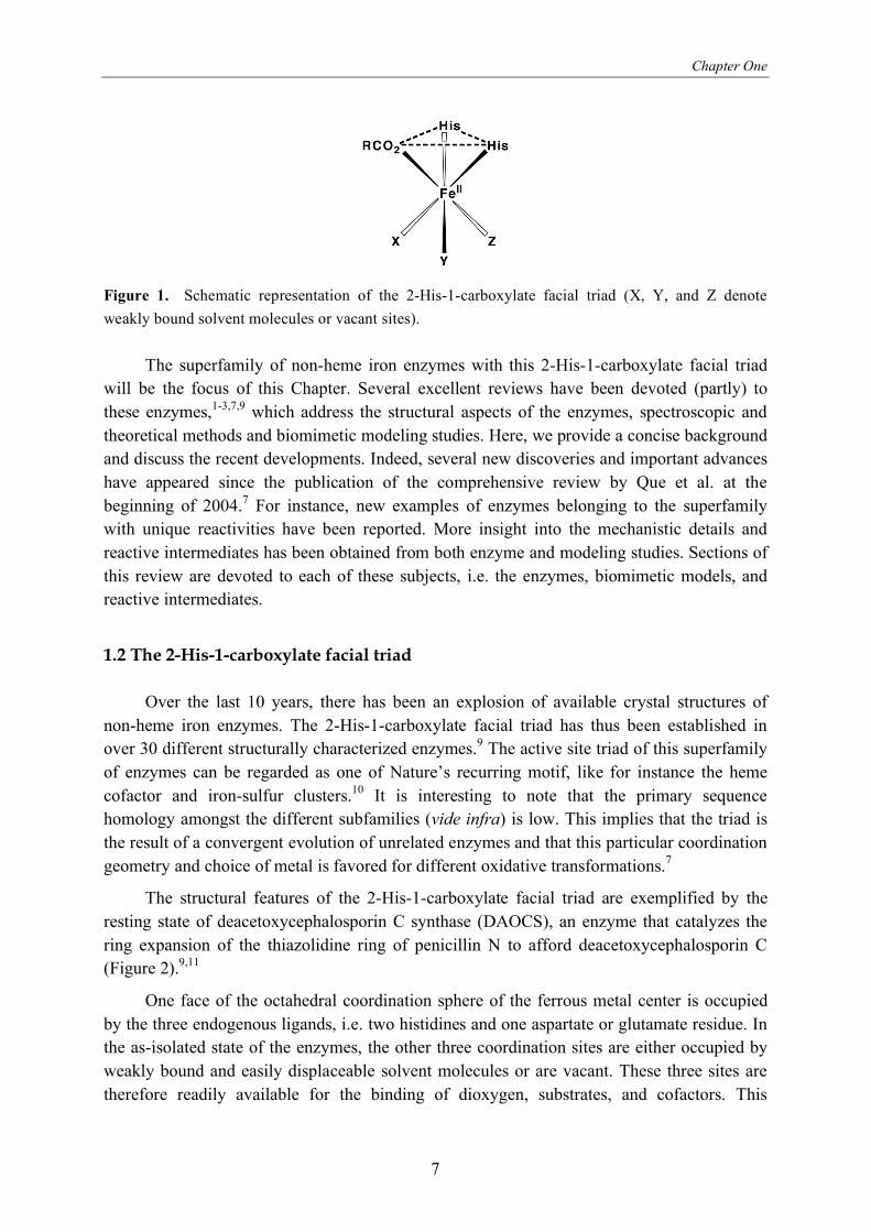
Figure 1. Schematic representation of the 2-His-1-carboxylate facial triad (X, Y, and Z denote
weakly bound solvent molecules or vacant sites).
The superfamily of non-heme iron enzymes with this 2-His-1-carboxylate facial triad
will be the focus of this Chapter. Several excellent reviews have been devoted (partly) to
these enzymes,1-3,7,9 which address the structural aspects of the enzymes, spectroscopic and
theoretical methods and biomimetic modeling studies. Here, we provide a concise background
and discuss the recent developments. Indeed, several new discoveries and important advances
have appeared since the publication of the comprehensive review by Que et al. at the
beginning of 2004.7 For instance, new examples of enzymes belonging to the superfamily
with unique reactivities have been reported. More insight into the mechanistic details and
reactive intermediates has been obtained from both enzyme and modeling studies. Sections of
this review are devoted to each of these subjects, i.e. the enzymes, biomimetic models, and
reactive intermediates.
1.2 The 2-His-1-carboxylate facial triad
Over the last 10 years, there has been an explosion of available crystal structures of
non-heme iron enzymes. The 2-His-1-carboxylate facial triad has thus been established in
over 30 different structurally characterized enzymes.9 The active site triad of this superfamily
of enzymes can be regarded as one of Nature’s recurring motif, like for instance the heme
cofactor and iron-sulfur clusters.10 It is interesting to note that the primary sequence
homology amongst the different subfamilies (vide infra) is low. This implies that the triad is
the result of a convergent evolution of unrelated enzymes and that this particular coordination
geometry and choice of metal is favored for different oxidative transformations.7
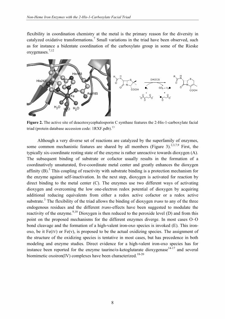
The structural features of the 2-His-1-carboxylate facial triad are exemplified by the
resting state of deacetoxycephalosporin C synthase (DAOCS), an enzyme that catalyzes the
ring expansion of the thiazolidine ring of penicillin N to afford deacetoxycephalosporin C
(Figure 2).9,11
One face of the octahedral coordination sphere of the ferrous metal center is occupied
by the three endogenous ligands, i.e. two histidines and one aspartate or glutamate residue. In
the as-isolated state of the enzymes, the other three coordination sites are either occupied by
weakly bound and easily displaceable solvent molecules or are vacant. These three sites are
therefore readily available for the binding of dioxygen, substrates, and cofactors. This
Non-Heme Iron Enzymes with the 2-His-1-Carboxylate Facial Triad
8
flexibility in coordination chemistry at the metal is the primary reason for the diversity in
catalyzed oxidative transformations.7 Small variations in the triad have been observed, such
as for instance a bidentate coordination of the carboxylato group in some of the Rieske
oxygenases.7,12
Figure 2. The active site of deacetoxycephalosporin C synthase features the 2-His-1-carboxylate facial
triad (protein database accession code: 1RXF.pdb).13
Although a very diverse set of reactions are catalyzed by the superfamily of enzymes,
some common mechanistic features are shared by all members (Figure 3).2,3,7,9 First, the
typically six-coordinate resting state of the enzyme is rather unreactive towards dioxygen (A).
The subsequent binding of substrate or cofactor usually results in the formation of a
coordinatively unsaturated, five-coordinate metal center and greatly enhances the dioxygen
affinity (B).3 This coupling of reactivity with substrate binding is a protection mechanism for
the enzyme against self-inactivation. In the next step, dioxygen is activated for reaction by
direct binding to the metal center (C). The enzymes use two different ways of activating
dioxygen and overcoming the low one-electron redox potential of dioxygen by acquiring
additional reducing equivalents from either a redox active cofactor or a redox active
substrate.3 The flexibility of the triad allows the binding of dioxygen trans to any of the three
endogenous residues and the different trans-effects have been suggested to modulate the
reactivity of the enzyme.9,10 Dioxygen is then reduced to the peroxide level (D) and from this
point on the proposed mechanisms for the different enzymes diverge. In most cases O–O
bond cleavage and the formation of a high-valent iron-oxo species is invoked (E). This iron-
oxo, be it Fe(IV) or Fe(v), is proposed to be the actual oxidizing species. The assignment of
the structure of the oxidizing species is tentative in most cases, but has precedence in both
modeling and enzyme studies. Direct evidence for a high-valent iron-oxo species has for
instance been reported for the enzyme taurine/ -ketoglutarate dioxygenase14-17 and several
biomimetic oxoiron(IV) complexes have been characterized.18-20

Chapter One
9
Fe
OH2H2O OH2
Fe
XX
Fe
XO2 X
Fe
X(R)O2 X
Fe
XO X
substrate/cofactor (X)
O2product(s)
(A) (B)
(C)(D)(E)
Figure 3. General mechanistic pathway for reactions catalyzed by the 2-His-1-carboxylate facial triad
superfamily of non-heme iron(II) enzymes. Picture adapted from Que et al.9
The enzymes featuring the 2-His-1-carboxylate facial triad can be classified into five
different groups based on their specific requirements for catalysis. These groups are the 1)
extradiol cleaving catechol dioxygenases, 2) Rieske oxygenases, 3) -ketoglutarate dependent
enzymes, 4) pterin dependent hydroxylases, and finally 5) a miscellaneous category. The
characteristics, recent developments, and illustrative examples of each group will be
discussed.
1. Extradiol cleaving catechol dioxygenases. Oxidative ring cleavage is a key
metabolic step in the biodegradation of aromatic compounds by bacteria.21 The common
metabolic pathway is the ring fission of catecholic substrates and is catalyzed by the extradiol
cleaving catechol dioxygenases.2,7,21,22 These enzymes utilize a non-heme iron(II) active site
(or in a few cases MnII)23 to cleave the C–C bond next to the two hydroxyl groups with
incorporation of both atoms of dioxygen in the substrate (Figure 4). Their intradiol
counterparts, which represent a minor pathway, utilize a non-heme iron(III) active site to
cleave the C–C bond in between the two hydroxyl groups. The extradiol cleaving
dioxygenases are more versatile and in addition to catecholic substrates also accept gentisate,
salicylate, hydroquinone and 2-aminophenol.21 The biological ins and outs of the catechol
cleaving dioxygenases have been very recently reviewed comprehensively.21
OH
OH2
12. Intradiol
dioxygenases,
FeIII, O2
O
H
OHOH
O
O
OH
O
OH
1. Extradioldioxygenases,
FeII, O2
Figure 4. The extradiol and intradiol catechol cleavage pathways catalyzed by the catechol cleaving
dioxygenases.
Three evolutionary independent classes of extradiol enzymes have been identified
which all share similar active sites and all feature the 2-His-1-carboxylate facial triad.21 In the
Non-Heme Iron Enzymes with the 2-His-1-Carboxylate Facial Triad
10
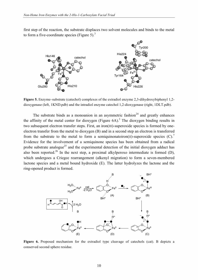
first step of the reaction, the substrate displaces two solvent molecules and binds to the metal
to form a five-coordinate species (Figure 5).3
Figure 5. Enzyme–substrate (catechol) complexes of the extradiol enzyme 2,3-dihydroxybiphenyl 1,2-
dioxygenase (left, 1KND.pdb) and the intradiol enzyme catechol 1,2-dioxygenase (right, 1DLT.pdb).
The substrate binds as a monoanion in an asymmetric fashion24 and greatly enhances
the affinity of the metal center for dioxygen (Figure 6A).3 The dioxygen binding results in
two subsequent electron transfer steps. First, an iron(III)-superoxide species is formed by one-
electron transfer from the metal to dioxygen (B) and in a second step an electron is transferred
from the substrate to the metal to form a semiquinonatoiron(II)-superoxide species (C).7
Evidence for the involvement of a semiquinone species has been obtained from a radical
probe substrate analogue25 and the experimental detection of the initial dioxygen adduct has
also been reported.26 In the next step, a proximal alkylperoxo intermediate is formed (D),
which undergoes a Criegee rearrangement (alkenyl migration) to form a seven-membered
lactone species and a metal bound hydroxide (E). The latter hydrolyzes the lactone and the
ring-opened product is formed.
FeII
O
OFeII
H2O
H2OH
B
FeIII
O
O
BH+
OO
BH+ BH+
FeII
O
O
B
O
OH+
FeII
O
O
B
OO
H+
FeII
O
O
B
OHO
cat-2 H2O
O2
2 H2OO
H
OHOH
O
(A) (B)
(C)(D)(E)
Figure 6. Proposed mechanism for the extradiol type cleavage of catechols (cat). B depicts a
conserved second sphere residue.

Chapter One
11
The origin of the respective regioselectivities of the extradiol and intradiol cleaving
catechol dioxygenases is not completely clear and is the subject of current research. The
active sites of the enzyme-substrate complexes of the extradiol and intradiol enzymes are
rather similar (Figure 5) and also for the intradiol cleaving enzymes a proximal alkylperoxo
intermediate is proposed. In the case of intradiol cleavage, however, a different Criegee
rearrangement would lead to the formation of an anhydride instead of a lactone. Several
different explanations have been offered for the observed difference in regiochemistry. For
instance, the exact coordination geometry,7 stereoelectronic factors,22 and acid-base chemistry
of highly conserved second-sphere residues27 have been suggested as decisive factors. Site-
directed mutagenesis studies have provided considerable evidence that the latter factor
determines the final outcome of the cleavage.26,28,29 The extradiol and intradiol cleaving
catechol dioxygenases are described in more detail in Chapters 3 and 4.
2. Rieske oxygenases. A common first step in the biodegradation of aromatic
compounds is their conversion into cis-dihydroxylated metabolites. The regio- and
stereospecific cis-dihydroxylation of arenas is catalyzed by Rieske non-heme iron
dioxygenases.7,12 Aromatic hydrocarbons are common contaminants of soil and groundwater
and the Rieske dioxygenases provide an attractive way of biodegradation of these pollutants.30
This, together with the fact that there is little precedent for its unique reactivity in synthetic
organic chemistry,7,31 has spurred a widespread interest in this group of enzymes. Rieske
oxygenases have furthermore been shown to be very versatile. Next to cis-dihydroxylation,
also other oxidations such as monohydroxylations, desaturation, sulfoxidation, O- and N-
dealkylation, and amine oxidation are catalyzed.7,32-35 Examples of some selected reactions
catalyzed by Rieske oxygenases are shown in Figure 7.
OH
OH NH
O NH
O
OH
ClNH2
NH
Cl
ClNO2
NH
Cl
Cl
Cl
O O
OHCl
Cl
OH O
OH
NDO
PrnD
OMO
DIC
Naphthalene dioxygenase 2-Oxoquinoline 8-monooxygenase
Aminopyrrolnitrin oxygenase Dicamba O-demethylase
Figure 7. Some examples of reactions catalyzed by different Rieske oxygenases.
The Rieske oxygenases are multicomponent enzymes and consist of a reductase, an
oxygenase, and in some cases also a ferredoxin component. Substrate oxidation takes place in
the oxygenase component, which contains both a Rieske-type [2Fe-2S] cluster and the
mononuclear non-heme iron active site. The Rieske cluster and the non-heme iron center are
too far apart in a single subunit to allow for electron transfer. However, the quaternary
trimeric structure allows for electron transfer of a Rieske cluster and a mononuclear iron
Non-Heme Iron Enzymes with the 2-His-1-Carboxylate Facial Triad
12
center from two different subunits. A key role in the electron transfer has been ascribed to a
fully conserved aspartic acid residue that bridges between the two metal sites.36 The two
additional electrons needed for the full reduction of dioxygen are supplied by NAD(P)H and
shuttled to the iron active site via the reductase and the Rieske cluster. The structural data on
the oxygenase component of Rieske oxygenases has long been limited to the crystal structure
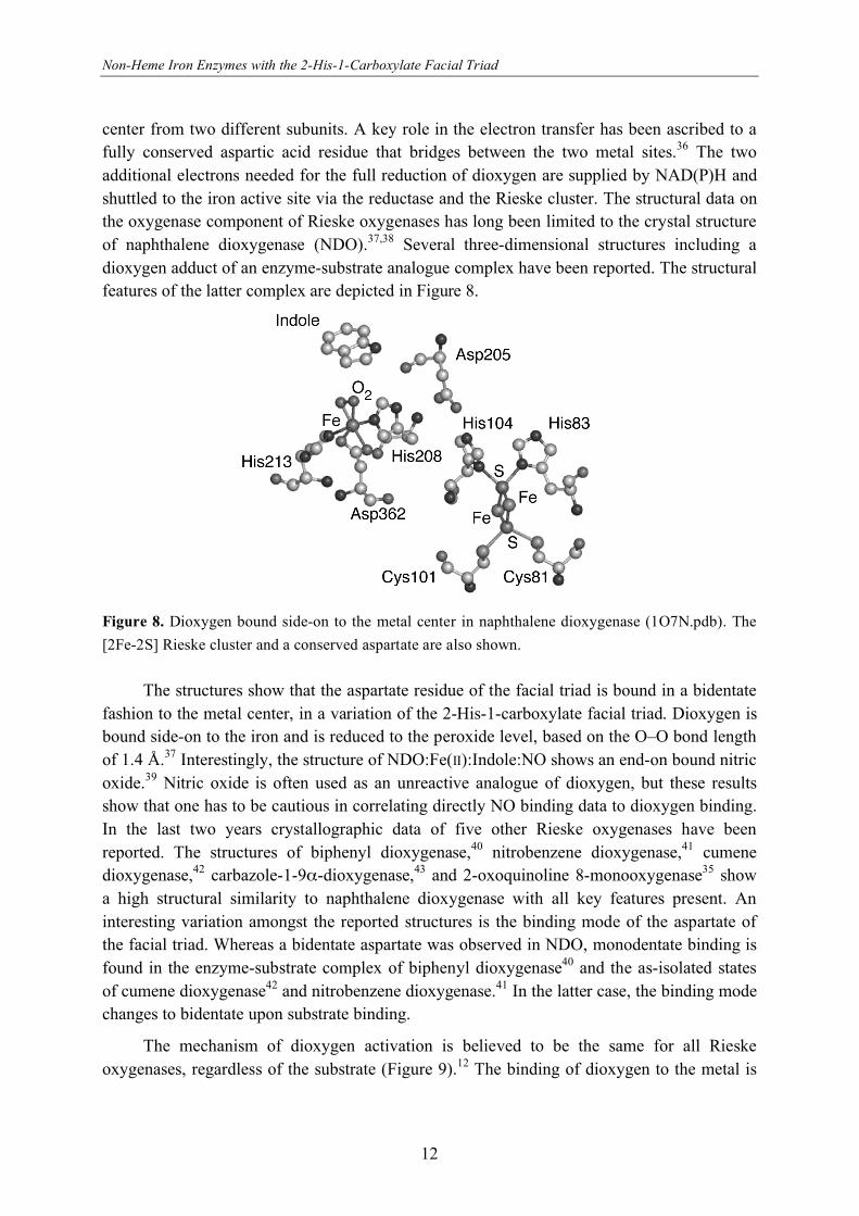
of naphthalene dioxygenase (NDO).37,38 Several three-dimensional structures including a
dioxygen adduct of an enzyme-substrate analogue complex have been reported. The structural
features of the latter complex are depicted in Figure 8.
Figure 8. Dioxygen bound side-on to the metal center in naphthalene dioxygenase (1O7N.pdb). The
[2Fe-2S] Rieske cluster and a conserved aspartate are also shown.
The structures show that the aspartate residue of the facial triad is bound in a bidentate
fashion to the metal center, in a variation of the 2-His-1-carboxylate facial triad. Dioxygen is
bound side-on to the iron and is reduced to the peroxide level, based on the O–O bond length
of 1.4 Å.37 Interestingly, the structure of NDO:Fe(II):Indole:NO shows an end-on bound nitric
oxide.39 Nitric oxide is often used as an unreactive analogue of dioxygen, but these results
show that one has to be cautious in correlating directly NO binding data to dioxygen binding.
In the last two years crystallographic data of five other Rieske oxygenases have been
reported. The structures of biphenyl dioxygenase,40 nitrobenzene dioxygenase,41 cumene
dioxygenase,42 carbazole-1-9 -dioxygenase,43 and 2-oxoquinoline 8-monooxygenase35 show
a high structural similarity to naphthalene dioxygenase with all key features present. An
interesting variation amongst the reported structures is the binding mode of the aspartate of
the facial triad. Whereas a bidentate aspartate was observed in NDO, monodentate binding is
found in the enzyme-substrate complex of biphenyl dioxygenase40 and the as-isolated states
of cumene dioxygenase42 and nitrobenzene dioxygenase.41 In the latter case, the binding mode
changes to bidentate upon substrate binding.
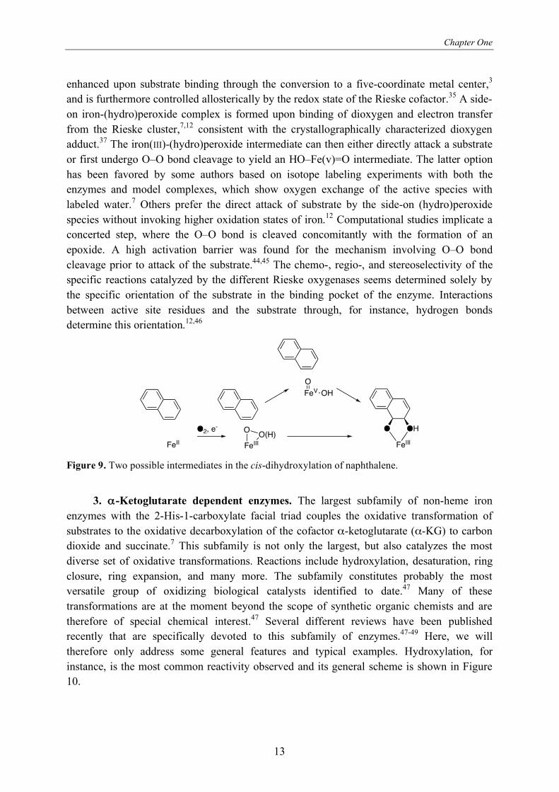
The mechanism of dioxygen activation is believed to be the same for all Rieske
oxygenases, regardless of the substrate (Figure 9).12 The binding of dioxygen to the metal is
Chapter One
13
enhanced upon substrate binding through the conversion to a five-coordinate metal center,3
and is furthermore controlled allosterically by the redox state of the Rieske cofactor.35 A side-
on iron-(hydro)peroxide complex is formed upon binding of dioxygen and electron transfer
from the Rieske cluster,7,12 consistent with the crystallographically characterized dioxygen
adduct.37 The iron(III)-(hydro)peroxide intermediate can then either directly attack a substrate
or first undergo O–O bond cleavage to yield an HO–Fe(v)=O intermediate. The latter option
has been favored by some authors based on isotope labeling experiments with both the
enzymes and model complexes, which show oxygen exchange of the active species with
labeled water.7 Others prefer the direct attack of substrate by the side-on (hydro)peroxide
species without invoking higher oxidation states of iron.12 Computational studies implicate a
concerted step, where the O–O bond is cleaved concomitantly with the formation of an
epoxide. A high activation barrier was found for the mechanism involving O–O bond
cleavage prior to attack of the substrate.44,45 The chemo-, regio-, and stereoselectivity of the
specific reactions catalyzed by the different Rieske oxygenases seems determined solely by
the specific orientation of the substrate in the binding pocket of the enzyme. Interactions
between active site residues and the substrate through, for instance, hydrogen bonds
determine this orientation.12,46
FeII FeIII
OO(H)
FeIII
O OH
FeVO
OH
O2, e-
Figure 9. Two possible intermediates in the cis-dihydroxylation of naphthalene.
3. -Ketoglutarate dependent enzymes. The largest subfamily of non-heme iron
enzymes with the 2-His-1-carboxylate facial triad couples the oxidative transformation of
substrates to the oxidative decarboxylation of the cofactor -ketoglutarate ( -KG) to carbon
dioxide and succinate.7 This subfamily is not only the largest, but also catalyzes the most
diverse set of oxidative transformations. Reactions include hydroxylation, desaturation, ring
closure, ring expansion, and many more. The subfamily constitutes probably the most
versatile group of oxidizing biological catalysts identified to date.47 Many of these
transformations are at the moment beyond the scope of synthetic organic chemists and are
therefore of special chemical interest.47 Several different reviews have been published
recently that are specifically devoted to this subfamily of enzymes.47-49 Here, we will
therefore only address some general features and typical examples. Hydroxylation, for
instance, is the most common reactivity observed and its general scheme is shown in Figure
10.

Non-Heme Iron Enzymes with the 2-His-1-Carboxylate Facial Triad
14
O
OO
O
O
OO
OOFeII, O2
R H R OH+ + + CO2
Figure 10. General hydroxylation reaction catalyzed by several -KG dependent oxygenases.
Two types of hydroxylation reactions have recently received increased attention out of
medical interest. Damage of RNA and DNA by nucleotide alkylation results in lesions that
are both cytotoxic and mutagenic.50 Several -KG dependent oxygenases have been shown to
repair these alkylated DNA and RNA bases. Escherichia coli AlkB and the homologous
human enzyme ABH3 fix the lesion by hydroxylating the alkyl group, after which
spontaneous deformylation yields the unmodified base (Figure 11A).50,51
NR
OR
NR
OR
OH
OP
HO HH
OP
HO OHH
NN
O
NH2
CH3O
R1OPO
POH/OH
NN
O
NH2
CH2OHO
R1OPO
POH/OH
NN
O
NH2
O
R1OPO
POH/OH
+O
HH
B C
A
NOHO2C
RN
OHO2C
R
OH
NO
O
CO2H
R
NO
O
CO2H
R
D
Figure 11. Some selected reactions catalyzed by -KG dependent oxygenases. A) Nucleobase
demethylation by DNA/RNA repair enzyme AlkB; B) Stereospecific proline hydroxylation by proline
4-hydroxylase; C) Hypophosphite hydroxylation by hypophosphite/ -KG dioxygenase HtxA; D)
Hydroxylation, ring closure and desaturation reactions in the synthesis of clavulanic acid by
clavaminate synthase CAS.
Other, related demethylation reactions catalyzed by -KG dependent enzymes, such as
histone demethylation, have been reported recently.52,53 The hydroxylation of specific
residues of protein side chains and more specifically in oxygen sensing in the cell has also
attracted recent interest. Hypoxia-inducible factor (HIF) is responsible for mediating the
mammalian response to low oxygen tension (hypoxia). -KG dependent HIF hydroxylases
have been implicated in this hypoxic response and are therefore interesting targets for the
development of new therapies for the different diseases associated with this system.54 Other
functions of the -KG dependent oxygenases include the biosynthesis of antibiotics and plant
products, lipid metabolism and biodegradation.48 Some selected examples are shown in
Figure 11.
Chapter One
15
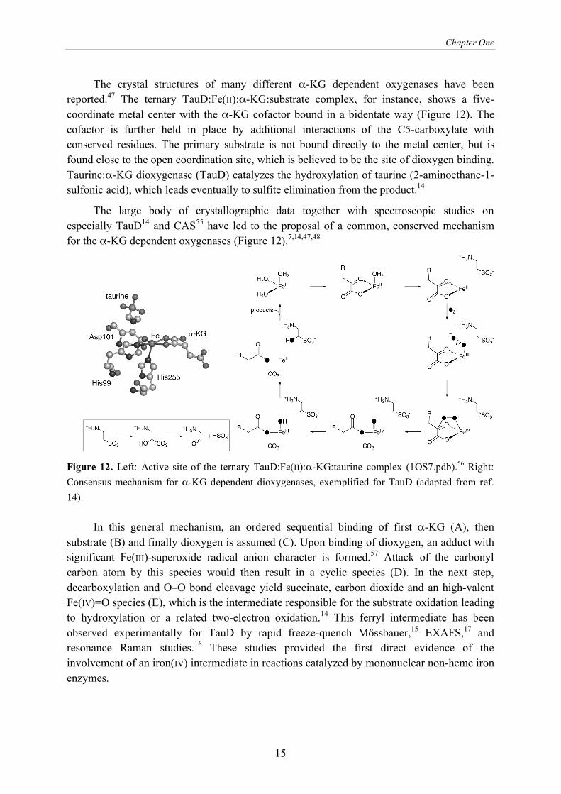
The crystal structures of many different -KG dependent oxygenases have been
reported.47 The ternary TauD:Fe(II): -KG:substrate complex, for instance, shows a five-
coordinate metal center with the -KG cofactor bound in a bidentate way (Figure 12). The
cofactor is further held in place by additional interactions of the C5-carboxylate with
conserved residues. The primary substrate is not bound directly to the metal center, but is
found close to the open coordination site, which is believed to be the site of dioxygen binding.
Taurine: -KG dioxygenase (TauD) catalyzes the hydroxylation of taurine (2-aminoethane-1-
sulfonic acid), which leads eventually to sulfite elimination from the product.14
The large body of crystallographic data together with spectroscopic studies on
especially TauD14 and CAS55 have led to the proposal of a common, conserved mechanism
for the -KG dependent oxygenases (Figure 12).7,14,47,48
Figure 12. Left: Active site of the ternary TauD:Fe(II): -KG:taurine complex (1OS7.pdb).56 Right:
Consensus mechanism for -KG dependent dioxygenases, exemplified for TauD (adapted from ref.
14).
In this general mechanism, an ordered sequential binding of first -KG (A), then
substrate (B) and finally dioxygen is assumed (C). Upon binding of dioxygen, an adduct with
significant Fe(III)-superoxide radical anion character is formed.57 Attack of the carbonyl
carbon atom by this species would then result in a cyclic species (D). In the next step,
decarboxylation and O–O bond cleavage yield succinate, carbon dioxide and an high-valent
Fe(IV)=O species (E), which is the intermediate responsible for the substrate oxidation leading
to hydroxylation or a related two-electron oxidation.14 This ferryl intermediate has been
observed experimentally for TauD by rapid freeze-quench Mössbauer,15 EXAFS,17 and
resonance Raman studies.16 These studies provided the first direct evidence of the
involvement of an iron(IV) intermediate in reactions catalyzed by mononuclear non-heme iron
enzymes.
Non-Heme Iron Enzymes with the 2-His-1-Carboxylate Facial Triad
16
The -KG-dependent non-heme iron enzymes (4-hydroxyphenyl)pyruvate dioxygenase
(HppD)58 and (4-hydroxy)mandalate synthase (HmaS)59 form an interesting pair in the sense
that they use the same substrate, (4-hydroxyphenyl)pyruvate which itself has an -keto acid
moiety (Figure 13). As a result both atoms of dioxygen are incorporated into the product,
similar to the extradiol cleaving dioxygenases. HppD yields the aromatic hydroxylated
product homogentisate using an electrophilic attack mechanism followed by an NIH shift,
whereas HmaS generates the benzylic hydroxylated product (S)-(4-hydroxy)mandalate via the
hydrogen abstraction mechanism.60 The two enzymes generate the same reactive Fe(IV)=O
intermediate, but are able to steer the reaction into two different directions. The decisive
factor was found to be the exact orientation of the substrate in the binding pocket.60
OH
O
OO
OH
OHO
O
OH
OH
O
OHmaS HppD
O2CO2 O2 CO2
Figure 13. Oxidative transformations of (4-hydroxyphenyl)pyruvate catalyzed by HmaS and HppD.
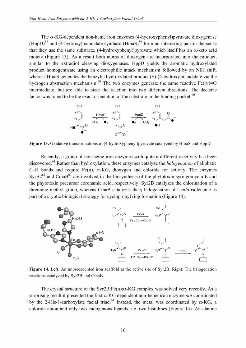
Recently, a group of non-heme iron enzymes with quite a different reactivity has been
discovered.61 Rather than hydroxylation, these enzymes catalyze the halogenation of aliphatic
C–H bonds and require Fe(II), -KG, dioxygen and chloride for activity. The enzymes
SyrB262 and CmaB63 are involved in the biosynthesis of the phytotoxin syringomycin E and
the phytotoxin precursor coronamic acid, respectively. Syr2B catalyzes the chlorination of a
threonine methyl group, whereas CmaB catalyzes the -halogenation of L-allo-isoleucine as
part of a cryptic biological strategy for cyclopropyl ring formation (Figure 14).
Figure 14. Left: An unprecedented iron scaffold at the active site of Syr2B. Right: The halogenation
reactions catalyzed by Syr2B and CmaB.
The crystal structure of the Syr2B:Fe(II): -KG complex was solved very recently. As a
surprising result it presented the first -KG dependent non-heme iron enzyme not coordinated
by the 2-His-1-carboxylate facial triad.64 Instead, the metal was coordinated by -KG, a
chloride anion and only two endogenous ligands. i.e. two histidines (Figure 14). An alanine
Chapter One
17
replaced the aspartate of the facial triad. The mechanism of halogenation is thought to be
similar to the common hydroxylation with chloride abstraction instead of hydroxyl abstraction
by the carbon radical in the final step.64 The non-heme iron halogenation catalysts are not
limited to these two enzymes as bioinformatic analysis has provided leads to additional
members of this interesting new group.61
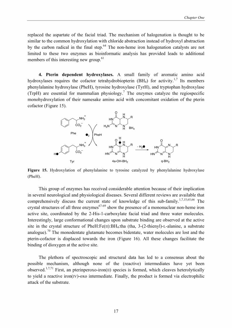
4. Pterin dependent hydroxylases. A small family of aromatic amino acid
hydroxylases requires the cofactor tetrahydrobiopterin (BH4) for activity.1,7 Its members
phenylalanine hydroxylase (PheH), tyrosine hydroxylase (TyrH), and tryptophan hydroxylase
(TrpH) are essential for mammalian physiology.7 The enzymes catalyze the regiospecific
monohydroxylation of their namesake amino acid with concomitant oxidation of the pterin
cofactor (Figure 15).
NH3
CO2
NH3
CO2HO
Phe
Tyr
HN
N NH
HN
H2N
O
R
HN
N NH
HN
HN
O
RO
H
HN
N NH
N
HN
O
R
O2 PheH
BH4
4a-OH-BH2 q-BH2
- H2O
Figure 15. Hydroxylation of phenylalanine to tyrosine catalyzed by phenylalanine hydroxylase
(PheH).
This group of enzymes has received considerable attention because of their implication
in several neurological and physiological diseases. Several different reviews are available that
comprehensively discuss the current state of knowledge of this sub-family.1,7,15,65,66 The
crystal structures of all three enzymes67-69 show the presence of a mononuclear non-heme iron
active site, coordinated by the 2-His-1-carboxylate facial triad and three water molecules.
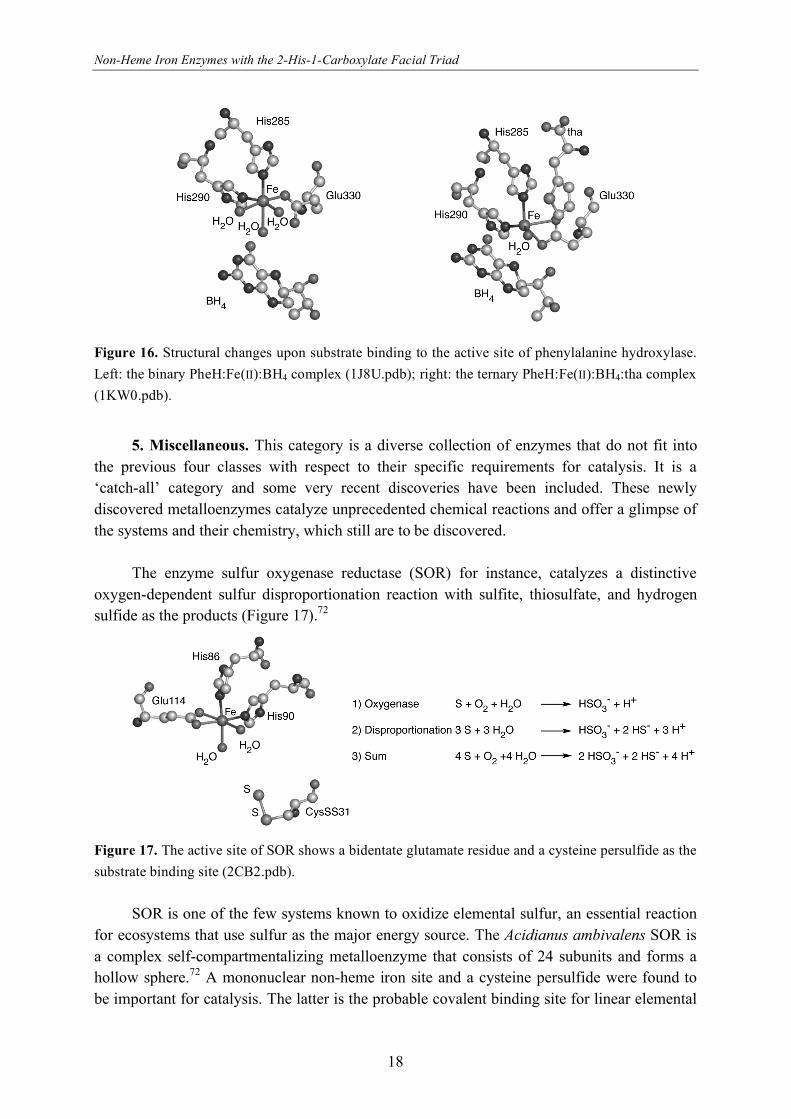
Interestingly, large conformational changes upon substrate binding are observed at the active
site in the crystal structure of PheH:Fe(II):BH4:tha (tha, 3-(2-thienyl)-L-alanine, a substrate
analogue).70 The monodentate glutamate becomes bidentate, water molecules are lost and the
pterin-cofactor is displaced towards the iron (Figure 16). All these changes facilitate the
binding of dioxygen at the active site.
The plethora of spectroscopic and structural data has led to a consensus about the
possible mechanism, although none of the (reactive) intermediates have yet been
observed.1,7,71 First, an pterinperoxo-iron(II) species is formed, which cleaves heterolytically
to yield a reactive iron(IV)-oxo intermediate. Finally, the product is formed via electrophilic
attack of the substrate.
Non-Heme Iron Enzymes with the 2-His-1-Carboxylate Facial Triad
18
Figure 16. Structural changes upon substrate binding to the active site of phenylalanine hydroxylase.
Left: the binary PheH:Fe(II):BH4 complex (1J8U.pdb); right: the ternary PheH:Fe(II):BH4:tha complex
(1KW0.pdb).
5. Miscellaneous. This category is a diverse collection of enzymes that do not fit into
the previous four classes with respect to their specific requirements for catalysis. It is a
‘catch-all’ category and some very recent discoveries have been included. These newly
discovered metalloenzymes catalyze unprecedented chemical reactions and offer a glimpse of
the systems and their chemistry, which still are to be discovered.
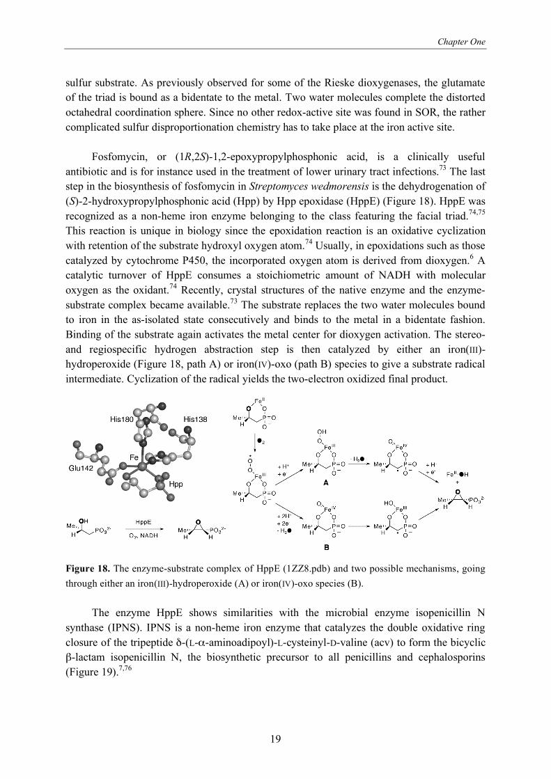
The enzyme sulfur oxygenase reductase (SOR) for instance, catalyzes a distinctive
oxygen-dependent sulfur disproportionation reaction with sulfite, thiosulfate, and hydrogen
sulfide as the products (Figure 17).72
Figure 17. The active site of SOR shows a bidentate glutamate residue and a cysteine persulfide as the
substrate binding site (2CB2.pdb).
SOR is one of the few systems known to oxidize elemental sulfur, an essential reaction
for ecosystems that use sulfur as the major energy source. The Acidianus ambivalens SOR is
a complex self-compartmentalizing metalloenzyme that consists of 24 subunits and forms a
hollow sphere.72 A mononuclear non-heme iron site and a cysteine persulfide were found to
be important for catalysis. The latter is the probable covalent binding site for linear elemental
Chapter One
19
sulfur substrate. As previously observed for some of the Rieske dioxygenases, the glutamate
of the triad is bound as a bidentate to the metal. Two water molecules complete the distorted
octahedral coordination sphere. Since no other redox-active site was found in SOR, the rather
complicated sulfur disproportionation chemistry has to take place at the iron active site.
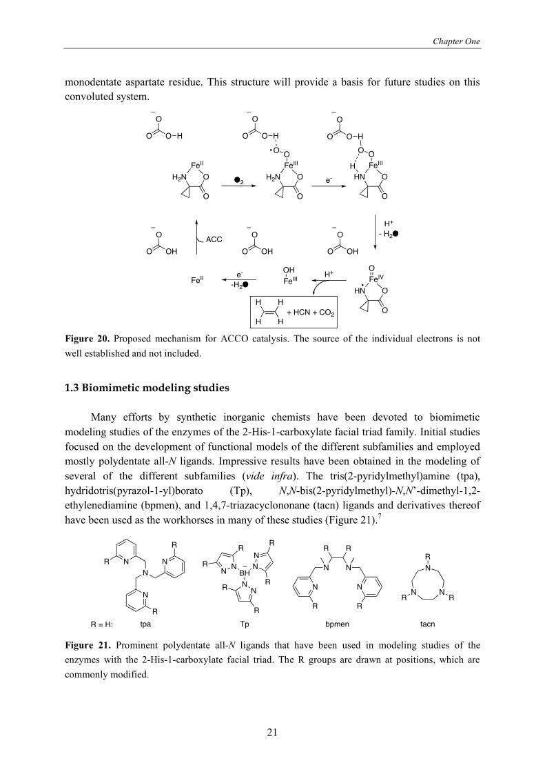
Fosfomycin, or (1R,2S)-1,2-epoxypropylphosphonic acid, is a clinically useful
antibiotic and is for instance used in the treatment of lower urinary tract infections.73 The last
step in the biosynthesis of fosfomycin in Streptomyces wedmorensis is the dehydrogenation of
(S)-2-hydroxypropylphosphonic acid (Hpp) by Hpp epoxidase (HppE) (Figure 18). HppE was
recognized as a non-heme iron enzyme belonging to the class featuring the facial triad.74,75
This reaction is unique in biology since the epoxidation reaction is an oxidative cyclization
with retention of the substrate hydroxyl oxygen atom.74 Usually, in epoxidations such as those
catalyzed by cytochrome P450, the incorporated oxygen atom is derived from dioxygen.6 A
catalytic turnover of HppE consumes a stoichiometric amount of NADH with molecular
oxygen as the oxidant.74 Recently, crystal structures of the native enzyme and the enzyme-
substrate complex became available.73 The substrate replaces the two water molecules bound
to iron in the as-isolated state consecutively and binds to the metal in a bidentate fashion.
Binding of the substrate again activates the metal center for dioxygen activation. The stereo-
and regiospecific hydrogen abstraction step is then catalyzed by either an iron(III)-
hydroperoxide (Figure 18, path A) or iron(IV)-oxo (path B) species to give a substrate radical
intermediate. Cyclization of the radical yields the two-electron oxidized final product.
Figure 18. The enzyme-substrate complex of HppE (1ZZ8.pdb) and two possible mechanisms, going
through either an iron(III)-hydroperoxide (A) or iron(IV)-oxo species (B).
The enzyme HppE shows similarities with the microbial enzyme isopenicillin N
synthase (IPNS). IPNS is a non-heme iron enzyme that catalyzes the double oxidative ring
closure of the tripeptide -(L- -aminoadipoyl)-L-cysteinyl-D-valine (acv) to form the bicyclic
-lactam isopenicillin N, the biosynthetic precursor to all penicillins and cephalosporins
(Figure 19).7,76

Non-Heme Iron Enzymes with the 2-His-1-Carboxylate Facial Triad
20
Figure 19. Active site of the ternary IPNS:Fe(II):acv:NO complex and the oxidative double ring
closure catalyzed by IPNS.
IPNS shows a high sequence homology to the -ketoglutarate dependent enzymes, but
does not require -ketoglutarate as cofactor and it does not incorporate oxygen into the
product.7 In the case of IPNS all four electrons required for the reduction of dioxygen to water
are provided by the substrate. Crystal structures are available for different stages of the
reaction,77-79 such as for instance the ternary IPNS:Fe(II):acv:NO complex.79 Nitric oxide
(NO) is often used as an unreactive O2-surrogate to get more insight into the interaction of
dioxygen with the metal. Based on this information and comprehensive studies with substrate
analogues,7,76 a mechanism has been proposed. The closure of the -lactam precedes the
formation of the thiazolidine ring, which is mediated by a reactive iron(IV)-oxo intermediate
(Figure 19). The enzymology of IPNS and other enzymes involved in the biosynthesis of -
lactam derived compounds has been recently reviewed.76
The enzyme 1-aminocyclopropane-1-carboxylic acid oxidase (ACCO) also shows a
high sequence homology to the -ketoglutarate dependent enzymes, but like IPNS does not
require -ketoglutarate as cofactor.7 ACCO is an enzyme that produces the plant hormone
ethylene, which regulates many aspects of plant growth and development. It couples the two-
electron oxidation of the unusual amino acid 1-aminocyclopropane-1-carboxylic acid (acc) to
give ethylene, CO2 and HCN.7 Continuous turnover requires the presence of ascorbate and
bicarbonate. Due to the lack of available structural information and the inherent complexity of
the system, the mechanism of ACCO is not yet fully understood.80,81 It is known that the
binding of substrate acc activates the iron center in the enzyme for dioxygen by converting it
from six- to five-coordinate.3,82 Indeed, the ternary ACCO:Fe(II):acc:NO complex has been
observed by ENDOR spectroscopy.83 Based on single turnover studies, ascorbate has been
ascribed the traditional role of preferred two-electron reductant.84 In addition, ascorbate might
serve a second role by binding at a specific effector site and thus facilitating the formation of
the ternary enzyme-substrate-dioxygen complex. Bicarbonate is then proposed to be involved
in the generation of the reactive species by a specific protonation step.84
The mechanism proposed by Que et al.84 is shown in Figure 20. Recently, the first
crystal structure of ACCO was reported,85 featuring the 2-His-1-carboxylate facial triad with a
Chapter One
21
monodentate aspartate residue. This structure will provide a basis for future studies on this
convoluted system.
O2O
O
H2N
FeII
O
O O
O
O
H2N
FeIII
O
O O
OO
HH
O
O
HN
FeIII
O
O O
OO
H
H
e-
O
O
HN
FeIV
O
O OH
O
H+
- H2O
FeIIIOH
FeII
H
H H
H+ HCN + CO2
H+
O
O OH
O
O OH
ACC
e-
-H2O
Figure 20. Proposed mechanism for ACCO catalysis. The source of the individual electrons is not
well established and not included.
1.3 Biomimetic modeling studies
Many efforts by synthetic inorganic chemists have been devoted to biomimetic
modeling studies of the enzymes of the 2-His-1-carboxylate facial triad family. Initial studies
focused on the development of functional models of the different subfamilies and employed
mostly polydentate all-N ligands. Impressive results have been obtained in the modeling of
several of the different subfamilies (vide infra). The tris(2-pyridylmethyl)amine (tpa),
hydridotris(pyrazol-1-yl)borato (Tp), N,N-bis(2-pyridylmethyl)-N,N’-dimethyl-1,2-
ethylenediamine (bpmen), and 1,4,7-triazacyclononane (tacn) ligands and derivatives thereof
have been used as the workhorses in many of these studies (Figure 21).7
N
N
N N
tpa
BHNN
N
N
N
N
NN
N N
R
R
R
R
R
R
R
R
R
R R
R R
N
N NR
R
R
Tp bpmen tacnR = H:
Figure 21. Prominent polydentate all-N ligands that have been used in modeling studies of the
enzymes with the 2-His-1-carboxylate facial triad. The R groups are drawn at positions, which are
commonly modified.
Non-Heme Iron Enzymes with the 2-His-1-Carboxylate Facial Triad
22
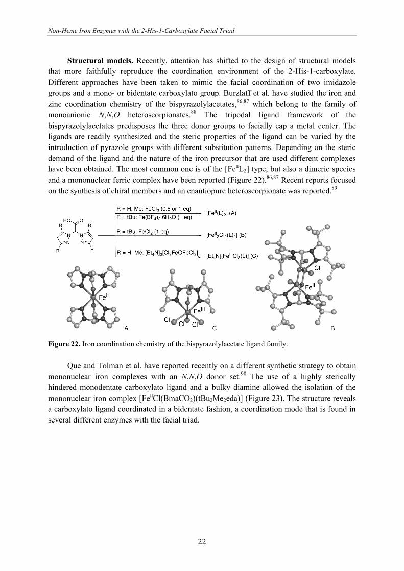
Structural models. Recently, attention has shifted to the design of structural models
that more faithfully reproduce the coordination environment of the 2-His-1-carboxylate.
Different approaches have been taken to mimic the facial coordination of two imidazole
groups and a mono- or bidentate carboxylato group. Burzlaff et al. have studied the iron and
zinc coordination chemistry of the bispyrazolylacetates,86,87 which belong to the family of
monoanionic N,N,O heteroscorpionates.88 The tripodal ligand framework of the
bispyrazolylacetates predisposes the three donor groups to facially cap a metal center. The
ligands are readily synthesized and the steric properties of the ligand can be varied by the
introduction of pyrazole groups with different substitution patterns. Depending on the steric
demand of the ligand and the nature of the iron precursor that are used different complexes
have been obtained. The most common one is of the [FeIIL2] type, but also a dimeric species
and a mononuclear ferric complex have been reported (Figure 22).86,87 Recent reports focused
on the synthesis of chiral members and an enantiopure heteroscorpionate was reported.89
Figure 22. Iron coordination chemistry of the bispyrazolylacetate ligand family.
Que and Tolman et al. have reported recently on a different synthetic strategy to obtain
mononuclear iron complexes with an N,N,O donor set.90 The use of a highly sterically
hindered monodentate carboxylato ligand and a bulky diamine allowed the isolation of the
mononuclear iron complex [FeIICl(BmaCO2)(tBu2Me2eda)] (Figure 23). The structure reveals
a carboxylato ligand coordinated in a bidentate fashion, a coordination mode that is found in
several different enzymes with the facial triad.

Chapter One
23
Figure 23. Assembly of a mononuclear iron complex from sterically hindered ligands.
The nitrogen donor atoms in these structural models are distinctly different from the
biological systems. The pyrazole rings of the bispyrazolylacetates, for instance, differ both in
size, and in chemical and electronic properties from the histidyl imidazole-side found in the
biological systems. To resemble also the electronic properties of the facial triad more closely
a new tripodal ligand system was developed by the group of Klein Gebbink91 and that of
Burzlaff.92 The family of substituted 3,3-bis(1-alkylimidazol-2-yl)propionates incorporate the
biologically relevant 1-methylimidazole and carboxylate donor groups into a tripodal,
monoanionic framework (Figure 24). The facial capping potential of these ligands was
illustrated by their copper,91 rhenium, and manganese92 complexes.
Figure 24. Left: the tripodal N,N,O binding 3,3-bis(1-methylimidazol-2-yl)propionate ligand family.
Right: Zeolite-immobilized copper complex with the parent ligand.
At the active site of the enzymes with the 2-His-1-carboxylate facial triad, the iron(II)
center is facially capped by the three protein residues and the other three sites are either
vacant or taken by solvent molecules. This situation is difficult to reproduce in a model
system. Indeed, both with the bispyrazolylacetates and 3,3-bis(1-alkylimidazol-2-
yl)propionates often coordinatively saturated [FeL2] type complexes are obtained.
Weckhuysen and Klein Gebbink et al. have reported a possible solution to this problem by
immobilizing a 1:1 copper(II) complex with 3,3-bis(1-methylimidazol-2-yl)propionate in a
Non-Heme Iron Enzymes with the 2-His-1-Carboxylate Facial Triad
24
zeolite supercage. The five-coordinate metal was shown to be facially capped by the ligand
and still catalytically active in the oxidation of benzyl alcohol (Figure 24).93 The
mononuclearity of the complex, which is difficult to achieve in solution, was illustrated by the
lack of activity in the oxidation of catechols, a reaction that typically requires two copper
centers in close proximity.
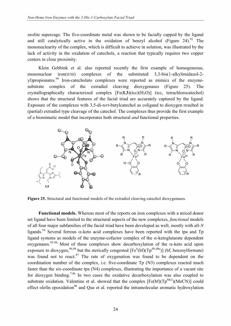
Klein Gebbink et al. also reported recently the first example of homogeneous,
mononuclear iron(II/III) complexes of the substituted 3,3-bis(1-alkylimidazol-2-
yl)propionates.94 Iron-catecholato complexes were reported as mimics of the enzyme-
substrate complex of the extradiol cleaving dioxygenases (Figure 25). The
crystallographically characterized complex [Fe(L3)(tcc)(H2O)] (tcc, tetrachlorocatechol)
shows that the structural features of the facial triad are accurately captured by the ligand.
Exposure of the complexes with 3,5-di-tert-butylcatechol as coligand to dioxygen resulted in
(partial) extradiol type cleavage of the catechol. The complexes thus provide the first example
of a biomimetic model that incorporates both structural and functional properties.
Figure 25. Structural and functional models of the extradiol cleaving catechol dioxygenases.
Functional models. Whereas most of the reports on iron complexes with a mixed donor
set ligand have been limited to the structural aspects of the new complexes, functional models
of all four major subfamilies of the facial triad have been developed as well, mostly with all-N
ligands.54 Several ferrous -keto acid complexes have been reported with the tpa and Tp
ligand systems as models of the enzyme-cofactor complex of the -ketoglutarate dependent
oxygenases.95-98 Most of these complexes show decarboxylation of the -keto acid upon
exposure to dioxygen,96,98 but the sterically congested [FeII(bf)(TpiPr,iBu)] (bf, benzoylformate)
was found not to react.97 The rate of oxygenation was found to be dependent on the
coordination number of the complex, i.e. five-coordinate Tp (N3) complexes reacted much
faster than the six-coordinate tpa (N4) complexes, illustrating the importance of a vacant site
for dioxygen binding.7,96 In two cases the oxidative decarboxylation was also coupled to
substrate oxidation. Valentine et al. showed that the complex [Fe(bf)(TpMe2)(MeCN)] could
effect olefin epoxidation96 and Que et al. reported the intramolecular aromatic hydroxylation
Chapter One
25
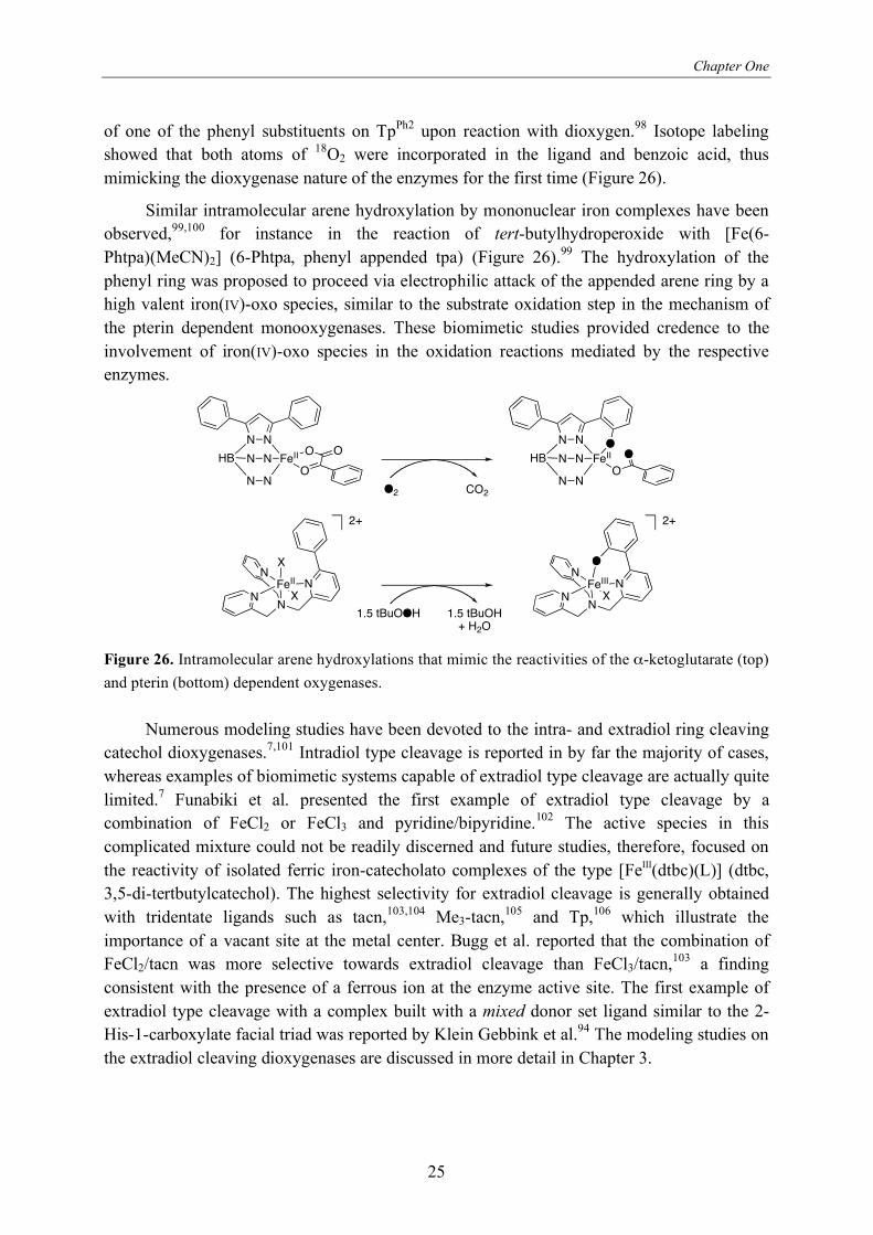
of one of the phenyl substituents on TpPh2 upon reaction with dioxygen.98 Isotope labeling
showed that both atoms of 18O2 were incorporated in the ligand and benzoic acid, thus
mimicking the dioxygenase nature of the enzymes for the first time (Figure 26).
Similar intramolecular arene hydroxylation by mononuclear iron complexes have been
observed,99,100 for instance in the reaction of tert-butylhydroperoxide with [Fe(6-
Phtpa)(MeCN)2] (6-Phtpa, phenyl appended tpa) (Figure 26).99 The hydroxylation of the
phenyl ring was proposed to proceed via electrophilic attack of the appended arene ring by a
high valent iron(IV)-oxo species, similar to the substrate oxidation step in the mechanism of
the pterin dependent monooxygenases. These biomimetic studies provided credence to the
involvement of iron(IV)-oxo species in the oxidation reactions mediated by the respective
enzymes.
N N
HBOO
OFeII
N
N
N
N
N N
HB OO
FeII
N
N
N
NO
O2 CO2
FeII
X
NN
N
XN FeIII
NN
N
XN
O
1.5 tBuOOH 1.5 tBuOH+ H2O
2+ 2+
Figure 26. Intramolecular arene hydroxylations that mimic the reactivities of the -ketoglutarate (top)
and pterin (bottom) dependent oxygenases.
Numerous modeling studies have been devoted to the intra- and extradiol ring cleaving
catechol dioxygenases.7,101 Intradiol type cleavage is reported in by far the majority of cases,
whereas examples of biomimetic systems capable of extradiol type cleavage are actually quite
limited.7 Funabiki et al. presented the first example of extradiol type cleavage by a
combination of FeCl2 or FeCl3 and pyridine/bipyridine.102 The active species in this
complicated mixture could not be readily discerned and future studies, therefore, focused on
the reactivity of isolated ferric iron-catecholato complexes of the type [FeIII(dtbc)(L)] (dtbc,
3,5-di-tertbutylcatechol). The highest selectivity for extradiol cleavage is generally obtained
with tridentate ligands such as tacn,103,104 Me3-tacn,105 and Tp,106 which illustrate the
importance of a vacant site at the metal center. Bugg et al. reported that the combination of
FeCl2/tacn was more selective towards extradiol cleavage than FeCl3/tacn,103 a finding
consistent with the presence of a ferrous ion at the enzyme active site. The first example of
extradiol type cleavage with a complex built with a mixed donor set ligand similar to the 2-
His-1-carboxylate facial triad was reported by Klein Gebbink et al.94 The modeling studies on
the extradiol cleaving dioxygenases are discussed in more detail in Chapter 3.
Non-Heme Iron Enzymes with the 2-His-1-Carboxylate Facial Triad
26
The functional models described above in most cases display stoichiometric, so-called
‘single turnover’ reactivities. Functional modeling studies on the Rieske dioxygenases,
however, have actually resulted in the discovery of a family of catalysts capable of cis-
dihydroxylation.107 Ferrous complexes of the ligands tpa and bpmen provided the first
example of cis-dihydroxylation catalyzed by a non-biological iron complex,108 a reaction that
usually requires reagents such as OsO4. Even enantioselective cis-dihydroxylation with an ee
up to 82% has been reported using a chiral analogue of bpmen.109 The complexes use
hydrogen peroxide as oxidant and catalyze the oxidation of a range of olefins with high
efficiency. At the same time, these complexes catalyze olefin epoxidation and the actually
observed epoxide to cis-diol ratios differ widely amongst the reported examples.107 These two
reactions were found to be closely related and the availability of two cis-positioned vacant
sites was proposed as a necessary requirement for both types of reactivities.110 The product
ratio was found to be dependent on the adopted ligand topology and the spin state of the metal
center.110,111 These factors influence the fate of an implicated FeIII–OOH intermediate such
that low spin intermediates generally yield more epoxide and high-spin intermediates more
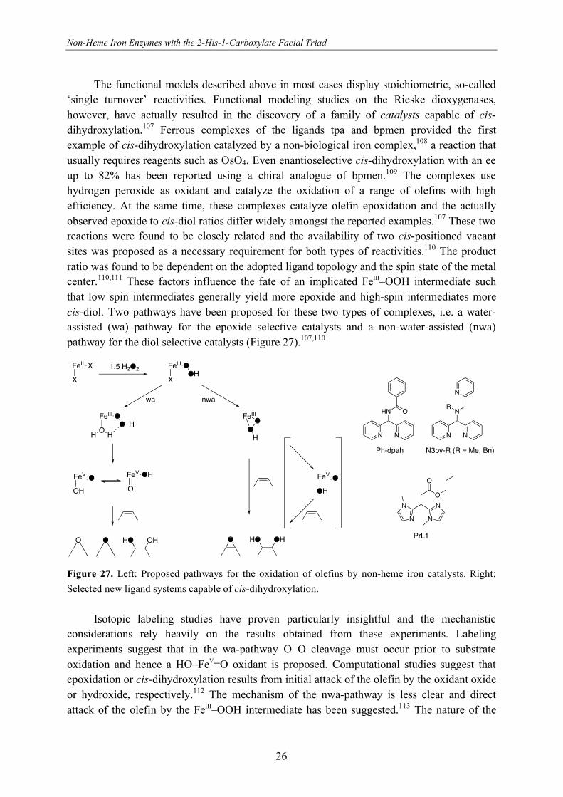
cis-diol. Two pathways have been proposed for these two types of complexes, i.e. a water-
assisted (wa) pathway for the epoxide selective catalysts and a non-water-assisted (nwa)
pathway for the diol selective catalysts (Figure 27).107,110
O
O
N
NN
N
PrL1
HN
N N
O
Ph-dpah
N
N N
N
R
N3py-R (R = Me, Bn)
FeII X
X
1.5 H2O2 FeIII O
XOH
FeIII O
OO H
HH
FeV O
OH
FeV OH
O
FeIIIO
OH
OHHO
FeV O
OH
wa nwa
O O OHHOO
Figure 27. Left: Proposed pathways for the oxidation of olefins by non-heme iron catalysts. Right:
Selected new ligand systems capable of cis-dihydroxylation.
Isotopic labeling studies have proven particularly insightful and the mechanistic
considerations rely heavily on the results obtained from these experiments. Labeling
experiments suggest that in the wa-pathway O–O cleavage must occur prior to substrate
oxidation and hence a HO–FeV=O oxidant is proposed. Computational studies suggest that
epoxidation or cis-dihydroxylation results from initial attack of the olefin by the oxidant oxide
or hydroxide, respectively.112 The mechanism of the nwa-pathway is less clear and direct
attack of the olefin by the FeIII–OOH intermediate has been suggested.113 The nature of the

Chapter One
27
two oxidants, however, differs significantly. Competition experiments have shown that the
oxidant in the wa-pathway has an electrophilic character, whereas in the nwa-pathway a
nucleophilic oxidant is generated.114
In addition to the tpa and bpmen complexes and derivatives thereof, other complexes
capable of cis-dihydroxylation have also been reported (Figure 27).115-118 The iron complex
derived from N3py-R reported by Feringa et al. shows comparable reactivity to the original
tpa and bmpen systems.116 Interestingly, the stereoselectivity of the reaction was solvent-
dependent, i.e. cis- or trans-diol was obtained from acetonitrile or acetone solutions,
respectively. Recently, examples have been reported that use tridentate ligands with an N,N,O
donor set. Oldenburg et al. use the ligand Ph-dpah to obtain the most selective cis-
dihydroxylation catalyst to date117 and in a related contribution Klein Gebbink et al. showed
that iron complexes of PrL1 also show good epoxidation and cis-dihydroxylation activity.115
More details on functional models of the Rieske dioxygenases can be found in Chapter 5.
The insights obtained from enzymology and functional models have played an
important role in the design of bio-inspired oxidation catalysts. The family of cis-
dihydroxylation/epoxidation catalysts mentioned before is just one example of many
biomimetic, non-heme iron oxidation catalysts that use hydrogen peroxide as the oxidant.
This topic has been reviewed very recently and will not be discussed here.119
1.4 Reaction intermediates
In many of the above studies of both the enzymes and the model systems high-valent
iron-oxo intermediates are invoked. Several efforts have been specifically devoted to the
study, isolation and characterization of these species to get more insight into the spectroscopic
and chemical properties of these intermediates. These studies are discussed below.
Iron(III)-hydroperoxide and -peroxide species. Iron(III)-hydroperoxide (FeIIIOOH)
and iron(III)-peroxide (FeIIIO2) species are often invoked as intermediates between the initial
binding of dioxygen to iron and the formation of the actual, active oxidant in many different
heme and non-heme iron biomolecules.7 Examples for the latter category include, for
instance, the crystallographically observed side-on bound O2 adduct of naphthalene
dioxygenase.37 In the Rieske dioxygenases, the group of enzymes to which naphthalene
dioxygenase belongs, this intermediate has been proposed by some authors to be the actual
oxidant rather than a precursor to a high valent iron-oxo oxidant (vide infra).12,44
Since the first report of the generation of an FeIIIOOH intermediate with a model
compound by Mascharak et al.,120 a number of mononuclear, non-heme iron(III)-
(hydro)peroxide complexes have been constructed with different, mostly neutral, polydentate
ligands such as N4py,121 tpa,122 H2bbpa,123 Rtpen,124 and bispidine-derived125 ligands (Figure
28).7,126 These species can be generated by the reaction of an iron(II) precursor with H2O2 at
Non-Heme Iron Enzymes with the 2-His-1-Carboxylate Facial Triad
28
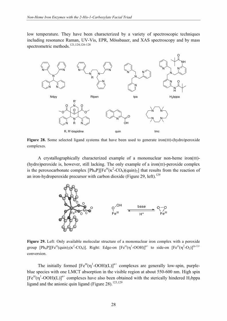
low temperature. They have been characterized by a variety of spectroscopic techniques
including resonance Raman, UV-Vis, EPR, Mössbauer, and XAS spectroscopy and by mass
spectrometric methods.121,124,126-128
N
NN
N N
N4py
NN
N
N
R
NN
N
N N
NH
O
NH
O
NR'
N
O
RN N
O
O
O
O
Rtpen H2bppa
R, R'-bispidine
N
N
N N
tpa
N N
NN
tmc
NO
OH
quin
Figure 28. Some selected ligand systems that have been used to generate iron(III)-(hydro)peroxide
complexes.
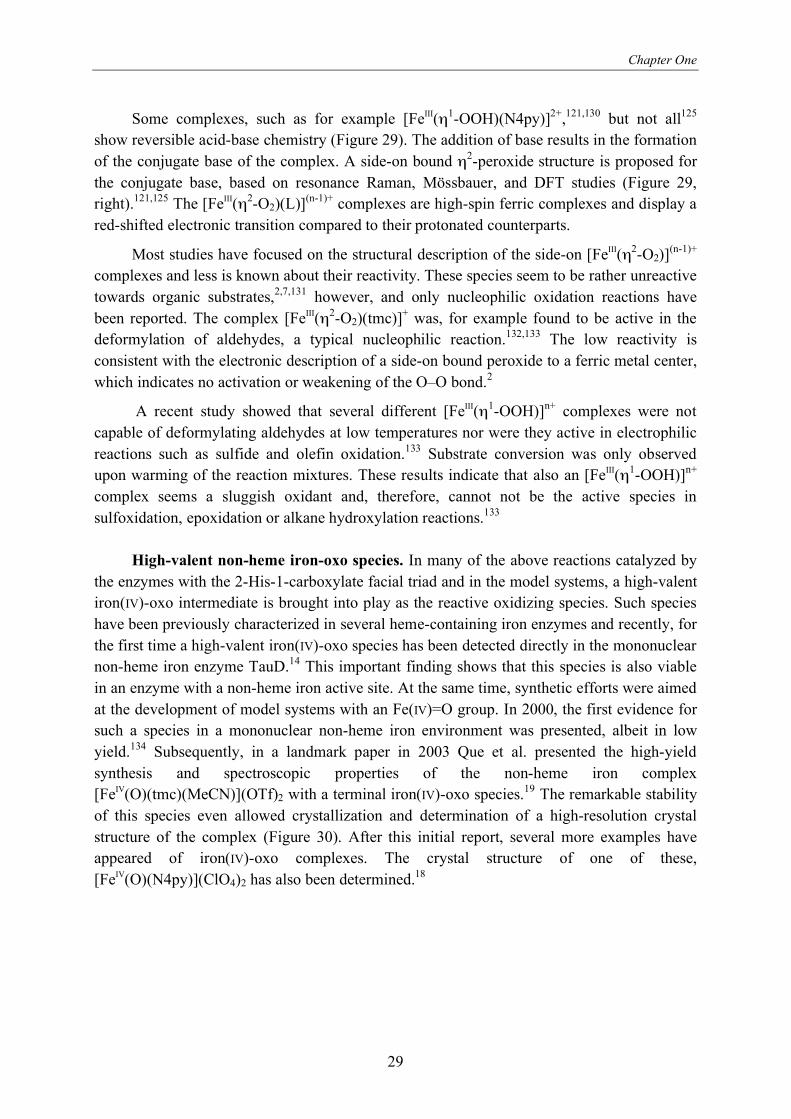
A crystallographically characterized example of a mononuclear non-heme iron(III)-
(hydro)peroxide is, however, still lacking. The only example of a iron(III)-peroxide complex
is the peroxocarbonate complex [Ph4P][FeIII( 2-CO4)(quin)2] that results from the reaction of
an iron-hydroperoxide precursor with carbon dioxide (Figure 29, left).129
Figure 29. Left: Only available molecular structure of a mononuclear iron complex with a peroxide
group [Ph4P][FeIII(quin)2(2-CO4)]. Right: Edge-on [FeIII( 1-OOH)]n+ to side-on [FeIII( 2-O2)]
(n-1)+
conversion.
The initially formed [FeIII( 1-OOH)(L)]n+ complexes are generally low-spin, purple-
blue species with one LMCT absorption in the visible region at about 550-600 nm. High spin
[FeIII( 1-OOH)(L)]n+ complexes have also been obtained with the sterically hindered H2bppa
ligand and the anionic quin ligand (Figure 28).123,129
Chapter One
29
Some complexes, such as for example [FeIII( 1-OOH)(N4py)]2+,121,130 but not all125
show reversible acid-base chemistry (Figure 29). The addition of base results in the formation
of the conjugate base of the complex. A side-on bound 2-peroxide structure is proposed for
the conjugate base, based on resonance Raman, Mössbauer, and DFT studies (Figure 29,
right).121,125 The [FeIII( 2-O2)(L)](n-1)+ complexes are high-spin ferric complexes and display a
red-shifted electronic transition compared to their protonated counterparts.
Most studies have focused on the structural description of the side-on [FeIII( 2-O2)](n-1)+
complexes and less is known about their reactivity. These species seem to be rather unreactive
towards organic substrates,2,7,131 however, and only nucleophilic oxidation reactions have
been reported. The complex [FeIII( 2-O2)(tmc)]+ was, for example found to be active in the
deformylation of aldehydes, a typical nucleophilic reaction.132,133 The low reactivity is
consistent with the electronic description of a side-on bound peroxide to a ferric metal center,
which indicates no activation or weakening of the O–O bond.2
A recent study showed that several different [FeIII( 1-OOH)]n+ complexes were not
capable of deformylating aldehydes at low temperatures nor were they active in electrophilic
reactions such as sulfide and olefin oxidation.133 Substrate conversion was only observed
upon warming of the reaction mixtures. These results indicate that also an [FeIII( 1-OOH)]n+
complex seems a sluggish oxidant and, therefore, cannot not be the active species in
sulfoxidation, epoxidation or alkane hydroxylation reactions.133
High-valent non-heme iron-oxo species. In many of the above reactions catalyzed by
the enzymes with the 2-His-1-carboxylate facial triad and in the model systems, a high-valent
iron(IV)-oxo intermediate is brought into play as the reactive oxidizing species. Such species
have been previously characterized in several heme-containing iron enzymes and recently, for
the first time a high-valent iron(IV)-oxo species has been detected directly in the mononuclear
non-heme iron enzyme TauD.14 This important finding shows that this species is also viable
in an enzyme with a non-heme iron active site. At the same time, synthetic efforts were aimed
at the development of model systems with an Fe(IV)=O group. In 2000, the first evidence for
such a species in a mononuclear non-heme iron environment was presented, albeit in low
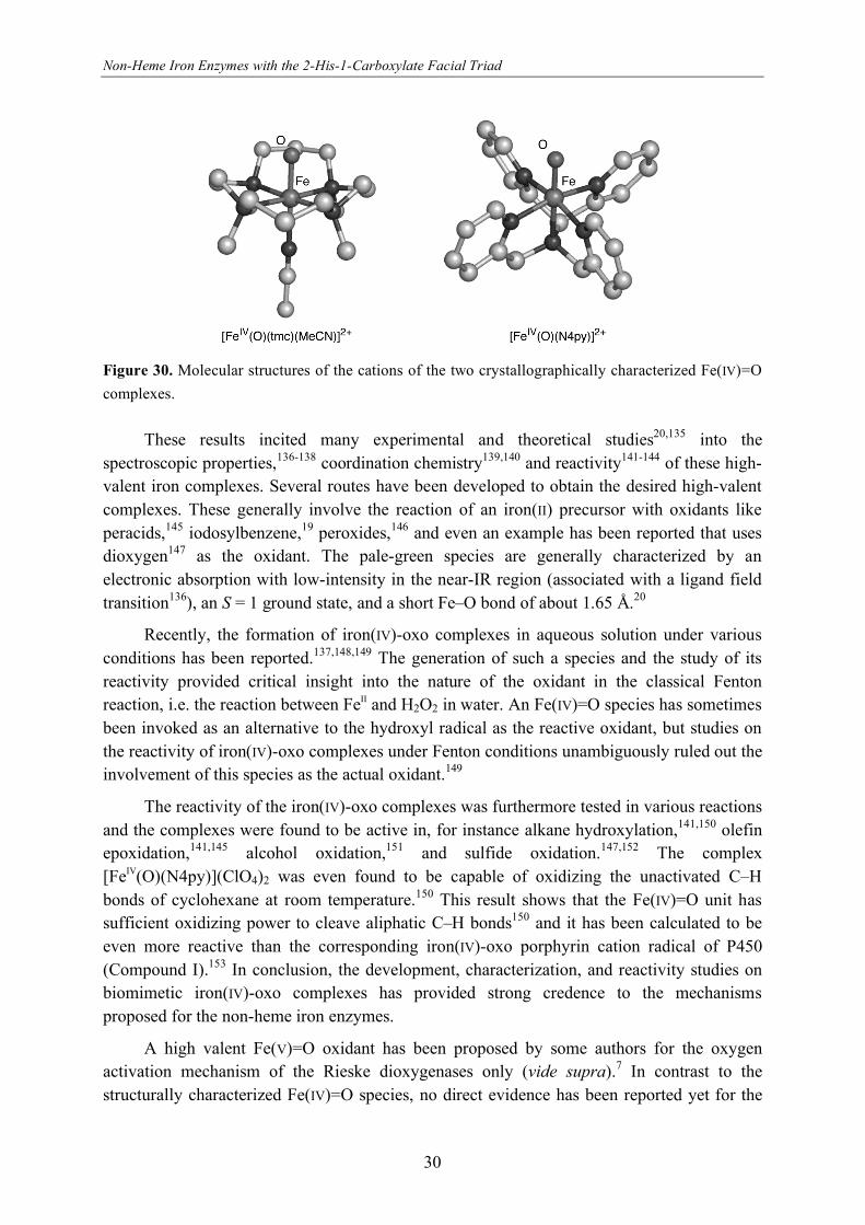
yield.134 Subsequently, in a landmark paper in 2003 Que et al. presented the high-yield
synthesis and spectroscopic properties of the non-heme iron complex
[FeIV(O)(tmc)(MeCN)](OTf)2 with a terminal iron(IV)-oxo species.19 The remarkable stability
of this species even allowed crystallization and determination of a high-resolution crystal
structure of the complex (Figure 30). After this initial report, several more examples have
appeared of iron(IV)-oxo complexes. The crystal structure of one of these,
[FeIV(O)(N4py)](ClO4)2 has also been determined.18
Non-Heme Iron Enzymes with the 2-His-1-Carboxylate Facial Triad
30
Figure 30. Molecular structures of the cations of the two crystallographically characterized Fe(IV)=O
complexes.
These results incited many experimental and theoretical studies20,135 into the
spectroscopic properties,136-138 coordination chemistry139,140 and reactivity141-144 of these high-
valent iron complexes. Several routes have been developed to obtain the desired high-valent
complexes. These generally involve the reaction of an iron(II) precursor with oxidants like
peracids,145 iodosylbenzene,19 peroxides,146 and even an example has been reported that uses
dioxygen147 as the oxidant. The pale-green species are generally characterized by an
electronic absorption with low-intensity in the near-IR region (associated with a ligand field
transition136), an S = 1 ground state, and a short Fe–O bond of about 1.65 Å.20
Recently, the formation of iron(IV)-oxo complexes in aqueous solution under various
conditions has been reported.137,148,149 The generation of such a species and the study of its
reactivity provided critical insight into the nature of the oxidant in the classical Fenton
reaction, i.e. the reaction between FeII and H2O2 in water. An Fe(IV)=O species has sometimes
been invoked as an alternative to the hydroxyl radical as the reactive oxidant, but studies on
the reactivity of iron(IV)-oxo complexes under Fenton conditions unambiguously ruled out the
involvement of this species as the actual oxidant.149
The reactivity of the iron(IV)-oxo complexes was furthermore tested in various reactions
and the complexes were found to be active in, for instance alkane hydroxylation,141,150 olefin
epoxidation,141,145 alcohol oxidation,151 and sulfide oxidation.147,152 The complex
[FeIV(O)(N4py)](ClO4)2 was even found to be capable of oxidizing the unactivated C–H
bonds of cyclohexane at room temperature.150 This result shows that the Fe(IV)=O unit has
sufficient oxidizing power to cleave aliphatic C–H bonds150 and it has been calculated to be
even more reactive than the corresponding iron(IV)-oxo porphyrin cation radical of P450
(Compound I).153 In conclusion, the development, characterization, and reactivity studies on
biomimetic iron(IV)-oxo complexes has provided strong credence to the mechanisms
proposed for the non-heme iron enzymes.
A high valent Fe(V)=O oxidant has been proposed by some authors for the oxygen
activation mechanism of the Rieske dioxygenases only (vide supra).7 In contrast to the
structurally characterized Fe(IV)=O species, no direct evidence has been reported yet for the

Chapter One
31
proposed HO–Fe(V)=O oxidant. Mechanistic studies, in particular isotopic labeling
experiments, on bio-inspired cis-dihydroxylation107 and alkane hydroxylation catalysts,116,154
however, strongly implicate the involvement of such an oxidant in these systems. The
involvement of an iron(V)-oxo has also been suggested in the self-hydroxylation of different
perbenzoic acids by a non-heme iron complex155 and in a regioselective ligand oxidation.156
Although no iron(V)-oxo species have been identified spectroscopically, a mononuclear non-
heme iron(V)-nitrido complex has been observed, which was generated by the photolysis of a
ferric bis(azido) complex.157,158 The isolation and spectroscopic or structural characterization
of an analogues oxo species, however, still awaits.
1.5 Concluding remarks
The recent explosion in crystallographically characterized non-heme iron enzymes has
firmly established the 2-His-1-carboxylate facial triad as a common platform for dioxygen
activation in Nature. The breadth of oxidative transformations is stunning and many reactions
do not have a precedent in synthetic organic chemistry. New members of the family are
reported at a remarkable pace, which illustrates both the ubiquity and the versatility of the
triad. This holds great promise for the future and without a doubt new systems that mediate
exciting new chemistry will be discovered. For these reactivities to be of practical interest the
mechanisms have to be understood at the molecular level. Great strides have been made by
complementary, parallel studies of enzymes and their biomimetic model complexes, with
notable achievements such as the detection and crystallization of iron(IV)-oxo species. Many
questions still remain, however, that concern the steps following initial dioxygen binding and
lead to product formation. The elaborate studies of synthetic functional analogues have
contributed greatly to our understanding of these enzymes. The recent developments towards
the synthesis of even more precise high-fidelity structural models promise further progress in
this field and may even ultimately lead to the development of synthetically useful catalysts.
1.6 References & Notes
1. Abu-Omar, M. M.; Loaiza, A.; Hontzeas, N. Chem. Rev. 2005, 105, 2227-2252.
2. Solomon, E. I.; Brunold, T. C.; Davis, M. I.; Kernsley, J. N.; Lee, S-K.; Lehnert, N.; Neese, F.; Skulan, A. J.;
Yang, Y-S.; Zhou, J. Chem. Rev. 2000, 100, 235-349.
3. Neidig, M. L.; Solomon, E. I. Chem. Commun. 2005, 5843-5863.
4. Solomon, E. I.; Chen, P.; Metz, M.; Lee, S.-K.; Palmer, A. E. Angew. Chem. Int. Ed. 2001, 40, 4570-4590.
5. Holm, R. H.; Kennepohl, P.; Solomon, E. I. Chem. Rev. 1996, 96, 2239-2314.
6. Denisov, I. G.; Makris, T. M.; Sligar, S. G.; Schlichting, I. Chem. Rev. 2005, 105, 2253-2277.
7. Costas, M.; Mehn, M. P.; Jensen, M. P.; Que, L., Jr. Chem. Rev. 2004, 104, 939-986.
8. Tshuva, E. Y.; Lippard, S. J. Chem. Rev. 2004, 104, 987-1012.
9. Koehntop, K. D.; Emerson, J. P.; Que, L., Jr. J. Biol. Inorg. Chem. 2005, 10, 87-93.
10. Que, L., Jr. Nat. Struct. Biol. 2000, 7, 182-184.

Non-Heme Iron Enzymes with the 2-His-1-Carboxylate Facial Triad
32
11. Valegård, K.; Terwisscha van Scheltinga, A. C.; Lloyd, M. D.; Hara, T.; Ramaswamy, S.; Perrakis, A.; Thompson,
A.; Lee, H.-J.; Baldwin, J. E.; Schofield, C. J.; Hajdu, J.; Andersson, I. Nature 1998, 394, 805-809.
12. Ferraro, D. J.; Gakhar, L.; Ramaswamy, S. Biochem. Biophys. Res. Commun. 2005, 338, 175-190.
13. Delano, W. L. The Pymol Molecular Graphics System, Delano Scientific: San Carlos, CA, USA, 2002.
14. Bollinger, J. M., Jr.; Price, J. C.; Hoffart, L. M.; Barr, E. W.; Krebs, C. Eur. J. Inorg. Chem. 2005, 4245-4254.
15. Price, J. C.; Barr, E. W.; Tirupati, B.; Bollinger, J. M., Jr.; Krebs, C. Biochemistry 2003, 42, 7497-7508.
16. Proshlyakov, D. A.; Henshaw, T. F.; Monterosso, G. R.; Ryle, M. J.; Hausinger, R. P. J. Am. Chem. Soc. 2004,
126, 1022-1023.
17. Riggs-Gelasco, P. J.; Price, J. C.; Guyer, J. C.; Brehm, J. H.; Barr, E. W.; Bollinger, J. M., Jr.; Krebs, C. J. Am.
Chem. Soc. 2004, 126, 8108-8109.
18. Klinker, E. J.; Kaizer, J.; Brennessel, W. W.; Woodrum, N. L.; Cramer, C. J.; Que, L., Jr. Angew. Chem. Int. Ed.
2005, 44, 3690-3694.
19. Rohde, J.-U.; In, J.-H.; Lim, M. H.; Brennessel, W. W.; Bukowski, M. R.; Stubna, A.; Münck, E.; Nam, W.; Que,
L., Jr. Science 2003, 299, 1037-1039.
20. Shan, X. P.; Que, L. J. Inorg. Biochem. 2006, 100, 421-433.
21. Vaillancourt, F. H.; Bolin, J. T.; Eltis, L. D. Crit. Rev. Biochem. Mol. Biol. 2006, 41, 241-267.
22. Bugg, T. D. H.; Lin, G. Chem. Commun. 2001, 941-952.
23. Que, L., Jr.; Reynolds, M. F. Met. Ions Biol. Syst. 2000, 37, 505-525.
24. Vaillancourt, F. H.; Barbosa, C. J.; Spiro, T. G.; Bolin, J. T.; Blades, M. W.; Turner, R. F. B.; Eltis, L. D. J. Am.
Chem. Soc. 2002, 124, 2485-2496.
25. Spence, E. L.; Langley, G. J.; Bugg, T. D. H. J. Am. Chem. Soc. 1996, 118, 8336-8343.
26. Groce, S. L.; Lipscomb, J. D. Biochemistry 2005, 44, 7175-7188.
27. Zhang, J.; Zheng, H.; Groce, S. L.; Lipscomb, J. D. J. Mol. Cat. A 2006, 251, 54-65.
28. Groce, S. L.; Lipscomb, J. D. J. Am. Chem. Soc. 2003, 125, 11780-11781.
29. Mendel, S.; Arndt, A.; Bugg, T. D. H. Chem. Commun. 2005, 666-668.
30. Gibson, D. T.; Parales, R. E. Curr. Op. Biotech. 2000, 11, 236-243.
31. Boyd, D. R.; Bugg, T. D. H. Chem. Commun. 2006, 181-192.
32. Gibson, D. T.; Resnick, S. M.; Lee, K.; Brand, J. M.; Torok, D. S.; Wackett, L. P.; Schocken, M. J.; Haigler, B. E.
J. Bacteriol. 1995, 177, 2615-2621.
33. Herman, P. L.; Behrens, M.; Chakraborty, S.; Chrastil, B. M.; Barycki, J.; Weeks, D. P. J. Biol. Chem. 2005, 280,
24759-24767.
34. Lee, J.; Zhao, H. Angew. Chem. Int. Ed. 2006, 45, 622-625.
35. Martins, B. M.; Svetlitchnaia, T.; Dobbek, H. Structure 2005, 13, 817-824.
36. Tarasev, M.; Pinto, A.; Kim, D.; Elliot, S. J.; Ballou, D. P. Biochemistry 2006, 45, 10208-10216.
37. Karlsson, A.; Parales, J. V.; Parales, R. E.; Gibson, D. T.; Eklund, H.; Ramaswamy, S. Science 2003, 299, 1039-
1042.
38. Kauppi, B.; Lee, K.; Carredano, E.; Parales, R. E.; Gibson, D. T.; Eklund, H.; Ramaswamy, S. Structure 1998, 6,
571-586.
39. Karlsson, A.; Parales, J. V.; Parales, R. E.; Gibson, D. T.; Eklund, H.; Ramaswamy, S. J. Biol. Inorg. Chem. 2005,
10, 483-489.
40. Furusawa, Y.; Nagarajan, V.; Tanokura, M.; Masai, E.; Fukuda, M.; Senda, T. J. Mol. Biol. 2004, 342, 1041-1052.
41. Friemann, R.; Ivkovic-Jensen, M. M.; Lessner, D. J.; Yu, C. L.; Gibson, D. T.; Parales, R. E.; Eklund, H.;
Ramaswamy, S. J. Mol. Biol. 2005, 348, 1139-1151.
Chapter One
33
42. Dong, X.; Fushinobu, S.; Fukuda, E.; Terada, T.; Nakamura, S.; Shimizu, K.; Nojiri, H.; Omori, T.; Shoun, H.;
Wakagi, T. J. Bacteriol. 2005, 187, 2483-2490.
43. Nojiri, H.; Ashikawa, Y.; Noguchi, H.; Nam, J. W.; Urata, M.; Fujimoto, Z.; Uchimura, H.; Terada, T.; Nakamura,
S.; Shimizu, K.; Yoshida, T.; Habe, H.; Omori, T. J. Mol. Biol. 2005, 351, 355-370.
44. Bassan, A.; Blomberg, M. R. A.; Siegbahn, P. E. M. J. Biol. Inorg. Chem. 2004, 9, 439-452.
45. Bassan, A.; Borowski, T.; Siegbahn, P. E. M. Dalton Trans. 2004, 3153-3162.
46. Ferraro, D. J.; Okerlund, A. L.; Mowers, J. C.; Ramaswamy, S. J. Bacteriol. 2006, 188, 6986-6994.
47. Clifton, I. J.; McDonough, M. A.; Ehrismann, D.; Kershaw, N. J.; Granatino, N.; Schofield, C. J. J. Inorg.
Biochem. 2006, 100, 644-669.
48. Hausinger, R. P. Crit. Rev. Biochem. Mol. Biol. 2004, 39, 21-68.
49. Hewitson, K. S.; Granatino, N.; Welford, W. D.; McDonough, M. A.; Schofield, C. J. Phil. Trans. R. Soc. A 2005,
363, 807-828.
50. Yu, B.; Edstrom, W. C.; Benach, J.; Hamuro, Y.; Weber, P. C.; Gibney, B. R.; Hunt, J. F. Nature 2006, 439, 879-
884.
51. Sundheim, O.; Vågbø, C. B.; Bjøras, M.; Sousa, M. M. L.; Talstad, V.; Aas, P. A.; Drabløs, F.; Krokan, H. E.;
Tainer, J. A.; Slupphaug, G. EMBO J. 2006, 25, 3389-3397.
52. Cloos, P. A. C.; Christensen, J.; Agger, K.; Maiolica, A.; Rappsilber, J.; Antal, T.; Hansen, K. H.; Helin, K. Nature
2006, 442, 307-311.
53. Tsukada, Y.-I.; Fang, J.; Erdjument-Bromage, H.; Warren, M. E.; Borchers, C. H.; Tempst, P.; Zhang, Y. Nature
2006, 439, 811-816.
54. McDonough, M. A.; Li, V.; Flashman, E.; Chowdhury, R.; Mohr, C.; Liénard, B. M. R.; Zondlo, J.; Oldham, N. J.;
Clifton, I. J.; Lewis, J.; McNeill, L. A.; Kurzeja, R. J. M.; Hewitson, K. S.; Yang, E.; Jordan, S.; Syed, R. S.;
Schofield, C. J. Proc. Natl. Acad. Sci. U. S. A. 2006, 103, 9814-9819.
55. Pavel, E. G.; Zhou, J.; Busby, R. W.; Gunsior, M.; Townsend, C. A.; Solomon, E. I. J. Am. Chem. Soc. 1998, 120,
743-753.
56. O'Brien, J. R.; Schuller, D. J.; Yang, V. S.; Dillard, B. D.; Lanzilotta, W. N. Biochemistry 2003, 42, 5547-5554.
57. Schenk, G.; Pau, M. Y. M.; Solomon, E. I. J. Am. Chem. Soc. 2004, 126, 505-515.
58. Moran, G. R. Arch. Biochem. Biophys. 2005, 433, 117-128.
59. Choroba, O. W.; Wiliams, D. H.; Spencer, J. B. J. Am. Chem. Soc. 2000, 122, 5389-5390.
60. Neidig, M. L.; Decker, A.; Choroba, O. W.; Huang, F.; Kavana, M.; Moran, G. R.; Spencer, J. B.; Solomon, E. I.
Proc. Natl. Acad. Sci. U. S. A. 2006, 103, 12966-12973.
61. Vaillancourt, F. H.; Yeh, E.; Vosburg, D. A.; Garneau-Tsodikova, S.; Walsh, C. T. Chem. Rev. 2006, 106, 3364-
3378.
62. Vaillancourt, F. H.; Yin, J.; Walsh, C. T. Proc. Natl. Acad. Sci. U. S. A. 2005, 102, 10111-10116.
63. Vaillancourt, F. H.; Yeh, E.; Vosburg, D. A.; O'Connor, S. E.; Walsh, C. T. Nature 2005, 436, 1191-1194.
64. Blasiak, L. C.; Vaillancourt, F. H.; Walsh, C. T.; Drennan, C. L. Nature 2006, 440, 368-371.
65. Fitzpatrick, P. F. Annu. Rev. Biochem. 1999, 68, 355-381.
66. Flatmark, T.; Stevens, R. C. Chem. Rev. 1999, 99, 2137-2160.
67. Goodwill, K. E.; Sabatier, C.; Marks, C.; Raag, R.; Fitzpatrick, P. F.; Stevens, R. C. Nat. Struct. Biol. 1997, 4, 578-
585.
68. Kobe, B.; Jennings, I. G.; House, C. M.; Michell, B. J.; Goodwill, K. E.; Santarsiero, B. D.; Stevens, R. C.; Cotton,
R. G. H.; Kemp, B. E. Nat. Struct. Biol. 1999, 6, 442-448.
69. Wang, L.; Erlandsen, H.; Haavik, J.; Knappskog, P. M.; Stevens, R. C. Biochemistry 2002, 41, 12569-12574.
70. Andersson, O. A.; Flatmark, T.; Hough, E. J. Mol. Biol. 2002, 320, 1095-1108.
Non-Heme Iron Enzymes with the 2-His-1-Carboxylate Facial Triad
34
71. Klinman, J. P. J. Biol. Inorg. Chem. 2001, 6, 1-13.
72. Urich, T.; Gomes, C. M.; Kletzin, A.; Frazão, C. Science 2006, 311, 996-1000.
73. Higgins, L. J.; Yan, F.; Liu, P.; Liu, H.-W.; Drennan, C. L. Nature 2005, 437, 838-844.
74. Liu, P.; Liu, A.; Yan, F.; Wolfe, M. D.; Lipscomb, J. D.; Liu, H.-W. Biochemistry 2003, 42, 11577-11586.
75. Yan, F.; Munos, J. W.; Liu, P.; Liu, H.-W. Biochemistry 2006, 45, 11473-11481.
76. Kershaw, N. J.; Caines, M. E. C.; Sleeman, M. C.; Schofield, C. J. Chem. Commun. 2006, 4251-4263.
77. Burzlaff, N. I.; Rutledge, P. J.; Clifton, I. J.; Hensgens, C. M. H.; Pickford, M.; Adlington, R. M.; Roach, P. L.;
Baldwin, J. E. Nature 1999, 401, 721-724.
78. Roach, P. L.; Clifton, I. J.; Hensgens, C. M. H.; Shibata, N.; Long, A. J.; Strange, R. W.; Hasnain, S. S.; Schofield,
C. J.; Baldwin, J. E.; Hajdu, J. Eur. J. Biochem. 1996, 242, 736-740.
79. Roach, P. L.; Clifton, I. J.; Hensgens, C. M. H.; Shibata, N.; Schofield, C. J.; Hajdu, J.; Baldwin, J. E. Nature 1997,
387, 827-830.
80. Brunhuber, N. M. W.; Mort, J. L.; Christoffersen, R. E.; Reich, N. O. Biochemistry 2000, 39, 10730-10738.
81. Thrower, J. S.; Blalock, R., III; Klinman, J. P. Biochemistry 2001, 40, 9717-9724.
82. Zhou, J.; Rocklin, A. M.; Lipscomb, J. D.; Que, L., Jr.; Solomon, E. I. J. Am. Chem. Soc. 2002, 124, 4602-4609.
83. Tierney, D. L.; Rocklin, A. M.; Lipscomb, J. D.; Que, L., Jr.; Hoffman, B. M. J. Am. Chem. Soc. 2005, 127, 7005-
7013.
84. Rocklin, A. M.; Kato, K.; Liu, H.-W.; Que, L., Jr.; Lipscomb, J. D. J. Biol. Inorg. Chem. 2004, 9, 171-182.
85. Zhang, Z.; Ren, J.-S.; Clifton, I. J.; Schofield, C. J. Chem. Biol. 2004, 11, 1383-1394.
86. Beck, A.; Barth, A.; Hübner, E.; Burzlaff, N. Inorg. Chem. 2003, 42, 7182-7188.
87. Beck, A.; Weibert, B.; Burzlaff, N. Eur. J. Inorg. Chem. 2001, 521-527.
88. Otero, A.; Fernández-Baeza, J.; Antiñolo, A.; Tejeda, J.; Lara-Sánchez, A. Dalton Trans. 2004, 1499-1510.
89. Peters, L.; Burzlaff, N. Polyhedron 2004, 23, 245-251.
90. Friese, S. J.; Kucera, B. E.; Que, L., Jr.; Tolman, W. B. Inorg. Chem. 2006, 45, 8003-8005.
91. Bruijnincx, P. C. A.; Lutz, M.; Spek, A. L.; van Faassen, E. E.; Weckhuysen, B. M.; van Koten, G.; Klein Gebbink,
R. J. M. Eur. J. Inorg. Chem. 2005, 779-787 (Chapter 2 of this thesis).
92. Peters, L.; Hübner, E.; Burzlaff, N. J. Organomet. Chem. 2005, 690, 2009-2016.
93. Kervinen, K.; Bruijnincx, P. C. A.; Beale, A. M.; Mesu, J. G.; van Koten, G.; Klein Gebbink, R. J. M.;
Weckhuysen, B. M. J. Am. Chem. Soc. 2006, 128, 3208-3217.
94. Bruijnincx, P. C. A.; Lutz, M.; Spek, A. L.; Hagen, W. R.; Weckhuysen, B. M.; van Koten, G.; Klein Gebbink, R.
J. M. J. Am. Chem. Soc., accepted for publication (Chapter 3 of this thesis).
95. Chiou, Y.-M.; Que, L., Jr. J. Am. Chem. Soc. 1995, 117, 3999-4013.
96. Ha, E. H.; Ho, R. Y. N.; Kisiel, J. F.; Valentine, J. S. Inorg. Chem. 1995, 34, 2265-2266.
97. Hikichi, S.; Ogihara, T.; Fujisawa, K.; Kitajima, N.; Akita, M.; Moro-oka, Y. Inorg. Chem. 1997, 36, 4539-4547.
98. Mehn, M. P.; Fujisawa, K.; Hegg, E. L.; Que, L., Jr. J. Am. Chem. Soc. 2003, 125, 7828-7842.
99. Jensen, M. P.; Lange, S. J.; Mehn, M. P.; Que, E. L.; Que, L., Jr. J. Am. Chem. Soc. 2003, 125, 2113-2128.
100. Mekmouche, Y.; Ménage, S.; Toia-Duboc, C.; Fontecave, M.; Galey, J.-B.; Lebrun, C.; Pécaut, J. Angew. Chem.
Int. Ed. 2001, 40, 949-952.
101. Yamahara, R.; Ogo, S.; Masuda, H.; Watanabe, Y. J. Inorg. Biochem. 2002, 88, 284-294.
102. Funabiki, T.; Mizoguchi, A.; Sugimoto, T.; Tada, S.; Tsuji, M.; Sakamoto, H.; Yoshida, S. J. Am. Chem. Soc. 1986,
108, 2921-2932.
103. Lin, G.; Reid, G.; Bugg, T. D. H. J. Am. Chem. Soc. 2001, 123, 5030-5039.
104. Dei, A.; Gatteschi, D.; Pardi, L. Inorg. Chem. 1993, 32, 1389-1395.
105. Jo, D.-H.; Que, L., Jr. Angew. Chem. Int. Ed. 2000, 39, 4284-4287.
Chapter One
35
106. Ogihara, T.; Hikichi, S.; Akita, M.; Moro-oka, Y. Inorg. Chem. 1998, 37, 2614-2615.
107. Oldenburg, P. D.; Que, L., Jr. Catal. Today 2006, 117, 15-21.
108. Chen, K.; Que, L., Jr. Angew. Chem. Int. Ed. 1999, 38, 2227-2229.
109. Costas, M.; Tipton, A. K.; Chen, K.; Jo, D.-H.; Que, L., Jr. J. Am. Chem. Soc. 2001, 123, 6722-6723.
110. Chen, K.; Costas, M.; Kim, J.; Tipton, A. K.; Que, L., Jr. J. Am. Chem. Soc. 2002, 124, 3026-3035.
111. Mas-Ballesté, R.; Costas, M.; van den Berg, T.; Que, L., Jr. Chem. Eur. J. 2006, 12, 3026-3035.
112. Bassan, A.; Blomberg, M. R. A.; Siegbahn, P. E. M.; Que, L., Jr. Angew. Chem. Int. Ed. 2005, 44, 2939-2941.
113. Quiñonero, D.; Morokuma, K.; Musaev, D.; Mas-Ballesté, R.; Que, L., Jr J. Am. Chem. Soc. 2005, 127, 6548-6549.
114. Fujita, M.; Costas, M.; Que, L., Jr. J. Am. Chem. Soc. 2003, 125, 9912-9913.
115. Bruijnincx, P. C. A.; Buurmans, I. L. C.; Gosiewska, S.; Lutz, M.; Spek, A. L.; van Koten, G.; Klein Gebbink, R. J.
M. to be submitted (Chapter 5 of this thesis).
116. Klopstra, M.; Roelfes, G.; Hage, R.; Kellogg, R. M.; Feringa, B. L. Eur. J. Inorg. Chem. 2004, 846-856.
117. Oldenburg, P. D.; Shteinman, A. A.; Que, L., Jr. J. Am. Chem. Soc. 2005, 127, 15673-15674.
118. Rotthaus, O.; Le Roy, S.; Tomas, A.; Barkigia, K. M.; Artaud, I. Inorg. Chim. Acta 2004, 357, 2211-2217.
119. Tanase, S.; Bouwman, E. Adv. Inorg. Chem. 2006, 58, 29-75.
120. Guajardo, R. J.; Hudson, S. E.; Brown, S. J.; Mascharak, P. K. J. Am. Chem. Soc. 1993, 115, 7971-7977.
121. Roelfes, G.; Vrajmasu, V.; Chen, K.; Ho, R. Y. N.; Rohde, J.-U.; Zondervan, C.; la Crois, R. M.; Schudde, E. P.;
Lutz, M.; Spek, A. L.; Hage, R.; Feringa, B. L.; Münck, E.; Que, L., Jr. Inorg. Chem. 2003, 42, 2639-2653.
122. Ho, R. Y. N.; Roelfes, G.; Feringa, B. L.; Que, L., Jr. J. Am. Chem. Soc. 1999, 121, 264-265.
123. Wada, A.; Ogo, S.; Nagamoto, S.; Kitagawa, T.; Watanabe, Y.; Jitsukawa, K.; Masuda, H. Inorg. Chem. 2002, 42,
616-618.
124. Hazell, A.; McKenzie, C. J.; Nielsen, L. P.; Schindler, S.; Weitzer, M. J. Chem. Soc., Dalton Trans. 2002, 310-317.
125. Bukowski, M. R.; Comba, P.; Limberg, C.; Merz, M.; Que, L., Jr.; Wistuba, T. Angew. Chem. Int. Ed. 2004, 43,
1283-1287.
126. Girerd, J.-J.; Banse, F.; Simaan, A. J. Struct. Bonding 2000, 97, 145-177.
127. Koehntop, K. D.; Rohde, J.-U.; Costas, M.; Que, L., Jr. Dalton Trans. 2004, 3191-3198.
128. Simaan, A. J.; Döpner, S.; Banse, F.; Bourcier, S.; Bouchoux, G.; Boussac, A.; Hildebrandt, P.; Girerd, J.-J. Eur. J.
Inorg. Chem. 2000, 1627-1633.
129. Hashimoto, K.; Nagamoto, S.; Fujinami, S; Furutachi, H.; Ogo, S.; Suzuki, M.; Uehara, A.; Maeda, Y.; Watanabe,
Y.; Kitagawa, T. Angew. Chem. Int. Ed. 2002, 41, 1202-1205.
130. Jensen, K. B.; McKenzie, C. J.; Nielsen, L. P.; Pedersen, J. Z.; Svendsen, H. M. Chem. Commun. 1999, 1313-1314.
131. Neese, F.; Solomon, E. I. J. Am. Chem. Soc. 1998, 120, 12829-12848.
132. Annaraj, J.; Suh, Y.; Seo, M. S.; Kim, S. O.; Nam, W. Chem. Commun. 2005, 4529-4531.
133. Park, M. J.; Lee, J.; Suh, Y.; Kim, J.; Nam, W. J. Am. Chem. Soc. 2006, 128, 2630-2634.
134. Grapperhaus, C. A.; Mienert, B.; Bill, E.; Weyhermüller, T.; Wieghardt, K. Inorg. Chem. 2000, 39, 5306-5317.
135. Decker, A.; Clay, M. D.; Solomon, E. I. J. Inorg. Biochem. 2006, 100, 697-706.
136. Decker, A.; Rohde, J.-U.; Que, L., Jr.; Solomon, E. I. J. Am. Chem. Soc. 2004, 126, 5378-5379.
137. Sastri, C. V.; Park, M. J.; Ohta, T.; Jackson, T. A.; Stubna, A.; Seo, M. S.; Lee, J.; Kim, J.; Kitagawa, T.; Münck,
E.; Que, L., Jr.; Nam, W. J. Am. Chem. Soc. 2005, 127, 12494-12495.
138. Rohde, J.-U.; Torelli, S.; Shan, X.; Lim, M. H.; Klinker, E. J.; Kaizer, J.; Chen, K.; Nam, W.; Que, L., Jr. J. Am.
Chem. Soc. 2004, 126, 16750-16761.
139. Rohde, J.-U.; Stubna, A.; L., Bominaar. E.; Münck, E.; Nam, W.; Que, L., Jr. Inorg. Chem. 2006, 45, 6435-6445.
140. Rohde, J.-U.; Que, L., Jr. Angew. Chem. Int. Ed. 2005, 44, 2255-2258.
Non-Heme Iron Enzymes with the 2-His-1-Carboxylate Facial Triad
36
141. Martinho, M.; Banse, F.; Bartoli, J.-F.; Mattioli, T. A.; Battioni, P.; Horner, O.; Bourcier, S.; Girerd, J.-J. Inorg.
Chem. 2005, 44, 9592-9596.
142. Sastri, C. V.; Oh, K.; Lee, Y. J.; Seo, M. S.; Shin, W.; Nam, W. Angew. Chem. Int. Ed. 2006, 45, 3992-3995.
143. Seo, M. S.; In, J.-H.; Kim, S. O.; Oh, N. Y.; Hong, J.; Kim, J.; Que, L., Jr.; Nam, W. Angew. Chem. Int. Ed. 2004,
43, 2417-2420.
144. Furutachi, H.; Hashimoto, K.; Nagamoto, S.; Endo, T.; Fujinami, S.; Watanabe, Y.; Kitagawa, T.; Suzuki, M. J.
Am. Chem. Soc. 2005, 127, 4550-4551.
145. Lim, M. H.; Rohde, J.-U.; Stubna, A.; Bukowski, M. R.; Costas, M.; Ho, R. Y. N.; Münck, E.; Nam, W.; Que, L.,
Jr. Proc. Natl. Acad. Sci. U. S. A. 2003, 100, 3665-3670.
146. Jensen, M. P.; Costas, M.; Ho, R. Y. N.; Kaizer, J.; Mairata i Payeras, A.; Münck, E.; Que, L., Jr.; Rohde, J.-U.;
Stubna, A. J. Am. Chem. Soc. 2005, 127, 9117-9128.
147. Kim, S. O.; Sastri, C. V.; Seo, M. S.; Kim, J.; Nam, W. J. Am. Chem. Soc. 2005, 127, 4178-4179.
148. Bautz, J.; Bukowski, M. R.; Kerscher, M.; Stubna, A.; Comba, P.; Lienke, A.; Münck, E.; Que, L., Jr. Angew.
Chem. Int. Ed. 2006, 45, 5681-5684.
149. Pestovsky, O.; Stoian, S.; Bominaar, E. L.; Shan, X.; Münck, E.; Que, L., Jr.; Bakac, A. Angew. Chem. Int. Ed.
2005, 44, 6871-6874.
150. Kaizer, J.; Klinker, E. J.; Oh, N. Y.; Rohde, J.-U.; Song, W. J.; Stubna, A.; Kim, J.; Münck, E.; Nam, W.; Que, L.,
Jr. J. Am. Chem. Soc. 2004, 126, 472-473.
151. Oh, N. Y.; Suy, Y.; Park, M. J.; Seo, M. S.; Kim, J.; Nam, W. Angew. Chem. Int. Ed. 2005, 44, 4235-4239.
152. Sastri, C. V.; Seo, M. S.; Park, M. J.; Kim, K. M.; Nam, W. Chem. Commun. 2005, 1405-1407.
153. Kumar, D.; Hirao, H.; Que, L., Jr.; Shaik, S. J. Am. Chem. Soc. 2205, 127, 8026-8027.
154. Chen, K.; Que, L., Jr. J. Am. Chem. Soc. 2001, 123, 6327-6337.
155. Oh, N. Y.; Seo, M. S.; Lim, M. H.; Consugar, M. B.; Park, M. J.; Rohde, J.-U.; Han, J.; Kim, K. M.; Kim, J.; Que,
L., Jr.; Nam, W. Chem. Commun. 2005, 5644-5646.
156. Nielsen, A.; Larsen, F. B.; Bond, A. D.; McKenzie, C. J. Angew. Chem. Int. Ed. 2006, 45, 1602-1606.
157. Aliaga-Alcalde, N.; DeBeer George, S.; Mienert, B.; Bill, E.; Wieghardt, K.; Neese, F. Angew. Chem. Int. Ed.
2005, 44, 2908-2912.
158. Meyer, K.; Bill, E.; Weyhermüller, T.; Wieghardt, K. J. Am. Chem. Soc. 1999, 121, 4859-4876.