montecrinanes a–c:triterpeneswith an unprecedented
TRANSCRIPT

& Structure Elucidation
Montecrinanes A–C: Triterpenes with an UnprecedentedRearranged Tetracyclic Skeleton from Celastrus vulcanicola.Insights into Triterpenoid Biosynthesis Based on DFT Calculations
Mart�n Purino,[a] Alejandro E. Ardiles,[a, b] Oliver Callies,[a] Ignacio A. Jim¦nez,[a] andIsabel L. Bazzocchi*[a]
Abstract: Three new triterpenoids with an unprecedented 6/
6/6/6-fused tetracyclic carbon skeleton, montecrinanes A–C(1–3), were isolated from the root bark of Celastrus vulcani-cola, along with known D :B-friedobaccharanes (4–6), and
lupane-type triterpenes (7–12). The stereostructures of thenew metabolites were elucidated based on spectroscopic
(1D and 2D NMR) and spectrometric (HR-EIMS and HR-ESIMS) techniques. Their absolute configurations were deter-mined by both NMR spectroscopy, with (R)-(¢)-a-methoxy-
phenylacetic acid as a chiral derivatizing agent, and bioge-
netic considerations. Biogenetic pathways for montecrinaneand D :B-friedobaccharane skeletons were proposed and
studied by DFT methods. The theoretical results support the
energetic feasibility of the putative biogenetic pathways, inwhich the 1,2-methyl shift from the secondary baccharenyl
cation represents a novel and key reaction step for a newmontecrinane skeleton.
Introduction
Plant triterpenoids are pharmacologically active and structural-ly diverse natural products, which biogenetically originate from
a common precursor, 2,3-(S)-oxidosqualene, through enzyme-initiated cation–olefin cyclization.[1] Regio- and stereochemical
alternatives that are produced in the cyclization–rearrange-ment process can generate over 100 different skeletal types,which are subsequently modified by cytochrome P450s, dehy-drogenases, reductases, and other modification enzymes to
produce more than 20 000 triterpenoids.[2] They contain mono-to pentacyclic structures,[3] but the most frequently occurringare pentacyclic 6/6/6/6/6 and 6/6/6/6/5 systems, and the tetra-cyclic 6/6/6/5 system.
The 6/6/6/6 tetracyclic triterpenoids are rare in nature
because few natural products with baccharane, D :B-friedo-baccharane, lemmaphyllane, or shionane skeletons have been
reported. The first 6/6/6/6 baccharane triterpene reported was
baccharis oxide isolated from Baccharis halimiolia,[4, 5] and sub-sequently, related metabolites have been isolated from differ-
ent species.[6–12] The first members of triterpenes with a D :B-
friedobaccharane framework (leonal and baruol (4)) were iso-lated from species of Celastraceae (Maytenus blepharodes andM. chiapensis).[13] Later, Zhang et al. reported three other new
metabolites from Salacia chinensis (Celastraceae).[14] As for lem-maphyllane-type triterpenes, only two metabolites have been
reported: lemmaphylla-7,21-diene,[15] and 3b-hydroxy-lemma-phylla-7,21-diene.[16] Regarding shionanes, the first example re-ported was russulaflavidin, which was isolated from Russulaflavida,[17] and afterwards, another three were isolated from the
rhizome of Aster tataricus.[18]
The enzymes that catalyze the formation of triterpenoids areknown as triterpene synthases,[1] and their high stereospecific-ity and selectivity lead to one of the most complex reactionsoccurring in nature.[19] The first plant oxidosqualene synthase/
cyclase (OSC) reported to yield a triterpene with a 6/6/6/6 tet-racyclic system was baruol synthase (BARS1) from Arabidopsis
thaliana, which produced 4 (90 %) and 22 minor products.[20]
Subsequently, baccharis oxide synthase (BOS) cloned fromStevia rebaudiana[5] and an A. tataricus OSC, yielding shionane
as the major product, were reported.[21]
In recent years, the commercial significance of terpenoids as
added-value products[22] has sparked interest in characterizingthe genes and enzymes involved,[23] as well as in strategies toelucidate new triterpene biosynthetic pathways.[24] Further-
more, due to the lack of experimental information at the mo-lecular level, quantum chemical calculation studies provide in-
formation to understand the complexity of the reactions.[25, 26]
Celastraceae species are a common source of pentacyclic tri-
terpenoids, including ursane, lupane, oleanane, and friedelane-type triterpenoids, whereas reports on taraxerane, glutinane,
[a] Dr. M. Purino, Dr. A. E. Ardiles, Dr. O. Callies, Dr. I. A. Jim¦nez,Prof. I. L. BazzocchiInstituto Universitario de Bio-Org�nica “Antonio Gonz�lez”and Departamento de Qu�mica, Universidad de La LagunaC/Astrof�sico Francisco S�nchez 2, 38206, La Laguna, Tenerife (Spain)E-mail : [email protected]
[b] Dr. A. E. ArdilesDepartamento de Qu�mica, Facultad de CienciasUniversidad de Chile, Las Palmeras 3425, ÄuÇoa, Santiago (Chile)
Supporting information for this article is available on the WWW underhttp ://dx.doi.org/10.1002/chem.201600294.
Chem. Eur. J. 2016, 22, 7582 – 7591 Ó 2016 Wiley-VCH Verlag GmbH & Co. KGaA, Weinheim7582
Full PaperDOI: 10.1002/chem.201600294

and dammarane compounds are scarce.[27] However, Cassinexylocarpa, a Salvadorian plant species, mainly biosynthesizes
a small group of triterpenoids with an olean-18-ene back-bone.[28] Moreover, species of this family are the first reported
sources of D :B-friedobaccharanes, the biosynthetic origin ofwhich has been postulated from the D :B-friedobaccharenyl
cation; an intermediate between baccharane and shionaneseries.[13, 29] Thus, the characterization of chemically novel triter-penoids from Celastraceae species could shed light on their
biosynthetic biodiversity.Previously, bioactive dihydro-b-agarofuran sesquiter-
penes[30–33] and friedelane,[34, 35] glutinane, olean-12-ene,[36] andD :B-friedobaccharane triterpenoids[29] from Celastrus vulcanicola
were reported. Herein, we report the isolation and stereostruc-ture of three triterpenoids with an unprecedented tetracyclic
6/6/6/6 backbone, named montecrinanes A–C (1–3), along
with three known biosynthetic related D :B-friedobaccharanes(4–6), and six known lupane-type triterpenes (7–12). A plausi-
ble biosynthetic pathway for the structurally unique montecri-nane framework was proposed and supported by DFT calcula-
tions. Finding novel carbon skeletons in natural products re-search is not common, and therefore, the isolation of a new
core may be indicative of alternative pathways for triterpene
biosynthesis.
Results and Discussion
Structural determination
The hexanes/Et2O extract of the root bark of C. vulcanicola,
a Salvadorian Celastraceae species, was subjected to multiple
chromatographic steps, involving vacuum liquid chromatogra-phy (VLC), medium-pressure liquid chromatography (MPLC),
and preparative TLC to yield three new triterpenoids, 1–3,along with the known related compounds, 4,[13] leonatriol
(5),[29] and leonatriolone (6).[29] Moreover, six known pentacycliclupane triterpenes (Figure 1) were isolated and identified as
nepeticin (7),[37] 2a,3b-dihydroxy-lup-20(29)-ene (8),[37] 3b,6b-di-
hydroxy-lup-20(29)-ene (9),[38] betulin (10),[37] 6b,28-dihydroxy-
3-oxolup-20(29)-ene (11),[37] and 6b-hydroxy-3-oxolup-20(29)-en-28-oic acid (12)[39] by comparison of their spectral data with
values reported in the literature. The structures of the newcompounds were deduced as described below.
Compound 1 was isolated as a colorless amorphous solid,with a specific rotation of ¢58.68 (c = 0.38 in CHCl3). The HR-
EIMS data show a molecular ion [M]+ at m/z 440.3654, whichindicates a molecular formula of C30H48O2 that requires 7 indi-ces of hydrogen deficiency. The EIMS results reveal signals at-
tributable to alcohol (m/z 422 [M+¢18]) and methyl (m/z 407[M+¢18¢15]) functions, and the IR spectrum presents charac-teristic absorption bands for hydroxyl (n= 3455 cm¢1), carbonyl(n= 1708 cm¢1), and alkene (n= 1459 cm¢1) groups. The1H NMR spectrum (Table 1) contains eight methyl signals, in-cluding two downfield singlets (dH = 1.62 (29-CH3), 1.71 ppm
(30-CH3)), and a doublet signal at dH = 0.87 ppm (d, J28,18 =
6.5 Hz; 28-CH3). Moreover, characteristic signals of methyleneprotons adjacent to a carbonyl group at dH = 2.28 (overlapping
signal ; H2a) and 2.78 ppm (dt, J2b,1b = 5.5 Hz, J2b,1a = 14.4 Hz;H2b), one hydroxymethine group at dH = 4.20 ppm (dd, J15a,16a =
5.8 Hz, J15a,16b = 9.2 Hz; H15), and two olefinic protons at dH =
5.11 (t, J21,20 = 6.9 Hz; H21) and 5.83 ppm (dd, J7,6 = 3.1, 6.4 Hz;
H7) were observed. The edited HSQC and the broadband-de-
coupled 13C NMR spectra established that compound 1 had 30carbon atoms classified by DEPT experiments as eight methyl,
eight methylene, seven methine, and seven quaternary carbonatoms. Further analysis revealed a carbonyl carbon signal at
dC = 216.7 ppm (C3), four sp2-carbon atoms at dC = 118.5 (7-CH), 124.9 (21-CH), 131.2 (C22) and 144.4 ppm (C8); this sug-
gested two trisubstituted double-bond functions and a methine
carbon bonded to oxygen (dC = 76.7 ppm (15-CH)) in a triter-pene scaffold. These data indicate that compound 1 is a triter-
pene containing four carboxylic rings as a scaffold. 1D and2D NMR spectroscopy experiments allow detailed structure
and complete 1H and 13C NMR signal assignments (Table 1 andFigures, Sections A1–A4 in the Supporting Information). Thus,the 1H NMR spectroscopy data, in combination with COSY and
TOCSY experiments, enable us to draw substructures A–D, withthe identification of four spin systems. Three of those are typi-
Figure 1. Structure of montecrinanes A–C (1–3), D :B-friedobaccharanes (4–6), and lupane triterpenes (7–12) isolated from C. vulcanicola.
Chem. Eur. J. 2016, 22, 7582 – 7591 www.chemeurj.org Ó 2016 Wiley-VCH Verlag GmbH & Co. KGaA, Weinheim7583
Full Paper

cal spin systems for the A (CH2¢CH2), B (CH¢CH2¢CH), and C(CH¢CH2¢CH2) rings of a triterpene scaffold. The other ob-served spin system from H-15 to CH3-28 (CH2¢CH2¢CH¢CH¢CH3) and CH-21 (CH2¢CH2¢CH¢CH2¢CH2) characterizes the par-tial structure of ring D with an unusual substituent rearrange-ment[3] and an olefinic side chain. The HMBC spectrum pro-vides correlations that support substructures A–D and gives in-
formation about the regiochemistry of compound 1 (seeFigure, Section A3 in the Supporting Information). Thus, 3JH,C
correlations of gem-dimethyl groups at dH = 1.07 and 1.14 ppmwith quaternary carbon atoms at dC = 52.3 (C5) and 216.7 ppmindicate that the carbonyl group is at C3. The secondary alco-
hol is sited at C15, as observed by 2, 3JH,C correlations from thesignal at dH = 4.20 ppm with carbon atoms C14, C8, C17, and
C26. In addition, 2, 3JH,C correlations from H7 (dH = 5.83 ppm) to6-CH2, 9-CH, and 5-CH, and those from H21 (dH = 5.11 ppm) to
29-CH3, 30-CH3, 20-CH2, 19-CH2, and C22 define the two
trisubstituted double bonds at D7 and D21.The relative configuration of 1 was derived from a combined
analysis of 3JH,H coupling constants and NOE correlations(Figure 2 and Table A4 in the Supporting Information). The
large 1,2-trans-diaxial J15ax,16ax = 9.2 Hz coupling along witha correlation from H15 to 27-CH3 further support the relative
orientation of the oxymethine proton at C15 as a, which indi-cates a b-equatorial disposition for the hydroxy group. Similar-
ly, the orientation of 18-CH3 and the side chain at C17 were as-
signed as a and b, respectively, based on correlations of H18/26-CH3 and H17/27-CH3. The established structure for com-
pound 1 consists of a carbon skeleton that belongs to a newclass of 6/6/6/6 tetracylic triterpenoid: the montecrinane
skeleton.[40] Thus, montecrinane A (1) is the first example of a
Table 1. NMR spectroscopic data[a] for montecrinanes A (1), B (2), and C (3) ; coupling constants are given in Hz in parentheses.
Position Montecrinane A (1) Montecrinane B (2) Montecrinane C (3)dH
[b] dC[c] dH
[b] dC[c] dH
[b] dC[c]
1 1.49 dt (4.2, 14.4), 2.00 38.5 t 1.15 dt (4.3, 14.2), 1.71 37.2 t 1.15 dt (4.3, 14.3), 1.69 37.2 t2 2.28, 2.78 dt (5.5, 14.4) 34.9 t 1.62, 1.70 27.7 t 1.63, 1.68 27.7 t3 216.7 s 3.27 dd (3.9, 11.3) 79.2 d 3.27 dd (4.3, 11.5) 79.2 d4 47.9 s – 39.0 s 38.9 s5 1.75 dd (6.2, 11.3) 52.3 d 1.34 50.6 d 1.34 dd (5.6, 12.3) 50.6 d6 2.14 24.3 t 2.01, 2.14 23.9 t 1.99, 2.17 23.9 t7 5.83 dd (3.1, 6.4) 118.5 d 5.76 dd (3.1, 6.4) 118.0 d 5.28 dd (3.0, 6.6) 118.0 d8 144.4 s 144.3 s 145.6 s9 2.28 48.7 d 2.18 49.1 d 2.22 48.8 d10 35.0 s 34.9 s 34.9 s11 1.60 18.3 t 1.55 18.1 t 1.55 18.1 t12 1.61, 1.89 34.6 t 1.61, 1.88 34.7 t 1.71, 1.48 33.7 t13 44.2 s 43.5 s14 52.2 s 52.2 s 51.2 s15 4.20 dd (5.8, 9.2) 76.7 d 4.19 dd (5.8, 9.2) 76.7 d 1.53, 1.82 dd (10.4, 13.4) 33.9 t16 1.71, 1.97 40.5 t 1.70, 1.95 40.5 t 1.31, 1.96 28.4 t17 1.34 35.7 d 1.33 35.7 d 1.44 35.5 d18 1.56 50.8 d 1.56 50.7 d 1.52 53.2 d19 1.04, 1.40 36.1 t 1.04, 1.40 36.1 t 1.24, 2.01 28.9 t20 1.89, 2.05 24.9 t 2.05 24.9 t 2.51 ddd (5.3, 9.5, 15.6) 32.9 t
2.60 ddd (6.5, 9.5, 15.6)21 5.11 t (6.9) 124.9 d 5.11 t (6.9) 125.0 d 214.8 s22 131.2 s 131.2 s 76.2 s23 1.07 s 24.5 q 1.00 s 27.6 q 0.99 s 27.6 q24 1.14 s 21.5 q 0.89[b] s 14.7 q 0.88 s 14.7 q25 1.03 s 12.8 q 0.78 s 13.1 q 0.77 s 13.1 q26 1.10 s 19.3 q 1.07 s 19.2 q 1.01 s 27.3 q27 0.89 s 22.1 q 0.89 s 22.1 q 0.86 s 21.9 q28 0.87 d (6.5) 18.1 q 0.88 d (6.5) 18.2 q 0.87 d (6.0) 18.5 q29 1.62 s 17.6 q 1.62 s 17.7 q 1.41 s 26.6 q30 1.71 s 25.7 q 1.71 s 25.8 q 1.41 s 26.6 q
[a] Spectra were recorded in CDCl3 at 25 8C (1H 500 MHz; 13C 125 MHz). [b] Signals without multiplicity are overlapping ones deduced by HSQC experi-ments. [c] Data are based on HSQC and HMBCexperiments.
Figure 2. Selected spatial correlations observed in a ROESY experiment(CDCl3, 500 MHz) of compound 1.
Chem. Eur. J. 2016, 22, 7582 – 7591 www.chemeurj.org Ó 2016 Wiley-VCH Verlag GmbH & Co. KGaA, Weinheim7584
Full Paper

metabolite with this unusual backbone, and its structurewas assigned as 15b-hydroxy-3-oxo-montecrinan-7,21-diene
(Figure 1).Montecrinane B (2) was isolated as a colorless amorphous
solid. It had a specific rotation of ¢53.1 (c 0.22, CHCl3) and themolecular formula was determined as C30H50O2 by HR-ESIMS
(calcd: m/z 442. 3811; found: m/z 442.3799). The IR spectrumshows an absorption band at n= 3401 cm¢1 that is characteris-tic of hydroxy groups. Based on the 1H and 13C NMR spectros-copy data (Table 1 and Table A5 in the Supporting Informa-tion), compound 2 is a tetracylic-type triterpenoid related to 1.The main difference is that the resonance of the carbonylgroup at C3 in 1 (dC = 216.7 ppm (s)) is replaced by those of an
oxymethine in 2 (dC = 79.2 ppm (d); dH = 3.27 ppm (1 H, dd,J3a,2a = 3.9 Hz, J3a,2b = 11.3 Hz)). This suggests that compound 2is the reduced derivative of 1, as verified by 2D NMR spectros-
copy data (Figure A6 in the Supporting Information). In partic-ular, long-range 3JC,H correlations between the oxymethine
carbon signal at dC = 79.2 ppm and those of 23-CH3 (dH =
1.00 ppm), 24-CH3 (dH = 0.89 ppm), H5 (dH = 1.56 ppm), and H1
(dH = 1.15 and 1.69 ppm) were observed in the HMBC experi-ment. The relative configuration of the oxymethine proton was
established on the basis of the coupling constants. Moreover,
it was also supported by a ROESY experiment, which showedan NOE with 23-CH3 and H5; this indicated an axial position for
H3 on the a face and an equatorial orientation for the hydroxygroup on the b face. These data support the structure of
compound 2 as 3b,15b-dihydroxy-montecrinan-7,21-diene.The molecular formula of montecrinane C (3) was deduced
as C30H50O3 by HR-ESIMS and 13C NMR spectroscopy data,
which indicated the presence of two fewer hydrogen atomsand one more oxygen atom than those in 2. The 13C NMR
spectroscopy data of 3 show two oxygenated carbon atoms atdC = 214.8 and 76.2 ppm, which exhibit long-range 2, 3JC,H corre-
lations with H20 (dH = 2.51 and 2.60 ppm), 29-CH3 (dH =
1.41 ppm), and 30-CH3 (d= 1.41 ppm). This pattern of signals is
in agreement with the presence of a ketone and a tertiary al-
cohol group at C21 and C22, respectively. A complete set of2D NMR spectra (COSY, ROESY, HSQC, and HMBC) was acquired(see Figures A8–A10 in the Supporting Information) to gain thecomplete and unambiguous assignment of the 1H and13C NMR resonances listed in Table 1. These data allowed us toestablish the structure of compound 3 as 3b,22-dihydroxy-21-
oxo-montecrinan-7-ene.The absolute configuration of stereocenter C3 of montecri-
nane C (3) was determined by NMR spectroscopy with (R)-(¢)-a-methoxyphenylacetic acid (MPA) as a chiral derivatizing
agent. The MPA ester 3 a was prepared by following Riguera’smethod.[41] The assignment of the R/S configuration was based
on the space-oriented anisotropic effect produced by the aro-matic group that selectively affected substituents of the secon-
dary chiral alcohol (Figure 3 and Tables A11–A14 in the Sup-porting Information). The 1H NMR chemical shifts of ester 3 awere assigned based on 2D NMR spectra (COSY and HMBC). Acomparative analysis of the 1H NMR spectroscopy data beforeand after saturation of the sample with Ba(ClO4)2 indicated
a positive Dd value for H2 and a negative one for 24-CH3 ; thisproved the S configuration for chiral center C3. Configurationof the remaining stereocenters C5, C9, C10, C13, C14, C17, andC18 were further assigned as S, R, S, S, S, R, S, and R,respectively, from the aforementioned spectroscopic data of 3.Attempts to determine the absolute configuration of montecri-
nanes A (1) and B (2) by the above methodology was
unsuccessful ; thus, they were assumed based on biosyntheticconsiderations, since they contained the same triterpenoid
core and specific rotation sign as montecrinane C (3).
Biosynthetic proposal for montecrinanes
Plant triterpenoids biogenetically originate from 2,3S-oxido-
squalene through cation–olefin cyclization, which is the firstdiversifying step in triterpenoid biosynthesis.[1, 24] Stable secon-
dary metabolites are released in the final reaction step by de-protonation, water addition, or oxidation, leading to a wide
array of different scaffolds. A key intermediate in the biosyn-thesis of triterpenoids is the dammarenyl cation (13),[42] which
is formed by cyclization of 2,3S-oxidosqualene with a specific
chair–chair–chair–boat conformation (Scheme 1). The D-ringexpansion of cation 13 through C16 migration led to a tetracy-
clic secondary cation: the baccharenyl cation (14). Thus, secon-dary cation 14 is responsible for the formation of tetra- and
pentacyclic scaffolds,[5, 24, 43–45] and this seems to be exceptionalwhen compared with the large number of terpenes obtainedvia tertiary carbocations.[46]
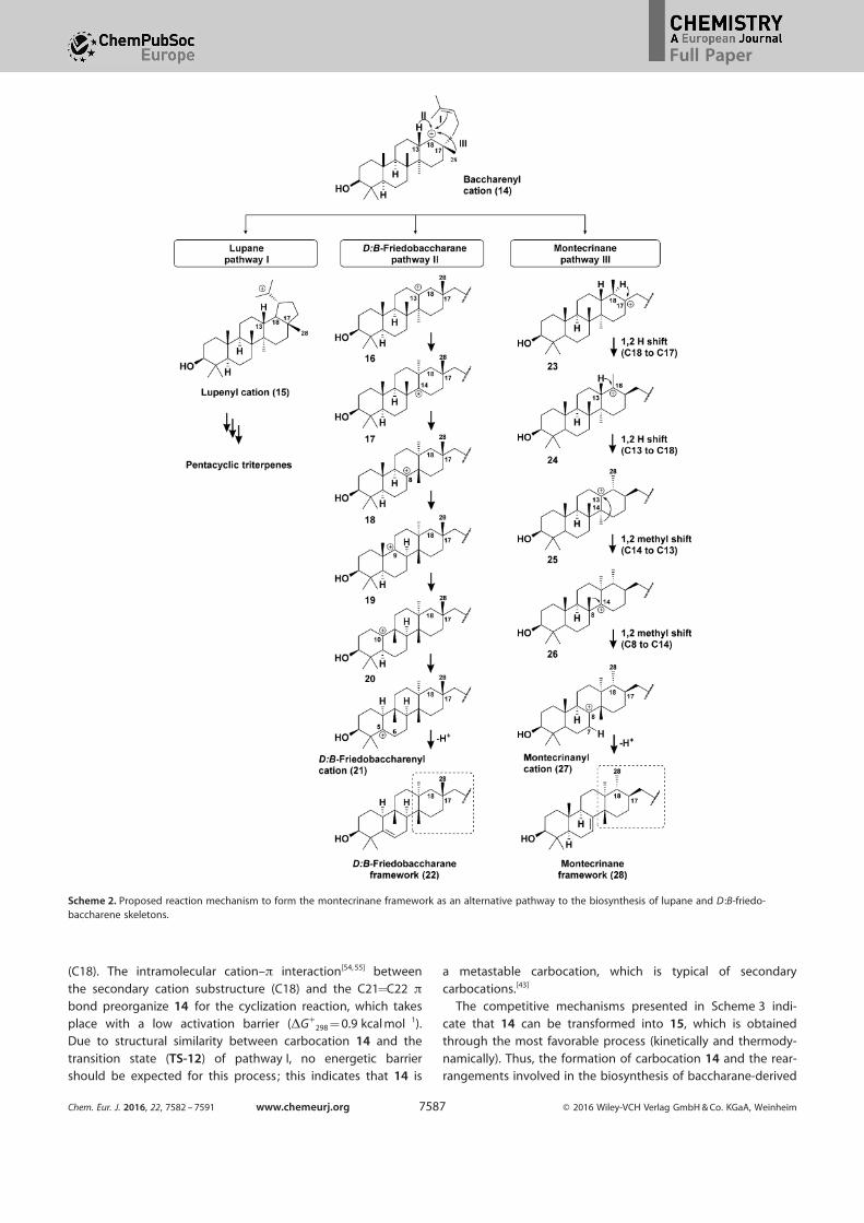
Intramolecular cyclization of 14 through E-ring closure tothe b-face of C18 by the olefinic bond generates the lupenylcation (15). This cation is the key intermediate for the forma-tion of pentacyclic triterpenes (Scheme 2, pathway I). However,
the biogenesis of 6/6/6/6 tetracyclic triterpenes occurs from 14through friedo rearrangement by alternative pathways through
1,2-hydride and methyl shifts.[1, 20] For instance, the 1,2-hydrideshift from C13 to C18 in carbocation 14, and successive rear-rangements, lead to the D :B-friedobaccharenyl cation (21;
Scheme 2, pathway II). The subsequent deprotonation of 21
Figure 3. Determination of the absolute configuration of the chiral center C3 in compound 3 from the DdHBa experimental values of the (R)-MPA derivative.
Chem. Eur. J. 2016, 22, 7582 – 7591 www.chemeurj.org Ó 2016 Wiley-VCH Verlag GmbH & Co. KGaA, Weinheim7585
Full Paper

from C6 gives D :B-friedobaccharan-5,21-diene triterpenes, such
as leonal and 4.[13] On the other hand, the formation of the un-precedented tetracyclic montecrinane skeleton can also be ra-
tionalized from intermediate 14. The novel reaction step in thebiosynthesis of the montecrinane framework (28) is the 1,2-
methyl (28-CH3) shift from C17b to C18, which generates
a more stable tertiary carbocation at C17 (intermediate 23 ;Scheme 2, pathway III).
A further 1,2-hydride shift (H18a to C17) leads to intermedi-ate 24, in which a new stereocenter at C17 is generated.[42] The
stereochemistry of the new chiral center locates the side chainin the b position, which is in agreement with experimental ob-
servations. As a result, cation 24 is an epimeric intermediate
compared with 14 and D :B-friedobaccharane derivatives. In thenext reaction steps, 1,2-hydride shift (H13 to C18) and further
methyl shifts (27-CH3 to C13 and 26-CH3 to C14) lead to themontecrinanyl cation (27), with a positive charge at C8. Subse-
quent deprotonation at C7 generates a new 6/6/6/6 tetracyclictriterpenene skeleton: montecrinane 28. Selective oxidation at
C3 and hydroxylation at C15 lead to montecrinanes A (1) and
B (2). Moreover, epoxidation of the D21-double bond followedby epoxide ring opening and water addition gave rise tomontecrinane C (3).
Theoretical studies on 6/6/6/6-tetracyclic triterpenebiosynthesis
Quantum chemical calculations have been used to describe
terpene-forming carbocation rearrangements, to predict geo-metric structures and relative energies of intermediates and
transition-state structures, as well as to support new biosyn-thetic pathways. Among DFT methods, the B3LYP functional
has become the most common method employed to calculate
the geometries of carbocation precursors of terpenes. Particu-larly, B3LYP geometries and mPW1PW91 single-point energies
(mPW1PW91/6-31 + G(d,p)//B3LYP/6-31 + G(d,p) methods) havebeen successfully used by Matsuda et al.[43] and Tantillo[25] to
compute carbocation reactions in the biosynthesis of naturalproducts.[26, 47–49]
To obtain mechanistic insights into the 6/6/6/6tetracyclic triterpene biosynthesis, theoretical studies
were carried out to compare the energetic profile ofthe possible pathways that led to these triterpenes,
starting from 14 (Scheme 2). Therefore, this study fo-cuses on the feasibility of the formation of cations
21 and 27 to understand energetic factors involvedin the mechanisms of tetracyclic triterpene skeleton
formation. Additionally, the formation of 15 from
carbocation 14 was studied because lupane-type tri-terpenes (compounds 7–12) were also isolated from
C. vulcanicola.First, a conformational study of 14 was carried out
by using PcModel 9.2.[50] Then, the minimized struc-tures were optimized at the B3LYP/6-31 + G(d,p)
level of theory in the gas phase by using Gaussi-
an 09.[51] The B3LYP functional tends to underesti-mate the relative stability of cyclic structures. Therefore, single-
point energy calculations were performed at the mPW1PW91/6-31 + G(d,p) level of theory by using the B3LYP geometries,
and the results were compared with those obtained by usingthe M06-2X/6-311 + G(d,p)//B3LYP/6-31 + G(d,p) method.[52, 53]
Conformational analysis shows that secondary carbocation
14 might exist as a true minimum in the potential energy sur-face. From this structure, a sequence of rearrangements (1,2-
hydride or -methyl shifts) were studied to compare the ener-getic changes of the proposed biosynthetic pathways. The re-
sults of these calculations are summarized in the energy dia-grams shown in Figure 4. The gas-phase model for the monte-
crinane framework formation is presented in Figure 4 A. The
Gibbs free energy profile shows that the 1,2-methyl shift from14 to tertiary carbocation 23 is the rate-determining step of
the process. The activation energy of this step (TS-1) isDG�
298 = 10.2 kcal mol¢1. Subsequent rearrangements from in-
termediate 23 lead to the formation of 27, which takes placedue to the high stability of the tertiary carbocations involvedin the rearrangements (intermediates 23–27) and the small ac-
tivation barriers for their formation (TS-2, TS-3, TS-4, and TS-5). The 1,2-hydride shift from carbocation 14 to tertiary carbo-cation 16, and subsequent rearrangements, generate the D :B-friedobaccharane skeleton (Figure 4 B). The activation energy
of the 1,2-hidryde shift (TS-6) is DG�298 = 6.3 kcal mol¢1, which
is 3.9 kcal mol¢1 lower than that of TS-1. This energetic differ-
ence could be due to the nature of the migration of the spe-cies (hydride or methyl group) involved in the rearrangementreaction. For hydride migration, there are interactions with thes orbital of the hydride, whereas in the case of methyl migra-tion sp3 orbitals are involved. The rate-determining step of the
whole process of going from 14 to 21 has an energetic barrierof DG�
298 = 8.5 kcal mol¢1 (TS-10), which is similar to that of
TS-1 (DG�298 = 10.2 kcal mol¢1). Based on the similar energetic
profiles predicted by theoretical calculations, the twotriterpene frameworks could occur in nature.
Finally, the reaction path connecting 14 and 15 was studiedand compared with previous mechanisms (Scheme 3). In sec-
ondary carbocation 14, the double bond of the side chain is lo-cated in the proximity of the carbon with electron deficiency
Scheme 1. Cyclization and rearrangement of oxidosqualene to form the dammarenylcation (13) ; a key intermediate in the biosynthesis of tetra- and pentacyclic triterpenes.Enz = enzyme.
Chem. Eur. J. 2016, 22, 7582 – 7591 www.chemeurj.org Ó 2016 Wiley-VCH Verlag GmbH & Co. KGaA, Weinheim7586
Full Paper
(C18). The intramolecular cation–p interaction[54, 55] between
the secondary cation substructure (C18) and the C21=C22 p
bond preorganize 14 for the cyclization reaction, which takes
place with a low activation barrier (DG�298 = 0.9 kcal mol¢1).
Due to structural similarity between carbocation 14 and the
transition state (TS-12) of pathway I, no energetic barriershould be expected for this process; this indicates that 14 is
a metastable carbocation, which is typical of secondary
carbocations.[43]
The competitive mechanisms presented in Scheme 3 indi-
cate that 14 can be transformed into 15, which is obtainedthrough the most favorable process (kinetically and thermody-
namically). Thus, the formation of carbocation 14 and the rear-rangements involved in the biosynthesis of baccharane-derived
Scheme 2. Proposed reaction mechanism to form the montecrinane framework as an alternative pathway to the biosynthesis of lupane and D :B-friedo-baccharene skeletons.
Chem. Eur. J. 2016, 22, 7582 – 7591 www.chemeurj.org Ó 2016 Wiley-VCH Verlag GmbH & Co. KGaA, Weinheim7587
Full Paper

terpenoids will depend on the enzymes present in the reactionmedia.[56, 57] For instance, a specific OSC, such as synthases re-
ported for related metabolites,[58, 59] could be responsible for
the formation of an unprecedented backbone in one sequenceof catalytic steps. Overall, the activation energies of the rate-
determining steps connecting 14 with cations 15 (0.9 kcalmol¢1), 21 (8.5 kcal mol¢1), and 27 (10.2 kcal mol¢1) are
consistent with the relative abundance of the lupane (7–12),D :B-friedobachharane (4–6), and montecrinane (1–3)
triterpenes described herein (see Table A15 in the SupportingInformation).
Conclusion
Complete carbocation cyclization/rearrangement pathwaysfrom 14 to montecrinane and D :B-friedobaccharane frame-
works were developed on the basis of the results of DFT calcu-lations. In the proposed mechanism towards montecrinanes,
Figure 4. Relative Gibbs free energy (DG�298 in kcal mol¢1) profile predicted at the mPW1PW91/6-31 + G(d,p)//B3LYP/6-31 + G(d,p) level of theory for montecri-
nane (A) and D :B-friedobaccharane (B) framework formation. Geometries, energies, and a comparison of mPW1PW91 and M06-2X functionals are given in theSupporting Information.
Chem. Eur. J. 2016, 22, 7582 – 7591 www.chemeurj.org Ó 2016 Wiley-VCH Verlag GmbH & Co. KGaA, Weinheim7588
Full Paper

the first reaction step is a novel methyl shift from C17 to C18,
which generates a tertiary carbocation at C17. Afterward, 1,2-hydride shift from C18 to C17 changes the configuration of the
side chain at C17. Subsequent rearrangements generate the
montecrinanyl cation, and the final step of the process is theelimination of a proton from C7. The montecrinane skeleton
might be indicative of a divergent pathway for tetracyclic tri-terpene biosynthesis from 14. The proposed biosynthetic path-
way is energetically feasible; therefore, more secondary metab-olites containing this skeleton are expected to be isolated
from Celastraceae species. The co-occurrence of D :B-friedobac-
charanes supports the hypothesis that Celastraceae speciesand, in particular, C. vulcanicola, possess specific OSCs that
lead to the biosynthesis of uncommon 6/6/6/6 tetracyclic tri-terpenoids. Hence, 1,2-methyl and -hydride shifts proposed for
the biosynthesis of montecrinanes might require a specificOCS capable of stabilizing carbocation intermediates.
Experimental Section
General
Optical rotations were measured on a PerkinElmer 241 automaticpolarimeter in CHCl3 at 20 8C. UV spectra were obtained ona JASCO V-560 spectrophotometer in absolute EtOH. IR (film) spec-tra were measured on a Bruker IFS 55 spectrophotometer. 1H(500 MHz) and 13C NMR (125 MHz) spectra were recorded ona Bruker Avance 500 spectrometer at 25 8C; chemical shifts werereferenced to the residual solvent signal (CDCl3 : dH = 7.26 ppm,dC = 77.0 ppm); DEPT, TOCSY, COSY, ROESY (spin lock field 2500 Hz),HSQC, and HMBC (optimized for J = 7.7 Hz) experiments were car-ried out with the pulse sequences given by Bruker. EIMS and HR-EIMS were obtained on a Micromass Autospec spectrometer, andHR-ESIMS spectra (positive mode) were performed on LCT PremierXE Micromass electrospray spectrometer. Silica gel 60 (particle size15–40 and 63–200 mm, Machery-Nagel) and Sephadex LH-20 (Phar-macia Biotech) were used for column chromatography, whereassilica gel 60 F254 (Machery-Nagel) was used for analytical and prepa-rative TLC. Centrifugal planar chromatography was performed bymeans of a Chromatotron instrument (model 7924T, Harrison Re-
search Inc. , Palo Alto, CA, USA) on manually coated silica gel 60GF254 (Merck) by using a 1, 2, or 4 mm plates. The spots were vi-sualized by UV light and heating silica gel plates sprayed withH2O/H2SO4/AcH (1:4:20). All solvents used were of analytical grade(Panreac). Optically pure MPA was purchased from Sigma, whereasthe other reagents were purchased from Aldrich and used withoutfurther purification.
Plant material
C. vulcanicola J. (Donnell Smith; Celastraceae) was collected in No-vember 2009 at Montecristo National Park (2051 m above sealevel), in the municipality of Metap�n, Province of Santa Ana, El Sal-vador. The plant was identified by Jorge Alberto Monterrosa Salo-mûn, Curator of the Herbarium at the Jard�n Bot�nico, La Laguna,El Salvador, and a voucher specimen (J. Monterrosa & R. Carballo412) was deposited in the Herbarium of Missouri Botanical Garden,St. Louis, MO, USA.
Extraction and isolation
The air-dried and powdered root bark of C. vulcanicola (0.8 kg) wasextracted with n-hexane/Et2O (1:1, 4.0 L, 48 h) in a Soxhlet appara-tus. Evaporation of the solvent under reduced pressure providedthe crude extract (24.0 g), which was subjected to column chroma-tography over silica gel (1.0 kg, 70–230 mesh) by using increasingpolarity mixtures of hexanes/EtOAc (0–100 %, 34.0 L) as the eluentto afford nine fractions (1–9). Fraction 2 (2.3 g; hexanes/EtOAc, 8:2)was separated by chromatography on Sephadex LH-20 (hexanes/CHCl3/MeOH, 2:1:1, 2.5 L) to afford 26 fractions, which were com-bined into six fractions (2 a–f) on the basis of their TLC profiles.Fractions 2 b (480.0 mg), 2 c (608.0 mg), and 2 f (127.0 mg) were fur-ther subjected to centrifugal planar chromatography on silica gelby using mixtures of CH2Cl2/Me2CO of increasing polarity (10.0 to8:2) to afford five (2 b1–5), eight (2 c1–8) and four (2 f1–4) subfrac-tions, respectively. Subfractions 2 b2, 2 c7, and 2 f3 were further pu-rified by preparative TLC by using mixtures of hexanes/EtO2 (8:2),CH2Cl2/Me2CO (9:1), and toluene/EtOAc (7:3), respectively, to yieldcompounds 1 (3.8 mg), 2 (2.2 mg), and 3 (9.7 mg), in addition tothe known compounds, 4 (8.5 mg), 5 (5.0 mg), and 6 (3.9 mg). Frac-tion 5 (1.6 g; hexanes/EtOAc, 7:3) was separated by chromatogra-phy on Sephadex LH-20 (hexanes/CHCl3/MeOH, 2:1:1), on silica gel(hexanes/EtOAc of increasing polarity, 8:2 to 3:7), by centrifugal
Scheme 3. Competitive reaction pathways from the baccharenyl cation (14) to tetra-/pentacyclic skeletons. Energies [kcal mol¢1] are given at themPW1PW91/6-31 + G(d,p)//B3LYP/6-31 + G(d,p) level of theory.
Chem. Eur. J. 2016, 22, 7582 – 7591 www.chemeurj.org Ó 2016 Wiley-VCH Verlag GmbH & Co. KGaA, Weinheim7589
Full Paper

planar chromatography (CH2Cl2/Me2CO of increasing polarity, 9:1 to7:3) and by preparative HPTLC (hexanes/Et2O, 3:7) to give theknown lupane triterpenes 7[36] (6.2 mg), 8[36] (4.8 mg), 9[37] (7.2 mg),10[36] (12.6 mg), 11[36] (7.8 mg), and 12[38] (4.1 mg).
Montecrinane A (1)
Colorless amorphous solid; [a]20>D =¢58.68 (c = 0.38 in CHCl3) ; for
1H and 13C NMR data, see Table 1; for 2D spectra, see Figures A2–A4 in the Supporting Information; IR (film) n= 3455, 2924, 2855,1708, 1459, 1381, 1267, 1046, 971, 755, 573 cm¢1; EIMS: m/z (%):440 (18) [M+] , 422 (4) [M+¢18(H2O)], 407 (19), 353 (6), 323 (9), 287(22), 273 (20), 257 (17), 245 (18), 231 (45), 203 (11), 185 (13), 159(27), 149 (26), 133 (38), 119 (48), 109 (78), 105 (44), 95 (68), 69(100), 55 (95); HR-EIMS: m/z calcd for C30H48O2 [M+]: 440.3654;found: 440.3654.
Montecrinane B (2)
Colorless amorphous solid; [a]20>D =¢53.18 (c = 0.22 in CHCl3) ; for
1H and 13C NMR data, see Table 1; for 2D spectra, see Figure A6 inthe Supporting Information; IR (film) n= 3401, 2923, 2853, 1460,1378, 1270, 1027, 974, 755, 501 cm¢1; EIMS: m/z (%): 442 (4), 424(7) [M+¢18(H2O)], 407 (8), 289 (9), 215 (20), 187 (32), 173 (30), 161(24), 145 (31), 121 (59), 109 (86), 107 (64), 105 (44), 95 (81), 69 (99),55 (100); HR-EIMS: m/z calcd for C30H50O2 [M+]: 442.3811; found:442.3799.
Montecrinane C (3)
Colorless amorphous solid; [a]20>D =¢12.58 (c = 0.97 in CHCl3) ; for
1H and 13C NMR data, see Table 1; for 2D spectra, see Figures A8–10 in the Supporting Information; IR (film) n= 3436, 2927, 2872,1709, 1462, 1377, 1279, 1166, 1067, 1033, 756 cm¢1; HR-ESIMS: m/zcalcd for C30H49O3 [M+¢Na¢H]: 472.3682; found: 472.3682.
Preparation of (R)-(¢¢)-a-methoxyphenylacetic ester (3 a)
A solution of (R)-MPA (5.0 mg), 3 (3.8 mg), dicyclohexylcarbodii-mide (DCC; 6.0 mg), and 4-dimethylaminopyridine (DMAP; 5.3 mg)in dry dichloromethane (1 mL) was stirred at room temperatureand under a nitrogen atmosphere for 24 h. The reaction mixturewas filtered through a short cotton-wool filter and the solvent wasremoved under reduced pressure to give a thick oil, which was pu-rified by preparative TLC on silica gel (n-hexane/EtOAc, 8:2) to givethe analytically pure ester 3 a (2.4 mg) as an amorphous solid.1H NMR (CD3CN, 500 MHz): d= 0.63 (d, J = 6.5 Hz, 3 H; 28-CH3), 0.73(s, 3 H; 25-CH3), 0.77 (s, 3 H; 27-CH3), 0.78 (s, 3 H; 24-CH3), 0.90 (s,3 H; 23-CH3), 0.95 (s, 3 H; 26-CH3), 1.33 (s, 3 H; 29-CH3), 1.42 (s, 3 H;30-CH3), 1.42 (m, 1 H; H2b), 1.48 (m, 1 H; H2a), 3.37 (s, 3 H; OMe-MPA), 4.52 (dd, J = 4.4, 11.7 Hz, 1 H; H3), 4.83 (s, 1 H; H2’-MPA), 5.24(dd, J = 3.2, 6.3 Hz, 1 H; H7), 7.21–7.48 ppm (m, 5 H; MPA); 13C NMR(CD3CN, 500 MHz): d= 12.1 (q, C25), 14.8 (q, C24), 17.4 (q, C28),18.1 (t, C11), 21.3 (q, C26), 22.3 (q, C29), 22.4 (q, C30), 23.3 (t, C6),24.4 (t, C2), 26.3 (q, C23), 26.4 (q, C26), 27.1 (t, C16), 28.9 (t, C19),31.6 (t, C20), 33.2 (t, C12), 33.3 (t, C15), 34.3 (s, C10), 35.0 (d, C17),35.8 (t, C1), 37.4 (s, C4), 43.6 (s, C13), 48.1 (d, C9), 49.9 (d, C5), 50.6(s, C14), 52.4 (d, C18), 81.2 (d, C3), 84.4 (s, C22), 115.8 (d, C7), 145.6(s, C8), 208.3 ppm (s, C21); MPA [56.4 (q), 82.2 (d), 126.6 (d), 128.0(2 Õ d), 128.7 (2 Õ d), 134.9 (s), 171.2 ppm (s)] ; 1H NMR (CD3CN,500 MHz, saturated with Ba(ClO4)2): d= 0.63 (s, 3 H; 28-CH3), 0.64 (s,3 H; 25-CH3), 0.74 (s, 3 H; 27-CH3), 0.82 (s, 3 H; 24-CH3), 0.86 (s, 3 H;23-CH3), 0.91 (s, 3 H; 26-CH3), 1.15 (m, 1 H; H2b), 1.24 (m, 1 H; H2a),1.30 (s, 3 H; 29-CH3), 1.48 (s, 3 H; 30-CH3), 3.32 (s, 3 H; OMe-MPA),
4.54 (dd, J = 3.9, 11.5 Hz, 1 H; H3), 5.08 (s, 1 H; H2’-MPA), 5.21 (dd,J = 3.6, 6.0 Hz, 1 H; H7), 7.39–7.51 ppm (m, 5 H; MPA); 13C NMR(CD3CN, 500 MHz, saturated with Ba(ClO4)2): d= 13.5 (q, C25), 14.9(q, C24), 18.1 (t, C11), 18.5 (q, C28), 21.8 (q, C27), 21.9 (q, C30), 23.1(q, C29), 23.5 (t, C6), 24.3 (t, C2), 26.7 (q, C23), 26.9 (q, C26), 27.1 (t,C16), 28.8 (t, C19), 31.7 (t, C20), 33.1 (t, C12), 33.6 (t, C15), 34.4 (s,C10), 34.4 (d, C17), 35.4 (t, C1), 37.4 (s, C4), 43.1 (s, C13), 49.2 (d,C9), 50.1 (d, C5), 50.9 (s, C14), 52.2 (d, C18), 84.1 (d, C3), 86.9 (s,C22), 118.3 (d, C7), 145.7 (s, C8), 209.7 ppm (s, C21); MPA (56.0 (q),82.4 (d), 126.6 (d), 128.0 (2 Õ d), 128.6 (2 Õ d), 135.0 (s), 171.1 ppm(s)) ; for 1H, 13C NMR, and 2D NMR spectra, see Figures A11, A12,and A14, respectively, in the Supporting Information,.
Computational details
The conformational space was searched by GMMX (included inPcModel 9.2[50]) by using a mixed method, which allowed the pro-gram to 1) randomly select a subset of all bonds for rotation; and2) randomly move a subset of all heavy and non-volatile atoms in3D space, which caused small changes in the shape of the mole-cule. The minimized structures obtained with PcModel 9.2[50] werereoptimized at the B3LYP/6–31 + G(d,p) level of theory by usingGaussian 09.[51] The geometric optimizations of ground and transi-tion states (as a first-order saddle point) and reaction profiles wereperformed without any symmetry restrictions. Harmonic vibrationalfrequency calculations were performed analytically to verify thecharacter of the stationary point obtained and to correct energiesfor the zero-point vibrations. Transition states were fully optimizedand characterized by the presence of only one imaginary frequen-cy. Notably, no basis set superposition error (BSSE) correction wascarried out during reaction profiles because hydride (or methylgroup) transfer occurred in the same molecule. Optimized coordi-nates and energies of all structures are included in the SupportingInformation. Images were produced by using the CYL viewprogram.[60]
Acknowledgements
This work was supported by the FP7-REGPOT-2012-CT2012-
316137-IMBRAIN and CTQ2011-28417-C02-01 (MINECO)
projects. We thank Jorge A. Monterrosa for collecting andidentifying the plant material, and the Servicio de Parques
Nacionales y Vida Silvestre, Direcciûn de Recursos Renovablesdel Ministerio de Agricultura y Ganader�a (MAG), and Funda-
ciûn Ecolûgica de El Salvador (SALVANATURA) for supplyingthe plant material. A.E.A. thanks CONICYT and FONDECYT3160414 of Chile for a fellowship.
Keywords: biosynthesis · density functional calculations ·natural products · structure elucidation · terpenoids
[1] R. Xu, G. C. Fazio, S. P. T. Matsuda, Phytochemistry 2004, 65, 261 – 291.[2] R. A. Hill, J. D. Connolly, Nat. Prod. Rep. 2015, 32, 273 – 327.[3] H. Sheng, H. Sun, Nat. Prod. Rep. 2011, 28, 543 – 593.[4] F. Mo, T. Anthonsen, T. Brunn, Acta Chem. Scand. 1972, 26, 1287 – 1288.[5] M. Shibuya, A. Sagara, A. Saitoh, T. Kushiro, Y. Ebizuka, Org. Lett. 2008,
10, 5071 – 5074.[6] N. Shoji, A. Umeyama, Z. Taira, T. Takemoto, K. Nomoto, K. Mizukawa, Y.
Ohizumi, J. Chem. Soc. Chem. Commun. 1983, 16, 871 – 873.[7] L. Pan, L. B. S. Kardono, S. Riswan, D. J. Newman, D. Kinghorn, J. Nat.
Prod. 2010, 73, 1873 – 1878.
Chem. Eur. J. 2016, 22, 7582 – 7591 www.chemeurj.org Ó 2016 Wiley-VCH Verlag GmbH & Co. KGaA, Weinheim7590
Full Paper

[8] B. Y. Hwang, B. N. Su, H. Chai, Q. Mi, L. B. S. Kardono, D. Kinghorn, J.Org. Chem. 2004, 69, 3350 – 3358.
[9] N. Stiti, S. Triki, M- A. Hartmann in Olives and Olive Oil in Heat and Dis-ease Prevention (Eds: V. R. Preedy, R. R. Watson), Elsevier, London, 2010,pp. 211 – 218.
[10] T. Fujioka, Y. Kashiwada, H. Okabe, K. Mihashi, K.-H. Lee, Bioorg. Med.Chem. Lett. 1996, 6, 2807 – 2810.
[11] K. Masuda, K. Shiojima, H. Ageta, Chem. Pharm. Bull. 1983, 31, 2530 –2533.
[12] S. ©ksìz, S. Serin, Phytochemistry 1997, 46, 545 – 548.[13] M. J. NfflÇez, M. R. Lûpez, I. A. Jim¦nez, L. M. Moujir, A. G. Ravelo, I. L.
Bazzocchi, Tetrahedron Lett. 2004, 45, 7367 – 7370.[14] Y. Zhang, S. Nakamura, T. Wang, H. Matsuda, M. Yoshikawa, Tetrahedron
2008, 64, 7347 – 7352.[15] T. Akihisa, K. Yasukawa, Y. Kimura, S. Takase, S. Yamanouchi, T. Tamura,
Chem. Pharm. Bull. 1997, 45, 2016 – 2023.[16] T. Akihisa, E. M. K. Wijeratne, H. Tokuda, F. Enjo, M. Toriumi, Y. Kimura, K.
Koike, T. Nikaido, Y. Tezuka, H. Nishino, J. Nat. Prod. 2002, 65, 158 – 162.[17] R. Frçde, M. Brçckelmann, B. Steffan, W. Steglich, R. Marumoto, Tetrahe-
dron 1995, 51, 2553 – 2560.[18] W. B. Zhou, J. Y. Tao, H. M. Xu, K. Li Chen, G. Z. Zeng, C. J. Ji, Y. M. Zhang,
N. H. Tan, Z. Naturforsch. Sect. B 2010, 65, 1393 – 1396.[19] M. Shibuya, T. Xiang, Y. Katsube, M. Otsuka, H. Zhang, Y. Ebizuka, J. Am.
Chem. Soc. 2007, 129, 1450 – 1455.[20] S. Lodeiro, Q. Xiong, W. K. Wilson, M. D. Kolesnikova, C. S. Onak, S. P. T.
Matsuda, J. Am. Chem. Soc. 2007, 129, 11213 – 11222.[21] S. Sawai, H. Uchiyama, S. Mizuno, T. Aoki, T. Akashi, S-i. Ayabe, T. Takaha-
shi, FEBS Lett. 2011, 585, 1031 – 1036.[22] S. C. Roberts, Nat. Chem. Biol. 2007, 3, 387 – 395.[23] S. T. Mugford, T. Louveau, R. Melton, X. Qi, S. Bakht, L. Hill, T. Tsurushima,
S. Honkanen, S. J. Rosser, G. P. Lomonossoff, A. Osbourn, Plant Cell2013, 25, 1078 – 1092.
[24] R. Thimmappa, K. Geisler, T. Louveau, P. O’Maille, A. Osbourn, Annu. Rev.Plant Biol. 2014, 65, 225 – 257.
[25] D. J. Tantillo, Nat. Prod. Rep. 2011, 28, 1035 – 1053.[26] D. J. Tantillo, Nat. Prod. Rep. 2013, 30, 1079.[27] N. Alvarenga, E. A. Ferro in Studies in Natural Products Chemistry, Vol. 33
(Ed. : Atta-ur-Rahman), Elsevier, Amsterdam, 2006, pp. 239 – 307.[28] A. A. Osorio, A. MuÇoz, D. Torres-Romero, L. M. Bedoya, N. R. Perestelo,
I. A. Jim¦nez, J. Alcam�, I. L. Bazzocchi, Eur. J. Med. Chem. 2012, 52, 295 –303.
[29] M. J. NfflÇez, A. E. Ardiles, M. L. Mart�nez, D. Torres-Romero, I. A. Jim¦nez,I. L. Bazzocchi, Phytochem. Lett. 2012, 5, 244 – 248.
[30] D. Torres-Romero, B. King-Diaz, I. A. Jim¦nez, B. Lotina-Hennsen, I. L.Bazzocchi, J. Nat. Prod. 2008, 71, 1331 – 1335.
[31] D. Torres-Romero, F. MuÇoz-Mart�nez, I. A. Jim¦nez, S. Castanys, F. Ga-marro, I. L. Bazzocchi, Org. Biomol. Chem. 2009, 7, 5166 – 5172.
[32] D. Torres-Romero, I. A. Jim¦nez, R. Rojas, R. H. Gilman, M. Lûpez, I. L.Bazzocchi, Bioorg. Med. Chem. 2011, 19, 2182 – 2189.
[33] O. Callies, M. P. S�nchez-CaÇete, F. Gamarro, I. A. Jim¦nez, S. Castanys,I. L. Bazzocchi, J. Nat. Prod. 2015, 78, 736 – 745.
[34] D. Torres-Romero, B. King-Diaz, R. J. Strasser, I. A. Jim¦nez, B. Lotina-Hennsen, I. L. Bazzocchi, J. Agric. Food Chem. 2010, 58, 10847 – 10854.
[35] A. E. Ardiles, A. Gonz�lez-Rodr�guez, M. J. NuÇez, N. R. Perestelo, V.Pardo, I. A. Jim¦nez, A. M. Valverde, I. L. Bazzocchi, Phytochemistry 2012,84, 116 – 124.
[36] M. J. NfflÇez, A. E. Ardiles, M. L. Mart�nez, D. Torres-Romero, I. A. Jim¦nez,I. L. Bazzocchi, Phytochem. Lett. 2013, 6, 148 – 151.
[37] M. J. NfflÇez, C. P. Reyes, I. A. Jim¦nez, L. Moujir, I. L. Bazzocchi, J. Nat.Prod. 2005, 68, 1018 – 1021.
[38] A. P. Dantanarayana, N. S. Kumar, M. U. S. Sultanbawa, S. J. Balasubrama-niam, J. Chem. Soc. Perkin Trans. 1 1981, 2717 – 2723.
[39] M. Kuroyanagi, M. Shiotsu, T. Ebihara, H. Kawai, A. Ueno, S. Fukushima,Chem. Pharm. Bull. 1986, 34, 4012 – 4017.
[40] The new compounds were named montecrinanes based on the placeof collection of the plant (C. vulcanicola), which was Montecristo Na-tional Park on the north-western tip of El Salvador.
[41] J. M. R. Seco, E. QuiÇoa, R. Riguera, Chem. Rev. 2004, 104, 17 – 117.[42] Q. Xiong, F. Rocco, W. K. Wilson, R. Xu, M. Ceruti, S. P. T. Matsuda, J. Org.
Chem. 2005, 70, 5362 – 5375.[43] S. P. T. Matsuda, W. K. Wilson, Q. Xiong, Org. Biomol. Chem. 2006, 4,
530 – 543.[44] W. K. Wilson, R. Xu, M. Ceruti, S. P. T. Matsuda, J. Org. Chem. 2005, 70,
6492.[45] I. Abe, Y. Sakano, H. Tanaka, W. Lou, H. Noguchi, M. Shibuya, Y. Ebizuka,
J. Am. Chem. Soc. 2004, 126, 3426 – 3427.[46] D. J. Tantillo, Chem. Soc. Rev. 2010, 39, 2847 – 2854.[47] Y. J. Hong, D. J. Tantillo, Nat. Chem. 2009, 1, 384 – 389.[48] Y. J. Hong, D. J. Tantillo, Org. Lett. 2011, 13, 1294 – 1297.[49] Y. J. Hong, D. J. Tantillo, Nat. Chem. 2014, 6, 104 – 111.[50] PCModel, version 9.2; Serena Software, Bloomington, IN.[51] Gaussian 09, Revision A.02, M. J. Frisch, G. W. Trucks, H. B. Schlegel, G. E.
Scuseria, M. A. Robb, J. R. Cheeseman, G. Scalmani, V. Barone, B. Men-nucci, G. A. Petersson, H. Nakatsuji, M. Caricato, X. Li, H. P. Hratchian,A. F. Izmaylov, J. Bloino, G. Zheng, J. L. Sonnenberg, M. Hada, M. Ehara,K. Toyota, R. Fukuda, J. Hasegawa, M. Ishida, T. Nakajima, Y. Honda, O.Kitao, H. Nakai, T. Vreven, J. A. Montgomery, Jr. , J. E. Peralta, F. Ogliaro,M. Bearpark, J. J. Heyd, E. Brothers, K. N. Kudin, V. N. Staroverov, R. Ko-bayashi, J. Normand, K. Raghavachari, A. Rendell, J. C. Burant, S. S. Iyen-gar, J. Tomasi, M. Cossi, N. Rega, J. M. Millam, M. Klene, J. E. Knox, J. B.Cross, V. Bakken, C. Adamo, J. Jaramillo, R. Gomperts, R. E. Stratmann,O. Yazyev, A. J. Austin, R. Cammi, C. Pomelli, J. W. Ochterski, R. L. Martin,K. Morokuma, V. G. Zakrzewski, G. A. Voth, P. Salvador, J. J. Dannenberg,S. Dapprich, A. D. Daniels, O. Farkas, J. B. Foresman, J. V. Ortiz, J. Cio-slowski, and D. J. Fox, Gaussian, Inc. , Wallingford CT, 2009.
[52] Y. Zhao, D. G. Truhlar, Theor. Chem. Acc. 2008, 120, 215 – 241.[53] Y. Zhao, D. G. Truhlar, Acc. Chem. Res. 2008, 41, 157 – 167.[54] Y. J. Hong, D. J. Tantillo, Chem. Sci. 2013, 4, 2512 – 2518.[55] Y. J. Hong, D. J. Tantillo, Org. Lett. 2015, 17, 5388 – 5391.[56] Y. J. Hong, D. J. Tantillo, J. Am. Chem. Soc. 2011, 133, 18249 – 18256.[57] R. P. Pemberton, K. C. Ho, D. J. Tantillo, Chem. Sci. 2015, 6, 2347 – 2353.[58] L. Kìrti, R.-J. Chein, E. J. Corey, J. Am. Chem. Soc. 2008, 130, 9031 – 9036.[59] Z. Wang, T. Yeats, H. Han, R. Jetter, J. Biol. Chem. 2010, 285, 29703 –
29712.[60] CYLview, 1.0b; Legault, C. Y. , Universit¦ de Sherbrooke, 2009 (http://
www.cylview.org).
Received: January 21, 2016
Published online on April 23, 2016
Chem. Eur. J. 2016, 22, 7582 – 7591 www.chemeurj.org Ó 2016 Wiley-VCH Verlag GmbH & Co. KGaA, Weinheim7591
Full Paper