moore et al-1994-australian and new zealand journal of surgery
TRANSCRIPT

8/16/2019 Moore Et Al-1994-Australian and New Zealand Journal of Surgery
http://slidepdf.com/reader/full/moore-et-al-1994-australian-and-new-zealand-journal-of-surgery 1/5
Aust N Z J .
Surg
1994) 64,
242-246
MANAGEMENT OF THE MALIGNANT COLORECTAL POLYP: THE
IMPORTANCEOF CLINICOPATHOLOGICALCORRELATION
JAMES
W.
E. MOORE,ESMOND. HOFFMANN*ND ROBERT OW L ND^
Colorectal Surgical Uni t, Royal Adelaide Hospital and tDepartment
of
Patholo gy, Institute
of
Medical and
Veterinary Science, Adelaide, South Australia, Australia
The results of management of colorectal adenomas removed endoscopically and found to contain invasive cancer seen in a
single institution over a 10 year period are presented. Clinical data were obtained retrospectively from patient case notes
and all specimens were reviewed by one pathologist. Fifty-four patients with malignant polyps were studied after exclusion
of others with polypoid carcinomas, epithelial misplacement and cases managed by primary segmental resection.
Of
the
various considered predictors of adverse outcome, only histologically incomplete excision proved significant. However,
when excision was considered macroscopically complete there was no significant association between incomplete
histological excision and adverse outcome. Consideration should be given to conservative management of such cases.
Key words: colonoscopy, colorectal carcinoma, colorectal polyp, polypectomy.
INTRODUCTION
The introduction of colonoscopic polypectomy in 1969I
revolutionized the managem ent of colorectal polyps and
has become the accepted practice in cases with lesions
proximal to the distal rectum. There is continued debate
regarding management of the endoscopically removed
polyp that con tains invasive carcin oma , despite almost 2
decades of discussion in the literature.*-14 t is clear tha t
there has been a significant shift towards conservative
managem ent and yet doub t still exists over the indications
for completion resection. This study presents the 10 year
experience of one Australian institution in the manage-
ment of malignant colorectal polyps, to determine the
prognostic significance of previously described clinical
and histopathological indicator s of ad verse outcom e after
treatment by polypectomy alone.
METHODS
A
record of all colorectal polyps containing invasive
malignancy seen at the Institute of Medical and Veteri-
nary Science
IMVS)
rom January 1982 to April 1992
was obtained by review of histopathology reports coded
by the System ized Nomenclatureof Medicine (SNOMED)
number. Only polyps removed by colonoscopic snare
excision were included. Invasive malignancy was defined
as invasion by malignant cells through the muscularis
mucosae. Cases of carcinoma in situ, intramucosal car-
cinoma and epithelial misplacement (pseudocarcinoma-
tous invasion 6.17 were excluded. Cases in which there
was no adeno mato us component in the mucosa adjac ent
Correspondence:J.
Moore,
Colorectal Surgical Unit, Royal Adelaide
Hospital, Adelaide. SA
SOOO
Australia.
Accepted for publication
30
September
1993
to the invasive tumour were termed polypoid carcinomas
and were excluded. Patient case notes were reviewed and
data regarding age, sex and date of polypectomy were
obtained . The site of the po lyp within the colon was taken
from the procedure report, and its size and morphology
(sessile
vs
pedunculated) were determined from the
procedure report or from the gross description
of
the
pathology specimen,
or
both. If the patient proceeded to
a completion resection (i.e. segme ntal colectomy, anterior
resection or abdo mino perinea l resection af ter polypec-
tomy), the pattern of residu al disease was recorded (none,
bowel wall, nodal, distal or a combination of these).
Follow up was ob tained by review of case notes, and by
general practitioner and patient interview. This was re-
corded as total and disease-free follow up in m onths post-
polypectomy. Disease-free status was determined by a
combination of clinical and endoscopic criteria, at last
review. If recurrence had occurred, the pattern and timing
of recurrence (local, regional or distal) was recorded.
Mortality was classified as cancer related or unrelated.
Patients were also assign ed to one of two groups:
(1) Adverse outcom e, where either recurrence occurred
after polypectomy alone or when residual or metastatic
disease was found at com pletion resection.
(2) Favourable outcom e, where no recurrence had been
documented after endoscopic polypectomy alone
or
when
completion resection revealed no evidence of residual
malignant disease (implying that polypectomy alone
would have produced a favourable outcome).
All pathology specimens were reviewed by a single
pathologist (R. Rowland). Details regarding the type of
polyp involved (tubular, comprising greater than 2/3
tubular architecture; villous, greater than 2/3 villous
architecture; and tubulovillous, neither of these) were
recorded. The carcinoma was graded as well, moderately
or poorly differentiated according to previously describ ed
8/16/2019 Moore Et Al-1994-Australian and New Zealand Journal of Surgery
http://slidepdf.com/reader/full/moore-et-al-1994-australian-and-new-zealand-journal-of-surgery 2/5
MANAGEMENT OF MALIGNANT
COLORECTAL
OLYP
243
riter ria. ̂ * ̂
The depth of invasion was recorded according
to the method described by Haggitt (Table
l).’
The
margin of clearance of invasive malignancy was meas-
ured on both lateral and deep margins in m illimetre s and
correlated with the endoscopic assessment
of
macro-
scopic clearance of the lesion. The presence or absence
of vessel invasion (defined as tumour within endothelial
lined spaces) was recorded but no attempt was m ade to
differentiate between lym phatic and venous invasion.
RESULTS
One hundred and twenty-nine pathology reports desc rib-
ing adenomatous polyps containing a focus of invasive
carcinoma were obtained after review of the records of
the IMVS. Thirty-two patients who underwent primary
resection were excluded. O ther exclusions were patients
with prior or synchronous bowel cancer (six cases),
polypoid carcinoma with no associated adenoma (five
cases), incomplete medical records
1
1
cases) and those
with seve re dysplasia
10
cases). An additional
1 1
cases
were reclassified after pathological review as having
mucosal misplaceme nt” and excluded. This left a study
group of
54
patients who had colorectal polyps with
invasive carcinoma . The mean age of the grou p was 66.7
years. The mean size of pedunculated polyps was
18.6
mm
(range
10-30)
and
21.3
mm (range
15-120)
for sessile
lesions. Median follow up was
34.5
months (range
4-1
10;
mean
39.5)
with two patients lost to follow up. Overall
70 of patients had follow up of greater than
24
months.
There was one cancer-related death and four patients
died of other causes at 5,
23, 24
and
62
months post-
Table 1.
Haggitt classification of depth of invasion
Level 1
Level
2
Level
3
Level 4
Invasive tumour (through muscularis mucosae)
confined to head
of
polyp
Invasive tumour to the junction
of
head and stalk
(i.e.
to
the ‘neck’ of polyp)
Invas ive tumour into part of the stalk
of
the polyp
Invasive tumour invading beyond the stalk, into the
submucosa of the bowel wall below the polyp but
not into the muscularis propria
A
carcinoma arising in
a
pedunculated polyp may
be
classified
as
Haggitt level 1-4 depending on the level
of
invasion seen, but a
carcinoma arising in a sess ile polyp is by definition level 4 as it involves
the submucosa
of
the underlying bowel.
Table
2
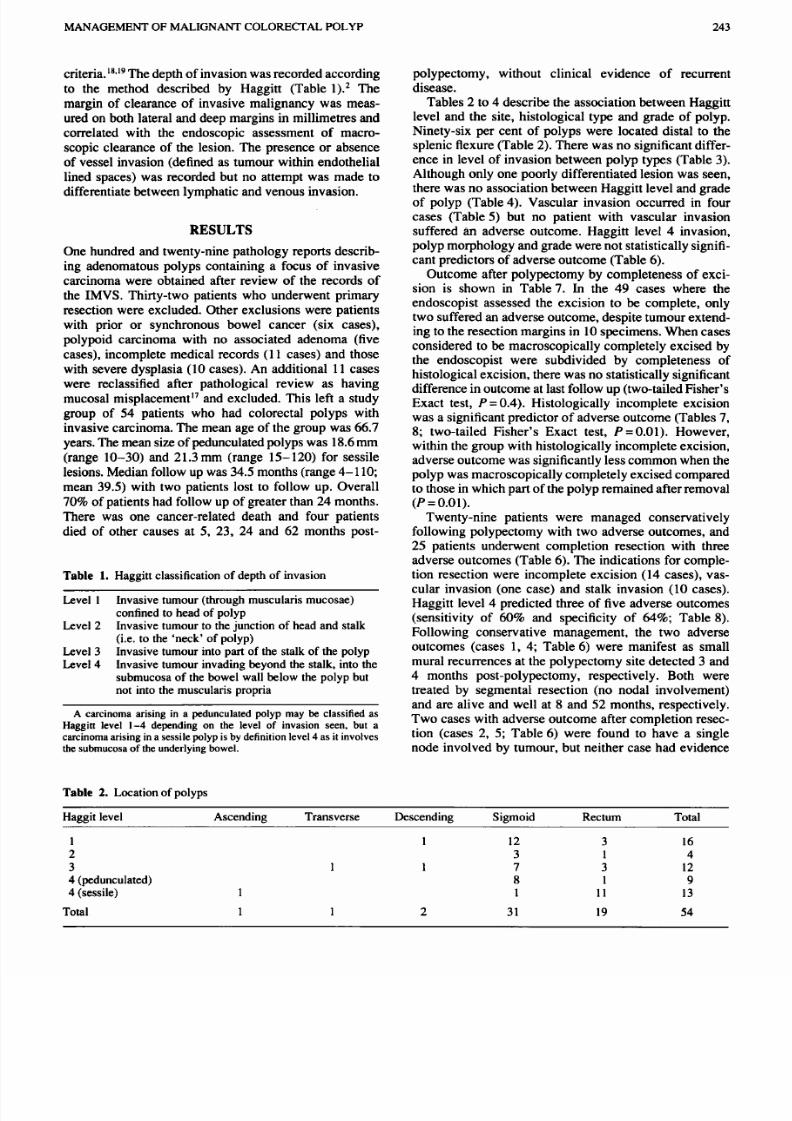
Locationof polyps
polypectomy, without clinical evidence of recurrent
disease.
Tables
2
to
4
describe the association between H aggitt
level and the site, histological type and grade of polyp.
Ninety-six per cent of polyps were located distal to the
splenic flexure (Table
2).
There was no significant differ-
ence in level of invasion between polyp types (Table 3).
Although only one poorly differentiated lesion was seen,
there was no association between Haggitt level and grade
of polyp (Table4). Vascular invasion occurred in four
cases (Table 5) but no patient with vascular invasion
suffered an adverse outcome. Haggitt level 4 invasion,
polyp morphology and grade were not statisticallysignifi-
cant predictors of adverse outcome (Table 6).
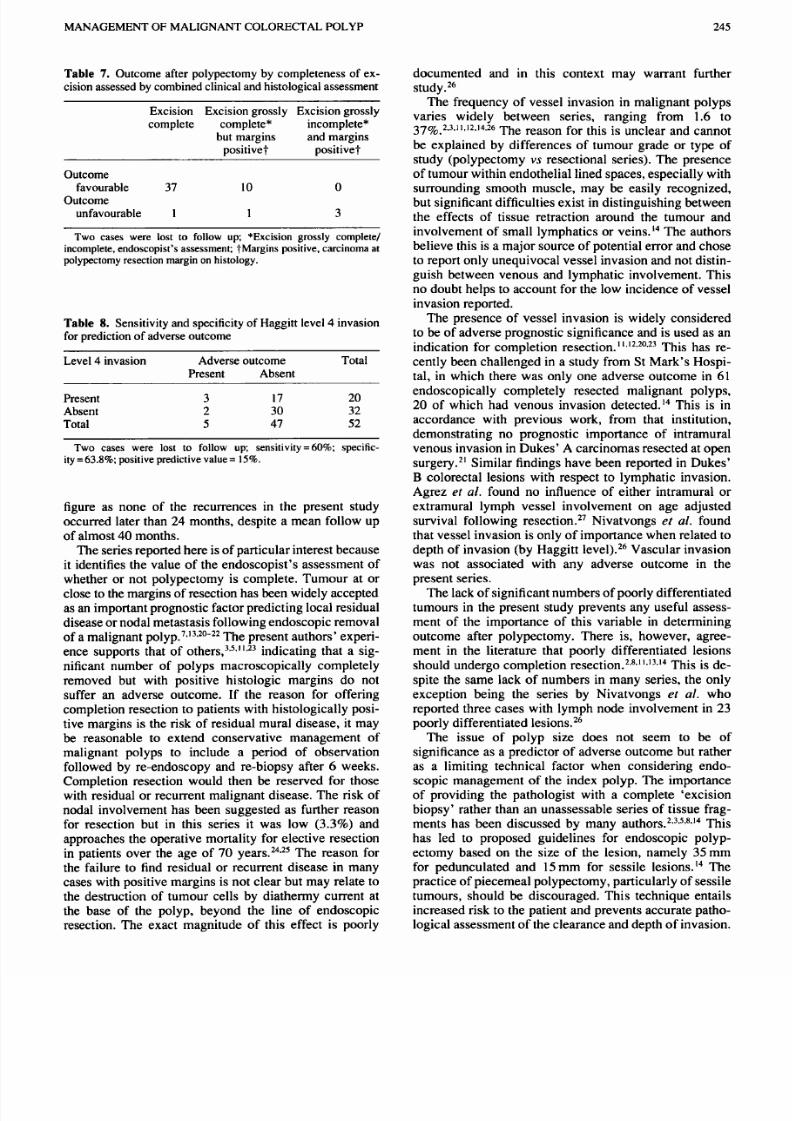
Outcome after polypectomy by completeness of exci-
sion is shown in Table7. In the
49
cases where the
endoscopist assessed the excision to be complete, only
two suffered an adverse outcome, despite tumour extend-
ing to the resection margins in 10specimens. When cases
considered to be macrosco pically com pletely excised by
the endoscopist were subdivided by completeness of
histologic al excisio n, there w as no s tatistically significant
difference in outcome at last follow up (two-tailed Fisher’s
Exact test, P
=0.4).
Histologically incomplete e xcision
was a significant predictor of adverse outcome (Tables 7,
8;
two-tailed Fisher’s Exact test, P
=0.01).
However,
within the group with histologically incomplete excision,
adverse outcom e was significantly less common when the
polyp was macroscopically completely excised compared
to those in which part of the polyp remained after removal
(P=0.01).
Twenty-nine patients were managed conservatively
following polypectomy with two adverse outcomes, and
25
patients underwent completion resection with three
adverse outcomes (Table 6). The indications for comple-
tion resection were incomplete excision 14 cases), vas-
cular invasion (one case) and stalk invasion
10
cases).
Haggitt level 4 predicted three of five adverse outcomes
(sensitivity of
60
and specificity of
64 ;
Table
8).
Following conservative management, the two adverse
outcomes (cases
1, 4;
Table 6) were manifest as small
mural recurrences at the polypectomy site detected
3
and
4
months post-polypectomy, respectively. Both were
treated by segmental resection (no nodal involvement)
and are alive and well at
8
and
52
months, respectively.
Two cases with adverse outcom e after completion resec-
tion (cases
2,
5; Table6) were found to have a single
node involved by tumour, but neither case had evidence
Haggit level Ascending Transverse Descending Sigmoid Rectum Total
1
1 12 3 16
2
3 1
4
3 1
1
7
3 12
4 (pedunculated) 8 1
9
4 (sessile) 1 1 1 1
13
Total
1 1
2 31
19
54

8/16/2019 Moore Et Al-1994-Australian and New Zealand Journal of Surgery
http://slidepdf.com/reader/full/moore-et-al-1994-australian-and-new-zealand-journal-of-surgery 3/5
244
MOORE ET AL..
of residual mural disease and are alive and well at 6 and
32 m onths. The third case underwent piecemeal resection
of a large sessile rectal lesion and subsequ ent completion
resection revealed no evidence of residual tumour. Multi-
ple hepatic m etastases developed at
12
months and this
patient died 15 months post-polypectomy.
Table
3. Histopathological type of polyp
Haggitt level Tubular Tubdovillou s Villous
1
9 1 6
2
3
3
1 1
4 pedunculated)
6 2
1
4 (sessile) 3
5 5
Total 3 10 3
Exact value = N S for all comparisons (Pearson Chi-squared test)
comparing polyp type by Haggitt level .
Table
4. Grade of polyps
Haggitt level Well Moderate Poor Total
1
12
4
16
2
3 1
4
3
5 7 12
4 (pedunculated) 3 5 1 9
4 (sessile) 3 10 13
Total 26 27 1 54
Table
5. Vascular invasion
Haggitt level Present Absent Total
1
1 15
16
2
4 4
3
12 12
4 (pedunculated)
2 7 9
4 (sessile)
1
12 13
Total 4 50
54
Table 6.
Details of cases with adverse outcome
DISCUSSION
Endoscopic polypectomy was introduced in
1969
by
Wolff and Shinya' and is now the preferred management
option for large bowel polyps proximal to the distal
rectum, but the management of the polyp containing
invasive malignancy remains controversial. Series reported
in the literature are often difficult to compare because
'invasive cancer' is not always clearly defined. In this
series, carcinoma was only diagnosed when there was
invasion beyond the muscularis mucosae. Cases of car-
cinoma
in situ
or intramucosal carcinoma were excluded
because of their apparent lack of metastasizing poten-
tial. Eleven adenom as initially reported as invasive
lesions were found, after review, to represent cases of
epithelial misplacement and were excluded. This high-
lights the difficulty in distinguishing misplacement from
invasive tumour.
6
It also emph asizes the need for carefu l
review of histology both in the individual patient in
whom a decision regarding resection is pending and when
assessing the published literature. By excluding patients
with polypoid carcinoma, the authors hope to avoid a
possible source of error (i.e. including cases that may not
have been suitable for endoscopic polypectomy in the
first instance).
Pathology reporting may have other influences on
published outcomes following endoscopic removal of
malignant polyps. The extent of excision, particularly of a
pedunculated lesion, can only
be
assessed by
a
combined
clinical and histopathological assessmen t, an issue often
not stressed in the literature. The endoscopist must be
sure that no residual polyp remains and that this is
conveyed to the pathologist. Otherwise, a pedunculated
polyp may
be
reported as sessile because little stalk has
been taken. Likewise, vessel invasion in the stalk of a
pedunculated polyp may be missed if only the head of
the polyp has been excised. The w ell-defined histologic
staging system of Hagg itt2 allows the stratification of
cases by depth of invasion and, when com bined with the
endoscopist's repo rt, overcomes these difficulties.
Almost all series have used similar outcome criteria to
those the present authors have used (i.e. recurrence on
follow up or residual disease on completion resection).
Wilcox has suggested that status after follow up be
reported only after 5 or preferably
10
years to overcome
the problem of late rec~rrence.'~his is a conservative
Case number and pattern of failure* Predictive
Local Nodal on M etastasis Local Nodal on outcome)
1
2
3 4
5
valuet (adverse
recurrence resection 12/12 recurrence resection
Haggitt level
1 2
3 4 4 NS
Morphology Ped Ped
Sess Sess Ped NS
Complete excision Yes No No No No P
=0.01
(histologically)
Grade
Moderate
Well Moderate Well Moderate NS
Vessel invasion No No No No No
*See text for details; tFisher's exact test;Ped. edunculated, Sess, sessile.
8/16/2019 Moore Et Al-1994-Australian and New Zealand Journal of Surgery
http://slidepdf.com/reader/full/moore-et-al-1994-australian-and-new-zealand-journal-of-surgery 4/5
MANAGEMENTOF MALIGNANT COLORJXTAL POLYP 245
Table 7.
Outcome after polypectomy by com pleteness of ex -
cision assessed by combined clinical and histological assessment
Excision Excision grossly Excision grossly
complete complete* incomplete*
but margins and margins
positive? positive?
Outcome
Outcome
favourable 37
10
0
unfavourable 1 1 3
Two cases were lost
to
follow up; *Excision grossly complete/
incomplete, endoscopist’s assessment; ?Margins positive, carcinoma at
polypectomy resection margin on histology.
Table 8. Sensitivity and specificity of H aggitt level 4 invasion
for prediction of adverse outcome
Level
4
invasion Adverse outcom e Total
Present Absent
Present
Absent
Total
3 17 20
2 30 32
5
47 52
Two cases were lost to follow
up; sensitivity =60 ; specific-
ity =63.8 ;positive predictive value
=
15 .
figure as none of the recurrences in the present study
occurred later than 24 months, despite a
mean
follow up
of almost
40
months.
The series reported here is of particular interest because
it identifies the value of the end oscop ist’s assessment of
whether or not polypectomy is complete. Tumour at or
close to the margins of resection has been widely accepted
as an im portant prognostic factor predicting local residual
disease
or
nodal m etastasis following endoscopic removal
of a malignant polyp.7.’3*20 -22he present authors’ experi-
ence supports that of others,3*5*11.23ndicating that a sig-
nificant number of polyps macroscopically completely
removed but with positive histologic margins do not
suffer an adverse outcome. If the reason for offering
completion resection to patients with histolo gically posi-
tive margins
is
the risk of residual mural disease,
it
may
be reasonable to extend conservative management of
malignant polyps to include a period of observation
followed by re-endoscopy and re-biopsy after 6 weeks.
Completion resection would then be reserved for those
with residual or recurrent malignant disease. The risk of
nodal involvement has been sug gested as further reason
for resection but in this series it was low
(3.3 )
and
approaches the operative mortality for elective resection
in patients over the age of 70 year^.^^**^ The reason for
the failure to find residual or recurrent disease in many
cases with positive margins is not clear but may relate to
the destruction of tumour cells by diathermy current at
the base of the polyp, beyond the line of endoscopic
resection. The exact magnitude of this effect is poorly
documented and in this context may warrant further
study.26
The frequency of vessel invasion in malignant polyps
varies widely between series, ranging from 1.6 to
37~0.2.3.11.12.14.26 h e reason for this is unclear and cannot
be explained by differences of tumour grade
or
type of
study (polypectomy vs resectional series). The presence
of tumour w ithin endothelia l lined spaces, espec ially with
surrounding smooth muscle, may be easily recognized,
but significant difficulties exist
in
distinguishing between
the effects of tissue retraction around the tumour and
involvement of small lymphatics or veins.I4The authors
believe this is a major sou rce of potential error and cho se
to report only unequivocal vessel invasion and not distin-
guish between venous and lymphatic involvement. This
no doubt helps to acco unt for the low incidence of vessel
invasion reported.
The presence of vessel invasion is widely considered
to be of adv erse prognostic significance and is used as an
indication for completion resection. 1312*20*23 This has re-
cently been challenged
in
a study from St Mark’s Hospi-
tal, in which there was only one adverse outcome
in
61
endoscopically completely resected malignant polyps,
20 of which had venous invasion detected.14 This is in
accordance with previous work, from that institution,
demonstrating no prognostic importance of intramural
venous invasion in D ukes’ A carcino mas resected at open
surgery.21Similar findings have been reported in Dukes’
B
colorectal lesions with respect to lymphatic invasion.
Agrez et al. found no influence of either intramural or
extramural lymph vessel involvement on age adjusted
survival following re ~e cti on .~ ’ ivatvongs ef
al.
found
that vessel invasion is only
of
impo rtance when related to
depth of invasion (by Haggitt Vascular invasion
was not associated with any adverse outcome in the
present series.
The lack of significant num bers of poorly differen tiated
tumours in the present study prevents any useful assess-
ment of the impo rtance of this variable in determinin g
outcome after polypectomy There is, however, agree-
ment in the literature that poorly differentiated lesions
should undergo completion resectio n.2+8*1 ’.13*14his is de-
spite the same lack of numbers in many series, the only
exception being the series by Nivatvongs
et
al who
reported three cases with lymph node involvement in 23
poorly differentiated lesions.26
The issue of polyp size does not seem to be of
significance as a predic tor of adverse outcom e but rather
as a limiting technical factor when considering endo-
scopic management of the index polyp. The importance
of providing the pathologist with a complete ‘excision
biopsy’ rather than an una ssessable series of tissue frag-
m ents has been discussed by many a ~ t h o r s . ~ . ~ * ~ * ~ * ~his
has led to proposed guidelines for endoscopic polyp-
ectomy based on the size of the lesion, namely 35mm
for pedunculated and
15
mm for sessile lesions.14 The
practice of piecemea l polypectom y, particularly of sessile
tumours, should be discouraged. This technique entails
increased risk to the patient and prevents accura te patho-
logical assessment of the clearance and depth of invasion.

8/16/2019 Moore Et Al-1994-Australian and New Zealand Journal of Surgery
http://slidepdf.com/reader/full/moore-et-al-1994-australian-and-new-zealand-journal-of-surgery 5/5
246
MOORE
ET
AL.
I t should be reserved for pa t ients whe re the
risks of
open
surgica l management are deemed prohibit ive and
no
practical alternative exists.
REFERENCES
1 Wolff WI. Shinya H. Polypectomy via the fiberoptic
colonoscope: Removal of lesions beyond reach of the
sigmoidoscope. N. Engl.
J.
Med. 1973;
288:
329-32.
2. Haggitt RC, Glotzbach RE, Soffer EE. “ruble LD.
Prognostic factors in colorectal carcinomas arising in
adenomas: Implications for lesions removed by endoscopic
polypectomy. Gastroenterology
1985;
89:
328-36.
3.
Cranley JP, Petras RE, Carey WD, Paradis K, Sivak MV.
When is endoscopic polypectomy adequate therapy for
colonic polyps containing invasive carcinoma? Gastro-
enterology
1986;
91:
419-27.
4. Fucini C, Wolff BG, Spencer RJ. An appraisal of endo-
scopic removal of malignant polyps. Mayo Clin. Proc.
5 Williams CB, Whiteway JE, Jass JR Practical aspects of
the management of malignant polyps. Endoscopy 1987; 19:
31-7.
6. Hermanek P, Gall
FP.
Early (microinvasive) colorectal
carcinoma. I n t
J. Colorect. Dis.
1986;
1:
79-84.
7. Frei JV. Endoscopic large bowel polypectomy: Adequate
treatment of som e completely removed, minimally invasive
lesions. Am.J. Surg. Patho l. 1985;
9
355-9.
8. Richards WO. W ebb
W A ,
Mom s SJ
et al.
Patient manage-
ment after endoscopic removal of the cancerous colon
adenoma.
Ann. Surg.
1987;
205:
665-72.
9. Colacchio TA, Forde KA, Scantlebury VP. Endoscopic
polypectomy Inadequate therapy for invasive colorectal
carcinoma.
Ann. Surg.
1981;
194:
704-7.
10.
Morson BC, Bussey HJR, Samoorian S. Policy of local
excision for early cancer of the colorectum.
Gut
1977;
18:
11. Coverlizza
S,
Risio M, Ferrari A Fenoglio-Preiser CM,
Rossini FP. Colorectal adenomas containing invasive car-
cinoma: Pathologic assessment of lymph node metastatic
potential.
Cancer
1989;
64:
1937-47.
12. Sugihara K. Muto T, Morioka Y. Management of patients
with invasive carcinoma removed by colonoscopic polyp-
ectomy.
Dis. Colon Rectum
1989;
32:
829-34.
13. Wilcox GM, Anderson PB, Colacchio TA. Early invasive
carcinoma in colonic polyps: A review of the literature with
1986; 61: 123-6.
1045-50.
emphasis on assessment of the risk of metastasis. Cancer
14.
Geraghty JM, Williams CB, Talbot IC. Malignant colo-
rectal polyps: Venous invasion and successful treatment
by endoscopic polypectomy. Gut 1991; 32: 774-8.
15. Rothwell DJ (Ed.).
Systemized Nomenclature
o
Medicine:
Microglossary for Surgical Pathology.
Illinois: College of
American Pathologists, 1987.
16. Greene
EL.
pithelial misplacement in adenomatou s polyps
of the colon and rectum.
Cancer
1974; 33: 206-17.
17. Muto T, Bussey HJR, Morson BC. Pseudocarcinomatous
invasion in adenomatous polyps of the colon and rectum.
J Clin. Path.
1973;
26:
25-31.
18. Jass
JR
Atkin WS, Cuzick J
et al.
The grading of rectal
cancer: Historical perspective and a multivariate analysis
of 447 cases. Histopathology 1986 10: 437-59.
19. Goh HS, Jass JR. DNA content and the adenoma carcinoma
sequence in adenomatous polyps of the colon and rectum.
J.
Clin. Path. 1986; 39: 387-92.
20. Wolff WL. Sh inya H, Cwern M , Hsu M. Cancerous colonic
polyps: ‘Hands on’
or
‘hands off’. Am. Surg.
1990
56:
21. Talbot IC, Ritchie
S,
Leighton MH, Hughes AO. Bussey
HJR, Morson BC. Spread of rectal cancer within veins.
Histologic features and clinical significance.
Am. J.
Surg.
22. Pines A, Bat L, Shemesh E et al. Invasive colorectal
adenomas: Surgery versus colonoscopic polypectomy.
J Surg. Oncol.
1990;
43:
53-5.
23. Conte CC. Welch JP, Tennant R, Forouhar F, Lundy J,
Bloom GP. Managem ent of endoscopically remov ed malig-
nant colon polyps.
J Surg. Oncol.
1987;
36:
116-21.
24. Bokey EL, Chapuis PH, Hughes WJ. Koorey SG Hinder
JM, Edwards R. Morbidity, mortality and survival follow-
ing resection for carcinoma of the rectum at Concord
Hospital. Aust. N.Z.J Surg.
1990;
6 :
253-9.
25. Corman ML. Veidenheimer MC, Coller
JA.
Colorectal
carcinoma: A decade of experience at the Lahey Clinic.
Dis . Colon Rectum
1979; 22: 477-9.
26. Nivatvongs
S,
Rojanasakul A, Reiman HM et al. The risk
of lym ph node metastasis in colorectal polyps w ith invasive
adenocarcinoma.
Dis. Colon Rectum
1991;
34:
323-8.
27. Agrez MV, Spagno lo D, Harvey J, House AK, O’Connell
D. Prognostic significance
of
lymphatic permeation in
Dukes B colorectal cancer.
Aust.
N .Z.
J. Surg.
1988;
58:
1986;
57:
160-71.
148-52.
1981; 141: 15-17.
39-42.