morpho-anatomical study of rhizome of limonium … brasileira de farmacognosia 25 (2015) 320–327...
TRANSCRIPT

O
M
TKMa
b
c
d
e
a
ARAA
KBGLPR
I
iifaacnmt
aRi
0
Revista Brasileira de Farmacognosia 25 (2015) 320–327
www . sb fgnos ia .org .br / rev is ta
riginal Article
orpho-anatomical study of rhizome of Limonium brasiliense
ânia Mara Antonelli-Ushirobiraa, Andressa Blainskia, Naiara Cássia Gancedob, Fernanda Gaburob,átia Aparecida Kern Cardosoc, Eneri Vieira de Souza Leite-Mellod, João Carlos Palazzo de Melloa,∗,aria Auxiliadora Milaneze-Gutierree
Programa de Pós-graduac ão em Ciências Farmacêuticas, Departamento de Farmácia, Universidade Estadual de Maringá, Maringá, PR, BrazilDepartamento de Farmácia, Universidade Estadual de Maringá, Maringá, PR, BrazilComplexo de Centrais de Apoio à Pesquisa, Universidade Estadual de Maringá, Maringá, PR, BrazilDepartamento de Ciências Morfológicas, Universidade Estadual de Maringá, Maringá, PR, BrazilDepartamento de Biologia e Museu Dinâmico Interdisciplinar, Universidade Estadual de Maringá, Maringá, PR, Brazil
r t i c l e i n f o
rticle history:eceived 5 June 2015ccepted 15 July 2015vailable online 6 August 2015
eywords:aicuruuaicuruimonium brasiliensehenolic compounds
a b s t r a c t
Limonium brasiliense (Boiss.) Kuntze, Plumbaginaceae, is an herb popularly known as guaicuru, guaicuráor baicuru. The species inhabits salt marshes from the coastal region of southern Brazil, including Riode Janeiro, to Uruguay and Argentina. Although widely used in folk medicine in the state of Rio Grandedo Sul to treat genitourinary infections and to regulate menstrual periods, L. brasiliense has been littlestudied. The present morpho-anatomical study was undertaken to resolve some doubts in the literatureas to the nature of the part of the plant that is used for medicinal purposes, a true rhizome or a root. Themorpho-anatomical characteristics were analyzed with the aid of light and scanning electron microscopy.The botanical material was characterized as a rhizome with internodes that are evident in the youngerbut not the older portions. Microscopic analysis revealed the presence of a multilayered periderm with
hizome a cortex, ray parenchyma, and pith, formed by collenchyma tissue with abundant intercellular spaces inthe outer portions of the cortex, responsible for the rigidity of the body, and with walls impregnated withphenolic compounds. The vascular bundles are collateral with elliptical to elongated cells, and with fewconducting and sclerenchymal elements. Groups of sclereids are dispersed through the cortex and pith.These morpho-anatomical characteristics define the structure as a rhizome.
© 2015 Sociedade Brasileira de Farmacognosia. Published by Elsevier Editora Ltda. All rights reserved.
ntroduction
Limonium Mill., the most species-rich in the Plumbaginaceae,ncludes about 350 species of herbs. The genus is best representedn the Mediterranean regions of Europe and in Asia, and can also beound in coastal regions of North and South America, South Africa,nd Australia (Mobot, 2015). In general, species of Limonium lives halophytes, including in alpine regions (Chant, 1993). In otherountries, such as China, stems and roots of various species of Limo-ium are used in folk medicine, and some of them are similar inorphology and anatomy, making them difficult to identify using
raditional methods (Ding et al., 2012).Limonium brasiliense (Boiss.) Kuntze is an herb popularly known
s guaicuru, guaicurá or baicuru (Dias da Silva, 1920) in the state ofio Grande do Sul, Brazil. It is grown and marketed by small farmers
n estuarine regions of the River Plate Basin. According to Simões
∗ Corresponding author.E-mail: [email protected] (J.C.P. de Mello).
http://dx.doi.org/10.1016/j.bjp.2015.07.010102-695X/© 2015 Sociedade Brasileira de Farmacognosia. Published by Elsevier Editora
et al. (1998), this herb is common in coastal salt marshes in south-ern Brazil, from Paraná to Rio Grande do Sul, and in Uruguay andArgentina; Zappi (2015) gave the range as extending from southernBrazil to the state of Rio de Janeiro.
Although it was described in the first edition of the BrazilianPharmacopoeia (1929), L. brasiliense is not included in the currentPharmacopoeia. It is popularly used to treat uterine and ovarianinflammation, vaginal discharge and dysmenorrhea (Moura et al.,1985), and is useful to regulate menstrual periods (Lifchitz, 1981),as well as having an antimicrobial effect (Rosito, 1975). Murrayet al. (2004) isolated five antioxidant compounds from extracts ofL. brasiliense roots. Their chemical composition includes hydrolyz-able and condensed tannins, 4-O-methyl gallic acid, sitosterol, andtriterpenic saponins, the structures of which have not been deter-mined (Rosito, 1975).
The first botanical description of L. brasiliense was contained in
the Flora brasiliensis (Martius, 1840–1906). Dias da Silva (1920) pro-vided a detailed anatomical description, noting the organolepticcharacteristics of fresh plants, i.e. a strong unpleasant odor thatdisappears upon dissection, and a spicy astringent flavor. MartiusLtda. All rights reserved.
rasilei
(mssaaahRsi(a
imuL
M
P
Koom3rcduh(riR
M
wim(bambwsssAt
asglct1
T.M. Antonelli-Ushirobira et al. / Revista B
1840–1906) named it Statice brasiliensis Boiss., an herb with aore or less scaly rhizome. In 1920, Dias da Silva, in describing the
pecies, initially referred to the rhizome as cylindrical-irregular,hort, thick, more or less covered with scales; but in describing thenatomy of this organ as used medicinally, reported that the “roots”re 1–2 cm in diameter, cylindrical-fusiform and crooked. The char-cterization as a “root” may have been a conceptual error, becauseis morphological description makes it clear that this is a rhizome.eitz (1965) described the species as having thick roots with redcales, and this error in terming the rhizome of L. brasiliense a roots also found in the studies of Corrêa (1952), Coimbra (1958), Cruz1982), Moura et al. (1985), Murray et al. (2004), Fenner et al. (2006)nd Blainski et al. (2013).
Given the possibility of exploitation of the species and thenexact categorization of the organ used, this study provided a
orpho-anatomical description of the part of the plant used in pop-lar medicine, contributing to the pharmacognostic evaluation of. brasiliense.
aterials and methods
lant material
This study used rhizomes of Limonium brasiliense (Boiss.)untze, Plumbaginaceae, collected in May 2010 and January 2013n the Ilha dos Marinheiros (31◦59′33′′ S, 052◦10′43′′ W) in the cityf Rio Grande, Rio Grande do Sul, Brazil. The collection of plantaterial is registered with IBAMA-SISBIO under number 11995-
of 2 November 2010, authentication code 46367613, under theesponsibility of João Carlos Palazzo de Mello. Access to the botani-al material was authorized and licensed by the Conselho Nacionale Desenvolvimento Científico e Tecnológico (CNPq), registerednder no. 010252/2015-0. Samples of the reproductive phase areeld in the Herbarium of the Universidade Estadual de MaringáHUEM) under registration numbers 21151 and 27725 for the mate-ial collected in 2010 and 2013, respectively. The plant material wasdentified by Prof. Dr. Lilian Auler Mentz (Universidade Federal doio Grande do Sul).
orpho-anatomical analysis
The macroscopic characterization of the rhizome of L. brasilienseas based on the notes of Oliveira et al. (1998). For the anatom-
cal analysis under light microscopy (LM) and scanning electronicroscopy (SEM), segments of rhizome with different diameters
from 0.77 to 1.53 cm) were used. The segments were rehydratedy boiling in a solution of 10% glycerin for 15–30 min (2 times)nd stored in 70% ethanol (Johansen, 1940), with weekly replace-ent of the ethanol to eliminate the excess red pigment released
y their tissues. Sections for light microscopy were made freehandith steel blades, on the standard planes for plant anatomy. The
ections were bleached with sodium hypochlorite (30%), double-tained with Astra blue (1%) and safranin (1%), and mounted onemi-permanent slides with glycerin gel as described by Kraus andrduin (1997). The same procedure was used to prepare slides with
he rehydrated powder from rhizomes.Histochemical tests were done with cross sections, prepared
s above, of samples hydrated in glycerin–water, which weretained with Lugol’s iodine solution to reveal the presence of starchrains; iodinated zinc chloride, for lignin; Sudan IV glycerin, for
ipophilic substances; ferric chloride, for polyphenols; and 60%hloral hydrate with 25% sulfuric acid, for calcium oxalate crys-als (Johansen, 1940; Berlyn and Miksche, 1976; Kraus and Arduin,997; Farmacopeia Brasileira, 2010).ra de Farmacognosia 25 (2015) 320–327 321
For analysis under scanning electron microscopy (SEM), the seg-ments of rhizomes were rehydrated and cut with steel blades into0.3 mm sections on different anatomical planes, and then fixed in1% glutaraldehyde in 0.1 M sodium phosphate buffer pH 7.2 (Krausand Arduin, 1997). After 72 h in glutaraldehyde, the samples weredehydrated in an ascending ethanol series (30, 50, 70, 90, 95%, v/v)for 15 min each, ending in absolute ethanol for 10 min twice, andthen critical point-dried with CO2 (Balzers CPD 30 critical-pointdryer) (Horridge and Tamm, 1969). Then the samples were pos-itioned on the different anatomical planes on metal stubs, attachedwith double-sided carbon tape, and sputter-coated with gold in aShimadzu IC-50 unit. The photomicrographs were obtained with anOlympus BX50 optical microscope, and the ultrastructural analysesused a Shimadzu SS 550 SEM (at 15 kV).
Results and discussion
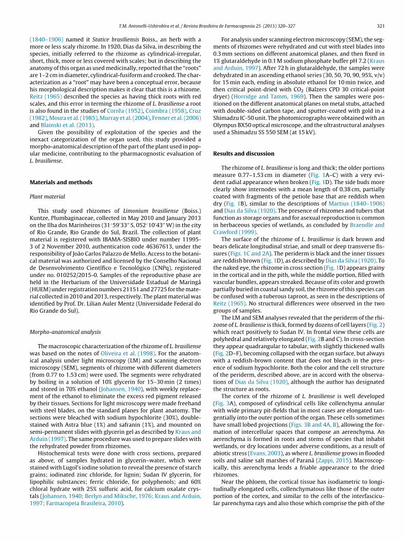
The rhizome of L. brasiliense is long and thick; the older portionsmeasure 0.77–1.53 cm in diameter (Fig. 1A–C) with a very evi-dent radial appearance when broken (Fig. 1D). The side buds moreclearly show internodes with a mean length of 0.38 cm, partiallycoated with fragments of the petiole base that are reddish whendry (Fig. 1B), similar to the descriptions of Martius (1840–1906)and Dias da Silva (1920). The presence of rhizomes and tubers thatfunction as storage organs and for asexual reproduction is commonin herbaceous species of wetlands, as concluded by Braendle andCrawford (1999).
The surface of the rhizome of L. brasiliense is dark brown andbears delicate longitudinal striae, and small or deep transverse fis-sures (Figs. 1C and 2A). The periderm is black and the inner tissuesare reddish brown (Fig. 1D), as described by Dias da Silva (1920). Tothe naked eye, the rhizome in cross section (Fig. 1D) appears grainyin the cortical and in the pith, while the middle portion, filled withvascular bundles, appears streaked. Because of its color and growthpartially buried in coastal sandy soil, the rhizome of this species canbe confused with a tuberous taproot, as seen in the descriptions ofReitz (1965). No structural differences were observed in the twogroups of samples.
The LM and SEM analyses revealed that the periderm of the rhi-zome of L. brasiliense is thick, formed by dozens of cell layers (Fig. 2)which react positively to Sudan IV. In frontal view these cells arepolyhedral and relatively elongated (Fig. 2B and C). In cross-sectionthey appear quadrangular to tabular, with slightly thickened walls(Fig. 2D–F), becoming collapsed with the organ surface, but alwayswith a reddish-brown content that does not bleach in the pres-ence of sodium hypochlorite. Both the color and the cell structureof the periderm, described above, are in accord with the observa-tions of Dias da Silva (1920), although the author has designatedthe structure as roots.
The cortex of the rhizome of L. brasiliense is well developed(Fig. 3A), composed of cylindrical cells like collenchyma annularwith wide primary pit-fields that in most cases are elongated tan-gentially into the outer portion of the organ. These cells sometimeshave small lobed projections (Figs. 3B and 4A, B), allowing the for-mation of intercellular spaces that compose an aerenchyma. Anaerenchyma is formed in roots and stems of species that inhabitwetlands, or dry locations under adverse conditions, as a result ofabiotic stress (Evans, 2003), as where L. brasiliense grows in floodedsoils and saline salt marshes of Paraná (Zappi, 2015). Macroscop-ically, this aerenchyma lends a friable appearance to the driedrhizomes.
Near the phloem, the cortical tissue has isodiametric to longi-tudinally elongated cells, collenchymatous like those of the outerportion of the cortex, and similar to the cells of the interfascicu-lar parenchyma rays and also those which comprise the pith of the

322 T.M. Antonelli-Ushirobira et al. / Revista Brasileira de Farmacognosia 25 (2015) 320–327
Fig. 1. Dried samples of Limonium brasiliense. Long thick rhizome (closed arrows) (A) with thinner lateral branches surrounded by reddish petiole bases (open arrows) (B);details of periderm (C) and internal tissues (D).
Fig. 2. Periderm of rhizome of Limonium brasiliense. General appearance of surface striae (A) and periderm cells (B and C); cross-sections of periderm (D and E) and detail ofthe cells (F). A, B and D: in SEM; C, E and F: in LM.

T.M. Antonelli-Ushirobira et al. / Revista Brasileira de Farmacognosia 25 (2015) 320–327 323
F hyma
sc
ttorp(iSrTeKp
Fp
ig. 3. Rhizome of Limonium brasiliense. General appearance (A) and detail of aerenc
tem, but always allowing the formation of relatively large inter-ellular spaces.
Sclereids with very varied dimensions are scattered throughhe cortex, individually (rarely, as in Fig. 4A) or in groups of fiveo a few dozen elements, positioned parallel to the major axisf the rhizome. These cells have a small to voluminous lumen,ounded, beveled or anomalous ends, but always with branchedits (Fig. 5A–C), similar to those described by Metcalfe and Chalk1950) in the cortex of the underground stems of Limonium bellid-folium (Gouan) Dumort and Limonium binervosum (G.E.Sm.) C.E.almon. The multiple layers of the secondary wall of this scle-enchymatic cell type become even more apparent in SEM (Fig. 5D).he presence of groups of sclereids was also reported by Grigore
t al. (2014) in the cortex of the rhizome of Limonium furfuraceumuntze, although in large groups. Similar groups of sclereids are alsoresent in the pith of the rhizome of L. brasiliense.ig. 4. More superficial strata of cortex of rhizome of Limonium brasiliense. Detail of tanrojections. sc: sclereid.
(B) in transversal sections under SEM. cx: cortex, p: periderm, vb: vascular bundles.
The vascular bundles of the rhizome of L. brasiliense are collateralwith an elliptical-elongated shape, short to very long, depending onthe specimen (Fig. 6); but always narrow and radially arranged, asdescribed by Dias da Silva (1920), composed of a small number ofconducting elements in relation to the parenchyma cells. Solitary,randomly positioned vascular bundles were observed in the cortexand in the pith (Fig. 6A), sometimes transverse to the major axis ofthe rhizome.
The vascular cambium was detected only within the vascularbundle (fascicular type), with a delicate and inconspicuous appear-ance (Fig. 7A), similar to the rhizome of Senecio juergensii Mattf.,a species of Asteraceae analyzed by Bagatini (2008), which, like L.brasiliense, occurs in flooded areas. No sclerified cells were found
in the phloem (Fig. 7), while in the xylem (Fig. 8), the vessel ele-ments, solitary or in small groups, may be accompanied only byparenchyma cells or by short fiber-sclereids with a large lumengential section (A) and radial section (B) under LM. Arrows indicate small lobed

324 T.M. Antonelli-Ushirobira et al. / Revista Brasileira de Farmacognosia 25 (2015) 320–327
Fig. 5. Group of sclereids in rhizome of Limonium brasiliense in radial section (A and C) and detail of secondary wall in cross section (B) with multiple layers (D). A and Bunder SEM, C and D under LM. sc: sclereid.
Fig. 6. General view of vascular bundles (highlighted) from different rhizome samples of Limonium brasiliense in cross section. Shorter, under SEM (A) or longer, under LM(B). cx: cortex, sc: sclereids, vb: vascular bundle. Arrows indicate anomalous vascular bundles in cortex and pith.
Fig. 7. Detail of some vascular bundles of rhizome of Limonium brasiliense under LM, indicating general aspect of delta phloem organization (A), details of parenchymatic ray(B) and phloematic cells (C). c: vascular cambium, ph: floem, pr: parenchymatic ray, ve: vessel element, xy: xylem.

T.M. Antonelli-Ushirobira et al. / Revista Brasileira de Farmacognosia 25 (2015) 320–327 325
Fig. 8. Xylem of rhizome of Limonium brasiliense. Overall appearance of unaligned vessel elements (A); details of fiber-sclereids accompanying vessel elements (B), both inr ts in tv
afnotats
adsos
Fp
adial section; detail of xylem elements in cross section (C and D), and vessel elemene: vessel element. A, B, C and F under LM; D and E under SEM.
nd beveled ends (Fig. 8B and C). These characteristics are very dif-erent from the stem of a species of Plumbago L., Plumbaginaceae,ative to South Africa (Galal et al., 2013), which has thick strandsf sclerenchyma surrounding the large vascular bundles; and fromhe species of Limonium analyzed by Colombo and Trapani (1992)nd the rhizome of L. furfuraceum (Grigore et al., 2014), althoughhe secondary xylem of this last species appears to be richer inclerenchyma than those of the others.
When examined in more detail, the phloem elements arerranged in the form of delta (Fig. 7A and B), with only a few con-
uctor elements (Fig. 7C). The xylem vessel elements (Fig. 8A) arehort and unaligned with the major axis of the organ, having sec-ndary walls with scalariform thickening with bordered pits and aimple perforation plate (Fig. 8B–F).ig. 9. Pith internal tissues (protoxylem and pith) of the rhizome of Limonium brasilienset: pith, pxy: protoxylem, sc: sclereids, vb: vascular bundle. Arrows indicate small lobed
angential section (E and F). fs: fiber-sclereids, rp: radial parenchyma, sc: esclereids,
The pith of the rhizome of L. brasiliense is bulky, comprised ofcollenchymatous cells similar to those of the cortex, allowing theformation of large intercellular spaces (Fig. 9A). Groups of sclereidsare visible (Fig. 9A), similarly to the three species of Plumbago ana-lyzed by Galal et al. (2013), P. auriculata Lam., P. indica L., and P.zeylanica L.; as well as anomalous vascular bundles, as describedabove (Figs. 6A and 9B).
The rehydrated powder of the rhizome of L. brasiliense (Fig. 10)contains easily identified fragments with collenchymatous cellscharacteristic of the cortex, parenchyma rays, and pith, as well
as groups of sclereids and vessel elements with their typical wallornamentation and simple perforated plate.All parenchyma cells of the rhizome in this species of Limo-nium show a strong reaction to ferric chloride, indicating that its
under LM (A) and detail of isolated vascular bundles presents in the pith SEM (B).projections in medullar parenchyma.

326 T.M. Antonelli-Ushirobira et al. / Revista Brasileira de Farmacognosia 25 (2015) 320–327
um br
wtLlCcGcer
C
iapfllcw
A
wpwoatrptm
C
A
ucT
Fig. 10. Fragments observed in rehydrated powder of rhizome of Limoni
alls are impregnated with polyphenols, similarly to the observa-ions of Colombo and Trapani (1992) on three species of Limonium,. albidum (Guss.) Pignatti, L. intermedium (Guss.) Brullo, and L.opadusanum Brullo, native to the Pelagic Islands (Italy). Lin andhou (2000) confirmed the presence of flavonoids and 20 phenolicompounds in the leaves and stem of L. sinense (Girard) Kuntze, andrigore et al. (2014) observed tannins impregnating the sclerifiedell walls of the rhizome of L. furfuraceum. No starch grains or otherrgastic substances were detected in the parenchyma cells of thehizome of L. brasiliense.
onclusion
The analyses confirmed that the organ of L. brasiliense usedn popular medicine is a rhizome, although the internodes, char-cteristic of this type of stem, are evident only in the youngerortions. The main pharmacognostic features observed in powderrom this species is the collenchymatous cortex tissue with smallobed projections, whose cell walls are impregnated with pheno-ic compounds; the elliptical-elongated vascular bundles with fewonducting elements and sclerenchyma; and groups of sclereidsith very thick walls and branched pits.
uthors’ contributions
TMAU assisted in the laboratory work, analysis, discussion, andriting and formatting the article. AB collected and dried thelant material, prepared the voucher specimen, and assisted withriting. NCG and FG (undergraduate students) conducted the lab-
ratory work, prepared the plant material for microscopic analysis,nd assisted with writing. KAKC contributed to the scanning elec-ron microscope analysis. EVSLM assisted in the project design andeviewed the manuscript. JCPM was responsible for conceiving theroject and assisted with the writing, review and supervision ofhe study. MAMG supervised the laboratory work, performed the
icroscopic analyses, and supervised the writing.
onflicts of interest
The authors declare no conflicts of interest.
cknowledgements
The authors thank Prof. Dr. M.H. Miranda Neto for allowing these of the light microscope with camera, and A. Arantes for techni-al support. Thanks are due to Dr. Janet W. Reid, JWR Associates,rumansburg, New York, for English revision. Financial support
asiliense under LM. Cortex cells (A), sclereids (B) and vessel element (C).
from CNPq, CAPES, FINEP, and Fundac ão Araucária. We are grate-ful to the Instituto Nacional de Ciência e Tecnologia para Inovac ãoFarmacêutica (INCT if) for a fellowship awarded to T.M. Antonelli-Ushirobira (Grant # 573663/2008-4). Dedicated to Professor Dr.Adolf Nahrstedt on the occasion of his 75th birthday.
References
Bagatini, K.P., (Dissertac ão de Mestrado) 2008. Ontogênese e anatomia da raiz,anatomia do rizoma e folhas de Senecio juergensii Mattf. (Asteraceae) (mar-garidinha do banhado) dos ambientes lêntico graminoso e lótico, e variac õesmorfoanatômicas dos estádios vegetativo e reprodutivo destes órgãos nos doisambientes. Programa de Pós-graduac ão em Botânica, Universidade Federal doParaná, Curitiba, 105 pp.
Berlyn, G.P., Miksche, J.P., 1976. Botanical Microtechnique and Cytochemistry. IowaState University, Ames.
Blainski, A., Lopes, G.C., de Mello, J.C.P., 2013. Application and analysis of the FolinCiocalteu method for the determination of the total phenolic content from Limo-nium brasiliense L. Molecules 18, 6852–6865.
Braendle, R., Crawford, R.M.M., 1999. Plants as amphibians. Pers. Plant Ecol. Evol.Syst. 2, 56–78.
Chant, S.R., 1993. Plumbaginaceae. In: Heywood, V.H. (Ed.), Flowering Plants of theWorld. B.T. Batsford, London.
Coimbra, R., 1958. Notas de Fitoterapia. Silva Araújo, Rio de Janeiro.Colombo, P., Trapani, S., 1992. Morpho-anatomical observations on three Limonium
species endemic to the Peladic Islands. Flora Medit. 2, 77–90.Corrêa, M.P., 1952. Dicionário das Plantas Úteis do Brasil e das Exóticas Cultivadas.
Ministério da Agricultura, Rio de Janeiro.Cruz, G.L., 1982. Dicionário das Plantas Úteis do Brasil. DIFEL, Rio de Janeiro.Dias da Silva, R.A., 1920. Plantas Medicinaes do Brasil, O Guaycurú. Bol. Ass. Bras.
Pharm. 1, 4–14.Dias da Silva, R.A., 1929. Statice brasiliensis Boissier, Plumbaginaceae. Nacional, São
Paulo.Ding, G., Zhang, D., Yu, Y., Zhang, B., Zhao, L., 2012. Genetic identification and rela-
tionship analysis of medicinal Limonium by rDNA ITS sequence, single nucleotidepolymorphism (SNP) and amplification refractory mutation system (ARMS). J.Med. Plants Res. 6, 4535–4539.
Evans, D.E., 2004. Aerenchyma formation. New Phytol. 161, 35–49.Farmacopeia Brasileira, 2010. Agência Nacional de Vigilância Sanitária. Brasília, DF.
Brasil.Fenner, R., Betti, A.H., Mentz, L.A., Rates, S.M.K., 2006. Plantas utilizadas na medicina
popular brasileira com potencial atividade antifúngica. Braz. J. Pharm. Sci. 42,369–394.
Galal, A.M., Raman, V., Avula, B., Wang, Y.H., Rumalla, C.S., Weerasooriya, A.D., Khan,I.A., 2013. Comparative study of three Plumbago L. species (Plumbaginaceae) bymicroscopy, UPLC-UV and HPTLC. J. Nat. Med. 67, 554–561.
Grigore, M.N., Ivanescu, L., Toma, C., 2014. Plumbaginaceae. In: Grigore, M.N., et,al. (Eds.), Halophytes: An Integrative Anatomical Study. Springer InternationalPublishing Switzerland.
Horridge, G.A., Tamm, S.L., 1969. Critical point drying for scanning electronmicroscopy study of ciliary motion. Science 163, 817–818.
Johansen, D.A., 1940. Plant Microtechnique. McGraw-Hill, New York.Kraus, J., Arduin, M., 1997. Manual Básico de Métodos em Morfologia Vegetal. EDUR,
Seropédica.Lifchitz, A., 1981. Plantas Medicinales; Guia Practico de Botânica Medicinal. Kier,
Buenos Aires.Lin, L.C., Chou, C.J., 2000. Flavonoids and phenolics from Limonium sinense. Planta
Med. 66, 382–383.Martius, K.F.P., 1878. Flora Brasiliensis, http://florabrasiliensis.cria.org.br/opus
(accessed January 2015).

rasilei
M
M
M
M
O
T.M. Antonelli-Ushirobira et al. / Revista B
etcalfe, C.R., Chalk, L., 1950. Anatomy of the Dicotyledons: Leaves, Stem, andWood in Relation to Taxonomy with Notes on Economic Uses. Clarendon Press,Oxford.
OBOT, 2015. Angiosperm Phylogeny Website, http://www.mobot.org/MOBOT/research/APweb/ (accessed January 2015).
oura, T.F.A.L., Schenkel, E.P., Schapoval, E.E.S., Simões, C.M.O., Santos, R.I.D., 1985.Estudos farmacológicos preliminares das raízes do Limonium brasiliense (Boiss.)Kuntze – Plumbaginaceae (Baicuru). Cad. Farm. 1, 45–54.
urray, A.P., Rodriguez, S., Frontera, M.A., Tomas, M.A., Mulet, M.C., 2004. Antioxi-dant Metabolites from Limonium brasiliense (Boiss.) Kuntze. Z. Naturforsch. 59,477–480.
liveira, F., Akisue, G., Akisue, M.K., 1998. Farmacognosia. Editora Atheneu, SãoPaulo.
ra de Farmacognosia 25 (2015) 320–327 327
Reitz, P.R., 1965. Plumbagináceas. Flora Ilustrada Catarinense. Herbário BarbosaRodrigues, Itajaí.
Rosito, J.F., 1975. Contribuic ão à análise das raízes de Limonium brasiliense (Boiss.).In: Moura, T.F.A.L., Schenkel, E.P., Schapoval, E.E.S., Simões, C.M.O., Santos,R.I.dos (Eds.), 1985. Estudos farmacológicos preliminares das raízes do Limo-nium brasiliense (Boiss.) Kuntze – Plumbaginaceae (Baicuru) (Dissertac ão deMestrado). Programa de pós graduac ão em Farmácia. Universidade Federal doRio Grande do Sul, Porto Alegre, 37 pp.
Simões, C.M.O., Mentz, L.A., Schenkel, E.P., Irgang, B.E., Stehmann, J.R., 1998. Plantasda Medicina Popular do Rio Grande do Sul. UFRGS, Porto Alegre.
Zappi, D.,2015. Limonium. In: Lista de Espécies da Flora do Brasil. Jardim Botânicodo Rio de Janeiro, http://floradobrasil.jbrj.gov.br/jabot/floradobrasil/FB12938(accessed January 2015).