morphological characterization and of …
TRANSCRIPT

7072
Volume 12 No. 7 December 2012
MORPHOLOGICAL CHARACTERIZATION AND IDENTIFICATION OF PHYTOPHTHORA SPECIES CAUSING CITRUS GUMMOSIS IN KENYA
Mounde LG1*
, Ateka EM2
, Kihurani AW 2 and L Wasilwa3
Leonard Mounde
*Corresponding author email: [email protected] 1 Pwani University College, P.O Box 195- 80108 Kilifi, Kenya 2Department of Horticulture, Jomo Kenyatta University of Agriculture and Technology. P.O Box 62000-00200, Nairobi, Kenya 3Kenya Agricultural Research Institute, P.O Box 57811 -00200, Nairobi, Kenya

7073
Volume 12 No. 7 December 2012
ABSTRACT Frequent outbreaks of citrus gummosis in Kenyan citrus orchards have been reported, yet the identity and distribution of the Phytophthora species causing the disease are unknown. Work was carried out to (i) characterize and identify Phytophthora species associated with citrus gummosis based on cultural and morphological traits and (ii) determine the distribution of these species associated with gummosis in different agroecological zones (AEZ). Some 59 plant and soil samples obtained from symptomatic trees and the rhizosphere were evaluated by direct isolation and baiting, respectively, using Phytophthora semi-selective media. Phytophthora species were identified on the basis of colony morphology, mycelial characteristics, cardinal growth temperatures, morphology and dimensions of sporangia, oogonia and antheridia. For colony morphology and growth temperature studies, a 5 mm diameter mycelial plug of each isolate was transferred to amended cornmeal agar (ACMA) and incubated at 5, 24 and 35°C for 7 days in the dark. Growth rates were evaluated based on daily records of mycelial growth for 7 days. The occurrence and distribution of these species were determined by recording the number of isolates recovered from samples from each AEZ. P. citrophthora was the most prevalent (76.3 %) of all the Phytophthora species identified in all the AEZs, followed by P. nicotianae (22 %). P. syringae was the least (1.7 %) prevalent. P. citrophthora was the only species present in all AEZs sampled whereas P. nicotianae was confined to the coastal lowlands although also present in other zones in a lower scale. P. syringae was present only in low midland zones and was the only species not found in coastal lowland zones. The forty five isolates of P. citrophthora, thirteen isolates of P. nicotianae and one isolate of P. syringae were tested for virulence on fruits of lemon var. rough lemon. The three most virulent isolates of P. citrophthora, two most virulent isolates of P. nicotianae and the only isolate of P. syringae were selected for pathogenicity testing on lemon seedlings. Based on these studies, it may be concluded that P. citrophthora, P. nicotianae (syn. P. parasitica) and P. syringae are the Phytophthora species associated with citrus gummosis in Kenya. Molecular characterization of the pathogens is recommended to confirm true genetic identity of the species. Key words: Phytophthora, morphological, characterization, virulence, pathogenicity
7074
Volume 12 No. 7 December 2012
INTRODUCTION Citrus gummosis is caused by several Phytophthora species [1]. Morphological differences between some of the species are few and variable, making it difficult to classify the species accurately. Misidentification or isolates identified simply as ‘unknown Phytophthora spp.’ can delay recognition of new disease threats and result in economic losses to growers. On the other hand, accurate identification is fundamental for implementation of effective disease control strategies [2]. Phytophthora species have historically been delimited by their morphology, cytology and biochemistry [3, 4, 5]. More than 50 species have been identified based on morphological characteristics [6]. Mycelial growth, oospores and sporangia characteristics, size and shape differences in reproductive structures are used based on taxonomic keys of Waterhouse [7] and Stamps et al. [6]. Frequent outbreaks of citrus gummosis in Kenyan citrus orchards have been reported, yet the identity and distribution of the Phytophthora species causing the disease are unknown. The objective of this study, therefore, was to identify and characterize the species and determine their distribution in the different ecological zones in Kenya. MATERIALS AND METHODS Isolation of Phytophthora A total of 59 bark and 9 soil samples were obtained from 70 affected orchards in 2007 and 2008 and used in the isolation of Phytophthora. Bark samples were labeled, placed in brown paper bags and taken to the laboratory. They were washed under running tap water, surface-sterilized in 70% ethanol for 5-10 seconds then dried on filter paper. Bark pieces, about 2-4mm-wide, were cut from the edge of the lesions and placed on cornmeal agar (CMA) (Sigma-Aldrich Chemie GmbH, Germany) amended with 10mg pimaricin, 200mg ampicilin, 10mg rifampicin, 10mg benomyl, 25mg pentachloronitrobenzene and 50mg hymexazol (PARBPH) [8]. Inoculated plates were incubated at 24oC in the dark and examined within 2–3 days. Pure cultures were obtained by subculturing hyphal tips onto amended corn meal agar (ACMA). About 500 cm3 soil samples were collected at 10–20 cm depth from severely affected trees. Four samples were bulked, mixed and small portions placed into wells, 10-mm wide and 15-mm deep, cut into apple fruits. Two wells per fruit and 2 fruits per sample were prepared following the method by Hendrix and Campbell [9] for isolation of Phytophthora. After three days, pieces of infected tissue were aseptically removed from the inoculated apples at the junction of the healthy and necrotic tissue and placed on PARBPH medium. A sterile wire loop was used to transfer fungal tips onto to CMA and V8 juice (20% Campbell’s Vegetable) agar for pathogen identification.

7075
Volume 12 No. 7 December 2012
Identification of isolates Identification was based on colony morphology, mycelial characteristics, cardinal growth temperatures, morphology and dimensions of sporangia, oogonia and antheridia as follows; Colony morphology and growth temperature: A 5-mm-diameter mycelial plug of each isolate was transferred to ACMA and incubated at 5, 24 and 35 °C for 7 days in the dark. Morphology was recorded as pattern, nature of margin and growth rate of isolates on ACMA. Growth rates were evaluated based on daily records of mycelium growth (mm /day; precision 0.5 mm) for 7 days. Sporangial form and dimensions: Sporangia were produced by cutting 5-mm-diameter disks from the advancing margin of a colony grown on V8 agar and floating these disks on 10 ml of 1·5% sterile soil extract for 4–5 days at 24 °C under fluorescent light as in Mitchell et al. [10]. Slide cultures were made by placing a plug of fresh V8 juice agar (13mm) at the centre of a sterile microscope slide placed over a bent glass rod in a Petri dish lined with moist filter paper. Mycelia was aseptically removed from the growing culture with an inoculating needle and smeared around the slide. Another sterile slide was placed on top of the agar block, covered with the top of the Petri dish, sealed with plastic tape to prevent desiccation, and incubated at 25oC under continuous 400 W/m2
white light provided by Phillips- Cleo Tubular Fluorescent tubes. After 5 days, the microscope slides were removed, a drop of sterile distilled water and placed on the fungal mycelia and covered with a glass cover slip. Sporangial morphology was examined under a compound microscope and the shape, size, presence or absence of papilla, proliferations and sporangiophore branching recorded. The descriptions of sporangial shape, measurement of papilla length, and length (l) and breadth (b) of 30 sporangia of each isolate were carried out at x400 magnification and their length to breadth (l/b) ratios calculated. These characteristics were utilized to trace the fungus to the species level through Heffer et al. [11] and Newhook et al. [12] analytical keys and to species level using specific monographs and publications such as Commonwealth Mycological Institute (CMI) Descriptions of Fungi and Bacteria No.32, 33 and No. 35 [13]. Distribution of Phytophthora species This was determined by recording the number of isolates recovered from samples obtained in each agro-ecological zone (AEZ) as described in Jaetzold and Schmidt [14]. Prevalence of each species was determined by expressing the number of isolates of each species recovered in all AEZs as a percentage of the total number of isolates collected there. Virulence tests Forty five isolates of P. citrophthora, thirteen isolates of P. nicotianae and one isolate of P. syringae were initially tested for virulence on lemon (var. rough lemon) fruits. Mature green fruits of uniform size were washed in running tap water and surface disinfected for 1 to 2 min in 75% ethanol. They were inoculated with a mycelial plug (5 mm in diameter) placed aseptically in a hole made with a cork borer. The experiment was replicated four times and laid in a completely randomized design (CRD) in a sterile humid plastic chamber at 20oC and 90% relative humidity. The diameter of the developing lesion was determined 7 days after inoculation.

7076
Volume 12 No. 7 December 2012
Pathogenicity tests With the help of the method described by Agrios [15], 3 most virulent isolates of P. citrophthora, (P.CIT1, P.CIT7, P.CIT41), 2 of P. nicotianae (P.NIC 11, P.NIC13) one of P. syringae (P.SYR) and the susceptible lemon (Citrus limon L). cv. Rough lemon [16] was used. The isolates were inoculated onto 1-year-old seedlings grown in a greenhouse in plastic pots (20 cm diameter ×25 cm deep) containing heat sterilized (121oC, 102 kPa, 60 min) field soil. Five-mm-diameter 2-day old mycelia (CMA) discs containing each isolate were used as inoculum. Healthy plants were surface sterilized using 75% ethanol for 1 to 2 min and the bark removed using a 5-mm cork borer to expose the cambium. Inoculations were made by placing the inoculum onto the wound. The inoculation site was then moistened with a drop of sterile water and sealed with a strip of Parafilm®. Each isolate was inoculated individually onto five seedlings. Control seedlings were inoculated with sterile discs. The experiment was arranged in a complete randomized design. Temperature was 25 to 32oC and relative humidity of approximately 60- 80%. Symptom development was monitored daily and resultant symptoms were compared with those described on naturally infected plant samples. After 3 weeks, the bark, just above and below the inoculation point, was removed and cultured onto PARBPH. When mycelia growth occurred, its culture characteristics were recorded and the isolate identified. The isolates with similar culture characteristics and disease symptoms on the inoculated plants to those observed originally, were identified as pathogens. Those that failed to produce disease symptoms on seedlings were identified as non-pathogenic to citrus. Data analysis The data, mean colony and hyphal diameter, length and breadth of sporangia, length of papilla, mean lesion diameter and length, was subjected to analysis of variance (ANOVA) using Statistical Package for Social Sciences (SPSS) computer software (version 15.0). Significant treatment means were separated by Student Newman Keuls (S-N-K) test at P<0.05. RESULTS Isolation and identification of isolates At the species levels, the Phytophthora isolates obtained were identified as P. citrophthora (76.3%), P. parastica (22%) and P. syringae (<2%). Colony diameter differed for each species. It was 55mm and 0 mm for P. citrophthora, 42mm and 60mm for P. nicotianae, and 37.5mm and 0mm for P. syringae at 24oC and 35oC, respectively. Fig. 1 shows the growth rates of the three Phytophthora species at 24oC.

7077
Volume 12 No. 7 December 2012
Figure 1: Diameter of P. citrophthora, P. nicotianae and P. syringae at 24oC
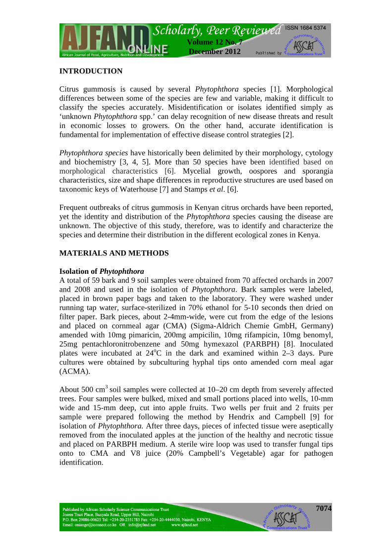
Growth rate is expressed as mm colony diameter/day. As shown in Plate 1A-F, P. citrophthora was characterized by a finely radiate, white rosette and slightly cottony colonies (A). Hyphae were 6 to 7nm (B). Sporangia were mostly laterally attached and were about 40 x 30µm. Shapes ranged from spherical, ovoid, obpyriform, obturbinate, to ellipsoidal, but mostly papillate (C and D). Some were asymmetrical (E) and others often had two divergent apices (F). Papilla measured upto 4µm long with an average length-breadth ratio of 1.4:1. Sporangiophores were irregularly branched singly or in a loose sympodium with a swelling at the point of branching. No chlamydospores or sexual structures were produced.
7078
Volume 12 No. 7 December 2012
Plate 1: Morphological characteristics of Phytophthora citrophthora. Finely
radiate cottony mycelium (A), coenocytic hyphae of up to 6-7nm in diameter (B), ovoid (C and D) and asymmetric (E) sporangium with prominent papilla. Sporangium with more than one papilla (F).Bar=10µm
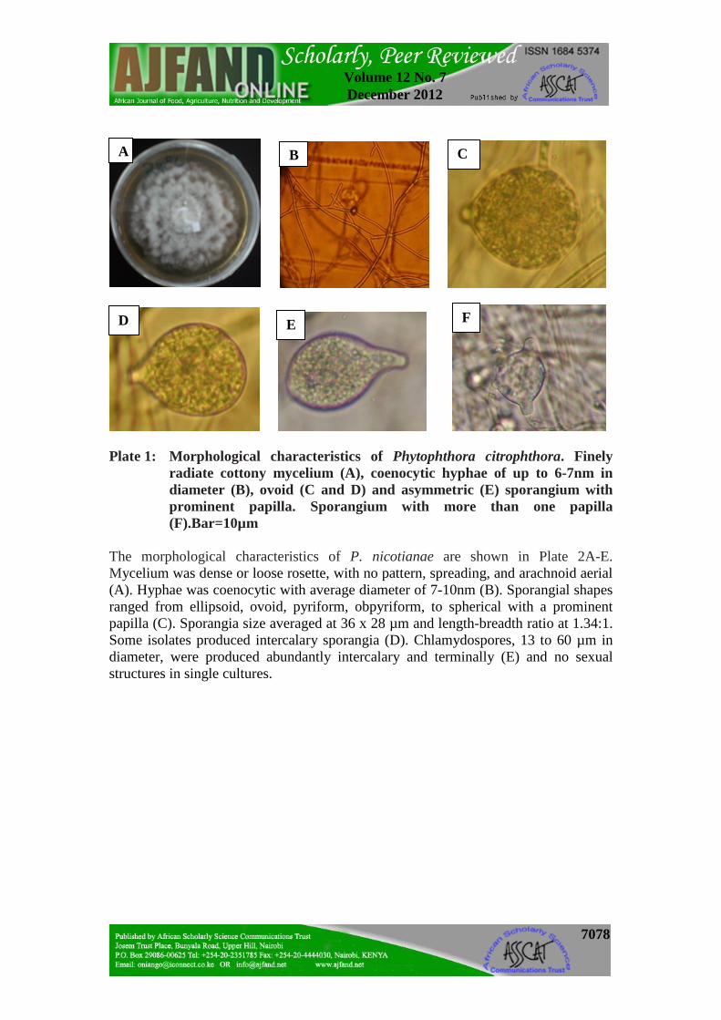
The morphological characteristics of P. nicotianae are shown in Plate 2A-E. Mycelium was dense or loose rosette, with no pattern, spreading, and arachnoid aerial (A). Hyphae was coenocytic with average diameter of 7-10nm (B). Sporangial shapes ranged from ellipsoid, ovoid, pyriform, obpyriform, to spherical with a prominent papilla (C). Sporangia size averaged at 36 x 28 µm and length-breadth ratio at 1.34:1. Some isolates produced intercalary sporangia (D). Chlamydospores, 13 to 60 µm in diameter, were produced abundantly intercalary and terminally (E) and no sexual structures in single cultures.
F E D
C B A

7079
Volume 12 No. 7 December 2012
Plate 2: Morphological characteristics of P. nicotianae (A) Dense rosette
spreading aerial mycelium, (B) coenocytic hyphae of up to 7-10nm in diameter, (C) ovoid terminal sporangia with prominent papilla, (D) intercalary sporangium, (E) chlamydospore. Bar=10µm
Plate 3A-D shows the characteristics of P. syringae. Dense or loose rosette spreading aerial mycelium with no pattern (A), slender coenocytic hyphae (B), with rounded or angular hyphal swellings, often in chains (catenulate) and delimited by septa (C). Sporangia were broadly ovoid or obpyriform, semipapillate, and persistent and formed in succession from a single sporangium (D). Average sporangial dimension was 39 x 26.5µm with length-breadth ratio varying from 1.32:1 to 1.85:1. No chlamydospores were produced on agar media.
A B C
D E

7080
Volume 12 No. 7 December 2012
Plate 3: Morphological characteristics of P. syringae (A) Loose rosette spreading aerial mycelium, (B) coenocytic hyphae with slender mycelium, (C) rounded hyphal swellings in chains (catenulate, shown with the arrow), (D) ovoid, semipapillate, and persistent sporangia. Bar=10µm.
Distribution of the Phytophthora species The distribution and prevalence of Phytophthora species associated with citrus gummosis in Kenya is shown in Table 1. P. citrophthora was the only species present in all AEZs sampled. Its distribution was highest in Lower Midland (LM) and lowest in Upper Midland (UM) zones. The distribution of P. nicotianae was highest in Coastal Lowlands (CL) zones. P. nicotianae and P. syringae, were absent in UM and Lower Highland (LH) zones. P. syringae was present only in LM zones and the only species not found in CL zones. P. citrophthora was the most prevalent in all the AEZs, and P. syringae the least prevalent. Virulence tests The Phytophthora species induced brown to reddish necrotic lesions on lemon fruits (Plate 4). Lesion diameters (LDs) were significantly (p<0.05) different among the isolates of P. citrophthora and P. nicotianae tested. In P. citrophthora, isolate P.CIT 1 was the most virulent while P.CIT 18 and P. CIT 35 were the least virulent. P. nicotianae, isolate P.NIC 11 was the most virulent and P.NIC 1 the least. The P. syringae isolate used (P.SYR) produced small necrotic lesions (Table 2).
B A
C D
7081
Volume 12 No. 7 December 2012
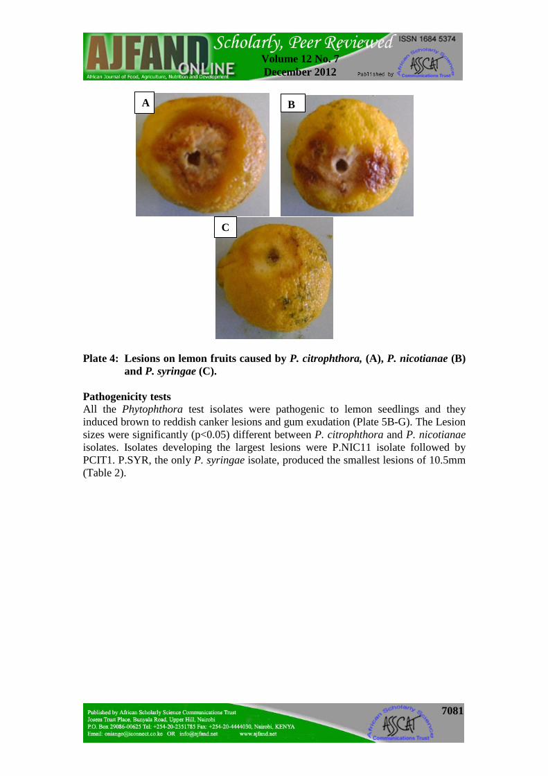
Plate 4: Lesions on lemon fruits caused by P. citrophthora, (A), P. nicotianae (B) and P. syringae (C).
Pathogenicity tests All the Phytophthora test isolates were pathogenic to lemon seedlings and they induced brown to reddish canker lesions and gum exudation (Plate 5B-G). The Lesion sizes were significantly (p<0.05) different between P. citrophthora and P. nicotianae isolates. Isolates developing the largest lesions were P.NIC11 isolate followed by PCIT1. P.SYR, the only P. syringae isolate, produced the smallest lesions of 10.5mm (Table 2).
C
B A

7082
Volume 12 No. 7 December 2012
Plate 5: One-year old lemon seedlings one day after inoculation (A), canker lesions caused by different Phytophthora test isolates 21 days after inoculation (B-G) and a non-innoculated control (H).
DISCUSSION Optimum temperature for growth was the main distinguishing feature between the test species with that of P. citrophthora being 24-28oC and for P. syringae and P. nicotianae 20oC and over 30oC, respectively. These results were consistent with those of another study that reported optimum temperature for P. citrophthora and P. nicotianae to be 24-28oC and 27-35oC, respectively [17]. The diversity in colony
H
F
G
E
D C
B
A

7083
Volume 12 No. 7 December 2012
morphology, rate and manner of growth of test isolates on CMA and sporangial size on V8 agar agreed with previous studies [3]. Diversity and similarities between the two test species, P. citrophthora and P. nicotianae, in the characteristics of their reproductive structures differentiated them from P. syringae and confirmed reports from other studies by Hall [18], Mchau and Coffey [19], and Waterhouse and Waterston [13]. The two species exhibited papillated sporangia; the main characteristic that differentiated them from P. syringae whose sporangia was semi-papillated. This was earlier reported in a similar study by Waterhouse and Waterston [13]. Similarly, the absence of chlamydospores in the two species P. syringae and P. citrophthora confirmed reports by Waterhouse and Waterston [13] and Ho and Jong [20], and the presence of intercalary and terminal chlamydospores in P. nicotianae was as reported by Hall [18]. The presence of rounded or angular hyphal swellings that were often in chains (catenulate) and delimited by septa is unique to P. syringae and facilitated its differentiation from P. nicotianae and P. citrophthora. The latter two species are heterothallic thus did not produce oospores and or sexual structures in single cultures. Colony and sporangial measurement combination provided a useful alternative to the characters given by Waterhouse [7] and Stamps et al. [6] for large-scale examination of Phytophthora species isolates under field and laboratory conditions. The occurrence of Phytophthora species in many citrus fields confirmed earlier studies that these pathogens are present in almost all orchards [21]. The high prevalence of P. citrophthora compared to P. nicotianae and P. syringae in all AEZs could be because citrus is grown in areas with average temperature at < 30oC, which is ideal for the pathogen. Similarly, the ideal temperature for P. nicotianae is 28oC and above, hence its high prevalence in AEZ-CL and other high temperature zones. These findings confirm that P. nicotianae is most active at high temperatures [17, 22]. Our results also confirm earlier findings by Ricci et al. [22] and Fagoaga et al. [23], which indicated that Phytophthora citrophthora is the most widely spread oomycete plant pathogen over all the citrus growing areas. The fact that more P. citrophthora species was isolated from soil samples compared to the other two species showed that this species is prevalent in the tropics and agrees with Ricci et al. [22] that the species is one of the most destructive citrus gummosis pathogen and predominates in very important regions of citrus production. The tropical regions are major citrus production areas just like the Mediterranean basin. The test isolates induced characteristic brown rot and gummosis symptoms on lemon fruits and seedlings, thereby confirming P. citrophthora, P. nicotianae, and P. syringae as the main causal agents of citrus gummosis in Kenya. This is in agreement with reports by Matheron et al. [24], Timmer, et al. [25] and Timmer [26].

7084
Volume 12 No. 7 December 2012
CONCLUSION AND RECOMMENDATIONS The Phytophthora species citrophthora, nicotianae and syringae are the causal agents of trunk gummosis and brown rot of citrus in Kenya. While P. citrophthora is prevalent in all AEZs, P. nicotianae is mainly confined within the coastal lowlands occurring only in a limited scale in the other zones. The distribution of P. syringae is limited in Kenya. Colony characteristics and growth rates were useful as a first step in identification of Phytophthora species by complementing sporangial characteristics in the differentiation of P. citrophthora, P. nicotianae and P. syringae. This study provided a baseline on the basis of which future studies can build on. The current findings have a practical importance with regard to management of gummosis. Since, the species that cause gummosis have been identified in this study, management approaches can be targeted at these three species. For instance the Phytophthora species exhibited different distribution patterns with P. citrophthora being prevalent in all AEZs; P. nicotianae only confined within the coastal lowlands and only in a limited scale in the other zones and that of P. syringae being very limited in Kenya. Thus, control measures such as development of resistance should be deployed with the distribution in mind. This study recommends further characterization of the three Phytophthora species at molecular level since many morphological characteristics of the sporangia, mycelia and colonies used in the differentiation of fungal species are plastic, influenced by environment and often overlap between species. It is also recommended that the species be subjected to selected Oomycete specific fungicides as an initial step in the identification of effective chemicals for the control of citrus gummosis in Kenya. ACKNOWLEDGEMENTS We thank the Ministry of Agriculture, Kenya, for financial support through the Agricultural Sector Programme Support (ASPS) and farmers and district agricultural officers in the surveyed area for their assistance. We appreciate the support of director, Kenya Forestry Research Institute (KEFRI), and his staff where part of the laboratory work was done.

7085
Volume 12 No. 7 December 2012
Table 1: Distribution and prevalence of Phytophthora species associated with citrus gummosis in Kenya.
Species Distribution* Prevalence ** (%)
LM CL LH UM P. citrophthora
30 7 5 3 76.3
P. nicotianae 4 9 0 0 22 P. syringae 1 0 0 0 1.7 *Distribution was determined by recording the number of isolates recovered from samples obtained in each AEZ. **Prevalence of each species was determined by expressing the number of isolates of each species recovered in all AEZs as a percentage of the total number of isolates collected.
Table 2: Lesions induced on lemon fruits and one-year old seedlings inoculated with Phytophthora isolates
Isolate Virulence Test Lesion size
(mm) Pathogenicity Test Lesion size (mm)
P. citrophthora PCT1 48.8± 1.76 19.0± 1.71 PCIT7 45.8± 1.76 13.5± 1.71 PCIT41 45.5± 1.76 13.8± 1.71 PCIT18 29.5± 1.76 - PCIT35 29.5± 1.76 - P. nicotianae PNIC11 30.0± 1.46 26.5± 2.0 PNIC13 28.3± 1.46 12.0± 2.0 PNIC1 17.5± 1.46 - P. syringae - PSYR 16.8 10.5

7086
Volume 12 No. 7 December 2012
REFERENCES
1. Vernière C, Cohen S, Raffanel B, Dubois A, Venard P and F Panabières Variability in Pathogenicity among Phytophthora spp. isolated from Citrus in Corsica. Phytopathology 2004; 152(8-9): 476–483.
2. CAB International. Crop protection Compendium CD-ROM Edition. Nosworthy way, Wallingford, United Kingdom, 2005.
3. Brasier CM and MJ Griffin Taxonomy of Phytophthora palmivora on cocoa. Transactions of the British Mycological Society 1979; 72: 111–43.
4. Kaosiri T and GA Zentmyer Protein, esterase, and peroxidase patterns in the Phytophthora palmivora complex from cacao. Mycologia 1980; 72: 988–1000.
5. Appiah AA, Flood J, Bridge PD and SA Archer Inter- and intraspecific morphometric variation and characterization of Phytophthora isolates from cocoa. Plant Pathol. 2003; 52 (2): 168.
6. Stamps DJ, Waterhouse GM, Newhook F and GS Hall Revised Tabular Key to the Species of Phytophthora. Mycol. Pap 1990; No. 162.
7. Waterhouse GM Key to the species of Phytophthora de Bary. Mycological Papers 92. Kew, Surrey, England: Commonwealth Mycological Institute, 1963.
8. Jeffers SN and SB Martin Comparison of two media selective for Phytophthora and Pythium species. Plant Dis. 1986; 70: 1038–43.
9. Hendrix FF and WA Campbell Distribution of Phytophthora and Pythium species in soils in the continental United States. Canadian Journal of Botany,1970; 48: 377–84.
10. Mitchell DJ, Kannwischer-Mitchell ME and GA Zentmyer Isolating, identifying and producing inoculum of Phytophthora spp. In: Methods for Evaluating Pesticides for Control of Plant Pathogens. Hickey, K. D. (ed.). The American Phytopathological Society, St. Paul, MN.USA, 1986.
11. Heffer V, Powelson ML and KB Johnson Oomycetes. The Plant Health Instructor. Laboratory Exercises in Plant Pathology. Oregon State University, USA. http://www.apsnet.org/education/ Lab Exercises/Oomycetes/Top.hmtl. Accessed in 4th July 2002
12. Newhook FJ, Stamps DJ, Waterhouse GM, Stamps DJ and GS Hall Revised tabular key to the species of Phytophthora. Commonw. Agric. Bur. Int. Mycol. Inst. Mycol. Pap, 1990; 162.
13. Waterhouse GM and JM Waterston Phytophthora nicotianae var. parasitica. CMI Descriptions of Pathogenic Fungi and Bacteria. No. 35. Common Mycol. Inst. Wallingford, UK, 1964.
7087
Volume 12 No. 7 December 2012
14. Jaetzold R and H Schmidt Natural conditions and farm management information. In: Farm Management Handbook of Kenya Vol. IIC. Ministry of Agriculture, Kenya and GTZ, Germany, 1983.15. Agrios GN Plant Pathology. 4th Edition. Academic press, 1997.
16. International Plant Protection Congress Where Chemistry meets Ecology Plant Protection towards the Third Millennium, 15th Edition. Jerusalem, Israel, 1999. http://www.kenes.co.il/IPPC/mon.htm. Accessed on 15th July 2007.
17. Erwin DC and OK Ribeiro Phytophthora Diseases Worldwide. APS Press. St. Paul, Minnesota, USA, 1996.
18. Hall G An integrated approach to the analysis of variation in Phytophthora nicotianae and a redescription of the species. Mycological Res. 1993; 97(5):559-574.
19. Mchau GRA and MD Coffey An integrated study of the morphological and isozyme patterns found within a worldwide collection of Phytophthora citrophthora and a redescription of the species. Mycology Res. 1994b; 98:1291-1299.
20. Ho HH and JC Jong P. hibernalis and P. syringae. Mycotaxon, 1993; 47: 439-460.
21. Ann PJ, Ko WH and HJ Su Interaction between Lukubin Bacterium and Phytophthora parastica in Citrus hosts. European Journal of Plant Pathology, 2004; 110: 1-6.
22. Ricci P, Pope-de-Vallavieille C, Panabieres F, Marais A and G Auge Characterization of the Phytophthora spp. attacking citrus. OEPP/EPPO Buletin, 1990; 20:19–28.
23. Fagoaga C, Rodrigo I, Conejero V, Hinarejos C, Tuset JJ, Arnau J, Pina JA, Navarro L and L Peña Increased tolerance to Phytophthora citrophthora in transgenic orange plants constitutively expressing a tomato pathogenesis related PR-5. Molecular Breeding, 2001; 7: 175-185.
24. Matheron ME, Wright G and M Porchas Resistance to Phytophthora citrophthora and P.parasitica and nursery characteristic of several citrus rootstocks. Plant Dis. 1998; 82:1217-1225.
25. Timmer LW, Garnsey SM and P Broadbent Diseases of Citrus. In: Diseases of Tropical Fruits Crops. R. C. Ploetz (ed.). CABI Publishing, CABI International, Wallingford, UK, 2003.
26. Timmer LW and JA Menge Phytophthora induced diseases. Pages 22-24 in: compendium of citrus diseases. Whiteside, J. O., Garnsey, S. M. and. Timmer, L.W. (eds.). APS Press, St.Paul, MN, 1988.