morphological hepatic alterations of cirrhosis: imaging...
TRANSCRIPT
Vol. 5 / Nº 14 - Septiembre 2016
Morphological hepatic alterations of cirrhosis: imaging findings Gustavo Raichholz, Sebastián Giménez, Cristian Froullet, Santiago Dumoulin, Hernán Brouver de Köning, José Luis Sañudo.
Theme review
Resumen
La cirrosis es una causa importante de morbilidad y
mortalidad en el mundo. El diagnóstico de cirrosis es
tradicionalmente establecido con resultados de biop-
sia, pero puede ser sugerido por los cambios morfoló-
gicos visualizados en imágenes. Su reconocimiento es
esencial para la caracterización de las lesiones focales
hepáticas. El objetivo de este trabajo es reconocer los
cambios morfológicos visualizados en imágenes en la
cirrosis hepática.
La cirrosis es comúnmente causada por la hepatitis
crónica de origen infeccioso o el abuso de alcohol,
si bien un gran número de desórdenes que causan
injuria hepática pueden llevar al desarrollo de esta
entidad. Patológicamente se define por tres caracte-
rísticas principales: fibrosis, transformación nodular y
distorsión de la arquitectura hepática.
Abstract
Cirrhosis is a major cause of morbidity and mortality
worldwide. The diagnosis of cirrhosis is traditionally es-
tablished through biopsy results, but it may be sugges-
ted by morphological changes displayed in images. Its
recognition is essential for the characterization of focal
liver lesions. The aim of this paper is to recognize the
morphological changes observed in images of cirrhosis.
Cirrhosis is commonly caused by chronic hepatitis due
to an infection or alcohol abuse, although a large
number of disorders that cause liver injuries may lead
to the development of this condition. Pathologically it is
defined by three main features: fibrosis, nodular trans-
formation and distortion of hepatic architecture.
Subtle morphological changes of the liver may be
the earliest detectable signs in images, including the
enlargement of the hilar periportal space, the interlobar
Contact information:
Gustavo Raichholz.
Diagnóstico por Imagen Junín - Santa Fe capital.
E-mail: [email protected]
Recibido: 22 de febrero de 2016 / Aceptado: 30 de marzo de 2016
Received: February 22, 2016 / Accepted: March 30, 2016
Revista Argentina de Diagnóstico por Imágenes Revista Argentina de Diagnóstico por Imágenes
Raichholz G. et al.
Morphological hepatic
alterations of cirrhosis
Sutiles cambios morfológicos del hígado pueden ser
los primeros signos detectables en imágenes, inclu-
yendo la ampliación del espacio periportal hiliar, de
la cisura mayor interlobar y el agrandamiento de la
fosa vesicular. Otros signos típicos son la atrofia del
segmento medial del lóbulo izquierdo y del segmento
anterior del lóbulo derecho o la hipertrofia del lóbulo
caudado y del segmento lateral del lóbulo izquierdo.
La nodularidad del borde hepático es otra caracterís-
tica visualizada en la cirrosis y está relacionada con
la presencia de nódulos de regeneración. También se
pueden ver signos derivados de la hipertensión portal,
incluyendo colaterales venosas, esplenomegalia, asci-
tis, entre otras.
Palabras clave: Cirrosis, alteraciones morfológicas.
fissure and the enlargement of the gallbladder fossa.
Other typical signs are atrophy of the medial segment
of the left lobe and the anterior segment of the right
lobe, or hypertrophy of the caudate lobe and lateral
segment of the left lobe.
Another characteristic of cirrhosis is the nodular edge of
the liver, which is related to the presence of regenerati-
ve nodules. Derivative signs of portal hypertension can
be observed, including venous collaterals, splenome-
galy, and ascites, among others.
Key words: Cirrhosis, morphological alterations.
IntroductionHepatic cirrhosis is one of the main health problems
in the world due to its high morbidity and mortali-
ty. It is a chronic and irreversible liver disease that
appears in the final stages of diverse pathologies.
The cellular lesion triggers an inflammation, regene-
ration and fibrosis cycle that leads to an alteration of
the intrahepatic circulation, portal hypertension and
cholestasis.
In anatomic pathology examinations, it is charac-
terized by an extensive fibrosis and the presence of
numerous regeneration nodules. Depending on the
size of these nodules, cirrhosis can be classified as
micronodular (smaller than 3 mm), macronodular
(bigger than 3 mm) and mixed (1).
Among the most common causes of cirrhosis, the-
re is excessive alcohol consumption and hepatitis
B or C virus infection. Less frequent causes include
chronic hemochromatosis, biliary obstruction and
hepatic congestion, use of pharmaceuticals and to-
xins, and hereditary disorders, such as Wilson’s di-
sease, alpha 1-antitrypsin deficiency and glycogeno-
sis type IV.
In images studies, cirrhosis is characterized by alte-
rations in the morphology, in the edges of the liver,
and in the parenchyma, with regeneration nodules
and fibrosis. There are also extrahepatic manifesta-
tions such as the development of portosystemic co-
llaterals, ascites and splenomegaly (2).
The main role of the radiologist consists of evalua-
ting the size of the liver and of its diverse segments,
perform a biometric analysis of the segments I and
IV in search of early signs of cirrhosis, analyze he-
patic edges exhaustively, and identify the effects of
portal hypertension. The presence of focal lesions in
a cirrhotic liver must be interpreted as hepatocellular
carcinoma in a first diagnosis but focal lesions in li-
ver without chronic pathology determine differential
diagnosis.
Vol. 5 / Nº 14 - Septiembre 2016
Morphological hepatic
alterations of cirrhosisRaichholz G. et al.
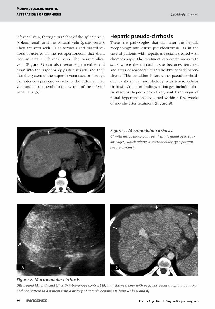
Analysis of the hepatic edge and parenchymaThe margins of the hepatic gland must be smooth,
but cirrhosis often makes them nodular due to the
existence of numerous regeneration nodules. The as-
pect of the hepatic edge will depend on the size of
these regeneration nodules. The edge can be nodular
and thin in cases of micronodular cirrhosis (Figure
1) or nodular and thick in cases of macronodular ci-
rrhosis (Figure 2). Hepatic regeneration nodules are
isodense with the glandular parenchyma in CT and
isointense in T1 and T2-weighted images in MRI. So-
metimes, they can be hyperattenuating in CT images
without contrast and hypointense in T1 and T2 se-
quences of MRI due to the presence of hemosiderin
(siderotic nodules).
A smaller amount of cirrhotic livers has a heteroge-
neous parenchyma in CT or MRI (2). The main cau-
ses of this heterogeneity are the presence of fibrosis,
irregular fatty liver and iron deposits. Hepatic fibrosis
is hypodense in relation to the parenchyma in CT
without intravenous contrast and can show a late en-
hancement after the administration of the contrast
agent (Figure 3). In MRI, it is often hypointense in
T1-weighted sequences and hyperintense in T2-wei-
ghted sequences.
Fibrosis can adopt several morphological patterns.
It can be patchy, thin, appear as thick perilobular
bands and/or perivascular cuffing producing an ox
eye pattern. An irregular fatty liver produces patchy
areas of less density in CT and it is frequent in pa-
tients with alcoholic liver cirrhosis who are still drin-
king alcohol (2). When the heterogeneity of the pa-
renchyma is caused by iron deposits, there are areas
of high density in CT without contrast that become
hypointense in MRI in T2-weighted sequences.
Signs of dimorphism: hepatic atrophy and hypertrophyApproximately 25% of cirrhotic livers in the final stage
have a normal size. 36% presents diffuse atrophy and
the rest of the patients present a combination of seg-
mental atrophy and hypertrophy.
An early sign of cirrhosis is the increase of hilar
periportal space, which is filled with fat content due
to the atrophy of segment IV. Normally, the hilar peri-
portal space measures less than 10 mm from the an-
terior edge of the right portal branch to the posterior
edge of the medial segment of the left lobe. A size
greater that 10 mm represents a sensitivity of 93% and
a specificity of 92% for the diagnosis of cirrhosis (3)
(Figure 4).
Focal atrophy is more common in the right hepatic
lobe and in the medial segment of the left hepatic
lobe (Figure 5). A sign produced by atrophy, which
is very specific of cirrhosis, is the presence of a clear
groove in the right posterior surface of the liver. This
groove is called posterior hepatic notch and it repre-
sents a sensitivity of 72% and a specificity of 98% for
the diagnosis of cirrhosis (Figure 6).
The segments that present hypertrophy with a grea-
ter frequency are the caudate lobe and the lateral seg-
ments of the left hepatic lobe. Biometry of segment
I has proved to be useful for the evaluation of the
relationship between segment I and the right lobe of
the liver. If this relationship is greater than 0.9, the-
re is a sensitivity of 71.1% and a specificity of 77%
for the diagnosis of cirrhosis. Biometry of segment
I is performed by drawing three vertical lines: one
through the internal edge of segment I, another one
through the external edge of the left liver and a last
imaginary line that goes through the right lateral wall
of the bifurcation of the main portal vein, parallel to
the middle sagittal plane of the body.
Portosystemic venous collaterals: signs of portal hypertensionIn patients with cirrhosis and portal hypertension,
part of the portal venous flow reverts its direction to
the systemic circulation through portosystemic anas-
tomosis. From the clinical point of view, the most
important venous collaterals are esophageal and
paraesophageal varices, due to the risk of digestive
hemorrhage. Esophageal varices are dilated veins lo-
cated in the wall of the lower part of the esophagus,
while paraesophageal varices are located outside the
digestive wall (Figure 7). Both receive the flow from
the left gastric vein, which is divided into anterior
branches to supply esophageal varices and posterior
branches to supply paraesophageal varices.
Another type of portosystemic shunts is that
which communicate the spleno-portal axis with the
10 Revista Argentina de Diagnóstico por Imágenes10 Revista Argentina de Diagnóstico por Imágenes
Raichholz G. et al.
Morphological hepatic
alterations of cirrhosis
left renal vein, through branches of the splenic vein
(spleno-renal) and the coronal vein (gastro-renal).
They are seen with CT as tortuous and dilated ve-
nous structures in the retroperitoneum that drain
into an ectatic left renal vein. The paraumbilical
vein (Figure 8) can also become permeable and
drain into the superior epigastric vessels and then
into the system of the superior vena cava or through
the inferior epigastric vessels to the external ilian
vein and subsequently to the system of the inferior
vena cava (5).
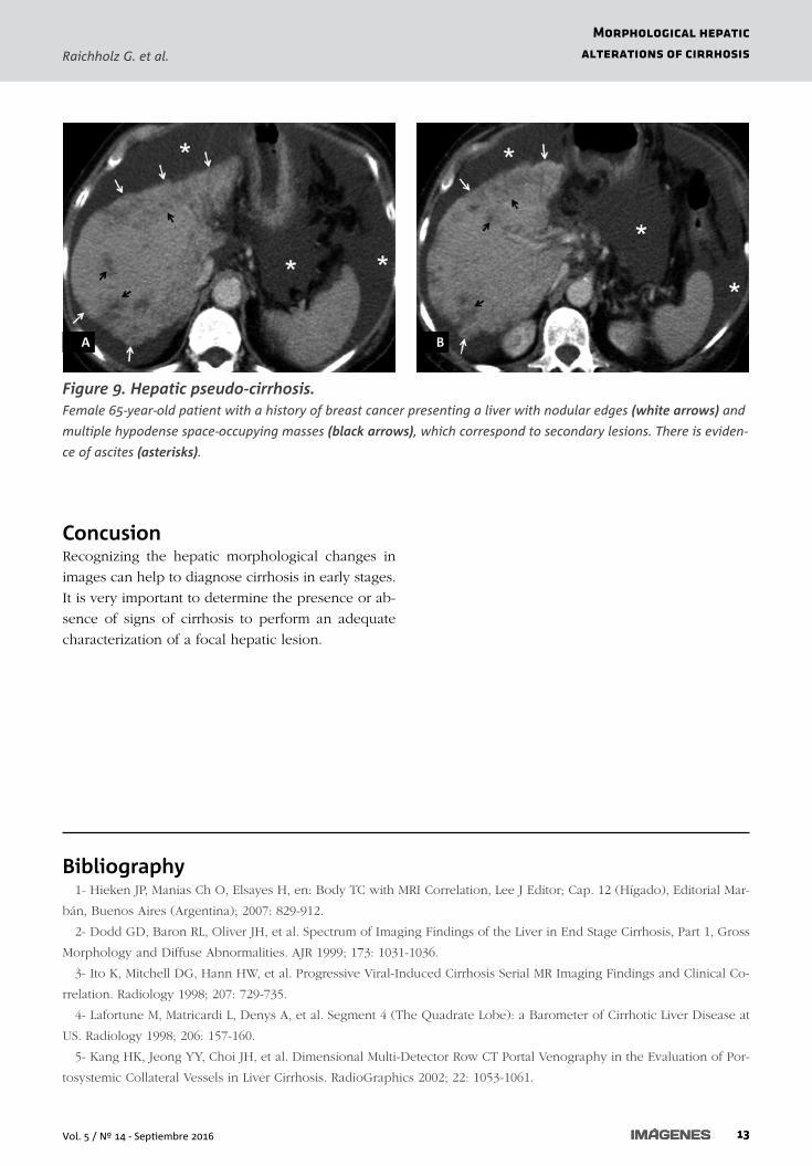
Hepatic pseudo-cirrhosisThere are pathologies that can alter the hepatic
morphology and cause pseudocirrhosis, as in the
case of patients with hepatic metastasis treated with
chemotherapy. The treatment can create areas with
scars where the tumoral tissue becomes retracted
and areas of regenerative and healthy hepatic paren-
chyma. This condition is known as pseudocirrhosis
due to its similar morphology with macronodular
cirrhosis. Common findings in images include lobu-
lar margins, hypertrophy of segment I and signs of
portal hypertension developed within a few weeks
or months after treatment (Figure 9).
Figure 1. Micronodular cirrhosis. CT with intravenous contrast: hepatic gland of irregu-
lar edges, which adopts a micronodular-type pattern
(white arrows).
Figure 2. Macronodular cirrhosis.Ultrasound (A) and axial CT with intravenous contrast (B) that shows a liver with irregular edges adopting a macro-
nodular pattern in a patient with a history of chronic hepatitis B (arrows in A and B).
A B
11Vol. 5 / Nº 14 - Septiembre 2016 11
Morphological hepatic
alterations of cirrhosisRaichholz G. et al.
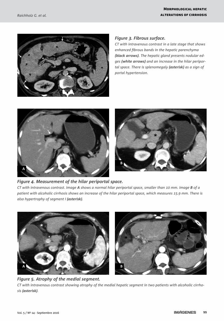
Figure 3. Fibrous surface. CT with intravenous contrast in a late stage that shows
enhanced fibrous bands in the hepatic parenchyma
(black arrows). The hepatic gland presents nodular ed-
ges (white arrows) and an increase in the hilar peripor-
tal space. There is splenomegaly (asterisk) as a sign of
portal hypertension.
Figure 4. Measurement of the hilar periportal space. CT with intravenous contrast. Image A shows a normal hilar periportal space, smaller than 10 mm. Image B of a
patient with alcoholic cirrhosis shows an increase of the hilar periportal space, which measures 15.9 mm. There is
also hypertrophy of segment I (asterisk).
A
B
Figure 5. Atrophy of the medial segment. CT with intravenous contrast showing atrophy of the medial hepatic segment in two patients with alcoholic cirrho-
sis (asterisk).
A B
12 Revista Argentina de Diagnóstico por ImágenesRevista Argentina de Diagnóstico por Imágenes
Raichholz G. et al.
Morphological hepatic
alterations of cirrhosis
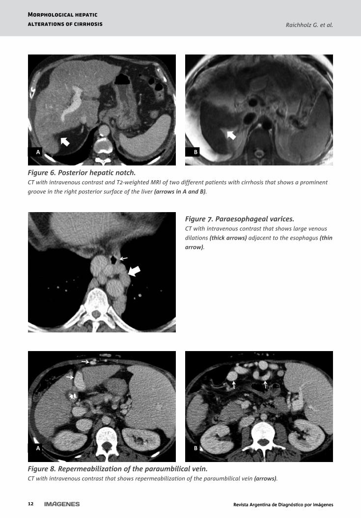
Figure 6. Posterior hepatic notch. CT with intravenous contrast and T2-weighted MRI of two different patients with cirrhosis that shows a prominent
groove in the right posterior surface of the liver (arrows in A and B).
A B
Figure 7. Paraesophageal varices. CT with intravenous contrast that shows large venous
dilations (thick arrows) adjacent to the esophagus (thin
arrow).
Figure 8. Repermeabilization of the paraumbilical vein. CT with intravenous contrast that shows repermeabilization of the paraumbilical vein (arrows).
A B
1Vol. 5 / Nº 14 - Septiembre 2016 1
Morphological hepatic
alterations of cirrhosisRaichholz G. et al.
Figure 9. Hepatic pseudo-cirrhosis. Female 65-year-old patient with a history of breast cancer presenting a liver with nodular edges (white arrows) and
multiple hypodense space-occupying masses (black arrows), which correspond to secondary lesions. There is eviden-
ce of ascites (asterisks).
A B
ConcusionRecognizing the hepatic morphological changes in
images can help to diagnose cirrhosis in early stages.
It is very important to determine the presence or ab-
sence of signs of cirrhosis to perform an adequate
characterization of a focal hepatic lesion.
Bibliography1- Hieken JP, Manias Ch O, Elsayes H, en: Body TC with MRI Correlation, Lee J Editor; Cap. 12 (Hígado), Editorial Mar-
bán, Buenos Aires (Argentina); 2007: 829-912.
2- Dodd GD, Baron RL, Oliver JH, et al. Spectrum of Imaging Findings of the Liver in End Stage Cirrhosis, Part 1, Gross
Morphology and Diffuse Abnormalities. AJR 1999; 173: 1031-1036.
3- Ito K, Mitchell DG, Hann HW, et al. Progressive Viral-Induced Cirrhosis Serial MR Imaging Findings and Clinical Co-
rrelation. Radiology 1998; 207: 729-735.
4- Lafortune M, Matricardi L, Denys A, et al. Segment 4 (The Quadrate Lobe): a Barometer of Cirrhotic Liver Disease at
US. Radiology 1998; 206: 157-160.
5- Kang HK, Jeong YY, Choi JH, et al. Dimensional Multi-Detector Row CT Portal Venography in the Evaluation of Por-
tosystemic Collateral Vessels in Liver Cirrhosis. RadioGraphics 2002; 22: 1053-1061.