morphological studie on ths maie branchen s of th radiae...
TRANSCRIPT
A C T A T H E R I O L O G I C A
VOL. XV, 12: 185—197. BIAOOWIEZA 30.IV.1970
Anna A R L A M O W S K A - P A L I D E R
Morphological Studies on the Main Branches of the Radial Nerve in Mammals
[With 5 Figs.]
Studies were made on 175 individuals representing the following orders: Marsupialia, Primates, Insectívora, Rodentia, Carnivora, Perisso-dactyla, Artiodactyla and Hyracoidea. The results of these observations showed that three different types of the radial nerve can be distingui-shed: Type I, occuring in marsupials — no cutaneous branches of the radial nerve before its entry into the canal of the supinator. Type II (lower placentals) — characterized by a well-developed cutaneous branch, that is, n. cutaneus antebrachii lateralis. Type III — characte-ristic of primates, with a new cutaneous branch, termed the superficial branch of the radial nerve in anthropotomy.
I. INTRODUCTION
The nomenclature of the main nerve trunks and their branches used in veteri-nary anatomy and in anatomical monographs on the lower mammals is based on the nomenclature of human anatomy. We thus distinguish in animals, as in the case of man, radial, ulnar, intermediary and other nerves. In describing the radial nerve E l l e n b e r g e r (1943), S i s s o n (1960), M a r t i n (1938), M a g i l t o n & G e t t y (1966), G h o s h a l & G e t t y (1967), R e i m e r s (1925), M i l -l e r (1964), T a y l o r & W e b e r (1951), T e r e n t e v (1952), K r a u s e (1884), H e i n z e & F r a n z k e (1960) and others state that in animals this nerve pierces from the medial side of the arm to the lateral side, passing between the heads of the triceps muscle of the arm, in the same way as in man and the pri-mates M a r c i n i a k (1965), C l a r a (1959), P r e u s c h o f t (1964), V l a t k o v i c (1966). Here also the radial nerve in lower mammals, in the opinion of the previously mentioned authors, sends out cutaneous branches identified as n. cutaneus brachii lateralis and n. cutaneus antebrachii lateralis corresponding to n. cutaneus brachii posterior and n. cutaneus antebrachii posterior in man. After sending out these branches the radial nerve splits into ramus superficialis and ramus profundus. The first of these runs to the hand, and there divides into the dorsal digital nerves ( P o v e á e n k o , 1963; S e r b a k o v , 1963). Ramus profundus penetrates below the brachioradial muscle and radial extensors of the carpus, where it innervates with its terminal branches the extensors of the carpus and fingers. Only S c h r e b e r (1956) in the case of the cow, and S e r w a t k a (1966) in the case of the European
[185]
186 A. Arlamowska-Palider
bison, when describing this nerve state that it sends out ramus superficialis, that is, n. radialis superficialis s. n. cutaneus antebrachii lateralis and later passes into ramus profundus. The radial nerve in the horse is presented in a similar way by B r a d l e y (1946). According to him n. cutaneus antebrachii lateralis runs from the radial nerve, and the main trunk of the nerve, entering into the interior of the muscle, sends out muscular branches only. In the opinion of the previously-men-tioned authors, in the horse and European bison the area served by the cutaneus nerves is reduced, since it reaches only to the region of carpus and proximal part of metacarpus.
The studies made on the nerves of the forearm in different mammals, however, give rise to certain doubts as to whether the branches of the radial nerve bearing the same names are in fact homological, since they exhibit completely different topography and even serve a different area. Thus it appears justifiable to discuss the topography and morphology of the main branches of the nerves of forelimbs in mammals on the basis of comparative anatomical examination. Such a discussion forms the purpose of this study, in which I have confined myself for the present to the radial nerve in different mammals.
II. MATERIAL
I made my examinations on: 1 individual of Metachirus nudicaudatus, 1 of Pe-taurus australis, 5 of Macropus rufus — representatives of Marsu,pialia; 4 indi-viduals of Erinaceus europaeus — representatives of Insectivora; 4 individuals of Cavia porcella, 6 of Myocastor coypus, 2 of Hystrix cristata, 5 of Hydrochoerus hydrochaeris, 3 of Sciurus vulgaris, 1 of Ondrata zibethica, 2 of Chinchilla lani-ger — representatives of Rodentia; 10 individuals of Canis familiaris, 8 of Alopex lagopus, 5 of Ursus arctos, 2 of Nasua narica, 5 of Martes foina, Martes martes, 10 of Mustela lutreola, 10 of Mustela putorius furo, 5 of Mustela putorius, 7 of Mustela nivalis, 1 of Felis pardalis, 1 of Felis concolor, 10 of Felis catus — repre-sentatives of Carnivora; 8 individuals of Bos taurus, 5 of Ovis aries, 3 of Capra hircus, 4 of Antilope cervicapra, Taurotragus oryx, Oryx gazella, Gazella dorcas, 3 of Lama glama, 1 of Moschus moschiferus, 6 of Equus caballus, 2 of Procavia habessinica — representatives of Perissodactyla, Artiodactyla and Hyracoidea, 2 of Pongo pygmaeus, 1 of Hylobates lar, 4 of Papio cynocephalus, 6 of Macaca mulatta, 3 of Cercopithecus cephus, 3 of Cercocephus torquatus, 1 of Macaca iris, 1 of Saimira sciureus, 2 of Cebus apella, 2 of Lemur catta, 4 of Galago senegalensis, 1 of Lori tardigradus, 1 of Arctocephus calabarensis, 3 of Tupaia glis — repre-sentatives of anthropoidal apes, catarrhine and platyrhine monkeys.
The study material came from the collections of the Zoological Museum of Wroclaw University, the Zoological Garden in Wroclaw and breeding farms.
III. RESULTS
The radial nerve in the marsupials examined emerges from the branchial plexus and is at first situated on the medial side of the arm, then further on, between the heads of the triceps of the arms, it penetra-tes to the lateral side, sending out several muscular branches on its way.

Main branches of the radial nerve in mammals 187
Continuing, the radial nerve enters under the bellies of brachioradial muscle and radial extensor of the carpus and sends out branches to them. The nerve then pierces through the supinator and after emerging from the canal of this muscle innervates the remaining muscles of the dorsal side of the forearm. The longest branch, running to the extensor of the long pollex and extensor of the index finger, can be considered as cor-responding to the posterior interosseous nerve of the forearm in man.
In the kangaroo I observed a cutaneous branch which runs from the radial nerve after the latter emerges from the canal of the supinator (Fig. 1). It serves the skin of the dorsal side of the lower half of the forearm up to the region of the carpus.
Fig. 1. Innervation of the dorsal side of the arm in the kangaroo.
1 — nervus medioradialis-, 2 — nervus radialis profun-dus; 2' — cutaneous branch of this nerve; 3 — dorsal branch of n. ulnaris; 4 — nn. digitales dorsales com-munes, a — m. supinator hrevis, b — amputated muscle
bellies of superficial layer. ii in
The dorsal common digital nerves I, II and III in the marsupials exa-mined originate not from the radial nerve, but from the nerve termed for the time being the medio-radial nerve, branching from the median nerve.
The skin of the dorsal surface of the forearm is served by an exceptio-nally strongly developed cutaneous branch of the axillary nerve.
A characteristic feature of the radial nerve in marsupials is, as can be seen, the absence of branches which could be identified with the lateral cutaneous nerve of the forearm in placentals, or the superficial branch
188 A. Arlamowska-Palider
in man and the primates. The whole nerve in these animals in fact corresponds only to ramus profundus n. radialis.
In placentals the case is different. The radial nerve always sends out one more cutaneous branches before its entry into the canal of the supi-nator. These branches are identified with n. cutaneus brachii posterior, n. cutaneus antebrachii posterior and ramus superficialis n. radialis in man, which would not however appear to be always accurate. In monkeys these branches behave almost the same as in man and it is therefore not difficult to identify them with the nerves in the latter.
In the anthropoid apes and catarrhine and platyrhine monkeys examin-ed the radial nerve on the lateral side of the arm first sends out a weak posterior cutaneous nerve of the arm, then a posterior cutaneous nerve of the forearm. It then enters under the brachioradial muscle, under the belly of which it divides into two terminal branches: ramus profundus and ramus superficialis. The posterior cutaneous nerve of the forearm pierces the fascia of the forearm and divides into two branches — lateral and medial — running superficially from all the muscles with the con-comitant subcutaneous veins. The medial branch, in the lower part of the forearm, may join with the superficial branch of the radial nerve. The latter runs under the brachioradial muscle, from under the belly of which it emerges superficially and divides into the common dorsal nerves of the fingers innervating fingers I, II and III.
Ramus profundus is strongly developed, as in the case of marsupials; it pierces through the supinator and sends branches to the muscles of the dorsal side of the forearm. Some of them run out before the entry to the canal of the supinator, and some immediately after emergence from it. One of them, the longest, forms m. interosseus antebrachii poste-rior, running between the tendons of the muscles: m. extensor digitalis longus and m. extensor indicis and extending to the capsule of the carpal articulation. The relations found on the left limb in the saimiri examined were, however, different namely the radial nerve sends out only the normal n. cutaneus antebrachii posterior, and then enters under the branchioradial muscle without sending out any further cutaneous nerves. As it continues it passes in a typical way through the canal of the supinator and extends into the strong developed n. interosseus ante-brachii posterior. The latter retains its typical position and course under the muscles of the superficial layer of the forearm, but in the carpal region appears on the exterior, superficially, and divides into the com-mon dorsal nerves of the fingers. The typical superficial branch is completely absent in this case, and the region which it innervates is taken over by the posterior interosseous nerve, which normally reaches only to the carpal articulation.

Main branches of the radial nerve in mammals 189
Among the lemurs and tarsiers examined I found relations typical of anthropoids in the former. N. cutaneus antebrachii posterior is present and behaves the same way as in monkeys, and also the typical superfi-cial branch extending to the fingers.
In the lori examined n. cutaneus antebrachii posterior behaves in the same way as in monkeys and lemurs. The typical superficial branch is, however, absent and the common dorsal nerves of the fingers originate from n. interosseus antebrachii posterior (A, Fig. 2), as in the case in the left limb of the saimiri.
In the galago, anguantibo and tupaya I observed the radial nerve to behave in yet another way. On the lateral side of the arm it sends out the strongly developed n. cutaneus antebrachii posterior, which separates from the main trunk before entry under the brachioradial muscle. This nerve runs superficially under the skin, in the same way as in monkeys and lemurs, but as distinct from the latter extends as far as the hand, where it divides into the common dorsal nerves of the fingers (B, Fig. 2). There is no typical superficial branch running under the brachioradial muscle. The radial nerve, after sending out the nerve described above, forms only ramus profundus, which sends out muscle branches only.
The above description shows that three branches reciprocally compete in the innervation of the hand in primates: this is mainly ramus superfi-cialis, sometimes n. cutaneus antebrachii posterior, or n. interosseus ante-brachii posterior.
In veterinary anatomy ramus superficialis of the radial nerve and n. cutaneus antebrachii lateralis are also distinguished.
The radial nerve in Carnivora behaves in a typical way, i. e. it threads its way between the heads of the triceps from the medial side of the arm to its lateral side. Here also it sends out a strongly formed cutaneous branch, which pierces the fascia and divides into two parts: the lateral and medial. They run superficially from all the muscles concomitantly with the subcutaneous veins immediately under the skin. Numerous ramifications intended for the skin of the forearm usually run from the lateral branch. The cutaneous branch of the axillary nerve anastomoses, in the majority of cases, with the medial branch near the elbow joint, and in the region of the carpal articulation this branch joins with the n. cutaneus antebrachii medialis 2) (cat and raccoons). In its course to-wards the fingers the medial branch passes into the common dorsal digital nerve I (dog, bear and martens). In the cat and raccoon kinds this nerve is formed from the junction of the medial branch and ramifi-
') Nervus cutaneus antebrachii posterior in man and the primates. s) Nervus cutaneus antebrachii lateralis in man and the primates.
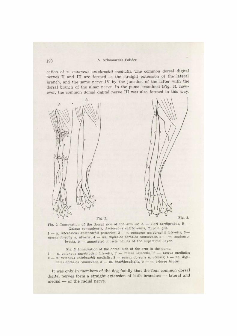
190 A. Arlamowska-Palider
cation of n. cutaneus antebrachii medialis. The common dorsal digital nerves II and III are formed as the straight extension of the lateral branch, and the same nerve IV by the junction of the latter with the dorsal branch of the ulnar nerve. In the puma examined (Fig. 3), how-
1 — n. interosseus antebrachii posterior; 2 — n. cutaneus antebrachii lateralis; 3 — ramus dorsalis n. ulnaris; 4 — nn. digitales dorsales communes, a — m. supinator
brevis, b — amputated muscle bellies of the superficial layer.
Fig. 3. Innervation of the dorsal side of the arm in the puma. 1 — cutaneus antebrachii lateralis, V — ramus lateralis, 1" — ramus medialis; 2 — n. cutaneus antebrachii medialis; 3 — ramus dorsalis n. ulnaris; 4 — nn. digi-
tales dorsales communes, a — m. brachioradialis, b — m. triceps brachii.
It was only in members of the dog family that the four common dorsal digital nerves form a straight extension of both branches — lateral and medial — of the radial nerve.

Main branches of the radial nerve in mammals 191
I observed a similar phenomenon in one of the house martens which was, however, caused by the complete absence of the dorsal branch of the ulnar nerve.
The cutaneous branch of the radial nerve in Carnivora described above forms n. cutaneus antebrachii lateralis, which innervates the fingers in a similar way to that observed in lemurs and tarsiers. The typical super-ficial branch in the sense used for primates, is absent. After sending out the above cutaneous branch the radial nerve enters under the brachio-radial muscle, or if the latter is absent, under the radial extensors of the carpus and then sends out muscular branches only.
The radial nerve behaves similarly in the hedgehog. It sends out a single cutaneous branch, which runs superficially under the skin con-comitantly with vena cephalica accessoria. On the forearm it sends out numerous ramifications intended for the skin of this region. In the direction of the fingers the cutaneous branch extends into the common dorsal nerves of fingers III and IV.
The radial nerve, after running through the canal of the supinator, passes into the posterior interosseous nerve of the forearm, which ex-tends right up to the hand as the common dorsal digital nerve II.
The radial nerve of rodents, before entering under the exceptionally strongly developed brachioradial muscle, sends out a typically behaving cutaneous branch. As in the hedgehog, and unlike Carnivora, this branch is single. Lying superficially from the muscle of the forearm, it sends out along its course ramifications to the skin and reaches the hand, where it divides into the common dorsal digital nerves. This branch may therefore be considered as n. cutaneus antebrachii lateralis, but there is no ramus superficialis n. radialis, in the sense used in primates.
In squirrels and chinchillas n. cutaneus antebrachii medialis joins the cutaneous branch, and together with the ramifications from the pre-viously mentioned branch passes into the common dorsal digital nerve T. In both the capibari and guinea pig there is a junction of this kind, from which, however, the common dorsal digital nerve II arises. In the majority of the rodents examined the cutaneous branch in its straight extension passes into the common dorsal digital nerve III, or sends out a very fine branch which, together with the dorsal branch of the ulnar nerve, form the common dorsal digital nerve IV, as in the case of the squirrel, porcupine and muskrat.
In the latter I noticed, in addition to the typical cutaneous branch, a second branch which separates from the trunk of the radial nerve im-mediately before the latter's entry into the canal of the supinator. It runs deeply between the bellies of the radial extensor muscles of the wrist and then in the medial part of the forearm emerges from under

192 A. Arlamowska-Palider
the superficially. Towards the fingers it joins with n. cutaneus ante-brachii medials and extends into the common dorsal digital nerve I.
Relations were approximately similar in the nutria examined (Fig. 4). In addition to the typical cutaneous branch running superficially under the skin on the dorsal side of the forearm and extending to the hand as the common dorsal digital nerves II, III and IV, I also observed a second,
Fig. 4. Innervation of the dorsal side of the arm in Myocastor coypus. 1 — n. cutaneus antebrachii lateralis, 1' — ramus accessorius; 2 — ramus dorsalis
n. ulnar is) 3 — nn. • digitales dorsales communes, a — m. triceps brachii.
Fig. 5. Innervation of the dorsal side of the arm in Hyracoidea. 1 _ n. cutaneus antebrachii lateralis; 1' — ramus accessorius; 2 — nn. digitales
dorsales communes, a — m. triceps brachii.
which also runs superficially but along the medial margin of the radial extensor muscles of the wrist. It reaches as far as the hand, forming the common dorsal digital nerve I.
The facts described above are to a certain extent similar to the relat-ions observed in the representative of Hyracoidea (Fig. 5). The radial

Main branches of the radial nerve in mammals 193
nerve on the lateral side of the arm sends out a cutaneous branch, from which a distinct ramification runs innervating the skin on the dorsal side of the forearm to the level of the metacarpus. In addition to the cutaneous branch already referred to the next cutaneous branch, situated more medially, separates from the radial nerve. Further along the fore-arm both branches send out small anastomoses, which join together and pass into the common dorsal digital nerve III. The main trunks of the two branches, however, extend into the common dorsal digital nerves II and IV.
The radial nerve behaves differently in representatives of Ruminantia and Tylopoda. On the lateral side in the region of the lower margin of the triceps it sends out a strongly formed cutaneous branch which may divide into two branches — lateral and medial (e. g. in deer, sheep and lamas). In Muntiacus muntiak the radial nerve sends out two entirely separate cutaneous branches, which may correspond to the lateral and medial branches in other species. During their further course the two branches interchange numerous anastomoses. Both the single cutaneous branch and the lateral branch in the region of the forearm send out numerous ramifications which innervate the fascia and skin of this region.
Emerging from beneath the lateral head of the triceps, the cutaneous branch pierces the fascia and runs superficially from all the muscles immediately under the skin. This typical topography argues in favour of its being considered as n. cutaneus antebrachii lateralis. The cutaneous branch in the upper half of the forearm may join with n. cutaneus ante-brachii anterior (from the axillary nerve) and in the region of the carpus in the cow and Muntiacus muntiak joins n. cutaneus antebrachii me- dialis. In the middle of metacarpus the cutaneous branch divides into the common dorsal digital nerve II and medial digital nerve IV. After sending out a cutaneous branch the radial nerve enters under the belly of the radial extensor muscles of the carpus and sends out muscular branches only. As the supinator is absent, the canal of the muscle is also absent.
This nerve behaves similarly in the horse. From under the lateral head of the triceps it sends out a cutaneous branch, which divides into several ramifications. They pierce the fascia and emerge superficially under the skin, contributing to the innervation of the skin on the dorso-la tera l side of the forearm. None of them, however, extends as far as the hand. The longest of them reaches only to the carpal region. After sending out this branch, which also, as in the case of the previously mentioned animals, should be treated as the lateral cutaneous nerve of the forearm, the trunk of the radial nerve forms only ramus profundus. 13 — Acta Theriologica

194 A. Arlamowska-Palider
The facts described above show that in the horse also there is no super-ficial branch of the radial nerve in the same sense as that used in case of primates.
III. DISCUSSION
The facts described above show that the radial nerve in marsupials differs markedly from this nerve in placentals. In the former the skin of the dorsal side of the forearm and hand is not innervated by ramificat-ions of the radial nerve, but by the axillary and medio-radial nerves, whereas in placentals this region is always innervated by ramifications of the radial nerve. Whether these ramifications are homologous is, how-ever, a debatable question. Branches exhibiting similar topography are given different names in different species, and also have different in-nervation areas. It would, seem, however, that not all the names used in anthropotomy can be transferred for use in veterinary anatomy. This applies primarily to the superficial branch of the radial nerve, which in the lower placentals does not correspond at all to the superficial branch in primates or man. In respect of the latter it is easy to distinguish three elements in the radial nerve: n. cutaneus antebrachii lateralis, ramus profundus and ramus superficialis. In the lower placentals, on the other hand, there are only two: one of them forms ramus profundus innervat-ing muscles and the second — the cutaneous branch — ramus super- ficialis, that is, n. cutaneus antebrachii lateralis.
In view of the foregoing I agree with the results obtained by S e r - w a t k a (1966) and S c h r e i b e r (1956), who identify ramus superficia- lis with n. cutaneus antebrachii lateralis, contrary to the large number of previously-mentioned authors who distinguish these two parts sepa-rately. M a y (1964) gives an even more exact definition of the problem of ramifications of the radial nerve in the sheep. According to him this nerve, after sending out the cutaneous branch, that is, n. cutaneus ante-brachii posterior, innervating the skin of the hand on the dorsal side, enters deep into the extensor muscles, which it innervates.
The fact of distinguishing only two parts of the redial nerve and the use of the term n. cutaneus antebrachii posterior point to the latter being a synonymous equivalent of the nerve in man or the primates, and that ramus superficialis as a separate ramification is absent. This question is given a different interpretation by G h o s h a l & G e t t y (1967). They distinguish n. cutaneus antebrachii nervi radialis, which separates from the main trunk before its division into ramus superficialis and ramus profundus. Ramus superficialis during its distal course sends out a large number of ramifications and these authors therefore propose, in order to

Main branches of the radial nerve in mammals 195
avoid misunderstandings, that these ramifications should be termed nervus cutaneus antebrachii lateralis.
The observations made show clearly that the topography of the so--called ramus superficialis in lower placentals (that is, n. cutaneus ante-brachii lateralis) is analogical to the topography of n. cutaneus ante-brachii posterior in the majority of the primates, but the area it in-nervates is larger, since in addition to the region of the forearm it also includes the hand, which in the majority of primates is innervated by ramus superficialis.
The fundamental problem then arises as to which can be considered as the significant criterion when defining homology of ramifications of the radial nerve in different species — the area of supply or topography.
The details observed in marsupials argue in favour of the second point of view. If the criterion of supply is used it would be necessary to consider ramus cutaneus nervi axillaris as corresponding to n. cutaneus antebrachii posterior in man, and the medio-radial nerve refered to above as corresponding to ramus superficialis nervi radialis. The relations ob-served in the hedgehog, saimiri and lori also argue in favour of the criterion of topography. The hand in these animals is reached by a nerve, identification of which with nervus interosseus antebrachii posterior raises no doubts. It would therefore seem that during philogenesis the various branches of the radial nerve change their areas of innervation competing reciprocally with each other and with other nerves. This does not of course refute the fact that fibres from the same neuromeres may reach the same areas, although they run in different paths.
The behaviour of the branches of the radial nerve makes it possible to distinguish three main types of this nerve:
(1) The type occurring in marsupials, in which there is total absence of cutaneous branches running from the trunk of the radial nerve before its entry into the canal of the supinator.
(2) The type characteristic of lower placentals, in which a strongly developed cutaneous branch occurs, that is, nervus cutaneus antebrachii lateralis, extending to the skin of the forearm and hand.
(3) The type characteristic of primates, in which nervus cutaneus antebranchii posterior is limited to innervating the skin of the forearm only. The hand is reached by a cutaneous branch, not occurring in other mammals, termed in anthropotomy ramus superficialis nervi radialis.
REFERENCES
1. B r a d l e y O., 1946: The topographical anatomy of the limbs of the horse. W. Green a. Son: 1—102. Edinburg.
196 A. Arlamowska-Palider
2. C l a r a M., 1959: Das Nervensystem des Menschen. Johann Ambrosius Barth: 132—207. Leipzig.
3. E l l e n b e r g e r - B a u m , 1943: Handbuch der vergleichenden Anatomie der Haustiere. Springer: 893—936. Berlin.
4. G h o s h a l N. S. & G e t t y R., 1967: Comparative study of the nomenclature of the nerves of the forearm and forefoot of the domestic animals. St. Univ. Vet., 29: 30—40. Iowa.
5. G h o s h a l N. S. & G e t t y R., 1967: Innervation of the forearm and foot in the ox, sheep and goat. Ibidem, 29: 19—29.
6. H e i n z e W. & F r a n z k e W., 1960: Zur Blutgefäss u. Nervenversorgung der Schulter u. Beckengliedmasse beim Reh. Anat. Anz., 109: 334—347, Jena.
7. K r a u s e W , 1884: Die Anatomie des Kaninchen. W. Engelman: 323—346. Leip-zig.
8. M a g i l t o n I. H. & G e t t y R., 1966: A comparative morphological study of the brachial plexus of the domestic animals (goat, sheep, ox, pig and horse). State Univ. of Science and Technology: 245—279. Ames, Iowa.
9. M a r c i n i a k T.: Anatomia prawidłowa człowieka. Państw. Zakł. Wyd. Lek., 3: 276—339. Warszawa.
10. M a r t i n P., 1938: Lehrbuch der Anatomie der Haustiere. Schickhardt-Ebner, 3: 479—493. Stuttgart .
11. M a y N., 1964: The anatomy of the sheep. Univ. of Queensland Press: 1—34. St. Lucia.
12. M i l l e r M., 1964: Anatomy of the dog. W. Saunders Co : 533—625. Philadelphia. 13. P o v e s e n k o M., 1963: Nervy piasti i palcev mielkogo rogatogo skota. Mater,
naucno-metod. konf. anat. gist, i embr., 1: 163—164. Moskva. 14. P r e u s c h o f t H., 1964: Die Nerven der Vorderextremität des Gorilla. Anat.
Anz., 115: 313—334. Jena. 15. R e i m e r s H., 1925: Die Innervation des Musculus brachialis der Haustiere.
Anat. Anz., 59: 289—301. Jena. 16. S c h r e i b e r J., 1956: Die anatomischen Grundlagen der Leitungsanästhesie
beim Rind. 3. Die Leitungsanästhesie der Nerven der Vorderextremität. Wien, tierärztl. Mschr., 43, 5: 273—287. Wien.
17. S e r w a t k a S., 1966: Nerwy rdzeniowe żubra, Bison bonasus (L.). Ph. D. thesis. Warszawa.
18. S i s s o n S. & G r o s s m a n J., 1960: The anatomy of the domestic animals. W. Saunders: 1—972. Philadelphia.
19. S e r b a k o v A., 1963: Nervy piasti i palcev krupnogo rogatogo skota. Mater, naućno-metod. konf. anat. gist, i embr., 1: 229—230. Moskva.
20. T a y l o r W. & W e b e r R., 1951: Functional mammalian anatomy (with special reference to the cat). D. Van Nostrand Co.: 1—575, New York.
21. T e r e n t e v P. & D u b i n i n W., 1952: Krolik. Sovetska Nauka: 207—221. Mo-skva.
22. V l a t k o v i c D., 1966: Die motorischen Nerven des Vorderbeins bei Galago senegalensis. Verhandl. d. Anat. Gesellsch. auf der 61 Versammlung in Basel. G. Fischer. Jena.
Received, July 7, 1969.
Department of Animal Anatomy, College of Agriculture, Wrocław 12, Kożuchowska 1.

Odgałęzienie nerwu promieniowego ssaków 197
Anna ARŁAMOWSKA-PALIDER
BADANIA MORFOLOGICZNE GŁÓWNYCH ODGAŁĘZIEŃ NERWU PROMIENIOWEGO (N. RADIALIS) SSAKÓW
Streszczenie
Celem niniejszej pracy było przeprowadzenie badań anatomo-porównawczych głównych odgałęzień nerwu promieniowego. Wykazano, że u niższych łożyskowców pień nerwu promieniowego ulega podziałowi tylko na dwa główne odgałęzienia: tj. gałąź głęboką — wchodzącą w głąb mięśni leżących po stronie grzbietowo-bocz-nej kończyny i gałąź skórną czyli nerw skórny przedramienia boczny unerwiający skórę przedramienia i ręki. Dopiero u naczelnych pojawia się jeszcze jedno odga-łęzienie noszące nazwę gałęzi powierzchownej. Gałąź ta unerwia tylko skórę ręki. Skóra przedramienia zaopatrzona jest nerwem skórnym przedramienia tylnym. Za utożsamieniem gałęzi skórnej z nerwem skórnym przedramienia bocznym, a nie uznawaniem jej za gałąź powierzchowną nerwu promieniowego przemawia fakt jej identycznej topografii z nerwem skórnym przedramienia tylnym naczelnych oraz jej liczne połączenia z innymi nerwami skórnymi (skórny przedramienia przedni, czy przyśrodkowy). Stosowanie nazwy gałąź powierzchowna może wprowa-dzić pewne niejasności czy komplikacje szczególnie przy prowadzeniu badań do-świadczalnych czy eksperymentalnych na zwierzętach przez lekarzy lub antropolo-gów; z tego względu, że gałąź powierzchowna naczelnych i człowieka nie znajduje odpowiednika u zwierząt.