morphology, feeding and behaviour of british magelona ... · morphology, feeding and behaviour of...
TRANSCRIPT

Memoirs of Museum Victoria 71: 177–201 (2014) Published December 2014
ISSN 1447-2546 (Print) 1447-2554 (On-line)http://museumvictoria.com.au/about/books-and-journals/journals/memoirs-of-museum-victoria/
Morphology, feeding and behaviour of British Magelona (Annelida: Magelonidae), with discussions on the form and function of abdominal lateral pouches
Kate MortiMer* (http://zoobank.org/urn:lsid:zoobank.org:author:F524276C-06D3-469A-AB33-0EA577F19F62) and andrew S.Y. MacKie (http://zoobank.org/urn:lsid:zoobank.org:author:3CCFC961-D3C6-48DD-A111-818870364429)
Department of Natural Sciences, Amgueddfa Cymru – National Museum Wales, Cathays Park, Cardiff CF10 3NP, Wales, UK ([email protected], [email protected])
* To whom correspondence and reprint requests should be addressed. E-mail: [email protected]
Abstract Mortimer, K. and Mackie, A.S.Y. 2014. Morphology, feeding and behaviour of British Magelona (Annelida: Magelonidae), with discussions on the form and function of abdominal lateral pouches. Memoirs of Museum Victoria 71: 177–201.
Observations were made on Magelona johnstoni Fiege, Licher & Mackie, 2000 and Magelona mirabilis (Johnston, 1865) maintained in a laboratory aquarium. Burrowing, feeding, palp regeneration, lateral pouch function, and behaviour were studied. The two morphologically similar (and co-occurring) species exhibited different behaviours and feeding strategies. Individuals of M. johnstoni were seen to undertake lateral sinuous movements of the thorax, both within and outside the burrow. These movements often occurred simultaneously in several animals, and on occasion, semi- emergent pairs also made direct thoracic contact. This behaviour generally took place between April and July and was likely associated with reproduction; published works suggest spawning may take place between May and August. The morphology and function of abdominal lateral pouches was investigated through SEM images, experimental observation, and consideration of literature accounts.
Keywords live observation, polychaete, burrowing, feeding, functional biology, lateral pouch, reproduction, palp regeneration, Magelona johnstoni, Magelona mirabilis
Introduction
The Magelonidae is a small family of polychaete worms, with around 70 species described worldwide. Most species are included in the genus Magelona F. Müller, 1858; however, two further genera have been described, Meredithia Hernández-Alcántara & Solís-Weiss, 2000 and Octomagelona Aguirrezabalaga, Ceberio & Fiege, 2001.
Magelonids are common in sands and muds, both intertidally and subtidally; most species occur in shallow waters (<100 m). They have a characteristic flattened prostomium, which gives rise to the group’s common name, the shovelhead worms. Two long papillated palps arise ventral to the prostomium, one either side of the mouth. Magelonid bodies are divided into two regions: a thorax of eight or nine segments, and an abdomen of many segments. Very little information about the biology, anatomy and behaviour of these animals exists; most existing knowledge comes from the works of McIntosh (1877, 1878, 1879, 1911, 1915, 1916) and Jones (1968). Filippova et al. (2005) investigated the musculature of Magelona cf. mirabilis by phalloidin labelling and confocal laser scanning microscopy (cLSM), while Dales (1962, 1977) and Orrhage (1973) provided details on the
magelonid buccal region and proboscis. Brasil (2003) examined the phylogeny of the Magelonidae based on external morphological features. Relatively little is known about the reproductive biology of the group (Rouse, 2001; Blake, 2006), and most knowledge of magelonid larval development comes from Wilson (1982).
Jones (1968) made observations on an unnamed species of Magelona collected near Woods Hole, stating that it was “more closely related to, but not identical with, the species referred to as M. papillicornis F. Müller by McIntosh (1877, 1878, 1879, 1911, and 1915) and other European workers”. This species was not subsequently formally described. Few other studies of living magelonids exist. The present study aims to increase our knowledge of several British magelonid species: primarily Magelona johnstoni Fiege, Licher & Mackie, 2000 and Magelona mirabilis (Johnston, 1865). Most European records of Magelona papillicornis Müller, 1858 (a Brazilian species) have been attributed to these two species, after the works of Jones (1977) and Fiege et al. (2000), and therefore the Magelona sp. of Jones (1968) is likely to share similarities with them and have great relevance to our study.

K. Mortimer & A.S.Y. Mackie178
One of the main diagnostic features within the Magelonidae is the presence or absence of lateral abdominal pouches. Fiege et al. (2000) described two types of lateral pouch present within the family:
• ∑-shaped pouches occur on the anterior abdomen and are generally paired on either side of the body. They are bounded, dorsally and ventrally, by a cuticular flap, containing a convoluted membrane, and open anteriorly.
• C-shaped pouches open posteriorly, occurring on median and posterior abdominal chaetigers. They are simple, pocket-like, and appear C-shaped when viewed in cross-section. They may be unpaired, alternating from one side of the body to the other, on alternate segments, or paired on consecutive segments.
Unfortunately, mention of magelonid pouches within species descriptions has been somewhat vague. Although Uebelacker and Jones (1984) stated: “In some species, lateral pouches occur between the parapodia of two consecutive anterior abdominal parapodia, or anterior to the parapodia of some or all segments farther back”, it was not until the work of Fiege et al. (2000) that different pouch morphologies in magelonids were described more fully. Many species descriptions prior to this noted only presence or absence, made no mention whatsoever, or incorrectly reported absence of pouches. The last two situations have been particularly true for species where the first pouch appears in the posterior region of the animal (e.g. Magelona filiformis Wilson, 1959 or Magelona dakini Jones, 1978—appearing after the 100th chaetiger, see appendix), or for species described from anterior fragments only. Reporting of anteriorly opening pouches was generally more reliable due to their conspicuous nature in comparison with posteriorly opening pouches. Patterns in pouch location distribution are reported more widely nowadays, and, more recently, additional pouch morphologies have been recognised: e.g. medial slits of posteriorly opening pouches (Mortimer, 2010: 22).
The function of these lateral pouches is unknown. Fiege et al. (2000) observed no independent motion of pouches for M. johnstoni, only contraction and expansion associated with movement. However, based on a personal communication from Leslie Harris, they reported irregular pouch contractions for Magelona sacculata Hartman, 1961, first on the dorsal side and then on the ventral side. Jones (1968) stated that the function of pouches in Magelona species would not seem to be related to reproduction, since they are present in males, females and juveniles, and neither Jones (1978) nor McIntosh (1911) found any communication from the interior of the animal through to the pouches.
To gain a better understanding of the biology of magelonids and to investigate the possible function of lateral pouches, detailed observation of live material was made in the laboratory. Additional study on pouch morphology was made using Scanning Electron Microscopy (SEM).
Materials and methods
Animal collection
Animals were collected over a 5-month period (November 2012 – April 2013) from three separate beaches (Rhossili Beach and Oxwich Bay, South Wales; and Berwick-upon-Tweed, Northumberland, north-east England) at low water (tide height of 0.9 m or less). Animals were gently removed from the sediment by hand using wash bottles and pliable forceps, after digging. Three species were collected: M. johnstoni (fig. 1), M. mirabilis (fig. 2) and M. filiformis. Animals were placed in small containers with seawater (a few individuals per container to prevent entanglement) and kept cool in iceboxes during transportation. The samples were processed within the laboratory as soon as possible after collection.
Tank and cooling system
An aquarium tank (45 × 20 × 20 cm), holding ~11 L of artificial seawater, was chilled by means of a closed water system (fig. 3A). Water was circulated by an AquaManta EFX 200 External Filter passed through a D-D DC300 aquarium cooler and into a coiled tube running along the bottom of the tank (kept in place between two layers of plastic mesh), before returning to the filter for circulation. A plastic shelf on top of the coiled pipe provided a flat surface on which smaller observation tanks were placed. Pipes between the filter, cooler and tank were lagged to prevent condensation and help maintain the experimental temperature. The water in the closed system was kept at a constant temperature (within ±1.5°C), with the aquarium water ~3–5°C higher (depending on the ambient temperature of the laboratory). The aquarium temperature was initially set to 6–7°C but was increased in parallel with sea surface temperatures for Northumberland as observations progressed (i.e. ranging from ~6°C in winter to ~14°C in summer). A standard aquarium pump and large air stone was employed to aerate the water and create a current within the tank.
Capillary tube observations
Two sizes of non-heparinised capillary/melting point tubes (80 mm in length, closed ends removed with a hand-held rotary tool), with internal diameters of 0.80 mm and 1.1 mm were used. In general, smaller diameter tubes were used for M. johnstoni and larger diameter tubes for M. mirabilis. It was important to select the right diameter tube for each individual; if too large, they were unable to crawl inside or would not remain inside. If the tubes were a good fit, then worms would quickly move up the inside, stopping ~3 cm from the end before looping their palps out (fig. 3B). Bubbles were removed before the addition of animals by placing a plastic pipette (cut to the right diameter) on the end of the capillary tube and sucking water through.
In initial experiments, animals were removed from the sediment and the prostomium placed gently into the end of the tube using forceps. The worms were then ‘encouraged’ to crawl in by gently tapping the posterior. However, in later experiments animals were left in the sediment and the end of a capillary tube placed near their prostomia. In most cases, they would crawl into the tubes after a short period of time, decreasing handling and the likelihood of damage during sediment removal. The capillary

Morphology, feeding and behaviour of Magelona 179
Figure 1. Magelona johnstoni Berwick-upon-Tweed (A, C, D, G: NMW.Z. 2013.037.0018; B: NMW.Z. 2013.037.0001; E, F: NMW.Z. 2013.037.0017; H: NMW.Z. 2013.037.0015): A, whole animal; B, anterior (dorsal view); C, anterior (ventral view); D, anterior (lateral view); E, palp; F, prostomium (ventral view, showing mouth); G, prostomium (ventral view, ‘proboscis’ everted); H, posterior section of female showing eggs. All MgCl2-relaxed. Photos: A.S.Y. Mackie.
K. Mortimer & A.S.Y. Mackie180
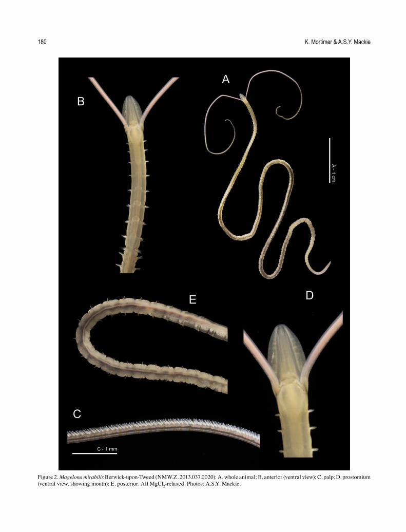
Figure 2. Magelona mirabilis Berwick-upon-Tweed (NMW.Z. 2013.037.0020): A, whole animal; B, anterior (ventral view); C, palp; D, prostomium (ventral view, showing mouth); E, posterior. All MgCl2-relaxed. Photos: A.S.Y. Mackie.

Morphology, feeding and behaviour of Magelona 181
tubes were then placed in small observation tanks within the main aquarium, some capillary tubes on the bottom of the tank and others held upright using a small plastic table-shaped holder. Capillary tubes were removed from the tank at intervals and viewed under a Leica MZ9.5 zoom microscope.
Additional observations within capillary tubes primed with a weak carmine or food colouring solution were carried
out under a microscope. Carborundum powder was also tested (particle size ~36 μm) but proved to be too coarse and dense.
In situ laboratory experiments
Sediment from the sampling site was sieved through a 0.5-mm sieve to remove macrofauna, while trying to retain the sediment characteristic of the sample. This was placed into a
Figure 3. Experimental set-up: A, aquarium tank and cooling system; B, Magelona johnstoni Berwick-upon-Tweed (NMW.Z. 2013.037.0001): live animal in capillary tube. Photo: A.S.Y. Mackie.

K. Mortimer & A.S.Y. Mackie182
small glass tank (internally 11.3 × 11.3 × 11.5 cm; volume ca. 1470 cm3) and allowed to settle before adding magelonids. Further sediment was placed on top and allowed to settle in a fridge before placing into the aquarium. In earlier trials, worms were placed directly onto the surface of the sediment, but many were unable to penetrate the surface so this second technique was adopted. Experiments were carried out in both still and flowing water to observe any potential differences in behaviour. The sediment level within the observation tanks was increased (to 5.5 cm deep, ca. 700 cm3 volume) during observations between April and June 2013, both to increase water flow across the sediment and allow a greater depth for burrowing.
Food was added to the tank at the sediment surface or around capillary tubes every 4–7 days, using plastic pipettes. Several food options were utilised: frozen marine invertebrate aquarium food (Dutch Select foods—food for invertebrates, marine) and SeAquariums Invertfood liquid diet (made up of plankton and other essential marine nutrients). Food was mixed with flocculent material collected from the surface of the sediment during sampling, enabling it to sink towards the sediment surface.
Animals were observed for seven months (April–October) during daylight hours; no observations were made at night. All experiments were filmed with a miniDV camcorder, and the resulting footage was observed both at full speed and in slow motion (10–50% slower). Separate glass tanks within the main aquarium were utilised, each containing only one of the species (M. mirabilis or M. johnstoni), allowing direct comparison of their behaviour. A further two smaller tanks were used, one containing animals that had lost both palps upon collection and one containing those that had lost only one. Palp regeneration was then followed over a period of 40 days for M. johnstoni. Animals were observed using a low-powered zoom microscope (×15–30) held horizontally towards the tank. Food colouring and carmine particles were added to the surface waters of small isolated tanks holding individual animals, in order to observe water flow.
Scanning electron microscopy (SEM)
Additional animals collected for SEM were fixed in ca. 6–8% formaldehyde or glutaraldehyde in seawater. Specimens were subsequently washed with fresh water, and transferred in an alcohol series through to 100% ethanol for critical point drying. They were then Sputter coated before imaging using a Jeol Neoscope JCM-5000 SEM. Specimens have been deposited in the National Museum of Wales (NMW), Cardiff.
Current knowledge of pouches in magelonids
All magelonid species descriptions and re-descriptions were examined for details of pouch presence/absence, pouch type (anteriorly or posteriorly opening), configuration (paired or unpaired), pattern (on alternating segments or consecutive segments) and the segment at which they first occur. The resulting information was then compiled to identify groups of species.
Observations and Discussion
Species presence and abundance
Each of the selected sampling sites varied in terms of sediment characteristics and consequently differed in the species present and their relative abundances. Magelona johnstoni was most abundant in the silty fine sands of Berwick-upon-Tweed, while M. filiformis dominated in the fine sands of Oxwich Bay. Magelona mirabilis was collected in low numbers at all sites, but M. johnstoni was absent from collections made at Oxwich Bay. Magelona were difficult to consistently collect on the Rhossili Bay shore due to its susceptibility to onshore winds and waves, though all three species were known to occur there, and sublittorally (Mackie et al. 2006). Hence, M. johnstoni and M. mirabilis were conveniently sourced from Berwick-upon-Tweed, and M. filiformis was collected at Oxwich Bay. Unfortunately all material of M. filiformis was small and delicate, and mortality occurred within several days. No observational data was obtained for this species.
Of the two remaining British species, Magelona alleni Wilson, 1958 was only recorded once during preliminary sampling at Mumbles Bay, Swansea (March 2012) and, from previous collecting (1998–2012), was known to be infrequent at Berwick-upon-Tweed. Magelona minuta Eliason, 1962 is an offshore muddy sediment species and was not encountered on any of the shores.
As previously mentioned, European records of the Brazilian M. papillicornis actually relate to M. mirabilis or M. johnstoni, or both. The same situation holds for any pre-2000 account of M. mirabilis (see Fiege et al. 2000). In the following text, an asterisk identifies these erroneous or suspect citations as M. papillicornis* or M. mirabilis*.
Burrowing
Burrowing observations for M. johnstoni essentially match those described by McIntosh (1878; 1911) for M. papillicornis* and Jones (1968) for Magelona sp. When burrowing, M. johnstoni moved its prostomium laterally from side to side, loosening the sediment in front and aiding movement forward. The everted ‘proboscis’ (see Mortimer et al., 2012 regarding terminology) was used as an anchor, allowing the body to be pulled towards the head. The ‘proboscis’ was then retracted, the prostomium moved forward and the process repeated. Jones (1968) felt that eversion of the ‘proboscis’ occurred primarily due to the hydrostatic pressure of the blood, but to a lesser extent via that of the coelomic fluid. The ‘proboscis’ is therefore totally essential for burrowing, and if compromised, would likely be fatal for the worm. This was recognised by McIntosh (1915), who suggested that the group’s preference for fine sands may help avoid sharp fragments of coarse gravel and sand that might damage their proboscides.
Jones (1968) postulated that the hollow cylindroid dorsal muscular ridges of the magelonid prostomium, which are provided with longitudinal muscles, were presumably fluid-filled and likely to provide rigidity during burrowing. During burrowing, the palps trailed behind the body, but once the worm was near the sediment surface, the palps looped out from underneath the body towards the opening. Both M. johnstoni

Morphology, feeding and behaviour of Magelona 183
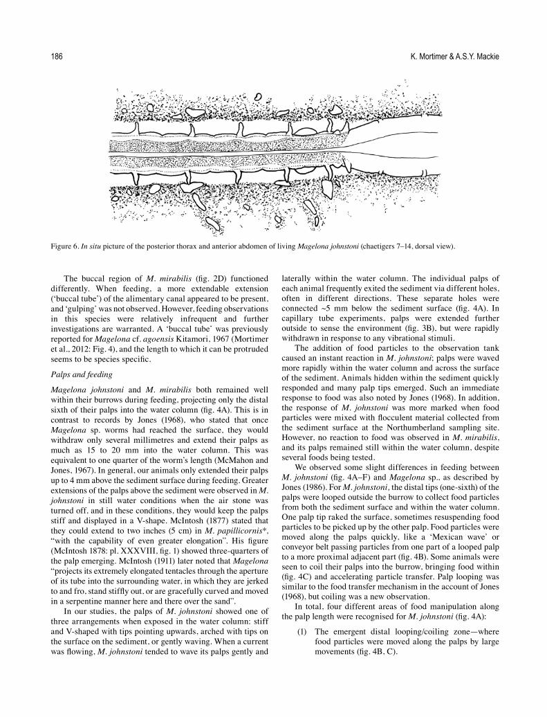
and M. mirabilis were observed to burrow directly to the surface of the sediment and then withdraw into the burrow. Alternatively, they stopped before the surface and moved their palps through the sediment to the water column. Palp length in living animals was extremely long (figs 1A–E; 2A), and the worms could stay well within the burrow with only the last distal sixth of the palps projecting into the water column (fig. 4A).
In the laboratory, M. johnstoni generally burrowed horizontally within the sediment. This was consistent with field observations, collected animals being found with the same orientation within the sediment at Berwick-upon-Tweed. To commence feeding, worms then burrowed upwards from their horizontal position towards the surface, thus, creating an arched or diagonally shaped burrow opening out into the water column (fig. 4A). Some variation in burrow shape was observed, although no U-shaped burrows were seen. McMahon and Jones (1967) and Jones (1968) suggested that Magelona sp. constructed vertical burrows. The latter author described animals burrowing directly downwards once initially placed into the observation chamber, then after reaching the bottom, they burrowed up to the surface. Although for our M. johnstoni, burrow shape was straighter in deeper sediment, strictly vertical burrows were not usually seen.
Differences in observed behaviours could be due to the contrasting experimental chambers and the methodology of both studies. The chamber used by McMahon and Jones (1967) was constructed from a U-shaped rubber tube clamped between two pieces of glass plate, which were no more than 0.7–1.0 cm apart (McMahon pers. comm.), and worms were introduced to the sediment surface. As stated above, M. johnstoni struggled to penetrate the sediment when placed directly onto the surface, therefore additional sediment was allowed to settle upon the worms after their placement into the cube-shaped tank. This may have affected the direction of initial travel; however, the much broader tank would not have constrained the direction of burrowing. The sediment volume in the observation tank used here was ~700 cm3, allowing ample space for movement in any direction, unlike the narrow tank of McMahon and Jones (1967) and Jones (1968). Nevertheless, once settled, M. johnstoni often burrowed against the glass of the observation tank, allowing them to be fully observed. Whether this was fortuitous, the worms were simply burrowing until they reached the glass, or it was due to an attraction to food accumulated against the tank sides, was not determined. The undescribed Magelona from Woods Hole was shown to have U-shaped burrows (McMahon, pers. comm.), with both ends at the surface. This warrants further investigation, particularly between species, and may depend on an ability to burrow backwards as well as forwards within a burrow.
During feeding and resting within the burrow, the bodies of both M. mirabilis and M. johnstoni were greatly stretched, their abdomens somewhat narrower than the thorax. In this region, only the lamellar tips were in contact with the sides of the burrow (fig. 6). If disturbed, contraction of their bodies enabled both species to withdraw quickly into their burrows. However, neither seemed able to actively burrow backwards. To change direction, bodies were retracted, bringing the prostomia under the sediment surface and new burrows were
formed in other directions. Before initiating a new burrow, individuals blocked the ends of their old burrows by shaking their prostomia laterally while everting their proboscides.
Burrows were observed to be temporary and worms moved around, periodically after several hours or days, making new burrows. Fauchald (1983) observed similar behaviour for M. sacculata living in sandy substrates off southern and central California. This species appeared to move through the sediment on a more or less continual basis. Movement to a new burrow may be initiated by the need to locate further food sources. However, M. mirabilis moved around less frequently than M. johnstoni and was generally much less active. Permanency of burrows may well be species-specific; species such as M. alleni and M. cincta Ehlers, 1908 build recognisable tubes (Mortimer and Mackie, 2009; Mortimer et al., 2012).
Burrows appeared to be maintained by mucus, something noted previously by Jones (1968) for Magelona sp. and Wilson (1982) for metamorphosing/metamorphosed M. filiformis, M. alleni and M. mirabilis*. Wilson further noted that some larvae used a band of mucus for adhesion in the approximate region of the ninth adult chaetiger. Interestingly, we have seen adults of M. alleni, separated from their usual red-purple papery tube (during the sieving of grab samples) quickly produce a loose mucus-bound sand tube (pers. obs.). Mucus secretions for M. johnstoni appeared much greater than those for M. mirabilis in the laboratory.
The longevity of magelonids kept in glass capillary tubes was much reduced in comparison with those living unconstricted in the aquarium sediment. Animals kept in capillary tubes within the aerated tank lasted about 4 days only. This was in marked contrast to the success others have had in maintaining various Spionida for long periods of time (over 4 years) in capillary tubes within petri dishes (Williams, 2002; Dualan and Williams, 2011). This dependence on sediment was recorded by McIntosh (1911), “sand is very necessary for the existence of this form, for though the animals survive a considerable period in captivity in vessels filled with pure sea-water, they thrive much longer amongst fine sand, with a few inches of water over it”. In our study, one individual kept in a capillary tube held upright in sediment, survived for over 8 weeks.
Buccal region
The buccal region of M. johnstoni has three lips, one larger triangular lip above two smaller lateral lips (fig. 1F, fig. 5A). These were seen to expand and separate, revealing a triangular-shaped mouth at their centre (fig. 4F). Animals displayed a ‘gulping’ action, apparently sucking in water on opening the mouth. The surface of the top lip and the area just above in M. johnstoni is speckled (fig. 1F).
Our observations agreed with those of the mouth of M. papillicornis* as described by McIntosh (1911), “a somewhat triangular or T-shaped slit surrounded by lips of mucous membrane, and situated between or very slightly in front of the bases of the tentacles. The anterior lip is sinuous but complete, while inferiorly there is a wide fissure (bounded laterally by prominent margins), which runs a considerable distance backwards. The lips are very mobile and in life frequently expand to gulp water.”

K. Mortimer & A.S.Y. Mackie184
Figure 4. Feeding in Magelona johnstoni: A, feeding position within the burrow (ventral view), indicating four zones where different methods are utilised to move food particles along the palp; B, looping of the palp at the surface (zone 1), in order to pass food particles along the palp (lateral view); C, similar process to that shown in B but utilising coiling of the palp (lateral view, papillae omitted for clarity); D, sequence showing the process of passing food particles from papillae to papillae along palp in zone 2; E, food particles being passed between papillae of both palps in zone 3; F, region where food particles are dropped towards mouth (ventral view) in zone 4.

Morphology, feeding and behaviour of Magelona 185
Figure 5. Magelona johnstoni Berwick-upon-Tweed: A, prostomium and first two chaetigers (ventral view), showing mouth surrounded by one upper (UL) and two lower lips (LL), and the proboscis (Pb, not everted) (NMW.Z.1999.021.0020a); B, papillae of mid-palp region (NMW.Z.2013.037.0008c); C, left-hand anteriorly opening pouch located between chaetigers 10 and 11 (lateral view, DF = dorsal flap, VF = ventral flap, LO = lateral organ, CM = convoluted membrane) (NMW.Z.2013.037.0011b); D, close-up view of convoluted membrane; E, transverse section through the body and anteriorly opening pouch situated between chaetigers 10 and 11 (posterior half of pouch and parapodia of chaetiger 11) (NotoL = notopodial lamellae, NeuroL = neuropodial lamellae) (NMW.Z.2013.037.0010c); F, anterior half of same pouch (NMW.Z.2013.037.0010b). Photos: K. Mortimer.
K. Mortimer & A.S.Y. Mackie186
The buccal region of M. mirabilis (fig. 2D) functioned differently. When feeding, a more extendable extension (‘buccal tube’) of the alimentary canal appeared to be present, and ‘gulping’ was not observed. However, feeding observations in this species were relatively infrequent and further investigations are warranted. A ‘buccal tube’ was previously reported for Magelona cf. agoensis Kitamori, 1967 (Mortimer et al., 2012: Fig. 4), and the length to which it can be protruded seems to be species specific.
Palps and feeding
Magelona johnstoni and M. mirabilis both remained well within their burrows during feeding, projecting only the distal sixth of their palps into the water column (fig. 4A). This is in contrast to records by Jones (1968), who stated that once Magelona sp. worms had reached the surface, they would withdraw only several millimetres and extend their palps as much as 15 to 20 mm into the water column. This was equivalent to one quarter of the worm’s length (McMahon and Jones, 1967). In general, our animals only extended their palps up to 4 mm above the sediment surface during feeding. Greater extensions of the palps above the sediment were observed in M. johnstoni in still water conditions when the air stone was turned off, and in these conditions, they would keep the palps stiff and displayed in a V-shape. McIntosh (1877) stated that they could extend to two inches (5 cm) in M. papillicornis*, “with the capability of even greater elongation”. His figure (McIntosh 1878: pl. XXXVIII, fig. 1) showed three-quarters of the palp emerging. McIntosh (1911) later noted that Magelona “projects its extremely elongated tentacles through the aperture of its tube into the surrounding water, in which they are jerked to and fro, stand stiffly out, or are gracefully curved and moved in a serpentine manner here and there over the sand”.
In our studies, the palps of M. johnstoni showed one of three arrangements when exposed in the water column: stiff and V-shaped with tips pointing upwards, arched with tips on the surface on the sediment, or gently waving. When a current was flowing, M. johnstoni tended to wave its palps gently and
laterally within the water column. The individual palps of each animal frequently exited the sediment via different holes, often in different directions. These separate holes were connected ~5 mm below the sediment surface (fig. 4A). In capillary tube experiments, palps were extended further outside to sense the environment (fig. 3B), but were rapidly withdrawn in response to any vibrational stimuli.
The addition of food particles to the observation tank caused an instant reaction in M. johnstoni; palps were waved more rapidly within the water column and across the surface of the sediment. Animals hidden within the sediment quickly responded and many palp tips emerged. Such an immediate response to food was also noted by Jones (1968). In addition, the response of M. johnstoni was more marked when food particles were mixed with flocculent material collected from the sediment surface at the Northumberland sampling site. However, no reaction to food was observed in M. mirabilis, and its palps remained still within the water column, despite several foods being tested.
We observed some slight differences in feeding between M. johnstoni (fig. 4A–F) and Magelona sp., as described by Jones (1986). For M. johnstoni, the distal tips (one-sixth) of the palps were looped outside the burrow to collect food particles from both the sediment surface and within the water column. One palp tip raked the surface, sometimes resuspending food particles to be picked up by the other palp. Food particles were moved along the palps quickly, like a ‘Mexican wave’ or conveyor belt passing particles from one part of a looped palp to a more proximal adjacent part (fig. 4B). Some animals were seen to coil their palps into the burrow, bringing food within (fig. 4C) and accelerating particle transfer. Palp looping was similar to the food transfer mechanism in the account of Jones (1968), but coiling was a new observation.
In total, four different areas of food manipulation along the palp length were recognised for M. johnstoni (fig. 4A):
(1) The emergent distal looping/coiling zone—where food particles were moved along the palps by large movements (fig. 4B, C).
Figure 6. In situ picture of the posterior thorax and anterior abdomen of living Magelona johnstoni (chaetigers 7–14, dorsal view).

Morphology, feeding and behaviour of Magelona 187
(2) The second zone, just below the surface, where each palp diverged within the sediment—food was transferred by very small loops of the palps in conjunction with direct movement by the papillae (figs 1E, 5A). Here, food particles were passed from papilla to papilla along each palp (fig. 4D).
(3) The third zone, further down the burrow, where the two palps aligned together in parallel—food particles were moved cooperatively between the papillae of both palps (fig. 4E). Particles could be moved using papillae of just one palp (as in zone 2), but the cooperative method predominated.
(4) The drop zone, coinciding with the non-papillated regions of the palps—food particles descended directly from the rapidly splayed palps to the mouth (fig. 4F). As they neared the mouth, a ‘gulping’ action aided consumption of the particles, and as transport of food particles within this zone was noticeably swift, it was likely that ‘gulping’ also created a current inward toward the mouth. No other forms of current generation (e.g. from pouch/lamellar movements, or lateral body movements) were observed during feeding.
Once consumed, food particles were readily observed through the body wall and moved rapidly through the thorax. However, the thoracic gut transition time increased as more food was consumed. McMahon and Jones (1967) and Jones (1968) postulated that there might be a mucus thread aiding the transport of food particles across the non-papillated region of the palps. They identified probable mucus-secreting cells at the bases of the papillae, along the proximal 20% of the palps and on the ventral surface of the prostomium. Jones (1968) also witnessed a coordinated movement between food particles moving to the mouth and along the gut, adding credence to the involvement of a mucus thread. However, we found no evidence for a mucus strand for M. johnstoni. Further, movement of particles through the thorax slowed as feeding progressed, yet movement along the palps continued as before. Food abundance had a major influence on feeding. For instance, if a glut of food was present at the sediment surface, animals continually brought food particles into the burrow, storing them just above the prostomium. One individual filled its entire burrow from prostomial tip to sediment surface with food, which was later fed upon more slowly. Despite burrows being temporary structures, the burrow structure around the palps was well maintained during feeding.
On occasion, individuals emerged slightly from the sediment and moved their palp tips towards the mouth region. This occurred simultaneously with ‘proboscis’ eversion that created a channel in which the palp tip was wiped across the mouth. The function of this behaviour was unclear.
Magelona mirabilis did not feed in the same manner as M. johnstoni. Its palps remained erect within the water column and made no response to the addition of food. Only relatively small movements of the palps were observed, even when water flow was increased. The species never waved its palps in the water column, as seen in M. johnstoni, even under comparable flow rates.
The palps of M. mirabilis (fig. 2A) (particularly the proximal third) were extremely stiff in comparison with those of M. johnstoni, even in relaxed animals. The blood vessels within the palps (compare figs 1F, G with 2B, D) were clearly much wider in M. mirabilis, and this may contribute to their rigidity. The general ‘feeding’ position within the burrow was similar to that of M. johnstoni, though M. mirabilis kept its palps closer together (fig. 7), the papillae (fig. 2C) of each extensively interlocked.
Then, on day 63, two animals were seen to consume large amounts of sediment, ignoring the recent addition of food. Their palps remained stiffly displayed within the water column, however long thin ‘pellets’ of sediment were brought down to the non-papillated region of their palps. These pellets were possibly formed as sediment was squeezed between the interlocked palps, but this could not be confirmed as the burrows were only partially visible. On several occasions, individuals were observed to move their palps backwards and forwards in a saw-like motion. Pellet movement was rather slow and often jerky, particularly in the region of the prostomium. This movement was unlike that observed for M. johnstoni. Again, no evidence of a mucus string was apparent, although sediment particles moved as if ‘tugged’ towards the mouth. Consumption of sediment was slow, with no quick ‘gulping’ action, and material built up around the mouth region. The ‘mouth’ region of M. mirabilis seemed to be more extendable than that of M. johnstoni, protruding more as a tube as it gathered in particles.
Apart from the apparently infrequent sediment ingestion, another possibility was that M. mirabilis preferentially fed on minute particles (and/or bacteria) in the water, which were unable to be seen using the techniques utilised here. This could explain why the papillae of its palps within the burrow were so interlocked. There was no evidence that the species employed a form of mucus-net suspension feeding, as found in certain other polychaetes (Riisgård and Larsen, 2010), though debris was seen to become lodged in between and along the length of the palp tips of M. mirabilis (fig. 7). All experimental individuals of this species survived over seven months within the tank. Further investigations with an increased number of animals are needed before any conclusions about its mode of feeding can be made.
Palps of M. johnstoni appear to be selective in what they pick up, using the papillae at the palp tips like fingers. Selectivity of the magelonid diet has been previously suggested (Hunt, 1925; Linke, 1939), and Fauchald and Jumars (1979) considered the group selective surface deposit feeders, and that selectivity may increase within poorly sorted sediments. However, the possibility of suspension feeding has been suggested by other authors (see Rouse, 2001). Our observations have shown M. johnstoni to both capture particles suspended within the water column as well as from the sediment surface, an idea previously suggested for M. papillicornis* (Wolff, 1973; Hartmann-Schröder, 1971), thus implying two different feeding modes.
The constituents of the magelonid diet have been noted by several authors (McIntosh, 1911; Hunt, 1925; Mare, 1942; Jones, 1968; Hartmann-Schröder, 1971; Wolff, 1973; Kühl, 1974) and include detritus, diatoms, organic debris, algal cysts, spores, foraminiferans, tintinnids, and the larvae of

K. Mortimer & A.S.Y. Mackie188
crustaceans, molluscs and worms. Bivalve and polychaete larvae, pelagic eggs and tintinnids have been recorded in the diets of magelonid larvae (Lebour, 1922; Thorson, 1946; Smidt, 1951; Kühl, 1974; Wilson, 1982). Although doubts exist as to whether natural predation on bivalve larvae is common (Johnson and Brink, 1998), prevalence may be higher in later-stage larvae (Wilson, 1982; Johnson and Brink, 1998).
McIntosh (1911) and Mare (1942) additionally reported the presence of sand, silt and debris, which likely concurs with our observations for M. mirabilis, and lends support for the presence of interspecific variation in feeding between co-existing magelonid species. This, and contributing factors such as behaviour, size, morphology, habitat, presence/absence of a tube, and palp morphology (e.g. stiffness/flexibility), warrants further investigation.
Palp regeneration
Several M. johnstoni lost their palps upon collection, and this provided an opportunity to monitor their regeneration. All animals initially stayed well within the sediment and were not visible for the first few days. By day 3, one individual began protruding the tip of its prostomium out of the sediment surface, occasionally everting its ‘proboscis’. This individual moved fairly swiftly around the tank, repeating this behaviour in different locations. While at the surface, it created an inward current toward the mouth by combining ‘gulping’ with everting and retracting the ‘proboscis’, enabling feeding despite the loss of palps. Capillary tube experiments confirmed that conspicuous inward currents could be produced in this manner.
Palp regeneration progressed at different rates between animals, and between palps on the same individual (table 1). By day 29, one pair of palp tips were noticeable protruding out of the sediment surface (palps now up to nine times prostomial length), and by day 31, one animal was using its palps to feed. Although these palps were thinner and shorter than those in intact animals, they were able to manipulate food particles
effectively and bring food to the mouth, as previously described for the species (see above). McIntosh (1911) described the rapidity by which magelonids regenerated their palps, noticing that within 3 days “the new organs appeared on each side as short blunt processes into which the blood entered”. This agrees well with the current observations, which saw short stumps on every animal within the same period.
During regeneration, individuals were seen to carry out lateral sinuous movements of the thorax within the burrow. This was first noticed on day 3 and continued sporadically up until day 24. The purpose of this behaviour was unclear, however, magelonid palps are thought to also have a respiratory function; this will be discussed more fully below.
Observations were also made on individuals that had lost only one palp. Initially, such animals were observed to stay close to the sediment surface, with the remaining palp extended into the water column and waved, as in normal behaviour. Palp regeneration followed a similar time-scale to that of animals lacking both palps. By day 8 they were a third of the length of the prostomium, and by day 18, about twice the length of the prostomium. By day 36, one regenerating palp was of similar length to the intact palp (less than one prostomium length’s difference in size). Single-palp individuals collected food particles effectively with their remaining palps, using a similar technique to that seen in zone 2 of intact animals (fig. 4D). Although transfer of food particles was slower, their feeding capability did not appear compromised in any other way.
These observations suggest the implication of palp loss in magelonids is lessened by their ability to continue to feed either with only one or no palps, and their ability to regenerate to fully functioning palps within ~30 days.
Sinuous lateral movements
On occasion, M. johnstoni made gentle, sinuous lateral movements of the thorax within the burrow, often for long periods. Jones (1968) also observed movements of the anterior 20 to 25 chaetigers of Magelona sp. (~85–100 times per
Figure 7. Various palp positions observed for Magelona mirabilis from in situ experiments. Third picture depicts debris collecting on and in between the palps.

Morphology, feeding and behaviour of Magelona 189
minute), which produced an inward-moving current. Jones noted that this behaviour was infrequent and postulated that it was linked to respiration, as occurrence and intensity of this behaviour increased if the water was allowed to become deoxygenated. Oxygen levels in our study were always maintained and we could not confirm this. However, sinuous movements were seen more frequently in individuals regenerating palps.
Both Jones (1968) and McIntosh (1911) believed that magelonid palps had, in part, a respiratory function, on the basis of their vascular nature and their placement into the water column. Additionally, when the current within the tank was halted, individuals extended their palps further into the water, holding them stiffly upwards. Therefore, a higher occurrence of these sinuous movements in individuals lacking palps could be a compensatory response for a loss of respiratory capacity. The relationship between burrow irrigation and body undulations has also been reported for other annelids. For example, Wells (1949) stated that Arenicola marina Linnaeus, 1758 irrigated their burrows, providing a supply of oxygenated water, using special waves that travelled along their bodies, usually from tail to head. Female A. marina could also use an altered form
of this irrigation behaviour during reproductive events (Hardege and Bentley, 1997), bringing sperm into the burrows.
Lateral movements also occurred outside of the burrow. These movements were brisk and quite marked, with the sides of the body almost touching the sediment on both sides, in contrast to the gentle undulations seen within the burrow. McIntosh (1911) stated that magelonids protrude their anterior region from the sand into the water column for aeration and food, suggesting that the modified chaetae of the 9th chaetiger aided emergence from the burrow. However, he made no mention of lateral movements of the body in conjunction with this behaviour, unlike Jones (1968) who noted this behaviour in individuals without palps, believing it related to respiration. This behaviour occurred “even when there appeared to be an adequate supply of fresh, oxygenated sea water”. This is in contrast to the current findings, as individuals without palps generally remained within the sediment, only bringing their prostomial tips above the sediment surface, and these movements occurred in individuals with intact palps and in aerated water, suggesting no link to respiration.
During lateral movements, the lateral abdominal pouches were generally kept flat against the body wall, and only slight
Table 1. Showing palp regeneration data for three Magelona johnstoni, all of which lacked palps on collection.
Day Palp length (in prostomium lengths) Notes1–2 – – – All animals within the sediment
3 Stumps noticeable
Stumps noticeable
Stumps noticeable
One prostomium protruding just out of the sediment. Very slight lateral movements of body observed.
4 1/6 – – All at or near sediment surface.
8 1/3 – –
10 2/3 – 1/6 All animals within the sediment; one just below the surface. Palps regenerating at different rates.
16 2 2 1/3 Two animals making slight lateral movements within burrow. Animal with shortest palps protruding prostomium tip just out of sediment.
17 – – – One animal continuing small lateral movements of the thorax within the burrow. Another protruding prostomium tip just out of sediment.
21 – – 3/4 One animal at surface with prostomium tip just emerging from sediment, while another was undergoing lateral movements of the thorax within burrow.
22 – 4, 1½ – Unequal regeneration of palps occurring on one animal. One animal making lateral movements within burrow.
23 – – – No animals at surface or undergoing lateral movements.
24 – – – One animal making lateral movements.
29 – – – First observation of palps emerging from the sediment surface. Palps relatively thin.
30 ~9 – 2 Palps appearing equal in length.
31 ~9 – – Palp tips at surface, first observation of an animal feeding using their palps, although palps still relatively thin.
35 ~12 – – Palp tips within water column.
36 – 8, 2 – Palp regeneration uneven within the same animal.

K. Mortimer & A.S.Y. Mackie190
movements in response to body movement were observed. While no sinuous lateral movements of the body were observed in M. mirabilis, further investigations with an increased number of individuals are warranted.
Reproductive behaviour?
As stated above, animals periodically extended their thoraxes out of their burrows/capillary tubes, generally to the thorax/abdominal junction (but occasionally to approximately chaetiger 15–20). Individuals would then display lateral sinuous movements of the thorax outside of the burrow, with sporadic eversion of the ‘proboscis’. These out-of-burrow movements often lasted for long periods of time, unless the animal was disturbed. However, this was an intermittent behaviour and generally took place during the months of April to July. No instances of this behaviour occurred after this period. Lateral movements were witnessed in animals with both palps intact, and occurred in both still and flowing, aerated water. Movements were often observed simultaneously in several animals, and during these periods pairs would often lean towards each other sometimes with bodies in direct contact (fig. 8).
During one observation in an isolated tank with slightly raised water temperature, an individual emerged from its burrow and commenced sinuous lateral movements of the thorax, directed towards a second individual believed to be female. Individual two was just below the surface, but the palps of each were in direct contact with those of the other. This occurred for several minutes before individual two retracted into its burrow, later emerging in another burrow some 3.5 cm away. Individual one emerged to the approximate level of chaetiger 15, stretching across the sediment towards the new position of individual two. Individual two appeared to respond to these lateral movements, by waving and looping the palps towards individual one. Individual two remained within the burrow, just below the sediment surface, but both individuals commenced entwinement of their palps until the tips became quite interlocked. After a period of time, individual two emerged from the burrow, to the approximate level of chaetiger 5. Cessation of palp entwinement occurred and individual two disappeared back into its burrow. Although the exact reason for this is unclear, the individual may have responded to vibrational stimuli. Individual one remained on the surface of the sediment for some time, continuing to stretch towards individual two, making lateral prostomial movements and eversion of the ‘proboscis’. Eventually, individual one withdrew into the sand and began burrowing in another direction. Simultaneously, in this isolated tank, another pair were observed making lateral movements of the body, one within the water column and one within the sediment. The latter individual later emerged from the burrow and continued moving the thorax outside the burrow. Four days later, a further two individuals (one female and one male) carried out lateral movements. Although, no release of reproductive products was ever confirmed, the synchronised/reactive behaviour of individuals outside of the burrow was strongly suggestive of an involvement in reproduction. The simultaneous spawning
of gametes in broadcast spawners would increase the probability of egg fertilisation.
Hardege and Bentley (1997) stated that synchronicity of gamete release within a population was particularly important for semelparous species, and that environmental factors such as photoperiod, temperature, lunar periodicity and tidal cycles may help in the synchronisation of broadcast spawners (believed to be the case for magelonids, see below). The observations of synchronised movements as described above during periods of increased water temperature suggests this is an important factor triggering this behaviour in magelonids. In addition, pheromones can play a final part in synchronising reproduction in both iteroparous and semelparous polychaete species (Hardege and Bentley, 1997).
One possibility is that lateral movements of the thorax may be involved in gamete release, either helping bring sperm into the burrow for egg fertilisation, as seen in female Arenicola marina (Hardege and Bentley, 1997), or dispersing both eggs and sperm. Jones (1968) showed that sinuous movements of the body within the burrow of Magelona sp. produced an inward current, suggesting that sperm released by the male could be drawn into the burrow for egg fertilisation. However, most records suggest that magelonid eggs and sperm are spawned directly into the water, and sperm structure would suggest fertilisation outside of the burrow (Blake, 2006; Rouse, 1999, 2006).
Our video footage of M. johnstoni clearly shows an exhalent current from the burrow during lateral movements outside the burrow, and it seems probable that if tubes are blind-ending, any inward current should circulate around the burrow and back out. If eggs and sperm are released from the posterior end into the burrow, then circulatory currents may help to push them from the burrow into open water.
Reproduction
Eggs were observed in M. johnstoni collected in November 2012 (Rhossili), and March and April 2013 (Berwick-upon-Tweed), with reproductive animals appearing more fragile abdominally. Reproductive females were white abdominally, in stark contrast to the conspicuous green gut (fig. 1H). Eggs were observed in M. mirabilis from November 2012 (Rhossili) and April 2013 (Berwick-upon-Tweed), while they were observed in animals of M. filiformis collected in January, February (Oxwich Bay) and April 2013 (Berwick-upon-Tweed).
Wilson (1982) collected mature eggs from M. mirabilis* between May and August, although the best fertilisations occurred in animals from July and August. This was in agreement with McIntosh (1877), who stated that M. papillicornis* was full of ova and sperm at St Andrews in June, and “the ova and spermatozoa … attain perfection in summer and autumn”. McIntosh (1911) stated that ova of a considerable size were present in large numbers at the end of June, but those that developed in late autumn did not successfully produce embryos. Kühl (1974) suggested that M. papillicornis* in Elbe, Cuxhaven, Scharnhörn and Gelbsand reproduced during the summer months. Wilson (1982) considered the spawning season for M. filiformis in Plymouth to peak during August, although mature gametes were

Morphology, feeding and behaviour of Magelona 191
collected between April and October, while M. alleni was likely to mature in late September or October.
The timing of spawning may be influenced by the availability of food. Kühl (1974) suggested that Magelona larvae were dependent on bivalve larvae of the right size. Magelona larvae were generally present in the Plymouth Sound plankton from April to November, but more commonly from July to October (Wilson, 1982). McIntosh (1915) encountered Magelona larvae from May–November in St Andrews, (locality not stated but assumed from McIntosh 1916: pl. XCIV, fig. 17), although numbers were generally much lower in October and November. Similar timings were noted by Kühl (1974) for M. papillicornis* larvae, present from May to October in polyhaline rivers discharging into the German North Sea (Elbe, Weser) and the Wadden Sea (Ems). Magelona juveniles may settle from the plankton by September, as indicated by samples collected in bottom-nets (McIntosh, 1915).
Nevertheless, very little is known about magelonid reproduction (Rouse, 2001). They are believed to be broadcast spawners, with ect-aquasperm (as defined by Jamieson and
Rouse, 1989) (Blake, 2006; Rouse, 1999; Rouse, 2006). The mechanism by which Magelona species shed gametes from their burrows is unknown. Several accounts have suggested magelonids only reproduce once, with mortality occurring after spawning. Ripe magelonids can be extremely fragile and, as also noted by McIntosh (1911), “it is possible that at the reproductive season degeneration of the organs [palps] may occur in some instances, or the animals themselves may perish”. Fauchald (1983) considered M. sacculata to be an annual species (monotelic/semelparous) with feeding larvae, based partly on the work by Hannan et al. (1977) in Monterey Bay.
Species activity
Observations show that M. johnstoni is a very active worm in comparison with the other two species investigated. Behavioural differences were obvious: M. filiformis was the most inactive and M. johnstoni the most active. The species differ markedly in terms of body shape and size: M. filiformis being very slender and long, comprising of many segments, while both M. mirabilis and M. johnstoni are broader animals. Magelona johnstoni moved around the environment more
Figure 8. Showing two individuals of Magelona johnstoni simultaneously making lateral sinuous movements of the thorax (outside the burrow) (dorsal views).

K. Mortimer & A.S.Y. Mackie192
frequently, while M. mirabilis, the larger of the two species, stayed very still, inhabiting burrows for much longer periods. Additionally, M. mirabilis appeared much less responsive to vibrational stimuli than M. johnstoni, which reacted to the slightest of knocks. Jones (1968) noted that the Magelona sp. was also extremely sensitive to vibrational stimuli, both within the sand and in the water column, and stated that its lateral organs (see figs 5 and 10) shared similarities with those vibration receptors found in ctenophores and chaetognaths. Mucus production in M. mirabilis was much lower than in M. johnstoni during observations. Magelona johnstoni placed in petri dishes with a small amount of sediment were shown to cover themselves in a mucus/sediment mixture very quickly, producing a rudimentary ‘tube’. McIntosh (1915) noted this behaviour, suggesting that this is “probably to compensate for the absence of its element”.
Pouches
The most obvious morphological feature separating M. johnstoni and M. mirabilis is the respective presence or absence of anteriorly opening abdominal lateral pouches on the anterior abdomen. The function of these (and other posteriorly directed pouches) in magelonids has never been resolved, despite much attention (McIntosh, 1878, 1911; Jones, 1968).
No significant movements of lateral abdominal pouches, either anteriorly or posteriorly opening forms, were observed for M. johnstoni during any capillary tube or in situ experiment. In general, the pouches were kept flat against the body. Any slight contractions of the anteriorly opening pouches were attributable to body movements. Lateral movements of the thorax would cause the lateral edges of the dorsal and ventral flaps to come together, and when animals lunged forward, pouches occasionally contracted slightly as the body elongated and narrowed. Slow-motion video footage also revealed small pouch contractions as the lumen/ventral vessel of the posterior region contracted, often propagating a wave down the abdomen. On occasion, the first pair of anteriorly opening pouches expanded against the sides of the capillary tubes, but this was generally restricted to a few individuals in poor condition.
Water flow throughout the capillary tube was produced by eversion and retraction of the ‘proboscis’, and through lateral movements of the body. Observations using carmine particles showed that water movement around the pouches was not significantly greater than that around parapodia and segments of other parts of the body. Water flowing along the dorsal and ventral edges of the body was directed laterally around parapodia and toward the opening of the pouches (fig. 9). No additional flow created by pouch function was evident when water flowed back out.
Possible function of abdominal lateral pouches
Anchor
One hypothesis is that pouch function may be related to anchorage, particularly during lateral body movements outside the burrow, from which M. johnstoni emerges to the approximate level of chaetiger 9 (just above the first pair of abdominal pouches). Pouches expanded against burrow sides
could help prevent worms being swept away by water movements. However, healthy M. johnstoni kept their lateral pouches flat against the body (fig. 6). In addition, burrow entrances become widened during this behaviour, making such anchorage unlikely. Posteriorly opening pouches were also kept flat against the body and were never shown to expand against the burrow sides.
Propulsion
Another hypothesis is that the contraction of anteriorly opening pouches could aid movement backwards (perhaps enhancing rapid retraction when under threat of predation), with posteriorly opening pouches enabling movement forwards. No evidence of pouch contraction could be seen in video of M. johnstoni for either slow or rapid movement, forwards or backwards. The presence of a medial slit in posteriorly opening pouches in some species also suggests that this is an unlikely function.
Reproduction, sperm storage, and brooding
Throughout this study, gametes were present within the body cavities of Magelona specimens, but no relationships between gametes and pouches were observed. Jones (1968) doubted any relationship between pouch function and reproduction because of their presence in both sexes and in juvenile forms. Conversely, McIntosh (1877, 1878, 1879) believed that ‘lateral organs’ (see below) appeared in ripe animals in summer and autumn. However, it is likely that he was examining two different species, M. mirabilis and M. johnstoni. As magelonids are thought to be broadcast spawners with ect-aquasperm, the likelihood that pouch function is related to sperm storage is low. Although sperm storage has been described for some members of the Spionida (see Blake, 2006), such as Streblospio benedicti Webster, 1879, Pseudopolydora kempi (Southern, 1921), Pseudopolydora paucibranchiata (Okuda, 1937) and Pygospio californica Hartman, 1936, sperm receptacles in these species differ greatly in morphology and show clear connections to the interiors of the animals concerned. No such connections have been found in magelonid pouches. Fauchald (1983) stated it unlikely that M. sacculata (a species with paired anteriorly opening pouches in the anterior abdomen) brooded its young due to its large reproductive effort. Nevertheless, pouch function could be seasonal, and without direct observation of spawning events, the link between the two cannot be completely refuted.
Burrow irrigation
Our observations suggest that magelonids use lateral movements of the thorax within the burrow (rather than contraction and expansion of pouches) to generate water circulation.
Morphology of pouches
Investigation of pouch morphology along the body of M. johnstoni supported a graduation between anteriorly and posteriorly opening pouches, as reported by Mortimer (2010). Understanding the form of the anteriorly opening pouches has been extremely difficult due to their apparently complex
Morphology, feeding and behaviour of Magelona 193
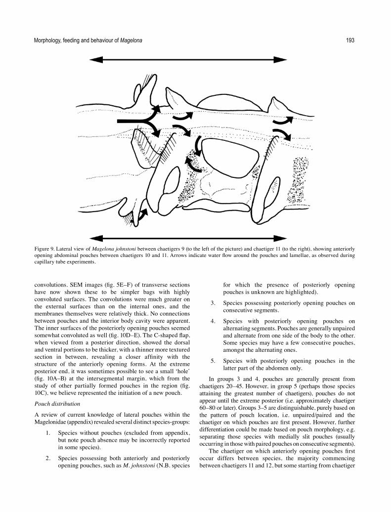
convolutions. SEM images (fig. 5E–F) of transverse sections have now shown these to be simpler bags with highly convoluted surfaces. The convolutions were much greater on the external surfaces than on the internal ones, and the membranes themselves were relatively thick. No connections between pouches and the interior body cavity were apparent. The inner surfaces of the posteriorly opening pouches seemed somewhat convoluted as well (fig. 10D–E). The C-shaped flap, when viewed from a posterior direction, showed the dorsal and ventral portions to be thicker, with a thinner more textured section in between, revealing a closer affinity with the structure of the anteriorly opening forms. At the extreme posterior end, it was sometimes possible to see a small ‘hole’ (fig. 10A–B) at the intersegmental margin, which from the study of other partially formed pouches in the region (fig. 10C), we believe represented the initiation of a new pouch.
Pouch distribution
A review of current knowledge of lateral pouches within the Magelonidae (appendix) revealed several distinct species-groups:
1. Species without pouches (excluded from appendix, but note pouch absence may be incorrectly reported in some species).
2. Species possessing both anteriorly and posteriorly opening pouches, such as M. johnstoni (N.B. species
for which the presence of posteriorly opening pouches is unknown are highlighted).
3. Species possessing posteriorly opening pouches on consecutive segments.
4. Species with posteriorly opening pouches on alternating segments. Pouches are generally unpaired and alternate from one side of the body to the other. Some species may have a few consecutive pouches, amongst the alternating ones.
5. Species with posteriorly opening pouches in the latter part of the abdomen only.
In groups 3 and 4, pouches are generally present from chaetigers 20–45. However, in group 5 (perhaps those species attaining the greatest number of chaetigers), pouches do not appear until the extreme posterior (i.e. approximately chaetiger 60–80 or later). Groups 3–5 are distinguishable, purely based on the pattern of pouch location, i.e. unpaired/paired and the chaetiger on which pouches are first present. However, further differentiation could be made based on pouch morphology, e.g. separating those species with medially slit pouches (usually occurring in those with paired pouches on consecutive segments).
The chaetiger on which anteriorly opening pouches first occur differs between species, the majority commencing between chaetigers 11 and 12, but some starting from chaetiger
Figure 9. Lateral view of Magelona johnstoni between chaetigers 9 (to the left of the picture) and chaetiger 11 (to the right), showing anteriorly opening abdominal pouches between chaetigers 10 and 11. Arrows indicate water flow around the pouches and lamellae, as observed during capillary tube experiments.

K. Mortimer & A.S.Y. Mackie194
Figure 10. Abdominal posteriorly opening pouches from several specimens of Magelona johnstoni: A–B, initiation of new pouches represented by small ‘holes’ (lateral view) (A: NMW.Z.2013.037.0008e; B: NMW.Z.2013.037.0010d); C, first pouch (~6 chaetigers) from pygidium (lateral view) (NMW.Z.2013.037.0011d); D, first pouch from a regenerating tail (ventral/posterior view) (NMW.Z.2013.037.0008c); E, third pouch (~10 chaetigers) from pygidium (posterior view) (NMW.Z.2013.037.0011d); F, posteriorly opening pouch of an abdominal fragment (lateral posterior view) (NMW.Z.1998.028). Photos: K. Mortimer.

Morphology, feeding and behaviour of Magelona 195
10 (chaetiger 9 is even reported in a small number of species). Most anteriorly opening pouches are paired; however, unpaired pouches are reported in some species, and this warrants further investigation, as does their number (some species only have one pair, while in others there are several). At present, details on the morphology of pouches in described species are insufficient to be able to further categorise the groups.
‘Lateral organs’ of McIntosh
Jones (1968) stated that the structures termed ‘lateral pouches’ were equivalent to the ‘lateral organs’ of McIntosh (1879, 1911, 1915). However, according to McIntosh’s accounts (1877, 1879), lateral organs appeared in ripe individuals, suggesting a connection with reproduction. In his 1877 account under a section headed ‘Reproductive Organs’, McIntosh states “the ova and spermatozoa are present in each sex in great abundance in the posterior region of the body, and attain perfection in summer and autumn. On the sides of the body, also, peculiar convoluted organs occur in processes composed of the cuticle, hypoderm, and basement-tissue”. Similarly in 1879, McIntosh writes “and in a male loaded with spermatozoa at the same season, and in which the lateral organs were present, the diaphanous tapering tips were extended forward nearly to the cuticle, and curved inward like the horns of the springbok”. McIntosh (1879) suggested that the appearance of ‘lateral organs’ caused ‘a curious change’, in which cephalic vessels became abbreviated and the direction of blood flow at the base of the prostomium was modified, further stating that there was a greater diversity in cephalic vessels in animals bearing ‘lateral organs’. McIntosh does not refer to lateral organs in his 1911 account, but does note ‘peculiar structures’ that occur on either side of the body in males and females with developed sexual products, on many of the posterior segments. Curiously, he states that these structures invariably occur on the segment immediately behind the mouth, stating: “and in this it first attains perfection”. ‘Lateral organs’ are figured in McIntosh (1878: pl. XXX, fig. 7) and clearly show anteriorly opening paired pouches located between the 10th and 11th chaetigers. Also figured, is a transverse section through the body wall and ‘lateral organ’ (pl. XXXIV, fig. 2) from the anterior abdominal region, which shows a dorsal and ventral flap with convoluted membrane. There is some doubt about which species McIntosh studied: although originally identified as M. papillicornis, most European records have been referred to either M. johnstoni or M. mirabilis. Fiege et al. (2000) reviewed specimens collected by McIntosh at St Andrews, referring them to M. mirabilis, and McIntosh’s 1916 drawing certainly shows an anterior abdomen lacking anteriorly opening pouches. Yet, the pouches drawn by McIntosh (1878) are indicative of M. johnstoni, although no locality was given for this particular specimen. McIntosh (1915) stated that “on the sides of the posterior region, from the twenty-fifth or twenty-sixth segment backward, are the peculiar glandular organs (pouch-like) which occupy the lateral region of each segment”. Abdominal pouches do not occur in M. mirabilis until approximately chaetiger 80 (see Fiege et al. 2000: 226 and Appendix), but posteriorly opening pouches are present in M. johnstoni from around chaetiger 20. As these two species were not differentiated until
2000, it is extremely likely that McIntosh was observing the two morphologically similar and co-existing species M. johnstoni and M. mirabilis under the name of M. papillicornis. Hence, the occurrence of ‘lateral organs’ was actually an unrecognised species-specific character, and not related to reproduction. Although McIntosh did not always state the location of specimen collection, references to St Andrews throughout his accounts exist (1877, 1878, 1911, 1915), and text clearly states that specimens possessing ‘lateral organs’ were present alongside specimens without.
Acknowledgements
The authors would like to thank Jason Williams (Hofstra University), Carol Anne Simon (Stellenbosch University), Greg Rouse (Scripps Institution of Oceanography) and Adriana Giangrande (Università del Salento) for their advice, Daniel Martin and João Gil (Centre d’Estudis Avançats de Blanes) for provision of papers, and Peter Howlett, Anna Holmes, Teresa Darbyshire and Catalena Angele (National Museum Wales) for their help with photography, SEM and care of animals under observation. Particular thanks go to Robert McMahon (The University of Texas at Arlington) for his personal communications on his observations of Magelona sp. from Woods Hole.
ReferencesAguirrezabalaga, F., Ceberio, A., and Fiege, D. 2001. Octomagelona
bizkaiensis (Polychaete: Magelonidae) a new genus and species from the Capbreton Canyon (Bay of Biscay, north-east Atlantic). Journal of the Marine Biological Association of the United Kingdom 81: 221–224.
Blake, J.A. 2006. Chapter 13. Spionida. Pp. 565–638 in: Rouse, G.W. and Pleijel, F. (vol. eds), Reproductive biology and phylogeny of Annelida. Science Publishers Inc.: Enfield New Hampshire.
Bolívar, G.A., and Lana, P.C. 1986. Magelonidae (Annelida: Polychaeta) do litoral sudeste do Brasil. Neritica 1: 131–147.
Brasil, A.C. dos S. 2003. Filogenia de Magelonidae Cunningham & Ramage, 1888 (Annelida–Polychaeta) com base na morfologia externa. Tese de Doutorado, Setor de Ciências Biológicas-Zoologia, Universidade Federal do Paraná. 113 pp.
Buzhinskaja, G.N. 1985. Polychaeta of the shelf off south Sakhalin and their ecology. Issledovaniya Fauny Morei 30: 72–224 (in Russian).
Clarke, D.T., Paterson, G.L.J., Florence, W.K., and Gibbons, M.J. 2010. A new species of Magelona (Polychaeta: Magelonidae) from southern Namibia. African Natural History 6: 77–82.
Dales, R.P. 1962. The polychaete stomodeum and the inter-relationships of the families of Polychaeta. Proceedings of the Zoological Society of London 139(3): 389−428.
Dales, R.P. 1977. The polychaete stomodeum and phylogeny. Pp. 525−546 in: Reish, D.J. and Fauchald, K. (eds), Essays on polychaetous annelids in memory of Dr. Olga Hartman. Allan Hancock Foundation, University of Southern California: Los Angeles.
Dualan, I.V., and Williams, J.D. 2011. Palp growth, regeneration, and longevity of the obligate hermit crab symbiont Dipolydora commensalis (Annelida: Spionidae). Invertebrate Biology 130(3): 264–276.
Ehlers, E. 1908. Die bodensässigen Anneliden aus dem Sammlungen der deutschen Tiefsee-Expedition. Wissenschaftliche Ergebnisse der Deutschen Tiefsee-Expedition auf dem Dampfer “Valdivia” 1898–1899 16: 1–167.

K. Mortimer & A.S.Y. Mackie196
Eliason, A. 1962. Undersökningar over Öresund. XXXXI Weitere Untersuchungen über die Polychaetenfauna des Öresunds. Lunds Universitets Årsskrift, N.F. Avd. 2 58: 1–98.
Fauchald, K. 1983. Life diagram patterns in benthic polychaetes. Proceedings of the Biological Society of Washington 96(1): 160–177.
Fauchald, K., and Jumars, P.A. 1979. The diet of worms: a study of polychaete feeding guilds. Oceanography and Marine Biology Annual Review 17: 193–284.
Fiege, D., Licher, F., and Mackie, A.S.Y. 2000. A partial review of the European Magelonidae (Annelida: Polychaeta): Magelona mirabilis redefined and M. johnstoni sp. nov. distinguished. Journal of the Marine Biological Association of the United Kingdom 80: 215–234.
Filippova, A., Purschke, G., Tzetlin, A.B., and Müller, M.C.M. 2005. Reconstruction of the musculature of Magelona cf. mirabilis (Magelonidae) and Prionospio cirrifera (Spionidae) (Polychaeta, Annelida) by phalloidin labeling and cLSM. Zoomorphology 124: 1–8.
Gallardo, V.A. 1968. Polychaeta from the Bay of Nha Trang, South Viet Nam. Naga Report 4(3): 35–279.
Glémarec, M. 1966. Les Magelonidae des Côtes de Bretagne. Description de Magelona wilsoni n. sp. Vie et Milieu 17: 1077–1085.
Gravier, C. 1905. Sur les annélides polychètes de la Mer Rouge (Cirratuliens, Spionidiens, Ariciens). Bulletin du Muséum d’Histoire Naturelle 11(1): 42–46.
Gravier, C. 1906. Contribution a l’étude des annélides polychaetes de la Mer Rouge. Nouvelles Archives du Muséum d’Histoire Naturelle, Paris, Sér. 4 8: 123–236, pls 1–9.
Hannan, C.A., Hulberg, L.W., Mawn, K.M., and Nybakken, J.W. 1977. A study to develop standard procedures for life history analysis of benthic invertebrates for biological monitoring in marine and estuarine environments. Moss Landing Marine Laboratories, California State University Consortium: Moss Landing, California. 217 pp.
Hardege, J.D., and Bentley, M.G. 1997. Spawning synchrony in Arenicola marina: evidence for sex pheromonal control. Proceedings of the Royal Society of London, Series B 264: 1041–1047.
Hartman, O. 1936. New species of Spionidae (Annelida Polychaeta) from the coast of California. University of California Publications in Zoology 41(6): 45–52.
Hartman, O. 1944. Polychaetous annelids from California, including two new genera and nine new species. Allan Hancock Pacific Expeditions 10: 239–304.
Hartman, O. 1961. Polychaetous annelids from California. Allan Hancock Pacific Expeditions 25: 1–226.
Hartmann-Schröder, G. 1971. Annelida, Borstenwürmer, Polychaeta. Die Tierwelt Deutschlands 58: 1–594.
Hernández-Alcántara, P., and Solís-Weiss, V. 2000. Magelonidae from the Mexican Pacific and Northern Gulf of Mexico, with the description of a new genus (Meredithia) and four new species. In: Reish, D.J. and Lana, P. (eds), Proceedings of the 6th International Polychaete Conference, Curitiba, Brazil, 1998. Bulletin of Marine Science 67: 625–644.
Hunt, J.D. 1925. The food of the bottom fauna of the Plymouth fishing grounds. Journal of the Marine Biological Association of the United Kingdom 13: 560–599.
Jamieson, B.G.M., and Rouse, G.W. 1989. The spermatozoa of the Polychaeta (Annelida): an ultrastructural review. Biological Reviews 64: 93–157.
Johnson, K.B., and Brink, L.A. 1998. Predation on bivalve veligers by polychaete larvae. Biological Bulletin 194: 297–303.
Johnston, G. 1865. A catalogue of the British non-parasitical worms in the collection of the British Museum. Trustees of the British Museum: London. 365 pp.
Jones, M.L. 1963. Four new species of Magelona (Annelida, Polychaeta) and a redescription of Magelona longicornis Johnson. American Museum Novitates 2164: 1–31.
Jones, M.L. 1968. On the morphology, feeding, and behavior of Magelona sp. Biological Bulletin 134: 272–297.
Jones, M.L. 1977. A redescription of Magelona papillicornis F. Müller. Pp. 247–266 in: Reish, D.J. and Fauchald, K. (eds), Essays on polychaetous annelids in memory of Dr. Olga Hartman. Printed for the Allan Hancock Foundation. University of Southern California: Los Angeles, CA.
Jones, M.L. 1978. Three new species of Magelona (Annelida, Polychaeta) and a redescription of Magelona pitelkai Hartman. Proceedings of the Biological Society of Washington 91: 336–363.
Kitamori, R. 1967. Magelonidae (Polychaetous annelids) from Japan, including the description of a new species. Bulletin of Tokai Regional Fisheries Research Laboratory 50: 49–54.
Kühl, H. 1974. Über Vorkommen und Nahrung der Larven von Magelona papillicornis O.F. Müller (Polychaeta Sedentaria) im Mündungsgebiet von Elbe, Weser und Ems. Berichte der Deutsche Wissenschaftlichen Kommission für Meeresforschung, Berlin 23(3): 296–301.
Lebour, M.V. 1922. The food of planktonic organisms. Journal of the Marine Biological Association of the United Kingdom 12: 644–677.
Linke, O. 1939. Die Biota des Jadebusenwattes. Helgoländer Wissenschaftliche Meeresuntersuchungen 1: 201–348.
Linnaeus, C. 1758. Systema naturae per regna tria naturae, secundum classes, ordines, genera, species, cum characteribus, differentiis, synonymis, locis. Editio decima, reformata. Laurentius Salvius: Holmiae. ii, 824 pp.
Mackie, A.S.Y., James, J.W.C., Rees, E.I.S., Darbyshire, T., Philpott, S.L., Mortimer, K., Jenkins, G.O., and Morando, A. 2006. The Outer Bristol Channel Marine Habitat Study. Studies in marine biodiversity and systematics from the National Museum of Wales. BIOMÔR Reports 4: 1–249 and A1–A227, + DVD-ROM (2007).
Mare, M.F. 1942. A study of a marine benthic community with special reference to the micro-organisms. Journal of the Marine Biological Association of the United Kingdom 25: 517–554.
McIntosh, W.C. 1877. On the structure of Magelona. Annals and Magazine of Natural History, Ser. 4 20: 147–152.
McIntosh, W.C. 1878. Beiträge zur Anatomie von Magelona. Zeitschrift für Wissenschaftliche Zoologie 31: 401–472.
McIntosh, W.C. 1879. On the circulatory system of Magelona. Journal of Anatomy and Physiology 13(3): 331–345.
McIntosh, W.C. 1911. On the structure of Magelona. Annals and Magazine of Natural History Ser. 8, 7: 417–457 (Translation of McIntosh, 1878).
McIntosh, W.C. 1915. A monograph of the British marine annelids. Vol. III. Part I. Text. Polychaeta. Opheliidae to Ammocharidae. The Ray Society: London.
McIntosh, W.C. 1916. A monograph of the British marine annelids. Vol. III. Part II. Plates. Polychaeta. Opheliidae to Ammocharidae. The Ray Society: London.
McMahon, R.F., and Jones, M.L. 1967. Observations on feeding in Magelona sp. Biological Bulletin of the Marine Biological Laboratory, Woods Hole 133: 476.
Mohammad, M.-B.M. 1970. On two new polychaetous annelids from Kuwait. Zoologischer Anzeiger 185: 303–307.
Mohammad, M.-B.M. 1973. New species and records of polychaete annelids from Kuwait, Arabian Gulf. Zoological Journal of the Linnean Society 52: 23–44.

Morphology, feeding and behaviour of Magelona 197
Monro, C.C.A. 1933. The Polychaeta Sedentaria collected by Dr. C. Crossland at Colón, in the Panama region, and the Galapagos Islands during the expedition of the S.Y. ‘St. George’. Proceedings of the Zoological Society of London 1933: 1039–1092.
Mortimer, K. 2010. Magelonidae (Polychaeta) from the Arabian Peninsula: a review of known species, with notes on Magelona tinae from Thailand. Zootaxa 2628: 1–26.
Mortimer, K., Cassà, S., Martin, D., and Gil, J. 2012. New records and new species of Magelonidae (Polychaeta) from the Arabian Peninsula, with a re-description of Magelona pacifica and a discussion on the magelonid buccal region. Zootaxa 3331: 1–43.
Mortimer, K., Gil, J., and Fiege, D. 2011. Portuguese Magelona (Annelida: Magelonidae) with a description of a new species, a re-description of Magelona wilsoni Glémarec, and a key to adult Magelonidae from European waters. Italian Journal of Zoology 78 (Suppl. 1): 124–139.
Mortimer, K., and Mackie, A.S.Y. 2003. The Magelonidae (Annelida: Polychaeta) from the Seychelles, with the description of three new species. In: Sigvaldadóttir, E., Mackie, A.S.Y., Helgason, G.V., Reish, D.J., Svavarsson, J., Steingrímsson, S.A. and Gudmundsson, G. (eds), Advances in polychaete research. Hydrobiologia 496(1–3): 163–173.
Mortimer, K., and Mackie, A.S.Y. 2006. The Magelonidae (Annelida: Polychaeta) from the Seychelles. 2. Description of four additional species, three new to science. In: Sardá, R., San Martín, G., López, E., Martin, D. and George, D. (eds), Scientific Advances in Polychaete Research. Scientia Marina 70(S3): 125–137.
Mortimer, K., and Mackie, A.S.Y. 2009. Magelonidae (Polychaeta) from Hong Kong, China; with discussions on related species and redescriptions of three species. In: Maciolek, N.J. and Blake, J.A. (eds), Proceedings of the Ninth International Polychaete Conference. Zoosymposia 2: 179–199.
Müller, F. 1858. Einiges über die Anneliden fauna der Insel Santa Catharina an der brasilianischen Küste. Archiv für Naturgeschichte 24: 211–220.
Nateewathana, A., and Hylleberg, J. 1991. Magelonid polychaetes from Thailand, the Andaman Sea, with descriptions of eight new species. In: Petersen, M.E. and Kirkegaard, J.B. (eds), Systematics, biology and morphology of world Polychaeta. Proceedings of the 2nd International Polychaete Conference, Copenhagen, 1986. Ophelia Suppl. 5: 169–184.
Okuda, S. 1937. Spioniform polychaetes from Japan. Journal of the Faculty of Science, Hokkaido University, Ser. 6 Zoology 5(3): 217–254.
Orrhage, L. 1973. Two fundamental requirements for phylogenetic scientific works as a background for an analysis for Dales (1962) and Webb (1969) theories. Zeitschrift fur Zoologische Systematik und Evolutionsforschung 11: 161−173.
Riisgård, H.U., and Larsen, P.S. 2010. Particle capture mechanisms in suspension feeding invertebrates. Marine Ecology Progress Series 418: 255−293.
Rouse, G.W. 1999. Polychaeta, including Pogonophora and Myzostomida. Pp. 81–124 in: Jamieson, B.G.M. (ed.), Reproductive biology of invertebrates, Vol. IXB. Progress in Male Gamete Ultrastructure and Phylogeny. Oxford and IBH Publishing Co.: New Delhi.
Rouse, G.W. 2001. Magelona Müller, 1858. Pp. 261–263 in: Rouse, G.W. and Pleijel, F. (authors), Polychaetes. Oxford University Press: Oxford.
Rouse, G.W. 2006. Chapter 3. Annelid sperm and spermiogensis. Pp. 45–76 in: Rouse, G.W. and Pleijel, F. (eds), Reproductive biology and phylogeny of Annelida. Science Publishers Inc.: Enfield New Hampshire.
Smidt, E.L.B. 1951. Animal production in the Danish Waddensea. Meddelelser fra Danmarks Fiskeri- og Havundersøgelser 11(6): 151 pp.
Southern, R. 1921. Polychaeta of the Chilka Lake and also of fresh and brackish waters in other parts of India. Memoirs of the Indian Museum 5: 563–659.
Thorson, G. 1946. Reproduction and larval development of Danish marine bottom invertebrates, with special reference to the planktonic larvae in the sound (Øresund). Meddelelser fra Danmarks Fiskeri- og Havundersøgelser. Ser.: Plankton 4: 7–523.
Uebelacker, J.M., and Jones, M.L. 1984. Family Magelonidae. 7.1–7.29 in: Uebelacker, J.M. and Johnson P.G. (eds), Taxonomic guide to the Polychaetes of the Northern Gulf of Mexico. Final report to the Minerals Management Service, contract 14-12-001-29091. Barry A. Vittor & Associates: Mobile, Alabama.
Webster, H.E. 1879. The Annelida Chaetopoda of New Jersey. Annual Reports of the New York State Museum of Natural History 32: 101–128.
Wells, G.P. 1949. Respiratory movements of Arenicola marina (L.): intermittent irrigation of the tube, and the intermittent aerial respiration. Journal of the Marine Biological Association of the United Kingdom 28: 447–464.
Wesenberg-Lund, E. 1949. Polychaetes of the Iranian Gulf. Danish Scientific Investigations in Iran 4: 247–400.
Williams, J.D. 2002. The ecology and feeding biology of two Polydora species (Polychaeta: Spionidae) found to ingest the embryos of host hermit crabs (Anomura: Decapoda) from the Philippines. Journal of Zoology, London 257: 339–351.
Wilson, D.P. 1958. The polychaete Magelona alleni n. sp. and a re-assessment of Magelona cincta Ehlers. Journal of the Marine Biological Association of the United Kingdom 37: 617–626.
Wilson, D.P. 1959. The polychaete Magelona filiformis sp. nov. and notes on other species of Magelona. Journal of the Marine Biological Association of the United Kingdom 38: 547–556.
Wilson, D.P. 1982. The larval development of three species of Magelona (Polychaeta) from localities near Plymouth. Journal of the Marine Biological Association of the United Kingdom 62: 385–401.
Wolff, W.J. 1973. The estuary as a habitat: an analysis of data on the soft-bottom macrofauna of the estuarine area of the Rivers Rhine, Meuse, and Scheldt. Zoologische Verhandelingen. Uitgegeven Door Het Rijksmuseum van Natuurlijke Histoire te Leiden 126: 1–242.
Zhou, J., and Mortimer, K. 2013. A new species of Magelona (Polychaeta: Magelonidae) from Chinese coastal waters. Journal of the Marine Biological Association of the United Kingdom 93(6): 1503–1510.

K. Mortimer & A.S.Y. Mackie198
Appendix.
All known information from literature records on the presence, morphology and pattern of lateral abdominal pouches within the Magelonidae. Key to categories: 1) species in which pouches are reported as absent (not included in table); 2) species possessing both anteriorly and posteriorly opening pouches (presence of posteriorly opening pouches unknown/not confirmed in some species+); 3) species with paired posteriorly opening pouches on consecutive segments; 4) species with unpaired posteriorly opening pouches alternating from one side of the body to the other, on alternate segments (sporadic pouches on consecutive segments may be present); 5) species with posteriorly opening pouches in the latter part of the abdomen only.
Species Notes Chaetiger of first appearance
Record Category
Magelona sp. of Jones (1968)
Paired anteriorly opening pouches between chaetigers 10 & 11.
10 Jones (1968) 2
Unpaired pouches, approximately every 4th chaetiger, alternating from one side of the body to the other.
20–23
Magelona sp. A Large, paired pouches. 11 Uebelacker and Jones (1984)
2
Posteriorly opening pouches, unpaired on alternate chaetigers, on alternating sides of the body.
26
*Notes only paired and unpaired C configuration pouches.
*Brasil (2003)
Magelona sp. B Large, paired pouches. 11 Uebelacker and Jones (1984)
2
Posteriorly opening pouches, unpaired on alternate chaetigers, on alternating sides of the body.
18–26
*Notes only paired and unpaired C configuration pouches.
*Brasil (2003)
M. cincta Ehlers, 1908
C configuration, unpaired on alternate chaetigers and alternate sides of the body, based on a single specimen from Mozambique. Not observed on holotype (specimen short anterior fragment).
19 Mortimer and Mackie (2009)
4
M. conversa Mortimer & Mackie, 2003
∑ configuration, paired (11, 14, 17, 20). Unpaired pouches on alternate chaetigers and alternate sides of the body.
11 Mortimer and Mackie (2003)
2
*Several unpaired pouches very large, more akin to ∑ configuration pouches.
23–26 Mortimer et al. 2012*
M. cornuta Wesenberg-Lund, 1949
C configuration, paired, on consecutive segments, medially slit, edges of which are surrounded by thicker cuticle (based on specimens from Hong Kong, not observed on short holotype).
~41 Mortimer and Mackie (2009)
3
M. crenulata Bolívar & Lana, 1986
Bolsas genitais pareadas no setígero 11 e não pareadas nos setígeros 20 e 28 [paired genital bags on setiger 11 and unpaired on chaetigers 20 to 28].
11; 20–28 Bolívar and Lana (1986)
2?
*Paired and unpaired ∑ configuration. *Brasil, 2003
M. crenulifrons Gallardo, 1968
C configuration, unpaired, on alternate chaetigers and alternate sides of the body. Not originally described, but present on type material.
25* based on Hong Kong specimen
Mortimer and Mackie (2009)
4

Morphology, feeding and behaviour of Magelona 199
Species Notes Chaetiger of first appearance
Record Category
M. dakini Jones, 1978 Unpaired, alternating from one side of the body to the other, irregularly located on chaetigers.
101–117 Jones (1978) 5
*Unpaired C configuration pouches. *Brasil (2003)
M. debeerei Clarke et al., 2010
∑ configuration, paired between chaetigers 10 & 11 and 14 & 15, unpaired pouches present between 13 & 14 in some specimens. C configuration not observed
10 Clarke et al. (2010) 2?
M. filiformis Wilson, 1959
C configuration occurring at the extreme posterior end, unpaired, on alternate segments and alternating from one side of the body to the other. Not recorded in original description and reported as absent in Fiege et al. (2000) and Brasil (2003).
This study 5
M. gemmata Mortimer & Mackie, 2003
C configuration, paired, on consecutive segments 42 Mortimer and Mackie (2003)
3?
Magelona sp. G Posteriorly opening pouches, paired, on consecutive segments.
27–28 Uebelacker and Jones (1984)
2/3?
*Paired, ∑ configuration pouches present. *Brasil (2003)
M. hartmanae Jones, 1978
Unpaired, initially on alternate segments and alternate sides of the body. However, variation in pattern occurs more posteriorly. Occasional pouches on consecutive segments.
42–48 Jones (1978) 4
*Unpaired C configuration pouches present. *Brasil (2003)
M. heteropoda Mohammad, 1973
∑, paired, *membrane on both sides of holotype presumed missing.
11 Mohammad (1973), synonymised with M. obockensis, see Mortimer (2010)
2
C configuration, unpaired, more or less alternating between chaetigers and side of the body. Pouches quite large, expanded more dorsally and ventrally, often convoluted.
17
M. johnstoni Fiege et al., 2000
∑, those between 10 & 11 paired, then several pouches occur either paired or unpaired. Some variation in patterns.
(9) 10 Fiege et al. (2000) 2
C configuration, unpaired, *on alternate segments and alternating sides of the body.
~20 *Present study
Magelona sp. L Posteriorly opening pouches, paired on consecutive segments.
28–31 Uebelacker and Jones (1984)
3
*Paired C configuration pouches. *Brasil (2003)
M. lusitanica Mortimer et al., 2011
Unpaired posteriorly opening pouches, alternating from one side of the body to the other, either on consecutive segments or every other. Pattern varies along body.
36 Mortimer et al. (2011) 4
M. mahensis Mortimer & Mackie, 2006
Unpaired C configuration pouches present, on alternate chaetigers, on alternate sides of the body, “Often more or less folded, with thicker cuticle on edges of fold and thinner cuticle inside. Edges of fold can be abutting or overlapping.”
38 Mortimer and Mackie (2006)
4

K. Mortimer & A.S.Y. Mackie200
Species Notes Chaetiger of first appearance
Record Category
M. mirabilis, (Johnston, 1865)
C configuration pouches present, paired, occurring on every 3 or 5 segments for the neotype.
~78 Fiege et al. (2000) 5?
*Paired C configuration pouches present. *Brasil (2003)
M. montera Mortimer et al., 2012
Posteriorly opening, paired pouches on consecutive segments. “Pouches appear as simple folds, split medially with thicker cuticle surrounding edges”.
38 Mortimer et al. (2012) 3
M. obockensis Gravier, 1905
∑, paired between chaetigers 11 & 12. Unpaired, anteriorly opening pouches present on one specimen, more closely resembling posteriorly opening pouches.
11 Mortimer (2010); Gravier (1906)
2
C configuration pouches, unpaired, alternating from one side of the body to the other, usually on alternate segments, “often quite large, more expanded both dorsally and ventrally, often convoluted”. Mortimer (2010) suggested this represented a graduation between the two pouch morphologies along the body.
17–271 1Based on senior author’s notes on syntype material
*Only paired C configuration pouches present. *Brasil (2003)
M. pacifica Monro, 1933
Paired posteriorly opening pouches, on consecutive segments. Medially split, with thicker cuticle surrounding edges.
36–40 Mortimer et al. (2012) 3
M. parochilis Zhou & Mortimer, 2013
Paired, anteriorly opening pouches between 11 & 12 and 14 & 15 (occasionally between 17 & 18).
11 Zhou and Mortimer (2013)
2
Unpaired posteriorly opening pouches, on alternate chaetigers, alternating from one side of the body to the other.
21
M. pectinata Nateewathana & Hylleberg, 1991
Large lateral pouches usually present between chaetigers 11 & 12 and 13 & 14. Other records of pouches present between chaetigers 10 & 11 and 12 & 13, 20 & 21, and 23 & 25. Smaller sporadic pouches are recorded for the posterior+.
10/11 Nateewathana and Hylleberg (1991)
2?
M. pitelkai Hartman, 1944
Posteriorly opening, unpaired, on alternate segments, alternating from one side of the body to the other.
64–84 Jones (1978) 5
M. pulchella Mohammad, 1970
C configuration, initially alternating and unpaired, then paired on consecutive segments. In the posteriormost region they are unpaired on consecutive segments, alternating from one side to the other.
39 Mortimer (2010) 3/4
M. rioja Jones, 1963 Pouches present in posterior region, occurring in a sporadic and irregular pattern; they “appear to be identical with similar structures described by Hartman (1961) for Magelona sacculata and others”.
Jones (1963) 2?
*Paired and unpaired ∑ configuration pouches. *Brasil (2003)
M. sacculata Hartman, 1961
“Conspicuous pouched membranes, first present behind the modified ninth segment, occur also between segments 10 and 11, and at irregular intervals in abdominal segments”. Note: original figure only shows pouches between segments 10 and 11 (paired).
9/10? Hartman, 1961 2?
*Paired and unpaired pouches of both morphologies present.
*Brasil (2003)

Morphology, feeding and behaviour of Magelona 201
Species Notes Chaetiger of first appearance
Record Category
M. sachalinensis Buzhinskaja, 1985
Large paired pouches between chaetigers 11–12 and further irregular pouches, which may occur either singly or paired+.
11 Buzhinskaja (1985) 2?
M. tinae Nateewathana & Hylleberg, 1991
∑, paired. 11 Nateewathana and Hylleberg (1991)
2
C configuration, unpaired, roughly on every other segment, alternating between sides of the body. Pouches quite large, often convoluted.
22 Mortimer (2010)
M. wilsoni Glémarec, 1966
Posteriorly opening, unpaired, alternating from one side of the body to the other. Pattern irregular, sometimes on consecutive segments, sometimes alternately (description based on a Gulf of Lions specimens, not observed in type material).
24 Mortimer et al. (2011) 4