morphometric characterization of the human neuroendocrine ... · morphometric characterization of...
TRANSCRIPT
Morphometric Characterization of the Human Neuroendocrine Merkel Cells
Andres Beiras, M .D., Ph.D., Tomas Ga rda-Caball ero, M .D ., Ph. D., Virginia Fernandez-Redondo, M .D., and Rosalia Ga ll ego, M . D . Departamento de C icncias Mo rfo logicas (Histologb Hum ana), Servicio de Microscopia Elec tronica, Faculrad de Med icin:!, Hospita l Genera l de Ga licia, Univers idad de Santiago de Co mpostda, Ga licia, Spai n
In this study, the neuroendocrin e M erkel cel ls (N EM Cs) from adu lt hu man epiderm is are defined mo rphometrica ll y, using the M OP 20 image analyze r to m easure 21 parameters of either the cell as a w hole, o r pa rticular cellular structures. Maximu m diameter (8.09 jLm), perimeter (26.51 jLl1l ), area (36.87 jL11l 2
) and for m fac to r (0.626) for the cell as a w hole, and maximum diameter (5.08 jLm ), perimeter (18.74 jLm), area (12.54 jLm 2
) and for m fac to r (0.452) fo r the nucleus were determined . Also measu red were nuclea rcytoplasmic ratio (0.5595), fil ament thickness (10 nm) , and g ranul ar llull1 eri ca l density (7.02 g ranules/ jLIl1 2
). M axi-
The fo ll owin g mo rpho metric va lues have been publi shed for the hu ma n neuroendocrin e M erkel cell s (N EMCs): 11 ± 2 J.Lm for maxim um cellula r diam eter (M CD) 111; 75-130 nm [1'/ or 89 ± 18 nm [2] fo r cellular neurosecreto ry (N S) gra nules, and 1. 6 X 0.4
nm fo r in tranu clea.r rods 13]. Fetal N S g ranu les of th e N E M Cs l11 easure SO-200 nm [4].
Mo re data are ava il able fo r the other anima l species. M CD fo r Cypril1l /s carp io NEM Cs is 7.5 J.L111 [51 and 16 J.L111 for Sa /al/la/l dra sa /al/lam/ra [6 /. T he nu clear d iameter (N O) of the rabbit N E M Cs is 9.5-10 J.L111 pJ. T he spine-li ke processes of An ura meas ure between 0.5 X 1.5 J.L m and 0.2 X 2 J.L m /6], and the interstitial processes of U rodcla N EM Cs can meas u re up to 4 J.L m [6].
N eurosecretory g ra nule d iameters measure 64 ± 17.9 nm in the N EMCs of the ad ul t ra t, and lIS ± 34.3 nm in the rat fe tus 18/; 120 n l11 in am phibians [6]; 93.30 ± 18.90 nm in th e pig sno ut 191; and 84 ± 13 [1 0], 60-120 nm [11/ or SO-100 nm [12J in un specified mamm als. Cy to plasm ic in te rmediate fila ments of rat N E M Cs have a thi ckness of 4.6 ± 0.1 nm [8], and 10 nm in
Ma nuscript received August 21, 1986; accepted for publica tion November 24, 1986.
Th is work was supported by g rant 86/11 55 from the Fondo de Investigaciones Sanitarias de Ia Seguridad Social, Ministe rio de Sanidad y ConSUIll O, Madrid , Spa in.
Reprint requests to: Professo r Andres ilciras, Servic io de M icroscopfa Electronic", Facu ltad de Med icina, C/ San Francisco s/n , 15075 Santiago de Co mpostda , La Con 1lla, Spain.
Abbreviations: FF: form fac tor GA: g ra nu la r area GMD: g ra nu lar max im um diamete r G ND: g"l nuiar nUlllerica l density MC D: ma xim ulll cellula r d iamete r N EMCs: neurocndocri ne Mcrkel ce ll s NS: neurosecretory
mum dia m eter, area, and for m fac to r of neurosecreto ry g ranules were 94.23 nm, 5020.05 nm2
, and 0.93, respectively. Length of desm osomes linkin g N EM Cs to keratinocytes w as determined (286.9 nm) and compared w ith that of interkera tinocyti c des mosomes (385 nm) . In addition , length and diameter of cellular processes (s pine-like processes (1. 58 jLm X 0.26 jLm) , interstitial processes (1. 39 jLl11 X 0.25 jLl11) , and microvilli (0.35 jLm X 0.25 jLm) w ere measured after separation and class ifica tion acco rding to the parti cular morphologic characteristi cs of each. J Invest Dennatol 88:766-768, 1987
am phib ia ns [6]. T hc fil amcntous ax is of thc processes is made up of 5 nm units [61. T he numerica l dcnsity of N S g ran ules (G N D) is 0.42-0.45 in the rat [S/, 3.1S ± 0. 15 in the rabbit [7], and S. 11 ± 2.51 in the pig snou t [9]. .
O ur o bjccti ve in thi s prescnt wo rk was to morpho metrica ll y dcfi ne th c hu ma n N E M Cs and to d isting uish them fro m th e N EM Cs of othcr species. T hesc morphometric data, it is ho ped, w il l also bc useful in the ultrast ructural knowledge of som e neuroendocrine hyperplas ias and d ysplas ias of N E M Cs o rig in .
M ATE RI A LS AN D M ETHODS
Sa m ples fro m th e epidermis of male and fe male patients between the ages of 33 and 70 years were taken fro m health y bo rders of biops ies o r fro m recent auto psies (2- 4 h pos tmortem). Fixatio n and postfixatio n were ca rried out in 2.5% g lutaraldehyde in sodiu m cacod ylate buffer and in 1 % OsO" in the sa me buffe r [13J. In cl usio n was done in Spurr 's epoxy res in [1 4J. Ultrathin sectio ns were stained w ith uranyl acetate and lead citrate.
Mo rph o metri c analysis was ca rried o ut w ith the MO P 20 (Ca rl Zciss, Oberkochcn, F. R.G.) w ith a constan t magnifi ca tion fo r each sa m ple. Real magni ficat ion was con tro lled w ith a calibrated g rid.
D iameter, peri meter, area, and fo rm facto r (FF) of the cell and nu cleus as wcll as ka ryocytoplasm ic ratio were es tab lished on the bas is of 50 cells exa mined.
Maxim um dia meter, area, and FF of N S granules, and granular numerical dcnsity (G N D) (nu mber of N S g ranules per J.Lm 2 of cytop las m ic area) were calculated in a 1026 N S g ranules sa m ple fro m 12 cells. T hc cri te ria for choosing these cells were as fo llows: (1) a minimum cellular surface area of 30 J.Lm 2, (2) appro priate ax ial o ri entat io n , and (3) a large nuclca r section.
Length of dcs moso mes betwcen N E M Cs and kcratin ocytes, and between keratinocytes was establi shed using 2 sa m ples of 30 des mosomes cacho For intermedi ate fil am ents 50 elements were exa mined. Length and diameter of processes were measured in 10 sp inc-li kc processes, 6 in terstiti al processes, and 15 microv ill i;
0022-202X/87/S03.50 Copyright © 1987 by T he Society ror Investigative Dermatology , Inc.
766
VOL. 88. NO.6 JUNE 1987
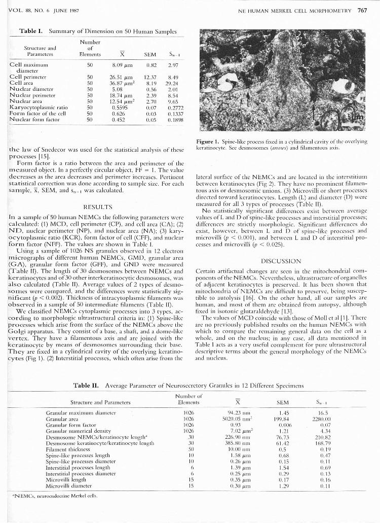
Table I. Summary of Dimension on 50 Human Samples
Number Structure and of
Parameters Elements X SEM 511 - 1
Cell maximum 50 8.09 /1- m 0.82 2.97 diameter
Cell perimeter 50 26.51 /1-m 12.37 8.49 Cel l area 50 36.87 /1-m2 8. 19 29.24 Nuclea r diameter 50 5.08 0.56 2.01 N uclca r perimeter 50 18.74 /1- m 2.39 8.54 Nuclear area 50 12.54 /1-111 2 2.70 9.65 KaJ:'yocy toplasmic ratio 50 0.5595 0.07 0.2772 Fa [']11 fa ctor of thc cell 50 0626 0.03 0. 1337 Nuclear form fa ctor 50 0.452 0.05 0. 1898
the law of Snedeco r was used fo r th e statisti ca l analysis of these processes [15] .
Form fa ctor is a ratio between the area and perim eter of the o"leasured o bj ect. In a perfectly circular obj ect, FF = 1. The va luc decreases as th e area decreases and 'perimeter in crcases. Pertin en t s tatisti cal co rrectio n was done acco rding to sample size. Fo r each sa nlple, X, SEM , and S,, _ I was calculated.
RESULTS
In a sample of 50 human NEM Cs the followin g parameters were calcu lated: (1) M CD, cell perimete r (CP), and cell area (CA); (2) ND, nuclear perimeter (NP), and nucl ear area (NA); (3) karyacytoplasl11i c ratio (KCR) , form factor of cell (C FF), and nuclea r [0['111 factor (NFF) . The va lues are shown in Tab le I.
Using a sam ple of 1026 N S gran ules observed in 12 electro n micrographs of different human NEM Cs, G MD, g ranul ar area (GA), g ranular form fa ctor (GFF) , and G ND were measured (Ta ble II). The length of 30 desmosomes betwecn N EM Cs and kcra tinocytes and of30 other interk eratinocytic desm oso m es , was a lso calcul ated (Table II) . Average va lues of 2 typcs of desl110-SOllles were compared, and the diffe rcnces wcre statist ica ll y significant (p < 0.002). Thickness of intracy toplas l11ic filam ents was observed in a sa mple of 50 intermed iate filaments (Tab lc II ).
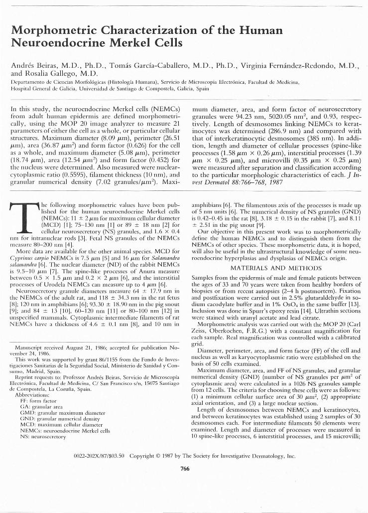
We class ified NEM Cs cytop lasm ic processes into 3 types, according to morphologic ultrast ru ctural criteria in: ('I) Spine-lik e processes which arise from the surface of the NEM Cs above th e Gol g i apparatus . They consist of a base, a shaft, and a dome-like vertex. They have a filamentous axi s and are joined wi th the keratinocyte by m ea ns of desmosomes surroundin g their base . T h ey are fixed in a cylindri ca l cavity of the overlyin g keratinocytes (Fig 1). (2) Interstit ial processes, w hi ch often arise from the
NE HUMAN MERKEL CE LL MORPHOMETRY 767
Figure 1. Spine-likc process fixed in a cylindrical cavity of tile overlying keratinocy te. See desl11osolllCS (a rl'O"',) and fil alllcntous ax is.
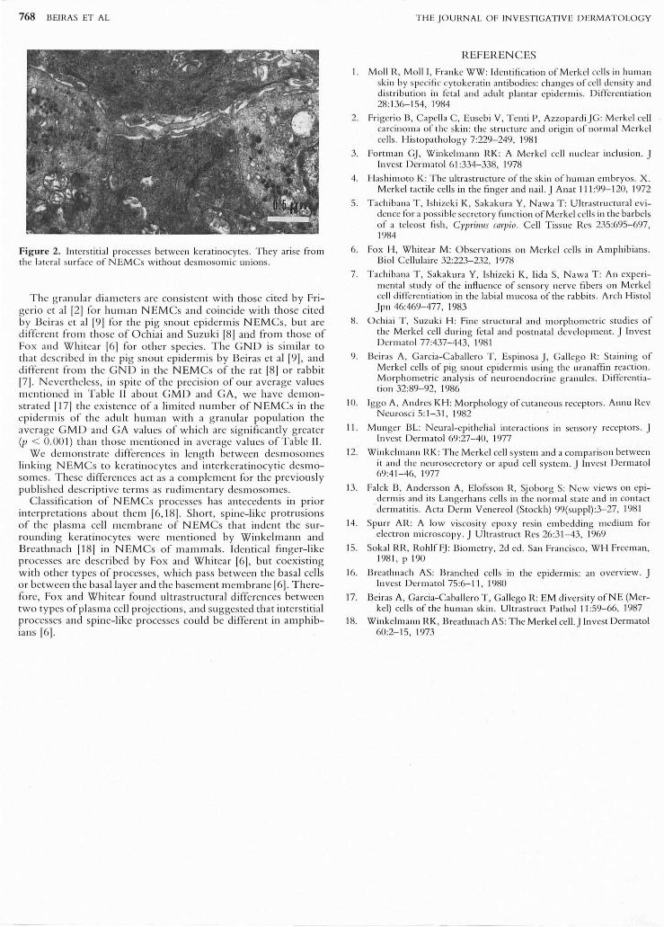
lateral surfa ce of the N EMCs and arc located in the interstitium bctwccn keratinocy tcs (Fig 2). They ha ve no pro min cnt filam cntou s axis o r dcsm oso mi c unio ns . (3) Mi crovi lli o r sho rt processes dirccted towa rd keratin ocy tcs . Lcn g th (L) and diamctcr (D ) wcre m cas ured fo r all 3 types of proccsses (Table II ).
N o stati sti cally signifi cant ditTcrcnccs cxist bctwecn avcrage va lucs ofL and D ofspi nc-likc proccsscs and intcrstitial processes; differenccs arc stri ctl y m o rpho log ic. Signifi ca nt differenccs do cx ist , howcvcr, bctween Land D o f spinc- likc proccsses and microvilli (p < 0 .001) , and bctwecn Land D o f in tcrstitial p roccsscs and mi crovill i (p < 0.025) .
DISC USS IO N
Ccrtai n artifac tual chan gcs arc scc n in the mi tocho ndri al components o fthc N EMCs. Nevcrtheless. ultrastru cturc o f o rganclles of adj acent ke ratinocy tcs is p reservcd . It has bccn shown that mitochondria of N E M Cs arc diffi cult to presc rvc, bcin g susceptib le to autolys is 11 61. O n thc other hand , all our sa mpl cs arc hum an , and most of th em are obtain ed fro m auto psy, alth oug h fixed in isoto nic g lutaraldeh ydc 1131 .
Thc va lu es o f M CD co in cidc w ith those of M oll ct al\I\ . There arc no prc vio usly publi shcd rcsults on th e human N EMCs w ith w hich to co mparc the rcmai nin g gcncral data o n thc ccll as a whole, and o n the nuclcus; in an y casc, all data menti oncd in Table I acts as a ve ry use ful co mplemcnt fo r pure ultrastru ctural descripti ve ter ms abo ut the genc ral mo rph o logy o f thc NEM es and nucleus.
Table II. Average Parameter of N curosecrcto ry G ranules in 12 Diffcrcnt Specimcns
NUl11ber of Structure and Parameters Ele l11ents X SE M 5,, _1
Gran ular ma ximum diam eter 1026 ':14.23 nlll 1.45 16.5 Gra llular area 1026 5020.05 11111 2 199.84 2280.00 Gra nular form factor IU26 0.93 0.006 O.()7 Granular num erical density 1026 7.02 /1-111 2 1. 21 4.34 Desmosome NEMCs/kcratinocyte length" 30 226.90 nm 76.73 210.82 Desmosome kcratillocy te /keratinocy te lell gth 30 385.80 11111 6 1. 42 168.7') Fi lament th ickncss 50 10.00 11111 0.5 0. 19 Spine-like processes lell gth 10 1.58 /1- 111 0.68 0.47 Spille-like processes diameter 10 0.26/1-111 0. 15 U. I I Interstitial processes length 6 1.39/1-111 1.54 D.m Interstitia l processes diameter 6 0.25 /1-111 0.29 0. 13 Microvi lli length 15 0.35 /1- m o 17 0.1 6 Microv illi diameter IS 0.30 /1-111 1. 29 0. 11
" N EMCs. neurocndocrinc Merkel cell s.
768 BEIRAS ET AL
Figure 2. Interstiti al proccsses betwecn kcratinocytes. Thcy arise from the btlTa l surfacc of N EMCs without desmosom ic unions.
Thc g ranular diameters are consistent w ith those cited by Frigeri o e t al [2] fo r hum an NEM Cs and co in cide with those cited by Bei ras et al 19 1 for th e pi g sno ut epide rmis N E M Cs, but are different fro m those of Ochiai and Suzuki 18 1 and from th ose of Fox and W hitea r 16] for other species. The G ND is simil ar to th at described in the pig sno ut epidermi s by Beiras et al 19]. and different from the G N D in the NEM Cs of the rat [8] or rabbit 171 . N everthe less, in spite of th e precisio n of o ur average va lues mentioned in Ta ble [[ abo ut G M D and G A, we have de m o nstrated 11 7 1 the ex istence of a limited number of N E M Cs in the epidermis o f the ad ult hum an with a g ranular po pulatio n the average G MD and G A va lu es of w hi ch are sig nifi cantl y g rea ter (p < 0.001) than those m enti o ned in avera ge va lues o f Table [I.
We dem o nstrate diffe rences in len g th between des moso m es lin kin g N E M Cs to ke ratin ocy tes and interk e ratinocyti c desm oso m es. T hese differen ces ac t as a co mpleI11en t for the previously publi shed descriptive term s as rudim enta ry desmoso m es.
C lassificat io n of N E M Cs processes has antecedents in prio r interpretations abo ut them 16, 18). Sho rt , spine- like protrusions of the pl asm a cell m embrane of N E M Cs that indent th e surro undin g keratinocy tes were m enti o ned by Winkelmann and Breathn ach 11 81 in N E M Cs of m ammals. Id enti cal fin ger-like processes are described by Fox and Whitear [6]. but coexis ting with o ther types of p rocesses, whi ch pass between th e basal cells o r between th e basa l layer and th e base m ent membrane [6]. T herefo re, Fox and Whitea r found ultrastru ctural differen ces between two types of p lasm a cell projecti o ns, and sugges ted th at interstitial processes and spine-like processes could be di ffe rent in amphibians [6].
T HE JO UI~NAL OF INV ESTIGATIV E DEnMATOLOGY
H.EFEH.ENCES
I. Moll R, Moll I, Franke WW : Identification of Merkel cells in human skin by specific cytokeratin antibodics: changes of cc ll dcnsity and distribution in fetal and adult plantar epidcrmis. Differcntiation 28 :1 36-154, 1984
2. Frige rio B, Capell a C, Eusebi V , Tenti P, Azzopa rdiJ G: Merkel cell ca rcino ma of the skin : th e structure and or igin of normal Mcrkel cell s. Histopathology 7:229-249, 1981
3. Fortman GJ, Winkelmann RK : A Merkcl ce ll nuclear inclusion. J In vest Dermatol 6 1 :334-338, 1978
4. Hashimoto K: The ultras tructure o f the skin of human embryos. X. Mcrkel tactile cells in the fin ger and nail. J Anat 111 :99-1 20, 1972
5. Tachibana T, Ishizeki K, Sakak ura Y, Nawa T: Ultras tru ctural evidence for a possible secretory fun ction of Merkel cells in the barbels of a telcos t fish, Cypri,II's carp io. Cell T issue Res 235:695-697, 1984
6. Fox H, Whitear M: O bserva tions on Merkel ce lls in Amphibians. Bioi Cellulairc 32:223-232, 1978
7. Tachi bana T, Sakakura Y, Ishizcki K, Iida S, Nawa T : An experimental stud y of the inAucnce of scnsory nerve fibers on Merkel cell differentiation in the labial mu cosa of the rabbits. Arch Histol Jpn 46:469-477, 1983
8. Ochiai T, SUZLIki H: Fine structura l and morphomctric studies of the Merkel cell durin g feta l and postnata l developmcnt. J In ves t Dermatol 77:437-443. 198 1
9. Bciras A, Ga rcia-Caba llero T, Espinosa J , Ga llego R: Staining of Merkel cells of pig snout epidermis using the uranaffin reaction. Morphometric analys is of ncuroendocrine granules. Differentiation 32:89-92, 1986
10. Iggo A, Andres KH: Mo rphology of cutaneous receptors. Annu Rev Neurosci 5: 1-31, 1982
II . Munger BL: Neural-epithelial interactions in sensory receptors. J Invest Derm atol 69:27-40, 1977
12. Winkelm ann RK: The Mcrkel ce ll sys tem and a comparison between it and the neurosecretory or apud ce ll system. J In ves t Dermatol 69:4 1-46, 1977
13. Falck B, Andcrsson A, Elofsson R, Sjoborg S: New views on epiderm is and its Langerhans cells in the norlll al state and in contact dermatitis. Acta Dcrm V enereol (S tockh) 99(suppl):3-27, 1981
14. Spurr AR: A low viscosity epoxy res in embedding medium for electron mi croscopy. J Ultrast ruct Res 26:3 1-43, 1969
15. Soka l RR, RohlfFJ : Biometry, 2d cd. San Francisco, WH Freeman, 198 1, pl 90
16. Breathnach AS: Branched cclls in the epidermis: an overview. J In ves t Dermatol 75:6-11, 1980
17. Beiras A, Ga rcia-Caballero T , Ga llego 1(: EM diversity of NE (Merkel) ce lls of the human skin. Ultrastruct Pathol 11 :59-66, 1987
18. Winkelmann RK, Breathnach AS: T he Merkel cell. J In vest Dermatol 60:2-15, 1973