morphometry study thinking to cerebral structures: an ... · pdf filerelating inter-individual...
TRANSCRIPT
Relating Inter-Individual Differences in Verbal CreativeThinking to Cerebral Structures: An Optimal Voxel-BasedMorphometry StudyFeifei Zhu1,2, Qinglin Zhang1,2, Jiang Qiu1,2*
1 Key Laboratory of Cognition and Personality (SWU), Ministry of Education, Chongqing, China, 2 School of Psychology, Southwest University, Chongqing,China
Abstract
Creativity can be defined the capacity of an individual to produce something original and useful. An importantmeasurable component of creativity is divergent thinking. Despite existing studies on creativity-related cerebralstructural basis, no study has used a large sample to investigate the relationship between individual verbal creativityand regional gray matter volumes (GMVs) and white matter volumes (WMVs). In the present work, optimal voxel-based morphometry (VBM) was employed to identify the structure that correlates verbal creativity (measured by theverbal form of Torrance Tests of Creative Thinking) across the brain in young healthy subjects. Verbal creativity wasfound to be significantly positively correlated with regional GMV in the left inferior frontal gyrus (IFG), which isbelieved to be responsible for language production and comprehension, new semantic representation, and memoryretrieval, and in the right IFG, which may involve inhibitory control and attention switching. A relationship betweenverbal creativity and regional WMV in the left and right IFG was also observed. Overall, a highly verbal creativeindividual with superior verbal skills may demonstrate a greater computational efficiency in the brain areas involved inhigh-level cognitive processes including language production, semantic representation and cognitive control.
Citation: Zhu F, Zhang Q, Qiu J (2013) Relating Inter-Individual Differences in Verbal Creative Thinking to Cerebral Structures: An Optimal Voxel-BasedMorphometry Study. PLoS ONE 8(11): e79272. doi:10.1371/journal.pone.0079272
Editor: Xi-Nian Zuo, Institute of Psychology, Chinese Academy of Sciences, China
Received June 15, 2013; Accepted September 22, 2013; Published November 5, 2013
Copyright: © 2013 Zhu et al. This is an open-access article distributed under the terms of the Creative Commons Attribution License, which permitsunrestricted use, distribution, and reproduction in any medium, provided the original author and source are credited.
Funding: This research was supported by the National Natural Science Foundation of China (31070900; 31271087), the Program for New CenturyExcellent Talents in University (2011) by the Ministry of Education, the Fundamental Research Funds for the Central Universities (SWU1209101) and theKey Discipline Fund of National 211 Project (TR201208-1). The funders had no role in study design, data collection and analysis, decision to publish, orpreparation of the manuscript.
Competing interests: The authors have declared that no competing interests exist.
* E-mail: [email protected]
Introduction
Creativity refers to the ability of an individual to producenovel and practical products or points of view [1]. Such ability isknown to serves as pillars of society, affecting all aspects ofhuman life [2,3]. The progress and innovation of human societyrely on the ability of individuals to step out of the box andbreathe new life to things [3]. Hence, creativity is essential tothe development of human civilization and plays a crucial rolein cultural life. Modern creativity research is attributed mainly toJoy Paul Guilford in 1950. Guilford indicated that creativethinking is the concrete manifestation of individual creativity,and that divergent and convergent thinking together constitutecomplete creative thinking, the core of which is divergentthinking [4]. Divergent thinking refers to the ability of anindividual to develop several solutions to a highly complexopen-ended problem [5]. This component of creative thinkinghas attracted considerable attention because it characterizes atechnique for pushing difficult problems into the realm of
empirical science [3]. This focus on divergent thinking has ledto the development of several standardized psychometricinstruments of creativity. The Torrance Test of CreativeThinking (TTCT) [6] is a well-established creativity test formeasuring creative thinking focusing on divergent thinking.Many experimental paradigms, developed from TTCT, havebeen applied in laboratory-based research on creativity. Withthe emergence of cognitive neuroscience, new researchtechniques, including high temporal resolution brainelectrophysiology such as electroencephalograph (EEG),event-related potential (ERP) and high spatial resolutionneuroimage techniques such as diffusion tensor imaging (DTI)and functional magnetic resonance imaging (fMRI) have beenapplied in cognitive psychology. These approaches, along withbehavioral assessment tools on creativity, can helpresearchers explore the neural mechanism of the creativethinking process.
Efforts have focused on creativity-related functional brainareas [7-15]. In their review, Fink and Benedek (2012) argued
PLOS ONE | www.plosone.org 1 November 2013 | Volume 8 | Issue 11 | e79272
that in focusing on one measure of brain activation (i.e., alphapower in the EEG), task- or event-related increases in alphasynchronization during creative ideation were often seen at theprefrontal and parietal and occipital sites [16,17]. For instance,Fink and Neubauer (2006) conducted an EEG study oncreativity, and aimed to explore the following two tasks derivedfrom TTCT: a) an unusual situation in that requires explanation,such as “a light in the darkness”; and b) a utopian situation thatwill never actually happen, such as “Imagine, there were acreeping plant rising up to the sky. What would you await at theend of this plant?” The participants of both tasks were asked toimagine themselves in the given situations and to visualizepossible causes and outcomes. The result of the studyreflected a frontal alpha synchronization during theperformance of the tasks, indicating that the generation ofnovel ideas involves an top-down inhibition function in theprefrontal cortex and a right parietal alpha synchronizationinvolved in vivid imaginative abilities during creative ideation[7]. In another review, Dietrich and Kanso (2010) suggestedthat in focusing on the functional aspects of brain activity, suchas fMRI, PET, and NIRS, and on the well-established creativethinking process (or more generally on divergent thinking),difficulties in identifying replicable brain correlates underlyingcreativity were apparent [3]. Despite the diversity of thefunctional activation patterns among these neuroimagingstudies, the activation of prefrontal regions was consistentlyreported [3]. The authors argued that the data permit theconclusion that the prefrontal cortex has a key role in divergentthinking. Beyond this rather general statement, however, amore specific premise is not possible [3].
Aside from the task-related functional research on creativitymentioned above, a few recent studies have focused on thecerebral structural basis of creativity and on the inter-individualdifferences in creativity [14,16,18-22]. Overall, similar to thediversity of the creativity-related functional brain areas, thecreativity-related brain structures were also scattered anddiffused in different studies. For example, Jung et al. (2010)observed a relationship between creativity and corticalthickness in several brain regions including the lingual gyrus,the right posterior cingulate, the left lateral orbitofrontal and theright angular gyrus [14]. In terms of white mattermicrostructure, creative performance was found to be positivelycorrelated with white matter integrity, as assessed by fractionalanisotropy (FA),in the left superior frontal gyrus [18]. Takeuchiet al. (2010a) reported a positive correlations between regionalGMV and individual creativity in several regions known to beassociated with the dopaminergic system, such as the rightdorsolateral prefrontal cortex (DLPFC), the bilateral striata, thesubstantia nigra, tegmental ventral area and periaqueductal[20]. Another study conducted by Takeuchi et al. (2010b)suggested that individual creativity is remarkably positivelycorrelated with FA in the bilateral prefrontal cortices, the bodyof the corpus callosum, the bilateral basal ganglia, the bilateraltemporo-parietal junction and in the right inferior parietal lobule[19]. Fink et al. (2013) reported that verbal creativity wassignificantly and positively associated with gray matter density(GMD) in clusters within the right cuneus and the rightprecuneus [16]. The inconsistency of these studies may be
primarily due to the broad diversity in measuring creativity aswell as to the diversity of experimental procedures andmethodologies used in this field of research. For instance, thediscrepancies between Takeuchi et al. (2010a) and Jung et al.(2010) might be due to the neuroimaging method used (GMVwith VBM in Takeuchi et al. 2010a vs. cortical thickness in Junget al2010), but also to the psychometric measures that theyused to assess ‘‘creativity’’ (verbal divergent thinking taskswere used to measure creativity in Takeuchi et al. 2010a vs.both verbal and figural divergent thinking tasks in Jung et al.2010). The discrepancies between Takeuchi et al. (2010a) andFink et al. (2013) might be due to the difference in the index ofbrain structures that were measured [GMV in Takeuchi et al.(2010a) vs. GMD in Fink et al. (2013)]. In addition, in closelyexamining these results, we could see that all these studies,except Fink et al. (2013), reported that prefrontal brain areascorrelated with creativity.
Creativity is not a prime example for a unitary orhomogeneous construct [3,17]; hence, neuroscientists havefaced many challenges to decompose the multifacetedconstruct of creativity into smaller and more definable units(i.e., verbal creativity, figural creativity, and art creativity), andto specifically relate these subcomponents of creativity to boththe structural and functional parameters of the brain. Severalstudies challenged this issue [e.g., Takeuchi et al. (2010a) andFink et al. (2013)]. In addition, one study [22], aimed atexamining the relationships between brain connectivity andvisuospatial divergent thinking (measured by the figural TTCT)chose the corpus callosum as a region of interest (ROI)because of the postulate that inter-hemispheric connectivity iscritical for divergent thinking. The authors found that thecreative visuospatial performance is inversely related to thesize of the corpus callosum. They suggested that decreasedcallosal connectivity enhances hemispheric specialization. Inparticular, the callosal sectioning prevented the visuospatialanalysis performed by the right hemisphere from reaching theverbal left hemisphere [22]. As a supplement for the neuralanatomy of creative visuospatial performance, Gansler et al.(2011) applied a statistics analysis of ROIs to explore the graymatter structure associated with creative visuospatialperformance (also assessed by the figural TTCT) the authorsfound that creative visuospatial performance is associated withGMV in the right parietal lobe, which is believed to be importantin visuospatial processing [21]. The study further reported thatvisuospatial divergent thinking requires some specializedcerebral functions (visuospatial imagery and skills) that may bemediated by the right parietal lobe.
In this study, we focused on verbal creativity measured bythe verbal TTCT and on the neural correlates of verbalcreativity to complement the structural brain research ondefinable units of creativity. The verbal TTCT comprises sevenverbal tasks or items. The tasks applied in Takeuchi et al.(2010a) and Fink et al. (2013) are only a part [three tasks inTakeuchi et al. (2010a) and one task in Fink et al. (2013)]developed from the verbal TTCT. We thought that using allseven tasks from the verbal TTCT might provide a highervalidity because in the same reliability, validity increases withthe increase in the items. In terms of the index of brain
Cerebral Structure Involved Verbal Creativity
PLOS ONE | www.plosone.org 2 November 2013 | Volume 8 | Issue 11 | e79272
structures, GMV, rather than GMD, was obtained with anoptimal voxel-based morphometry (VBM), which is anautomated technique for assessing structural changes in thebrain. (see Images preprocessing for VBM, Method in details).One purpose of our study was to verify whether these braincorrelates underlying verbal creative thinking in Takeuchi et al.(2010a) and Fink et al. (2013) can be stably manifested in ourreplicable experiment. Conversely, we wondered that if healthyparticipants with greater visuospatial creativity have largerGMVs in the right parietal lobe, then people with differentverbal creative ability may also demonstrate differences in thecortical regions involving language cognition and divergentthinking. Specifically, the bilateral prefrontal cortices might bethe cortical areas where these differences may likely bemanifested. As mentioned above, the prefrontal cortex isknown to play an important role in creative thinking. A recentstudy associated the left inferior frontal gyrus (IFG) activationwith novelty-based representations originating from thedevelopment and selection of semantic relatedness [23]. Inaddition, language production and comprehension are knownto be mediated by Broca’s area (BA44 and BA45), which is apart of the left IFG [24-26]. Based on the idea that the volumeof cortical tissue devoted to a certain function influences thequality of a person’s ability to perform that function, verbalcreative performance was proposed to be associated withcerebral (GMVs) or/and white matter volumes (WMVs) in thebrain regions that are believed to be important in verbalprocessing and divergent thinking, such as Broca’s area or theleft IFG and bilateral prefrontal cortices.
Methods
ParticipantsA total of 285 subjects (130 men, aged 17-26 years, mean
=20.18 ± 1.27 years; 155 women, aged 17-24 years, mean =19.68 ± 1.01 years) participated in the study as part of ourongoing project investigating the associations among brainimaging, cognition, emotion and personality. The data collectedfrom the participants in this work will be used in other studiesexploring a different subject matter. All participants were right-handed, had normal or corrected-to-normal vision, and had nohistory of neurological or psychiatric illness. All participantsrecruited are undergraduates or graduates in SouthwestUniversity. They gave their informed written consent and wealso obtained informed written consent from the two youngestparticipants’ (aged 17 years old) guardians who were theircollege instructors. The local ethics committee of SouthwestUniversity (Chongqing, China) approved this consentprocedure and the experiment procedure which were inaccordance with the standards of the Declaration of Helsinki.
Assessment of creativityThe TTCT was designed as a measure of divergent thinking,
which is a central aspect of creativity [27]. The TTCT containsverbal, figural and auditory tests [27]. In this study, the verbalTTCT was used to assess individual divergent thinking abilities[28-30]. The verbal TTCT comprises seven tasks. Three of thetasks required participants to generate questions, causes and
consequences in response to a scenario presented pictorially(given 5 minutes, respectively). The fourth task requiredparticipants to propose creative ideas to improve a toy elephant(e.g., remake this toy elephant as a money-box or pillow; given10 minutes). The fifth task asked participants to generatedifferent ideas for the use of cardboard boxes (e.g., thecardboard boxes can be used as feeder or bathtub for animals;given 10 minutes). The sixth task required participants to thinkof questions relating to a carton (e.g., why people make carton;given 5 minutes). Finally, the seventh task asked participants toimagine the consequences of an imaginary scenario (given 5minutes) [31]. Scoring comprised three components: fluency(the number of meaningful and relevant responses, which isassociated with the ability to generate and consider otherpossibilities), flexibility (the number of different categories ofresponses, which reflects the ability to shift betweenconceptual fields) and originality (the degree of originality of theresponses, which is associated with thinking “outside the box”)[31]. The participates’ responses were evaluated according tothe norming scoring guides [32-34]. More specifically, thefluency represents the number of meaningful responses, theflexibility represents how many different categories to which theresponses are belong (e.g., the cardboard boxes can be usedas shoes or clothes which are belong to dress category,therefore, the two responses only scoring 1 point), and theoriginality represents the degree of originality of the responses(e.g., the cardboard boxes can be used as postcard scoring 2point, but the response the cardboard boxes can be used asvase scoring 0 point). Three raters took part in the evaluation;the inter-rater correlation coefficient was 0.9. In the currentstudy, the total scores (sum of fluency, flexibility, and originalityscores) were used as the creativity index. Heausler andThompson (1988) revealed that the total score was highlycorrelated with the scores of the three components (fluency,flexibility, and originality), and the scores of the threecomponents were highly correlated with each other (allcorrelations between the scores had simple correlationcoefficients of >0.81). Heausler and Thompson (1988)suggested that the high correlations among the threesubscales of the TTCT could not provide meaningfully differentdata, and therefore, the total score of TTCT can be used asvalid creativity index [35].
Assessment of general intelligenceIn order to examine intellectual ability, participants completed
the Combined Raven’s Test (CRT)-the Chinese revised edition.The reliability coefficient was 0.92 [36,37]. The CRT includedthe Raven's standard progressive matrix (C, D, E sets) andRaven's colored progressive matrix (A, B, AB sets), consistedof 72 items revised by the Psychology Department of EastChina Normal University in 1989. The score of this test (thenumber of correct answers given in 40 minutes) was used as apsychometric index of individual intelligence. In line withstandard practice, the current study focused on the total scoreof the test [38] [39].
Cerebral Structure Involved Verbal Creativity
PLOS ONE | www.plosone.org 3 November 2013 | Volume 8 | Issue 11 | e79272
Images acquisitionAll images were collected using a 3-T Siemens Trio MRI
scanner (Siemens Medical, Erlangen, Germany). The high-resolution T1-weighted structural images were acquired using amagnetization-prepared rapid gradient echo (MPRAGE)sequence. The parameters were as follows: TR = 1900ms, TE= 2.52 ms, TI = 900 ms, FA = 9 degrees, 256 × 256 matrix, 176slices, 1.0 mm slice thickness, voxel size = 1×1×1 mm.
Images preprocessing for VBMAll images were processed using the SPM8 (Wellcome
Department of Cognitive Neurology, London, UK;www.fil.ion.ucl.ac/spm) implemented in Matlab 7.8 (MathWorksInc., Natick, MA, USA). First, each MR image was displayed inSPM8 to monitor artifacts or obvious anatomical abnormalities.For enhanced registration, the reorientation of the images wasmanually set to the anterior commissure. Then, VBM withdiffeomorphic anatomical registration was performed usingexponentiated lie algebra (DARTEL) [40]. DARTEL is provenas be an optimal VBM procedure that generates a moreprecise registration than the standard VBM procedure [41]. TheNew Segment Toolbox from SPM8 was applied to every T1-weighted MR image to extract tissue maps corresponding togray matter, white matter, and cerebral spinal fluid in nativespace. This algorithm is an improved version of the unifiedsegmentation algorithm [42]. It uses a Bayesian framework torespectively carry out the probabilistic tissue classification andspatial non-linear deformation to Montreal Neurological Institute(MNI) space. The segmented images of gray and white matterwere aligned and warped to a template space. The imageswere then resampled to 1.5 mm isotropic voxels. Using theDARTEL template-creation toolbox, the resliced images of grayand white matter were then registered to a subject-specifictemplate to improve inter-subject alignment. Subsequently, thenormalization function in the DARTEL toolbox was used tonormalize the individual images of gray and white matter toMNI space (1.5 mm isotropic voxel). Finally, the gray and whitematter map of each subject were warped using theircorresponding smooth, reversible deformation parameters tothe custom template space and then to the MNI standardspace. As for GMV and WMV, the warped images of gray andwhite matter were modulated by calculating the Jacobiandeterminants derived from the special normalization step andby multiplying each voxel by the relative change in volume [43].The modulation step was carried out to correct any volumechanges during nonlinear normalization. The warpedmodulated images of gray and white matter were smoothenedthrough the convolution of a 10-mm full-width at half-maximumisotropic Gaussian kernel.
Statistical analysisIn the whole brain analysis, a multiple regression model in
SPM8 was applied to determine the GMV or WMV thatdisplayed considerable covariation with verbal creativitymeasured by TTCT respectively. In terms of GMV, to deal withindividual differences in brain size, the value of total graymatter volume, obtained by extracting values in the non-normalized segmented images of gray matter, was used to
adjust for individual differences in brain size. In terms of WMV,the value of total white matter volume, obtained by extractingvalues in the non-normalized segmented images of whitematter, was used to adjust for individual differences in brainsize. For each regression model, repressors of gender, age,general intelligence, and total gray matter volume or total whitematter volume were incorporated into the design matrix ascovariates of no interest. Any effects correlated with theaforementioned factors were thus regressed. Explicit maskingwas also applied with population-specific optimal thresholdachieved by the masking toolbox in SPM8 to restrict the searchvolume within gray and white matter (http://www0.cs.ucl.ac.uk/staff/g.ridgway/masking/). Contrary to absolute or relativethreshold masking, explicit masking reduced the risk of falsenegatives caused by overly restrictive masking, as potentiallyinteresting voxels are excluded from the statistical analysis[44]. The T-statistic maps that show the correlation betweencreativity and regional GMV and WMV were reported. Inaddition, the voxels that survived a cluster-level statisticalthreshold (corrected for non-stationary, p < 0.05) with anunderlying uncorrected voxel level of p < 0.0001 wereidentified. Non-stationary cluster-size correction can be appliedto data known to be non-stationary data (in another words, notuniformly smooth data), such as VBM data [45]. In this non-stationary cluster-size correction of random field theory, a lowercluster determining threshold resulted in a more conservativetest [45].
Next, interest in sex effect, whether interaction effectsexisted between sex and verbal creativity on GMV, wasinvestigated. In the whole brain analysis, a voxel-wise analysisof covariance (ANCOVA) was used, and added sex differenceas a group factor using the full factorial option of SPM8 [46].Age, general intelligence, the total scores of TTCT, and totalgray matter volume or total white matter volume were enteredas covariates of no interest. The interaction effect between sexand the total score of TTCT on GMV was assessed using t-contrasts. The voxels that survived a cluster-level statisticalthreshold (corrected for non-stationary, p < 0.05) with anunderlying uncorrected voxel level of p < 0.0001 wereidentified.
Results
Behavioral DataThe intelligence measured by CRT was not significantly
correlated with age, the three sub-dimensional scores of verbalTTCT (originality, flexibility, and fluency), and with the totalscore of verbal TTCT. Meanwhile, the correlations amongoriginality, flexibility, fluency, and the total score of verbal TTCTwere high (r > 0.57, p = 0.000), which is consisting with findingsof Heausler and Thompson [35]. The distribution of the totalscore of verbal TTCT is close to normal distribution (Skewness= 0.53, Std. Error of Skewness = 0.14; Kurtosis = -0.08, Std.Error of Kurtosis = 0.29). Extreme values were detected by thecriterion, which is range within Mean plus or minus Standarddeviation multiplied by three. We initially calculated the Meanand Standard deviation of the total scores of TTCT, thenobtained the upper limit by obtaining Mean plus Standard
Cerebral Structure Involved Verbal Creativity
PLOS ONE | www.plosone.org 4 November 2013 | Volume 8 | Issue 11 | e79272
deviation multiplied by three, and the lower limit by obtainingMean minus Standard deviation multiplied by three. We deletedthe values greater than the upper limit and less than the lowerlimit. No extreme values in the measures on verbal creativitywere derived. The same criterion was used to detect extremevalues on general intelligence. Two extreme values less thanthe lower limit (the score <56) were derived; thus, we deletedthe two participants’ data corresponding to the two extremevalues. Finally, data on 283 participants were retained toperform subsequent analysis. Table 1 shows the descriptivestatistics of the 283 study participants.
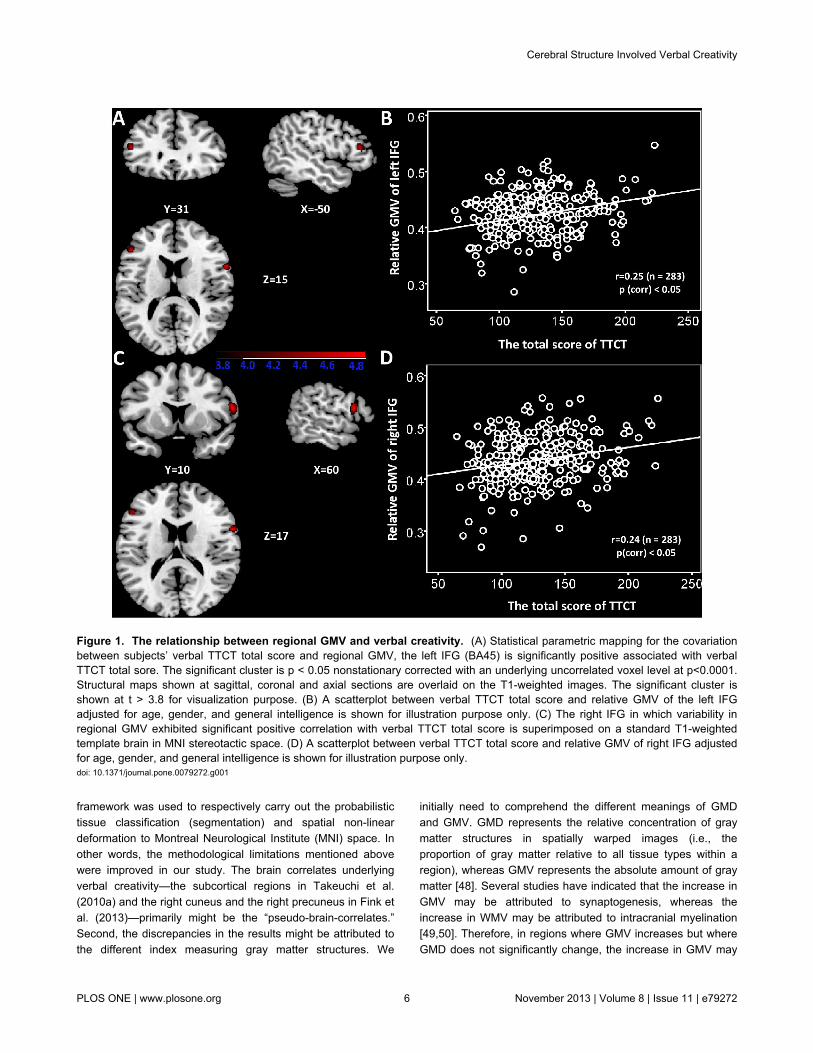
Correlation between GMVs and verbal creativityAfter control for age, gender, general intelligence, and total
gray matter volume in the multiple regression analysis, thecorrelation of GMVs with verbal creative thinking measured byverbal TTCT was determined (Table 2). As shown in Table 2,significantly positive correlation was found between the verbalTTCT score and the left IFG, pars triangularis (BA45: x = -50, y= 31, z = 15; see also Figure 1A), and the right IFG (x =60, y =10, z = 17; see also Figure 1C). All voxels survived at cluster-level non-stationary correction for multiple comparisons (p <0.05). The average local GMVs (left IFG/BA 45, right IFG) werethen obtained using the “Extract ROIs signal” utilitiesimplemented in REST [47]. To control for individual differencesin whole brain size, the average relative GMVs obtained bydividing local GMVs by total gray matter volume wascalculated. These average relative GMVs were then used asthe dependent variables for all bivariate correlations (Figures1B and 1D).
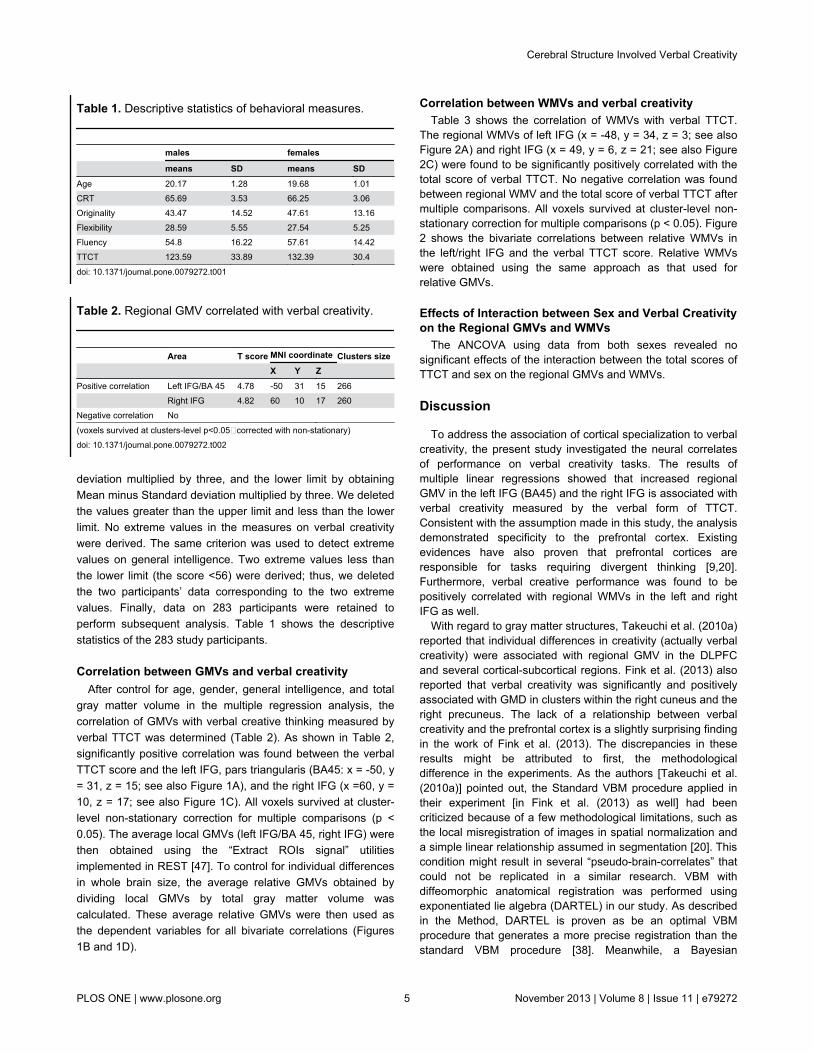
Table 1. Descriptive statistics of behavioral measures.
males females
means SD means SDAge 20.17 1.28 19.68 1.01CRT 65.69 3.53 66.25 3.06Originality 43.47 14.52 47.61 13.16Flexibility 28.59 5.55 27.54 5.25Fluency 54.8 16.22 57.61 14.42TTCT 123.59 33.89 132.39 30.4
doi: 10.1371/journal.pone.0079272.t001
Table 2. Regional GMV correlated with verbal creativity.
Area T score MNI coordinate Clusters size X Y Z Positive correlation Left IFG/BA 45 4.78 -50 31 15 266 Right IFG 4.82 60 10 17 260Negative correlation No
(voxels survived at clusters-level p<0.05,corrected with non-stationary)doi: 10.1371/journal.pone.0079272.t002
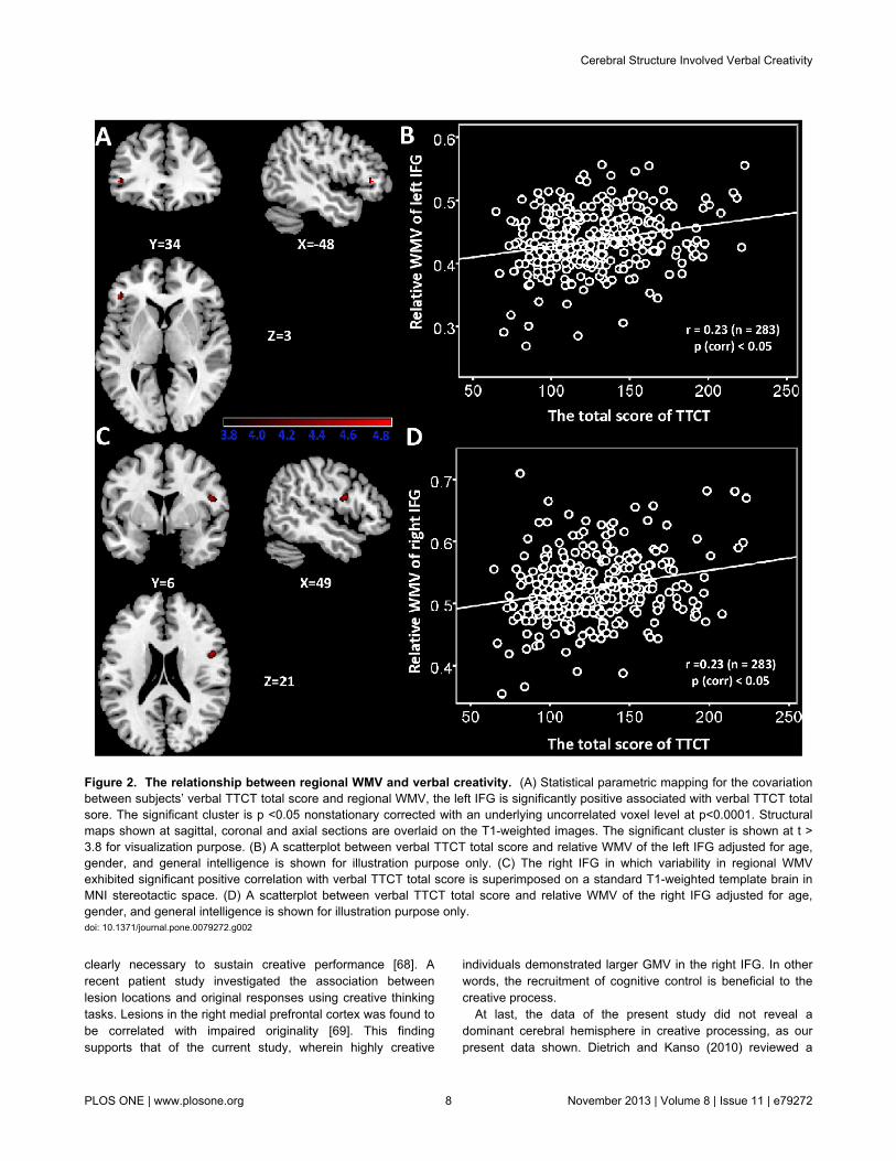
Correlation between WMVs and verbal creativityTable 3 shows the correlation of WMVs with verbal TTCT.
The regional WMVs of left IFG (x = -48, y = 34, z = 3; see alsoFigure 2A) and right IFG (x = 49, y = 6, z = 21; see also Figure2C) were found to be significantly positively correlated with thetotal score of verbal TTCT. No negative correlation was foundbetween regional WMV and the total score of verbal TTCT aftermultiple comparisons. All voxels survived at cluster-level non-stationary correction for multiple comparisons (p < 0.05). Figure2 shows the bivariate correlations between relative WMVs inthe left/right IFG and the verbal TTCT score. Relative WMVswere obtained using the same approach as that used forrelative GMVs.
Effects of Interaction between Sex and Verbal Creativityon the Regional GMVs and WMVs
The ANCOVA using data from both sexes revealed nosignificant effects of the interaction between the total scores ofTTCT and sex on the regional GMVs and WMVs.
Discussion
To address the association of cortical specialization to verbalcreativity, the present study investigated the neural correlatesof performance on verbal creativity tasks. The results ofmultiple linear regressions showed that increased regionalGMV in the left IFG (BA45) and the right IFG is associated withverbal creativity measured by the verbal form of TTCT.Consistent with the assumption made in this study, the analysisdemonstrated specificity to the prefrontal cortex. Existingevidences have also proven that prefrontal cortices areresponsible for tasks requiring divergent thinking [9,20].Furthermore, verbal creative performance was found to bepositively correlated with regional WMVs in the left and rightIFG as well.
With regard to gray matter structures, Takeuchi et al. (2010a)reported that individual differences in creativity (actually verbalcreativity) were associated with regional GMV in the DLPFCand several cortical-subcortical regions. Fink et al. (2013) alsoreported that verbal creativity was significantly and positivelyassociated with GMD in clusters within the right cuneus and theright precuneus. The lack of a relationship between verbalcreativity and the prefrontal cortex is a slightly surprising findingin the work of Fink et al. (2013). The discrepancies in theseresults might be attributed to first, the methodologicaldifference in the experiments. As the authors [Takeuchi et al.(2010a)] pointed out, the Standard VBM procedure applied intheir experiment [in Fink et al. (2013) as well] had beencriticized because of a few methodological limitations, such asthe local misregistration of images in spatial normalization anda simple linear relationship assumed in segmentation [20]. Thiscondition might result in several “pseudo-brain-correlates” thatcould not be replicated in a similar research. VBM withdiffeomorphic anatomical registration was performed usingexponentiated lie algebra (DARTEL) in our study. As describedin the Method, DARTEL is proven as be an optimal VBMprocedure that generates a more precise registration than thestandard VBM procedure [38]. Meanwhile, a Bayesian
Cerebral Structure Involved Verbal Creativity
PLOS ONE | www.plosone.org 5 November 2013 | Volume 8 | Issue 11 | e79272
framework was used to respectively carry out the probabilistictissue classification (segmentation) and spatial non-lineardeformation to Montreal Neurological Institute (MNI) space. Inother words, the methodological limitations mentioned abovewere improved in our study. The brain correlates underlyingverbal creativity—the subcortical regions in Takeuchi et al.(2010a) and the right cuneus and the right precuneus in Fink etal. (2013)—primarily might be the “pseudo-brain-correlates.”Second, the discrepancies in the results might be attributed tothe different index measuring gray matter structures. We
initially need to comprehend the different meanings of GMDand GMV. GMD represents the relative concentration of graymatter structures in spatially warped images (i.e., theproportion of gray matter relative to all tissue types within aregion), whereas GMV represents the absolute amount of graymatter [48]. Several studies have indicated that the increase inGMV may be attributed to synaptogenesis, whereas theincrease in WMV may be attributed to intracranial myelination[49,50]. Therefore, in regions where GMV increases but whereGMD does not significantly change, the increase in GMV may
Figure 1. The relationship between regional GMV and verbal creativity. (A) Statistical parametric mapping for the covariationbetween subjects’ verbal TTCT total score and regional GMV, the left IFG (BA45) is significantly positive associated with verbalTTCT total sore. The significant cluster is p < 0.05 nonstationary corrected with an underlying uncorrelated voxel level at p<0.0001.Structural maps shown at sagittal, coronal and axial sections are overlaid on the T1-weighted images. The significant cluster isshown at t > 3.8 for visualization purpose. (B) A scatterplot between verbal TTCT total score and relative GMV of the left IFGadjusted for age, gender, and general intelligence is shown for illustration purpose only. (C) The right IFG in which variability inregional GMV exhibited significant positive correlation with verbal TTCT total score is superimposed on a standard T1-weightedtemplate brain in MNI stereotactic space. (D) A scatterplot between verbal TTCT total score and relative GMV of right IFG adjustedfor age, gender, and general intelligence is shown for illustration purpose only.doi: 10.1371/journal.pone.0079272.g001
Cerebral Structure Involved Verbal Creativity
PLOS ONE | www.plosone.org 6 November 2013 | Volume 8 | Issue 11 | e79272

be attributed to an increase in the number of synapses perneuron; a similar increase in the WMV due to myelinationoccurs simultaneously in these regions [48]. Both GMVs andWMVs in the bilateral prefrontal regions apparently increasedwith verbal creativity in the present study, whereas GMD in theregions did not significantly change with verbal creativity in Finket al. (2013).
With regard to the white matter structures, theinconsistencies with previous studies were also observed. Theparadoxical results of white structure related to creativity mightbe attributed to first, the difference in the experimental tasks. InJung et al. (2010), both verbal and visuospatial divergent-thinking capacities were assessed, whereas in the presentstudy and in Takeuchi et al. (2010b), only the verbal divergent-thinking capacity was assessed. Second, the paradoxicalresults might be attributed to the methodological difference inthe experiments [FA derived from DTI in Jung et al. (2010) andTakeuchi et al. (2010b) vs. WMV derived from VBM in thepresent study]. A direct link between macroscopic neuralstructure (WMV) and microscopic neural structure (FA) in thehuman brain has yet to be established. What is known is thatwhite matter structures are largely composed of nerve fiberscalled axons and fatty myelin sheaths around the axons [51].As mentioned above, the increase in WMV may be attributed tointracranial myelination [46,47]. The myelination process allowsneural signals to propagate more swiftly and with less signalloss. This aspect enhances connectivity within specific brainregions (the bilateral IFG here) and improves broader neuralpathways connecting spatially separate regions required formany sensory, cognitive, and motor functions. The neuralpathways are extremely complicated and require furtherresearch. Overall, the greater GMVs, together with the greaterWMVs in the bilateral IFG, may reveal a greater neuralcomputational efficiency in the regions.
The bilateral IFG (including BA 45) was the only region foundto be associated with verbal creative thinking in the presentstudy. To our knowledge, BA45, together with BA44, comprisesBroca’s area, a region that is mainly involved in languageproduction and comprehension, such as semantic generationtasks [52] and verbal fluency [53]. In one study,neuropsychological evidence revealed that patients with leftprefrontal lesions exhibited impairment of the generation ofverbal fluency [54]. Moreover, the role of the left inferiorprefrontal cortex in ideas generation has been supported by aconsiderable amount of neuroimaging data [55,56]. In thepresent study, regional GMV in BA45 exhibited a positivecorrelation with verbal creativity. In other words, individuals
Table 3. Correlation between WMV and TTCT total score.
Area T score MNI coordinate Clusters size X Y Z Positive correlation Left IFG 4.50 -48 34 3 21 Right IFG 4.43 49 6 21 44Negative correlation No
(voxels survived at clusters-level p<0.05 corrected with non-stationary)doi: 10.1371/journal.pone.0079272.t003
with larger GMV in BA45 exhibited higher verbal creative thanthose with smaller GMV. Larger cortical volume is often linkedto the improved computational efficacy of such region [57].That is, the generation of verbal creative ideas may require acerebral structural basis that can support effective semantic orconcept generation. This view is consistent with the idea thatcreative processing requires cognitive control over conceptualknowledge networks as that are facilitated by the leftventrolateral prefrontal cortices [58]. In addition, the triangularispart of the left IFG has also been shown to play a role in thecognitive control of memory [59,60]. According to the “two-part”model of memory retrieval [60], we try to retrieve information ina top-down manner when we confront problems or tasks thatmust be solved. The retrieved knowledge is initially stored inthe lateral temporal cortex, such as in the middle temporalgyrus. Recalling the top-down retrieval depends on consciouscognitive control. One can assume that a technique may beemployed to exclude irrelevant data from the retrievalknowledge. To hone in on the desired information, someselection must occur. This selection is thought to occur post-retrieval in the left mid-ventral lateral prefrontal cortex (VLPFC),which corresponds to the location of the left IFG, specificallythe pars triangularis [60]. Apparently, the process of excludingirrelevant information in top-down memory retrieval plays acrucial role in creative problems solving. The creative processentails activating remote conceptual networks selectively andinhibiting related semantic information [61]. For instance, aprevious fMRI study suggested that the left IFG is recruited inthe selection of more semantically distant demand [62]. Thefinding is in line with the idea that the generation of creativeideas involves remote conceptual association [23]. In anotherfMRI study (Fink et al., 2009), Alternative Use Task and nameinvention task asking for generating as original names aspossible were applied. The two tasks were found to elicitconsistently strong activation in the left IFG [9]. The authorssuggested that the left IFG is responsible for the emergence ofa new semantic representation in divergent thinking. Based onthe findings of previous studies, the left IFG is believed to playa major role in the generation of semantic relatedness and inthe selection of semantic relatedness. Generally, researchersbelieve in the existence of structural bases that underliesspecific cognitive function. Hence, highly creative individual inthe present study had larger GMV in the left IFG (i.e., parstriangularis).
The right IFG is commonly known to play a role in responseinhibition [63-65]. Specifically, the right IFG is involved ininhibitory control over cognitive mental processing rather thanin motor response control [65]. In addtion, strong evidencesuggests that the right IFG also plays a role in attentionswitching [66,67]. As noted by Hampshire et al. (2010), theright IFG facilitated attention switching by inhibiting thepreviously attended object, location, or dimension. In this case,the attention shifts. To overcome the pre-potent response, agreater degree of inhibition is therefore necessary whenswitching attention away from a previous routine response, forexample, during the generation of creative ideas. Flexiblecognitive control, which is in the form of resistance fromdistraction (inhibitory control) and anttentional switch may be
Cerebral Structure Involved Verbal Creativity
PLOS ONE | www.plosone.org 7 November 2013 | Volume 8 | Issue 11 | e79272
clearly necessary to sustain creative performance [68]. Arecent patient study investigated the association betweenlesion locations and original responses using creative thinkingtasks. Lesions in the right medial prefrontal cortex was found tobe correlated with impaired originality [69]. This findingsupports that of the current study, wherein highly creative
individuals demonstrated larger GMV in the right IFG. In otherwords, the recruitment of cognitive control is beneficial to thecreative process.
At last, the data of the present study did not reveal adominant cerebral hemisphere in creative processing, as ourpresent data shown. Dietrich and Kanso (2010) reviewed a
Figure 2. The relationship between regional WMV and verbal creativity. (A) Statistical parametric mapping for the covariationbetween subjects’ verbal TTCT total score and regional WMV, the left IFG is significantly positive associated with verbal TTCT totalsore. The significant cluster is p <0.05 nonstationary corrected with an underlying uncorrelated voxel level at p<0.0001. Structuralmaps shown at sagittal, coronal and axial sections are overlaid on the T1-weighted images. The significant cluster is shown at t >3.8 for visualization purpose. (B) A scatterplot between verbal TTCT total score and relative WMV of the left IFG adjusted for age,gender, and general intelligence is shown for illustration purpose only. (C) The right IFG in which variability in regional WMVexhibited significant positive correlation with verbal TTCT total score is superimposed on a standard T1-weighted template brain inMNI stereotactic space. (D) A scatterplot between verbal TTCT total score and relative WMV of the right IFG adjusted for age,gender, and general intelligence is shown for illustration purpose only.doi: 10.1371/journal.pone.0079272.g002
Cerebral Structure Involved Verbal Creativity
PLOS ONE | www.plosone.org 8 November 2013 | Volume 8 | Issue 11 | e79272

total of 63 literatures and explored the neural underpinnings ofcreative behavior using either electroencephalographictechniques or neuroimaging techniques. Notably, these studiesdid not associated creativity with the right hemisphere or withany part of the right hemisphere, because creative processingdoes not rely on only a single, simple mental process or brainregion. The present study supported this conclusion from abrain structural perspective. Regardless of the indicators ofbrain structure, i.e. GMVs or WMVs, both the left and rightbrain regions exhibited an obvious association with creativity.
Author Contributions
Conceived and designed the experiments: FZ JQ QZ.Performed the experiments: FZ. Analyzed the data: FZ.Contributed reagents/materials/analysis tools: FZ JQ. Wrotethe manuscript: FZ.
References
1. Sternberg RJ, Lubart TI (1996) Investing in creativity. Am Psychol 51:677. doi:10.1037/0003-066X.51.7.677.
2. Fink A, Grabner RH, Gebauer D, Reishofer G, Koschutnig K et al.(2010) Enhancing creativity by means of cognitive stimulation:Evidence from an fMRI study. NeuroImage 52: 1687-1695. doi:10.1016/j.neuroimage.2010.05.072. PubMed: 20561898.
3. Dietrich A, Kanso R (2010) A review of EEG, ERP, and neuroimagingstudies of creativity and insight. Psychol Bull 136: 828-848. PubMed:20804237.
4. Guilford JP (1950) Creativity research: Past, present and future. AmPsychol 5: 444-454. doi:10.1037/h0063487. PubMed: 14771441.
5. Guilford JP (1967) The nature of human intelligence. New York, NY,US: McGraw-Hill.
6. Torrance EP (1974) Torrance Test of Creative Thinking. Lexington, MA:Personal Press.
7. Fink A, Neubauer AC (2006) EEG alpha oscillations during theperformance of verbal creativity tasks: differential effects of sex andverbal intelligence. Int J Psychophysiol 62: 46-53. doi:10.1016/j.ijpsycho.2006.01.001. PubMed: 16503062.
8. Grabner RH, Fink A, Neubauer AC (2007) Brain correlates of self-ratedoriginality of ideas: evidence from event-related power and phase-locking changes in the EEG. Behav Neurosci 121: 224-230. doi:10.1037/0735-7044.121.1.224. PubMed: 17324067.
9. Fink A, Grabner RH, Benedek M (2009) The creative brain:Investigation of brain activity during creative problem solving by meansof EEG and fMRI. Hum Brain Mapp 30: 734-748. doi:10.1002/hbm.20538. PubMed: 18266217.
10. Razumnikova OM, Volf NV, Tarasova IV (2009) Strategy and results:Sex differences in electrographic correlates of verbal and figuralcreativity. Hum Physiol 35: 285-294. doi:10.1134/S0362119709030049.
11. Jung RE, Gasparovic C, Chavez RS, Flores RA, Smith SM et al. (2009)Biochemical support for the "threshold" theory of creativity: a magneticresonance spectroscopy study. J Neurosci 29: 5319-5325. doi:10.1523/JNEUROSCI.0588-09.2009. PubMed: 19386928.
12. Fink A, Koschutnig K, Benedek M, Reishofer G, Ischebeck A et al.(2011) Stimulating creativity via the exposure to other people's ideas.Hum Brain Mapp: n/a-n/a.
13. Fink A, Schwab D, Papousek I (2011) Sensitivity of EEG upper alphaactivity to cognitive and affective creativity interventions. Int JPsychophysiol 82: 233-239. doi:10.1016/j.ijpsycho.2011.09.003.PubMed: 21930162.
14. Jung RE, Segall JM, Jeremy Bockholt H, Flores RA, Smith SM et al.(2010) Neuroanatomy of creativity. Hum Brain Mapp 31: 398-409.PubMed: 19722171.
15. Fink A, Grabner RH, Benedek M, Neubauer AC (2006) Divergentthinking training is related to frontal electroencephalogram alphasynchronization. Eur J Neurosci 23: 2241-2246. doi:10.1111/j.1460-9568.2006.04751.x. PubMed: 16630071.
16. Fink A, Koschutnig K, Hutterer L, Steiner E, Benedek M et al. (2013)Gray matter density in relation to different facets of verbal creativity.Brain Struct Funct, 218: 1-7. PubMed: 22286950.
17. Fink A, Benedek M (2012) EEG alpha power and creative ideation.Neurosci Biobehav Rev. PubMed: 23246442
18. Jung RE, Grazioplene R, Caprihan A, Chavez RS, Haier RJ (2010)White matter integrity, creativity, and psychopathology: disentanglingconstructs with diffusion tensor imaging. PLOS ONE 5: e9818. doi:10.1371/journal.pone.0009818. PubMed: 20339554.
19. Takeuchi H, Taki Y, Sassa Y, Hashizume H, Sekiguchi A et al. (2010)White matter structures associated with creativity: evidence fromdiffusion tensor imaging. NeuroImage 51: 11-18. doi:10.1016/j.neuroimage.2010.02.035. PubMed: 20171286.
20. Takeuchi H, Taki Y, Sassa Y, Hashizume H, Sekiguchi A et al. (2010)Regional gray matter volume of dopaminergic system associate withcreativity: evidence from voxel-based morphometry. NeuroImage 51:578-585. doi:10.1016/j.neuroimage.2010.02.078. PubMed: 20226253.
21. Gansler DA, Moore DW, Susmaras TM, Jerram MW, Sousa J et al.(2011) Cortical morphology of visual creativity. Neuropsychologia 49:2527-2532. doi:10.1016/j.neuropsychologia.2011.05.001. PubMed:21600905.
22. Moore DW, Bhadelia RA, Billings RL, Fulwiler C, Heilman KM et al.(2009) Hemispheric connectivity and the visual-spatial divergent-thinking component of creativity. Brain Cogn 70: 267-272. doi:10.1016/j.bandc.2009.02.011. PubMed: 19356836.
23. Zhang H, Liu J, Zhang Q (2013) Neural representations for thegeneration of inventive conceptions inspired by adaptive featureoptimization of biological species. Cortex. PubMed: 23582377
24. Skipper JI, Goldin-Meadow S, Nusbaum HC, Small SL (2007) Speech-associated gestures, Broca’s area, and the human mirror system. BrainLanguage 101: 260-277. doi:10.1016/j.bandl.2007.02.008. PubMed:17533001.
25. Caplan D (2006) Why Is Broca'S Area Involved in Syntax? Cortex 42:469-471. doi:10.1016/S0010-9452(08)70379-4. PubMed: 16881251.
26. Grewe T, Bornkessel I, Zysset S, Wiese R, von Cramon DY et al.(2005) The emergence of the unmarked: A new perspective on thelanguage-specific function of Broca's area. Hum Brain Mapp 26:178-190. doi:10.1002/hbm.20154. PubMed: 15929098.
27. Huang P, Qiu L, Shen L, Zhang Y, Song Z et al. (2012) Evidence for aleft-over-right inhibitory mechanism during figural creative thinking inhealthy nonartists. Hum Brain Mapp.
28. Torrance EP (1987) Teaching for creativity. Frontiers of creativityresearch: Beyond the basics: 189-215.
29. Carson DK, Bittner MT, Cameron BR, Brown DM, Meyer SS (1994)Creative thinking as a predictor of school-aged children's stressresponses and coping abilities. Creativity Res J 7: 145-158. doi:10.1080/10400419409534520.
30. Kim KH, Cramond B, Bandalos DL (2006) The latent structure andmeasurement invariance of scores on the Torrance Tests of CreativeThinking–Figural. Educ Psychol Meas 66: 459-477. doi:10.1177/0013164405282456.
31. de Souza LC, Volle E, Bertoux M, Czernecki V, Funkiewiez A et al.(2010) Poor creativity in frontotemporal dementia: a window into theneural bases of the creative mind. Neuropsychologia 48: 3733-3742.doi:10.1016/j.neuropsychologia.2010.09.010. PubMed: 20868703.
32. DeHaan RL (2009) Teaching creativity and inventive problem solving inscience. CBE-Life Sciences Educ 8: 172-181. doi:10.1187/cbe.08-12-0081. PubMed: 19723812.
33. Runco MA, Millar G, Acar S, Cramond B (2010) Torrance tests ofcreative thinking as predictors of personal and public achievement: Afifty-year follow-up. Creativity Res J 22: 361-368. doi:10.1080/10400419.2010.523393.
34. Silvia PJ (2011) Subjective scoring of divergent thinking: Examining thereliability of unusual uses, instances, and consequences tasks.Thinking Skills Creativity 6: 24-30. doi:10.1016/j.tsc.2010.06.001.
35. Heausler NL, Thompson B (1988) Structure of the Torrance Tests ofcreative thinking. Educ Psychol Meas 48: 463-468. doi:10.1177/0013164488482021.
36. Dan Li, Peng CG (1989) Combined Reven's teat (CRT) - Chineserevised version (in Chinese). Shanghai: East China Normal University.
37. Ming WDQ (1989) Revision on the Combined Raven's Test for theRural in China [J]. Psychol Sci 5: 004.
38. Takeuchi H, Taki Y, Hashizume H, Sassa Y, Nagase T et al. (2011)Cerebral blood flow during rest associates with general intelligence and
Cerebral Structure Involved Verbal Creativity
PLOS ONE | www.plosone.org 9 November 2013 | Volume 8 | Issue 11 | e79272
creativity. PLOS ONE 6: e25532. doi:10.1371/journal.pone.0025532.PubMed: 21980485.
39. Takeuchi H, Taki Y, Sassa Y, Hashizume H, Sekiguchi A et al. (2011)Regional gray matter density associated with emotional intelligence:Evidence from voxel-based morphometry. Hum Brain Mapp 32:1497-1510. doi:10.1002/hbm.21122. PubMed: 20740644.
40. Ashburner J (2007) A fast diffeomorphic image registration algorithm.NeuroImage 38: 95-113. doi:10.1016/j.neuroimage.2007.07.007.PubMed: 17761438.
41. Klein A, Andersson J, Ardekani BA, Ashburner J, Avants B et al. (2009)Evaluation of 14 nonlinear deformation algorithms applied to humanbrain MRI registration. Neuroimage 46: 786–802. doi:10.1016/j.neuroimage.2008.12.037. PubMed: 19195496.
42. Ashburner J, Friston KJ (2005) Unified segmentation. NeuroImage 26:839-851. doi:10.1016/j.neuroimage.2005.02.018. PubMed: 15955494.
43. Good CD, Johnsrude IS, Ashburner J, Henson RN, Fristen K et al.(2002) A voxel-based morphometric study of ageing in 465 normaladult human brains. IEEE Pp: 16.
44. Ridgway GR, Omar R, Ourselin S, Hill DL, Warren JD et al. (2009)Issues with threshold masking in voxel-based morphometry ofatrophied brains. NeuroImage 44: 99-111. doi:10.1016/j.neuroimage.2008.08.045. PubMed: 18848632.
45. Hayasaka S, Phan KL, Liberzon I, Worsley KJ, Nichols TE (2004)Nonstationary cluster-size inference with random field and permutationmethods. NeuroImage 22: 676-687. doi:10.1016/j.neuroimage.2004.01.041. PubMed: 15193596.
46. Takeuchi H, Taki Y, Hashizume H, Sassa Y, Nagase T et al. (2012)The Association between Resting Functional Connectivity andCreativity. Cereb Cortex, 22: 2921–9. PubMed: 22235031.
47. Song X-W, Dong Z-Y, Long X-Y, Li S-F, Zuo X-N et al. (2011) REST: atoolkit for resting-state functional magnetic resonance imaging dataprocessing. PLOS ONE 6: e25031. doi:10.1371/journal.pone.0025031.PubMed: 21949842.
48. Taki Y, Hashizume H, Thyreau B, Sassa Y, Takeuchi H et al. (2012)Linear and curvilinear correlations of brain gray matter volume anddensity with age using voxel-based morphometry with the Akaikeinformation criterion in 291 healthy children. Hum Brain Mapp: n/a-n/a.
49. Paus T (2005) Mapping brain maturation and cognitive developmentduring adolescence. Trends Cogn Sci 9: 60-68. doi:10.1016/j.tics.2004.12.008. PubMed: 15668098.
50. Sowell ER, Thompson PM, Tessner KD, Toga AW (2001) Mappingcontinued brain growth and gray matter density reduction in dorsalfrontal cortex: inverse relationships during postadolescent brainmaturation. J Neurosci 21: 8819-8829. PubMed: 11698594.
51. Kanai R, Dong MY, Bahrami B, Rees G (2011) Distractibility in daily lifeis reflected in the structure and function of human parietal cortex. JNeurosci 31: 6620-6626. doi:10.1523/JNEUROSCI.5864-10.2011.PubMed: 21543590.
52. Vidorreta JG, Garcia R, Moritz-Gasser S, Duffau H (2010) Doubledissociation between syntactic gender and picture naming processing:a brain stimulation mapping study. Hum Brain Mapp 32: 331-340.PubMed: 21319264.
53. Costafreda SG, Fu CH, Lee L, Everitt B, Brammer MJ et al. (2006) Asystematic review and quantitative appraisal of fMRI studies of verbalfluency: role of the left inferior frontal gyrus. Hum Brain Mapp 27:799-810. doi:10.1002/hbm.20221. PubMed: 16511886.
54. Stuss DT, Alexander MP, Hamer L, Palumbo C, Dempster R et al.(1998) The effects of focal anterior and posterior brain lesions on verbalfluency. J-Int Neuropsychol Soc 4: 265-278. PubMed: 9623001.
55. Bechtereva NP, Korotkov AD, Pakhomov SV, Roudas MS, StarchenkoMG et al. (2004) PET study of brain maintenance of verbal creativeactivity. Int J Psychophysiol 53: 11-20. doi:10.1016/j.ijpsycho.2004.01.001. PubMed: 15172131.
56. Gazzaniga MS (2000) Cerebral specialization and interhemisphericcommunication Does the corpus callosum enable the human condition?Brain 123: 1293-1326. doi:10.1093/brain/123.7.1293. PubMed:10869045.
57. Kanai R, Rees G (2011) The structural basis of inter-individualdifferences in human behaviour and cognition. Nat Rev Neurosci 12:231-242. doi:10.1038/nrn3000. PubMed: 21407245.
58. Chrysikou EG, Thompson-Schill SL (2011) Dissociable brain stateslinked to common and creative object use. Hum Brain Mapp 32:665-675. doi:10.1002/hbm.21056. PubMed: 20533561.
59. Badre D, Wagner AD (2007) Left ventrolateral prefrontal cortex and thecognitive control of memory. Neuropsychologia 45: 2883-2901. doi:10.1016/j.neuropsychologia.2007.06.015. PubMed: 17675110.
60. Badre D, Poldrack RA, Paré-Blagoev EJ, Insler RZ, Wagner AD (2005)Dissociable controlled retrieval and generalized selection mechanismsin ventrolateral prefrontal cortex. Neuron 47: 907–918. doi:10.1016/j.neuron.2005.07.023. PubMed: 16157284.
61. Heilman KM, Nadeau SE, Beversdorf DO (2003) Creative innovation:possible brain mechanisms. Neurocase 9: 369-379. doi:10.1076/neur.9.5.369.16553. PubMed: 14972752.
62. Green AE, Kraemer DJ, Fugelsang JA, Gray JR, Dunbar KN (2010)Connecting long distance: semantic distance in analogical reasoningmodulates frontopolar cortex activity. Cereb Cortex 20: 70-76. doi:10.1093/cercor/bhp081. PubMed: 19383937.
63. Aron AR, Robbins TW, Poldrack RA (2004) Inhibition and the rightinferior frontal cortex. Trends Cogn Sci 8: 170-177. doi:10.1016/j.tics.2004.02.010. PubMed: 15050513.
64. Chikazoe J, Konishi S, Asari T, Jimura K, Miyashita Y (2007) Activationof right inferior frontal gyrus during response inhibition across responsemodalities. J Cogn Neurosci 19: 69-80. doi:10.1162/jocn.2007.19.1.69.PubMed: 17214564.
65. Hampshire A, Chamberlain SR, Monti MM, Duncan J, Owen AM (2010)The role of the right inferior frontal gyrus: inhibition and attentionalcontrol. NeuroImage 50: 1313-1319. doi:10.1016/j.neuroimage.2009.12.109. PubMed: 20056157.
66. Hampshire A, Duncan J, Owen AM (2007) Selective tuning of the bloodoxygenation level-dependent response during simple target detectiondissociates human frontoparietal subregions. J Neurosci 27:6219-6223. doi:10.1523/JNEUROSCI.0851-07.2007. PubMed:17553994.
67. Cools R, Clark L, Owen AM, Robbins TW (2002) Defining the neuralmechanisms of probabilistic reversal learning using event-relatedfunctional magnetic resonance imaging. J Neurosci 22: 4563-4567.PubMed: 12040063.
68. Zabelina DL, Robinson MD (2010) Creativity as flexible cognitivecontrol. Psych Aesthet Creativity and Arts 4: 136-143. doi:10.1037/a0017379.
69. Shamay-Tsoory SG, Adler N, Aharon-Peretz J, Perry D, Mayseless N(2011) The origins of originality: The neural bases of creative thinkingand originality. Neuropsychologia 49: 178-185. doi:10.1016/j.neuropsychologia.2010.11.020. PubMed: 21126528.
Cerebral Structure Involved Verbal Creativity
PLOS ONE | www.plosone.org 10 November 2013 | Volume 8 | Issue 11 | e79272