mosaic expression of pluripotency-related proteins oct-3/4 ... · advanced studies in biology, vol....
TRANSCRIPT
Advanced Studies in Biology, Vol. 5, 2013, no. 4, 157 - 172 HIKARI Ltd, www.m-hikari.com
Mosaic Expression of Pluripotency-Related
Proteins Oct-3/4 and Alkaline Phosphatase in
Human Pancreatic Carcinoma Cell PANC-1
Masahiro Sato*
Section of Gene Expression Regulation, Frontier Science Research Center, Kagoshima University, Kagoshima 890-8544, Japan
Shin-ichi Maeda
Department of Surgery II, Graduate School of Medical and Dental Sciences,
Kagoshima University, Kagoshima 890-8544, Japan
Emi Inada
Department of Pediatric Dentistry, Graduate School of Medical and Dental Sciences, Kagoshima University, Kagoshima 890-8544, Japan
Issei Saitoh
Division of Pediatric Dentistry,Graduate School of Medical and Dental Science,
Niigata University, Niigata 951-8514, Japan
Naoko Kubota
Department of Pediatric Dentistry, Graduate School of Medical and Dental Sciences, Kagoshima University, Kagoshima 890-8544, Japan
*Corresponding author. e-mail: [email protected]
Abstract
Most current research on cancer stem cells (CSCs) associated with human tumors has focused on the molecular and cellular analysis of hematopoietic lineage markers (e.g., CD44, CD138, and CD 133), which can also serve as important

158 Masahiro Sato et al CSC markers in a variety of cancers. However, these markers are generally expressed at late stages in embryonic development. Oct-3/4, a member of the family of POU-domain transcriptional factors, and alkaline phosphatase (ALP) are known to be expressed in the inner cell mass of blastocysts, germ cells, and pluripotent embryonic stem cells. We thus consider Oct-3/4 and ALP to be promising markers for CSC. Herein, we examined expression of Oct-3/4 and ALP using 6 established human pancreatic carcinoma cell lines. RT-PCR analysis revealed the presence of Oct-3/4 and ALP mRNA in those cells. Immunocytochemical and cytochemical staining revealed that both Oct-3/4 and ALP proteins are present as mosaics in PANC-1 cell line, one of those 6 cell lines (23% and 19%, respectively). However, Oct-3/4-positive PANC-1 cells did not exhibit overt ATP-binding cassette transporter G2 (ABCG2) activity, as revealed by Hoechst 33342 dye exclusion assay. Transfection of PANC-1 cells with an Oct-3/4 promoter-directed, enhanced green fluorescent protein (EGFP) construct confirmed the presence of Oct-3/4-positive cells. These findings indicate that in PANC-1 cells there are at least 2 subset populations, namely Oct-3/4-positive and ALP-positive cells. However, it remains unknown whether expression of these 2 markers overlaps. Enrichment of Oct-3/4- or ALP-positive cells by gene transfer and subsequent drug selection will be helpful for further characterization of these cells as possible CSCs. Keywords: ABCG2, Alkaline Phosphatase, Cancer stem cell, Oct-3/4, PANC-1, Pancreatic Carcinoma 1. Introduction
Recent data have demonstrated that tumors contain a small subpopulation of cells, namely, cancer stem cells (CSCs), which exhibit a self-renewing capacity and are responsible for tumor maintenance and metastasis.1-5 Although many exceptions have been reported, cell-surface molecules such as CD133 (prominin-1), CD44, and CD138 (syndecan-1) are generally considered markers for CSCs.6 However, these molecules are expressed in the later stages of embryonic cells, such as blastocysts (a stage just prior to implantation), post-implanted early fetuses, and hematopoietic progenitor populations. For example, CD133/prominin-1 is expressed in trophoblasts, but not in cells of the inner cell mass (ICM) at the blastocyst stage.7 The CD44 molecules are present in abundance in the liver of post-implantation fetuses at E (embryonic day) 10.5. Furthermore, as early as E 9.5, CD44-positive cells can be detected in circulating blood.8 In the developing mouse, CD138/syndecan-1 is first detected throughout the embryo at the 4-cell stage.9
Oct-3/4, a member of the family of POU-domain transcriptional factors, is expressed in pluripotent embryonic stem (ES) and germ cells, including primordial germ cells (PGC) and spermatogonia.10 Its expression commences at the earlier stages (including oocytes, early-cleavage stage embryos, and ICM

Mosaic expression of pluripotency-related proteins Oct-3/4 159
cells) of preimplantation development and is abundant in the embryonic ectoderm of post-implanted egg cylinder-stage embryos in mice.10-12 Inactivation of the Oct-3/4 gene by means of gene-targeting technique causes early lethality due to the lack of ICM formation, indicating that Oct-3/4 plays a critical role in early embryonic development.13 Tai et al.14 further demonstrated that Oct-3/4 is abundantly expressed in somatic stem cells, such as neuronal progenitors and adipogenic progenitors. Oct-3/4 has also been shown to activate transcription via octamer motifs and regulates several of its down-stream genes. These include Fgf4, Utf1, Spp1/Opn, Fbxo15/Fbx15, Sox2, Pdgfa/PDGFa, Cga/a, bhCG, Ifngr1/tINF, Zfp42/Rex1, Otx2, Lefty1, Upp1/Upp, Dppa5/Esg1, and Nanog.15
These findings suggest that Oct-3/4 functions as a master switch during differentiation by regulating the pluripotent fates of stem cells, thus playing a pivotal role in mammalian development.16 Furthermore, Oct-3/4 has been found to possess oncogenic potential17,18 and anti-apoptosis effects.19 It has also been found to play a role in the regulation of tumor progression.20 Oct-3/4 is overexpressed in a variety of cancers, including retinoblastoma,21 bladder cancer,17.22 and non-small cell lung cancer.23 As mentioned previously, pre-existing CSC markers, such as the CD series, appear to be lineage-restricted markers, most likely regulated by genes functioning downstream of the Oct-3/4 gene. Thus, the Oct-3/4 gene likely sits at the top of a gene-cascade hierarchy.24 Given this background, we hypothesized that Oct-3/4 could serve as a CSC marker, if tumor cells express it.
Alkaline phosphatase (ALP) is an enzyme that hydrolyzes a broad range of monophosphate esters at alkaline pH optima. In humans, there are at least 4 distinct forms of APL: placental, intestinal, germ cell, and liver/bone/kidney (L/B/K) ALP.25 The latter, L/B/K ALP, is expressed in numerous adult tissues, and therefore, is also called tissue non-specific ALP (TNAP). Aside from its expression in adult tissues, ALP is abundantly expressed in undifferentiated cells, such as ES cells, preimplantation embryos [2-cell embryos to blastocysts (ICM)] and embryonic ectoderm at the egg-cylinder stage, PGCs, and immature spermatogenic cells.26-28 It is also a marker of neuronal progenitor cells29 and of mesenchymal stem cells (MSCs) in bone marrow.30 Given that the cell/tissue localization pattern of ALP resembles that of Oct-3/4 and ALP is recognized as a marker for some progenitor cells, it is likely that ALP can serve as a CSC marker, if tumor cells express ALP.
To investigate the possibility that both Oct-3/4 and ALP could serve as CSC markers, we focused our study on proteins’ expression in PANC-1 cells, one of the human pancreatic carcinoma cell lines, using immunocytochemical, cytochemical, and molecular biological approaches. 2. Materials and Methods 2.1. Cell lines and culture
Two mouse cell lines, including nullipotent embryonal carcinoma (EC) cell line F931 and fibroblastic cell line NIH3T3 (CRL-1658™; purchased from

160 Masahiro Sato et al American Type Culture Collection (ATCC), Manassas, VA, USA), were used as controls. Normal human dermal fibroblast line HDFa (#106-05a) from Cell Applications, Inc. (San Diego, CA, USA) was also used as a control. Human pancreatic cell lines PANC-1 (CRL-1469™), MIA PaCa-2 (CRL-1420™), SW1990 (CRL-2172™), AsPC-1 (CRL-1682™), and Capan-1 (HTB-79™) were purchased from ATCC. PK-45H (RCB1973) was obtained from RIKEN BioResource Center Cell Bank (Tsukuba, Ibaraki, Japan). All these cells were grown in high glucose-DMEM (#11995; Invitrogen Co., Carlsbad, CA, USA) with 10% fetal bovine serum (FBS) and 100 U/mL penicillin/streptomycin at 37°C in an atmosphere of 5% CO2 in air.
2.2. RT-PCR
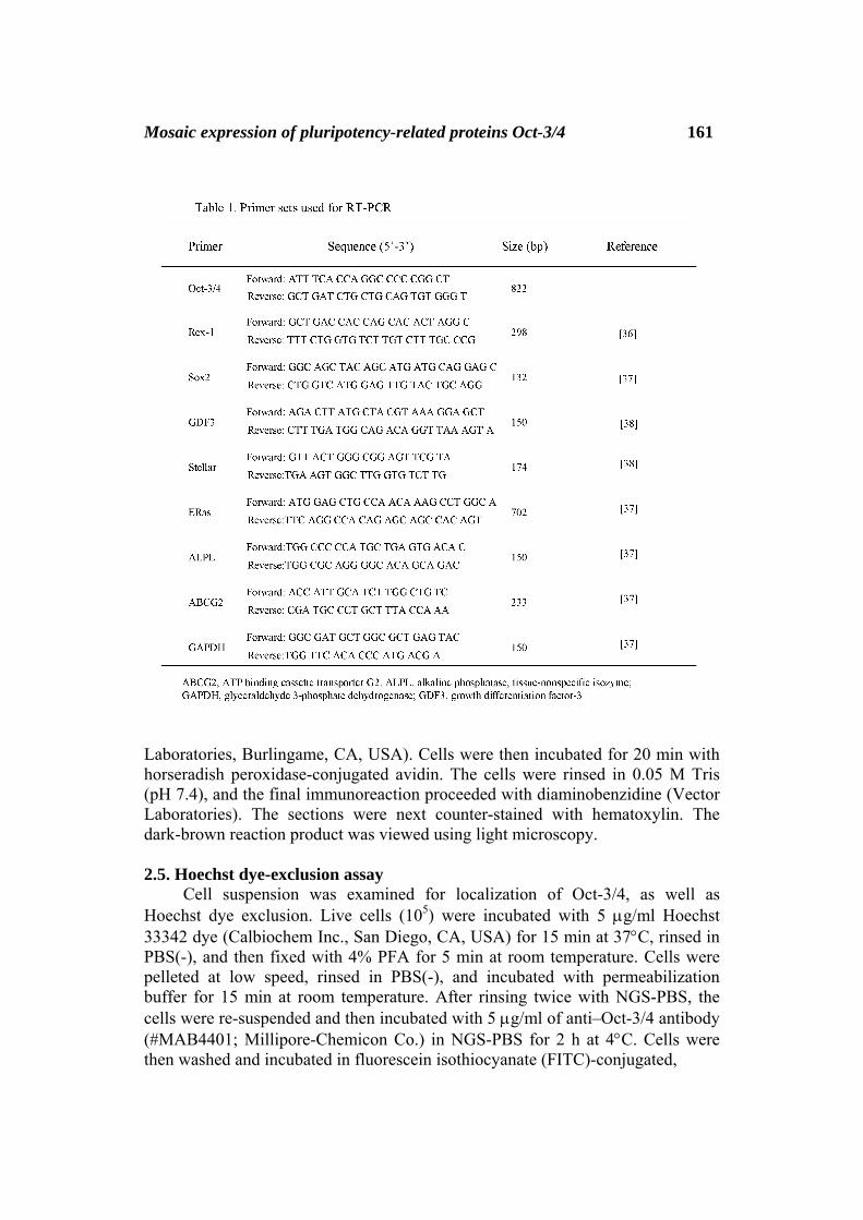
Total RNA was isolated using ISOGEN (Wako Pure Chemicals Inc., Tokyo, Japan). Four micrograms of total RNA from the cell lines was reverse transcribed into cDNA using SuperScript® II Reverse Transcriptase (Invitrogen Co.) and Oligo(dT)20 primer (#FSK-201; Toyobo Co. Ltd., Osaka, Japan) in a total of volume of 20 μL. The cDNAs were amplified from undiluted cDNA samples (1 μL) by 32 cycles of 45 s denaturation at 94°C, 45 s annealing at 58°C, and 60 s extension at 72°C, in a PC708 thermal cycler (Astec, Fukuoka, Japan). A negative, no-template control (designated as -RT) was included for each reaction. Information concerning the PCR primers used is listed in Table 1. The resulting RT-PCR products were subjected to electrophoresis in 2% agarose gels and visualized after staining with ethidium bromide.
2.3. Cytochemistry
The ALP assay was performed using the Leukocyte Alkaline Phosphatase kit (#85L3R; Sigma-Aldrich Co., St. Louis, MO) following the manufacturers’ instructions.
2.4. Immunocytochemistry
Cells (2 × 10⁵) were seeded onto a well of the Lab-Tek® Chamber Slide™ System (#177399; Nalge Nunc International, Naperville, IL, USA) with 300 µL of medium 1 day before immunocytochemical staining. After fixation with 4% paraformaldehyde (PFA) in phosphate-buffered saline without Ca2+ or Mg2+ at pH 7.4 [PBS(-)] at room temperature for 5 min, cells were permeabilized with permeabilization buffer [PBS(-) containing 10% normal goat serum (NGS) and 0.1% Triton X-100] for 15 min at room temperature. After rinsing with 2% NGS in PBS(-) (hereafter referred to as NGS-PBS), the cells were incubated with anti-Oct-3/4, monoclonal antibody (clone 10H11.2; #MAB4401, Millipore-Chemicon Co., Temecula, CA, USA) and diluted to 5 μg/mL for overnight treatment at 4°C. After rinsing 3 times with NGS-PBS, the cells were incubated with 1 μg/mL of biotynylated goat anti-mouse IgG (Zymed/Invitrogen Co.) for 1 h using a biotin-avidin immunostaining kit (Elite kit; Vector
Mosaic expression of pluripotency-related proteins Oct-3/4 161
Laboratories, Burlingame, CA, USA). Cells were then incubated for 20 min with horseradish peroxidase-conjugated avidin. The cells were rinsed in 0.05 M Tris (pH 7.4), and the final immunoreaction proceeded with diaminobenzidine (Vector Laboratories). The sections were next counter-stained with hematoxylin. The dark-brown reaction product was viewed using light microscopy. 2.5. Hoechst dye-exclusion assay
Cell suspension was examined for localization of Oct-3/4, as well as Hoechst dye exclusion. Live cells (105) were incubated with 5 μg/ml Hoechst 33342 dye (Calbiochem Inc., San Diego, CA, USA) for 15 min at 37°C, rinsed in PBS(-), and then fixed with 4% PFA for 5 min at room temperature. Cells were pelleted at low speed, rinsed in PBS(-), and incubated with permeabilization buffer for 15 min at room temperature. After rinsing twice with NGS-PBS, the cells were re-suspended and then incubated with 5 μg/ml of anti–Oct-3/4 antibody (#MAB4401; Millipore-Chemicon Co.) in NGS-PBS for 2 h at 4°C. Cells were then washed and incubated in fluorescein isothiocyanate (FITC)-conjugated,
162 Masahiro Sato et al anti-mouse IgG antibodies (1 μg/ml; #AP503F, Millipore-Chemicon Co.) for 1 h at 37°C. Next, the cells were pipetted onto a slide and cover-slipped for microscopic viewing. Fluorescent cells were visualized using an Olympus BX10 microscope with epifluorescent filters for Hoechst and FITC. Microphotographs were obtained using a digital camera (FUJIX HC-300/OL; Fuji Film, Tokyo, Japan) and were printed using a digital color printer (CP700DSA; Mitsubishi, Tokyo, Japan).
2.6. Transfection with Oct-3/4 promoter-directed EGFP construct
The reporter plasmid pOEIN used consists of 5.4-kb mouse Oct-3/4 promoter, EGFP cDNA, poly(A) sites, rabbit β-globin gene–derived HS4 insulator, and neo (neomycin resistance gene) expression unit.32 For transient transfection, pOEIN DNA (circular form; 2 μg) was mixed with 10 μL of FuGENE HD transfection reagent (Roche Applied Science, Indianapolis, IN, USA) in 90 μL of Opti-MEM (Invitrogen Co.); the resultant mixture was added onto the cells (8 × 10⁴ cells) in a 24-well plate (Iwaki Co. Ltd., Tokyo, Japan). The plasmid pmaxGFP (Lonza GmbH, Cologne, Germany) was also used as a positive control. At 3 h after incubation at 37°C, the medium was removed and replaced by fresh medium. One day after transfection, the cells were inspected for the presence of EGFP-derived fluorescence under an Olympus BX10 fluorescence microscope. 3. Results 3.1. Expression of mRNA for pluripotency-related marker genes
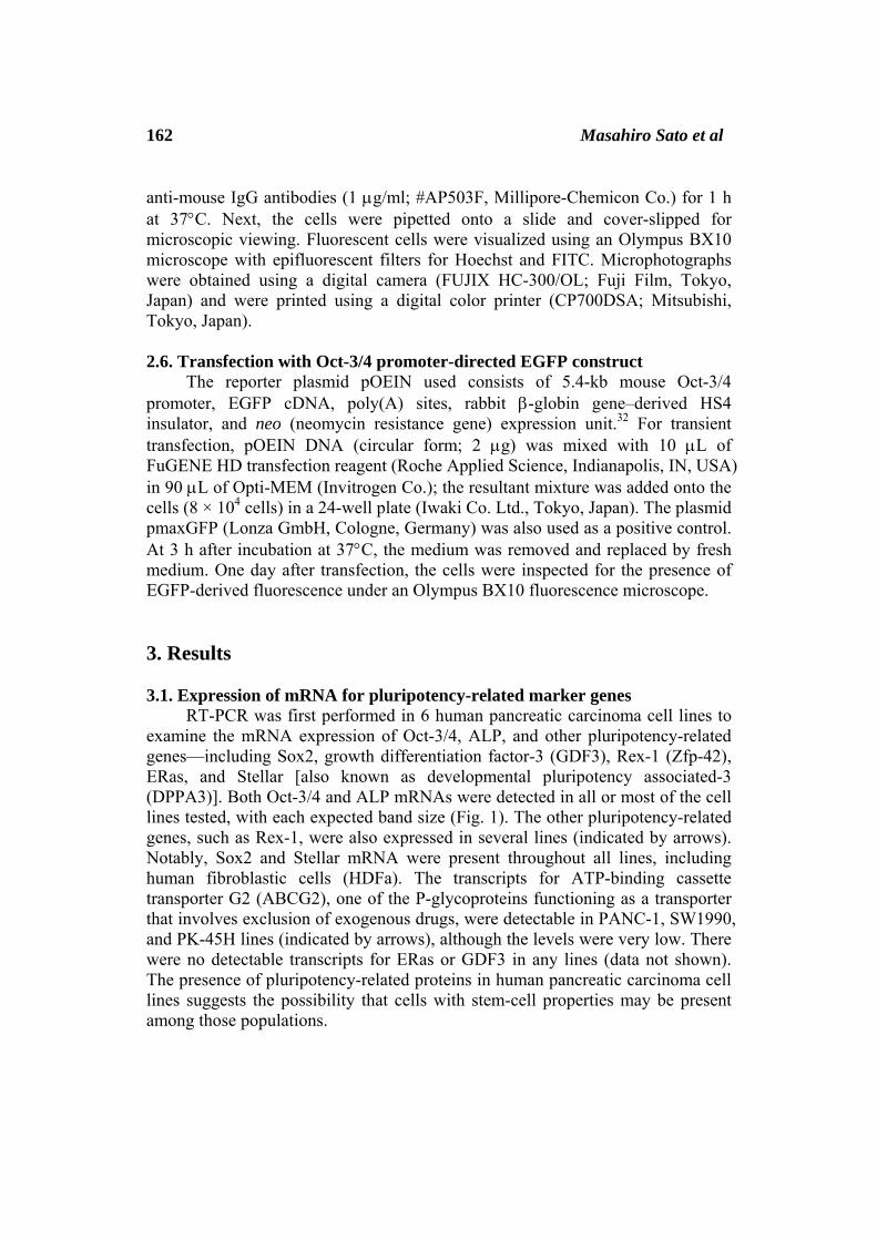
RT-PCR was first performed in 6 human pancreatic carcinoma cell lines to examine the mRNA expression of Oct-3/4, ALP, and other pluripotency-related genes—including Sox2, growth differentiation factor-3 (GDF3), Rex-1 (Zfp-42), ERas, and Stellar [also known as developmental pluripotency associated-3 (DPPA3)]. Both Oct-3/4 and ALP mRNAs were detected in all or most of the cell lines tested, with each expected band size (Fig. 1). The other pluripotency-related genes, such as Rex-1, were also expressed in several lines (indicated by arrows). Notably, Sox2 and Stellar mRNA were present throughout all lines, including human fibroblastic cells (HDFa). The transcripts for ATP-binding cassette transporter G2 (ABCG2), one of the P-glycoproteins functioning as a transporter that involves exclusion of exogenous drugs, were detectable in PANC-1, SW1990, and PK-45H lines (indicated by arrows), although the levels were very low. There were no detectable transcripts for ERas or GDF3 in any lines (data not shown). The presence of pluripotency-related proteins in human pancreatic carcinoma cell lines suggests the possibility that cells with stem-cell properties may be present among those populations.
Mosaic expression of pluripotency-related proteins Oct-3/4 163
3.2. Mosaic expression of Oct-3/4 and ALP proteins in PANC-1 pancreatic carcinoma cell line
To investigate further evidence for gene expression at the protein level, PANC-1 was examined for expression of Oct-3/4 and ALP proteins. Immunostaining using anti-Oct-3/4 monoclonal antibody revealed that a small population of these two cell lines was immunoreactive to this antibody: 23% (13/57 cells examined) of cells was positive for Oct-3/4 expression in PANC-1 (indicated by arrows in Fig. 2A-c). Murine EC cell line F9 cells (positive control) were positively stained with the antibody (Fig. 2A-a), while mouse fibroblastic cell line NIH3T3 (negative control) was negative for Oct-3/4 expression (Fig. 2A-b). Cytochemical staining for ALP activity revealed that 19% (12/64 cells examined) of cells was positive for ALP expression in PANC-1 (indicated by arrows in Fig. 2A-f). As expected, F9 cells (but not NIH3T3 cells) exhibited distinct ALP activity (Fig. 2A-d vs. -e). 3.3. Oct-3/4-positive PANC-1 cells failed to exclude Hoechst 33342 Some CSCs have been shown to exhibit reduced staining with Hoechst 33342 as a stem cell characteristic for side population.33 This is because Hoechst 33342 dye could be excluded based on expression of the drug transporter/stem cell marker ABCG2. To test whether Oct-3/4-positive pancreatic tumor cell lines possess such

164 Masahiro Sato et al
a property, we incubated live PANC-1 cells with Hoechst 33342 and then fixed them for immunostaining with anti-Oct-3/4 antibody. As expected, some cells reacted with the antibody (arrowheads in Fig. 2B-a,b). However, these Oct-3/4-positive cells were distinctly stained with Hoechst 33342, a characteristic that was indistinguishable from the Oct-3/4-negative cells (arrows vs. arrowheads in Fig. 2B-a,b). This finding suggests that Oct-3/4 expression does not correlate

Mosaic expression of pluripotency-related proteins Oct-3/4 165 with the ability to exclude Hoechst 33342 dye in the PANC-1 cell line we tested.
3.4. Transfection with Oct-3/4 promoter–directed EGFP construct also demonstrates the presence of Oct-3/4-positive cells in PANC-1 pancreatic carcinoma cell line
The final goal of this study was to investigate if Oct-3/4 or ALP-positive pancreatic carcinoma cells exhibit CSC-specific properties (i.e., the ability to
166 Masahiro Sato et al differentiate into other types of cell, resistance to anti-cancer drugs, and a high degree of proliferative activity). This investigation required the enrichment Oct-3/4- or ALP-positive pancreatic carcinoma cells, which can be done using at least 2 different approaches: a fluorescence activated cell sorting (FACS)-based approach or a gene engineering-based approach. The former method requires appropriate cell-surface markers or an Oct-3/4- or ALP-specific fluorescent substance, but those are presently unavailable. Therefore, we employed the latter approach, which depends upon transfection of a plasmid harboring a marker gene (i.e., EGFP or a drug-resistance gene), whose expression is controlled by Oct-3/4 or ALP promoter. To test the feasibility of the latter approach, PANC-1 cells were liposomally transfected with a pOEIN plasmid (Fig. 3A) harboring 5.4-kb mouse Oct-3/4 promoter, EGFP cDNA, poly(A) sites, HS4 insulator, and the neo expression unit. Upon inspection 1 day after transfection, a small number of cells (0.8%; 3/360 cells examined) were found to be fluorescent (arrow in Fig. 3B-b). This was in contrast with transfection using pmaxGFP, which yielded 5% (25/472 cells examined) fluorescent cells (Fig. 3B-d). In the control groups, some F9 cells were fluorescent, as expected (arrows in Fig. 3B-f), while no fluorescence was observed in NIH3T3 cells (Figure 3B-j), indicating the fidelity of the 5.4-kb mouse Oct-3/4 promoter used in this study. The presence of fluorescent PANC-1 cells following transfection with pOEIN suggests that Oct-3/4-positive cells exist among the PANC-1 cell population and that they can be enriched following cultivation in the presence of G418, a neomycin analog. 4. Discussion
In this study, we have shown that certain subpopulations of human pancreatic carcinoma cell line PANC-1 express pluripotency-related genes such as Oct-3/4 and ALP. However, the Oct-3/4-positive PANC-1 cells failed to exhibit a high degree of ABCG2 activity, as revealed by the Hoechst 33342-exclusion assay, although they did express ABCG2 mRNA very weakly. A preliminary transfection experiment using an Oct-3/4 promoter-directed EGFP construct confirmed the presence of Oct-3/4-positive cells among the PANC-1 cell population, suggesting the possible enrichment of Oct-3/4-positive cells using this construct.
Consistent with the present findings, expression of pluripotency-related genes such as Oct-3/4 and Nanog in human pancreatic carcinoma cell lines has been reported previously.14 However, the subcellular localization of these proteins remains unknown. In this study, we demonstrate for the first time that cells expressing at least 2 proteins, Oct-3/4 and ALP, are present as subset populations of cells in the established cell line (Fig. 2). Unfortunately, it remains unknown whether both proteins are co-expressed within a cell or not. Since these 2 proteins are abundantly expressed in undifferentiated cells such as ES cells and early embryos, and Oct-3/4 is known to play an important role in maintaining the

Mosaic expression of pluripotency-related proteins Oct-3/4 167 undifferentiated state of a cell, cancer cells expressing those proteins may be progenitor cells with immature features. The characterization of Oct-3/4 as a marker for CSC was recently suggested by Sajithlal et al.34 and Wang et al. 35 The former group demonstrated that the Oct-3/4-expressing human breast cancer cell line MCF7 possesses a high degree of proliferative activity following transplantation into immunocompromized mice, further exhibiting resistance against antitumor drugs, such as 5-fluorouracil and cis-platinum. These cells express ABCG2 mRNA strongly. The latter group used chemotherapeutic drugs to select drug-resistant hepatocellular carcinoma (HCC), which displayed CSC features, such as increased self-renewal ability, cell motility, and levels of Oct-3/4 mRNA. By contrast, the possibility that ALP may serve as a marker for CSC has not yet been confirmed. Considering the similarity between Oct-3/4 expression and ALP in view of their cell type-specificity, we consider that ALP-positive cancer cells may be CSCs.
As mentioned above, we showed in this study that subpopulations of pancreatic cancer cells expressed Oct-3/4 and ALP. It is important to determine whether these Oct-3/4- or ALP-positive cells are labile—in other words, if they can revert to Oct-3/4- or ALP-negative cells or continue to express Oct-3/4 or ALP. To address this point in more detail, Oct-3/4- or ALP-positive cells must be enriched from the heterogeneous populations of pancreatic carcinoma cell lines. In an attempt to enrich those cells, we introduced pOEIN plasmid [carrying EGFP cDNA (whose expression is controlled by Oct-3/4 promoter) and a neo expression unit] into PANC-1 cells, and found that a small number of cells were fluorescent (see arrow in Fig. 3B-b). If these transfected cells survived after selection with G418, they would be used to test the potential of Oct-3/4-positive cells. Unfortunately, the present transfection efficiency in PANC-1 cells using FuGENE HD is very low (<1%). We are now attempting to transfect those cells using a more efficient gene transfer system (e.g., a nucleofection system provided by Lonza GmbH). As for obtaining ALP-positive cancer cells, we are constructing a reporter plasmid that harbors 1.2-kb human ALP promoter36 linked to a red fluorescent protein cDNA and drug-resistance gene.
Notably, Chen et al.37 previously isolated CD133-positive cells from clinical samples and lung cancer cell lines, finding that these cells exhibited strong proliferation and invasive capabilities both in vitro and in vivo. Interestingly, Oct-3/4 expression was upregulated in these cells. Furthermore, siRNA-mediated knock down of Oct-3/4 expression in these cells eliminated their proliferative activity. These findings indicate that Oct-3/4 plays a crucial role in maintaining self-renewal. More importantly, these data suggest a close relationship between Oct-3/4 and CD133 expression. In this context, it will be worthwhile to explore the possible linkage between Oct-3/4 or ALP and other known surface markers, such as CD133.
Some CSCs, such as melanoma CSCs, have been found to highly co-express CD133 and ABCG2 markers, possessing enhanced tumorigenic potential.34 To correlate ABCG2 and Oct-3/4 expression in pancreatic tumor cells, we first examined expression of ABCG2 mRNA; we found trace amounts of this mRNA
168 Masahiro Sato et al in some lines, including PANC-1 (see Fig. 1). However, direct assessment using the Hoechst 33342 dye-exclusion assay failed to reveal a link between Oct-3/4 expression and ABCG2 transporter activity (see Fig. 2). Interestingly, Wang et al.35 have reported that Oct-3/4 overexpression induces the activation of ABCG2 in liver cancer cells. Corresponding with these results, forced Oct-3/4 overexpression in PANC-1 cells may induce elevated expression of ABCG2.
5. Conclusion
We demonstrated that the pluripotency-related proteins Oct-3/4 and ALP are expressed in human pancreatic carcinoma cell lines and that cells expressing Oct-3/4 or ALP exist as subpopulations in at least one line, namely, PANC-1. Enrichment of Oct-3/4- or ALP-positive cells will be helpful for the further characterization of these cells needed to define them as possible pancreatic CSCs. Acknowledgements
This work was supported by a Grant-in-Aid for Scientific Research (C) from the Ministry of Education, Science, Sports, Culture, and Technology of Japan. References [1] T. Lapidot, C. Sirard, J. Vormoor, B. Murdoch, T. Hoang, J. Caceres-Cortes,
M. Minden, B. Paterson, M. A. Caligiuri and J. E. Dick, A cell initiating human acute myeloid leukaemia after transplantation into SCID mice, Nature, 367(1994), 645–648.
[2] D. Bonnet and J. E. Dick, Human acute myeloid leukemia is organized as a
hierarchy that originates from a primitive hematopoietic cell, Nature Medicine, 3(1997), 730–737.
[3] S. K. Singh, C. Hawkins, I. D. Clarke, J. A. Squire, J. Bayani, T. Hide, R. M.
Henkelman, M. D. Cusimano and P. B. Dirks, Identification of human brain tumour initiating cells, Nature, 432(2004), 396–401.
[4] M. Al-Hajj and M. F. Clarke, Self-renewal and solid tumor stem cells,
Oncogene, 23(2004), 7274–7282. [5] C. T. Jordan, M. L. Guzman and M. Noble, Cancer stem cells, New England
Journal of Medicine, 355(2006), 1253–1261.

Mosaic expression of pluripotency-related proteins Oct-3/4 169 [6] J. E. Visvader and G. J. Lindeman, Cancer stem cells in solid tumours:
accumulating evidence and unresolved questions, Nature Reviews Cancer, 8(2008), 755–768.
[7] G. Kania, D. Corbeil, J. Fuchs, K. V. Tarasov, P. Blyszczuk, W. B. Huttner, K.
R. Boheler and A. M. Wobus, Somatic stem cell marker prominin-1/CD133 is expressed in embryonic stem cell-derived progenitors, Stem Cells, 23(2005), 791–804.
[8] S. C. Wheatley, C. M. Isacke and P. H. Crossley, Restricted expression of the
hyaluronan receptor, CD44, during postimplantation mouse embryogenesis suggests key roles in tissue formation and patterning, Development, 119(1993), 295–306.
[9] A. E. Sutherland, R. D. Sanderson, M. Mayes, M. Seibert, P. G. Calarco, M.
Bemfield and C. H. Damsky, Expression of syndecan, a putative low affinity fiboblast growth factor receptor, in the early mouse embryo, Development, 113(1991), 339–351.
[10] S. Kellner and N. Kikyo, Transcriptional regulation of the Oct4 gene, a
master gene for pluripotency, Histology and Histopathology, 25(2010), 405–412.
[11] M. H. Rosner, M. A. Vigano, K. Ozato, P. M. Timmons , F. Poirie, P. W. J.
Rigby and L. M. Staudt, A POU-domain transcription factor in early stem cells and germ cells of the mammalian embryo, Nature, 345(1990), 686–692.
[12] H. R. Schöler, S. Ruppert, N. Suzuki, K. Chowdhury and P. Gruss, New type
of POU domain in germ line-specific protein Oct-4, Nature, 344(1990), 435–439.
[13] J. Nichols, B. Zevnik, K. Anastassiadis, H. Niwa, D. Klewe-Nebenius, I.
Chambers, H. Schöler and A. Smith, Formation of pluripotent stem cells in the mammalian embryo depends on the POU transcription factory Oct4, Cell, 95(1998), 379–391.
[14] M. H. Tai, C. C. Chang, M. Kiupel, J. D. Webster, L. K. Olson and J. E.
Trosko, Oct4 expression in adult human stem cells: evidence in support of the stem cell theory of carcinogenesis, Carcinogenesis, 26(2005), 495–502.
[15] R. Matoba, H. Niwa, S. Masui, S. Ohtsuka, M. G. Carter, A. A. Sharov and
M. S. Ko MS, Dissecting Oct3/4-regulated gene networks in embryonic stem cells by expression profiling, PLoS ONE, 1(2006), e26.

170 Masahiro Sato et al [16] M. Pesce, X. Wang, D. J. Wolgemuth and H. Schöler, Differential expression
of the Oct-4 transcription factor during mouse germ cell differentiation, Mechanism of Development, 71(1998), 89–98.
[17] S. Gidekel, G. Pizov, Y. Bergman and E. Pikarsky, Oct-3/4 is a
dose-dependent oncogenic fate determinant, Cancer Cell, 4(2003), 361–370. [18] K. Hochedlinger, Y. Yamada, C. Beard and R. Jaenisch, Ectopic expression
of Oct-4 blocks progenitor-cell differentiation and causes dysplasia in epithelial tissues, Cell, 121(2005), 465–477.
[19] T. Hu , S. Liu, D. R. Breiter, F. Wang, Y. Tang and S. Sun, Octamer 4 small
interfering RNA results in cancer stem cell-like cell apoptosis, Cancer Research, 68(2008), 6533–6540.
[20] C. C. Chang, G. S. Shieh, P. Wu, C. C. Lin, A. L. Shiau and C. L. Wu,
Oct-3/4 expression reflects tumor progression and regulates motility of bladder cancer cells, Cancer Research, 68(2008), 6281–6291.
[21] G. M. Seigel, A. S. Hackam and A. Ganguly, Human embryonic and
neuronal stem cell markers in retinoblastoma, Molecular Vision, 13(2007), 823–832.
[22] M. T. Sung, T. D. Jones, S. D. Beck, R. S. Foster and L. Cheng, OCT4 is
superior to CD30 in the diagnosis of metastatic embryonal carcinomas after chemotherapy, Human Pathology, 37(2006), 662–667.
[23] G. Karoubi, M. Gugger, R. Schmid, A. Dutly, G. Karoubi, M. Gugger, R.
Schmid and A. Dutly, OCT4 expression in human non-small cell lung cancer: implications for therapeutic intervention, Interactive CardioVascular and Thoracic Surgery, 8(2009), 393−397.
[24] M. Pesce and H. Schöler, Oct-4: gatekeeper at the beginnings of mammalian
development, Stem Cells, 19(2001), 271–278. [25] E. Mornet, E. Stura, A.-S. Lia-Baldini, T. Stigbrand, A. Ménez and M.-H. Le
Du, Structural evidence for a functional role of human tissue nonspecific alkaline phosphatase in bone mineralization, Journal of Biological Chemistry, 276(2001), 31171–31178.
[26] M. L. Lepire and C. A. Ziomek, Preimplantation mouse embryos express a
heat-stable alkaline phosphatase, Biology of Reproduction, 41(1989), 464–473.

Mosaic expression of pluripotency-related proteins Oct-3/4 171 [27] A. C. Hahnel, D. A. Rappolee, J. L. Millan, T. Manes, C. A. Ziomek, N. G.
Theodosiou, Z. Werb, R. A. Pedersen and G. A. Schultz, Two alkaline phosphatase genes are expressed during early development in the mouse embryo, Development, 110(1990), 555–564.
[28] M. Ginsburg, M. H. Snow and A. McLaren, Primordial germ cells in the
mouse embryo during gastrulation, Development, 110(1990), 521–528. [29] D. Langer, Y. Ikehara, H. Takebayashi, R. Hawkes and H. Zimmermann, The
ectonucleotidases alkaline phosphatase and NTPDase2 are associated with subsets progenitor cell populations in the mouse embryonic, postnatal and adult neurogenic zones, Neuroscience, 150(2007),863–879.
[30] V. L. Battula, S. Treml, P. M. Bareiss, F. Gieseke, H. Roelofs, P. de Zwart, I.
Müller, B. Schewe, T. Skutella, W. E. Fibbe, L. Kanz and H. J. Bühring, Isolation of functionally distinct mesenchymal stem cell subsets using antibodies against CD56, CD271, and mesenchymal stem cell antigen-1, Haematologica, 94(2009), 173–184.
[31] E. G. Bernstine, M. L. Hooper, S. Grandchamp and B. Ephrussi, Alkaline
phosphatase activity in mouse teratoma, Proceedings of the National Academy of Sciences of the United States of America, 70(1973), 3899–3903.
[32] K. Miyoshi, H. Mori, Y. Mizobe, E. Akasaka, A. Ozawa, M. Yoshida and M.
Sato, Development of a noninvasive monitoring system for evaluation of Oct-3/4 promoter status in miniature pig somatic cell nuclear transfer embryos, Journal of Reproduction and Development, 55(2009), 661–669.
[33] S. Zhou, J. D. Schuetz, K. D. Bunting, A. M. Colapietro, J. Sampath, J. J.
Morris, I. Lagutina, G. C. Grosveld, M. Osawa, H. Nakauchi and B. P. Sorrentino, The ABC transporter Bcrp1/ABCG2 is expressed in a wide variety of stem cells and is a molecular determinant of the side-population phenotype, Nature Medicine, 7(2001), 1028–1034.
[34] G. S. Sajithlal, K. Rothermund, F. Zhang, D. J. Dabbs, J. J. Latimer, S. G.
Grant and E. V. Prochownik, Permanently blocked stem cells derived from breast cancer cell lines, Stem Cells, 28(2010), 1008–1018.
[35] X. Q. Wang, W. M. Ongkeko, L. Chen, Z. F. Yang, P. Lu, K. K. Chen, J. P.
Lopez, R. T. Poon and S. T. Fan, Octamer 4 (Oct4) mediates chemotherapeutic drug resistance in liver cancer cells through a potential Oct4-AKT-ATP-binding cassette G2 pathway, Hepatology, 52(2010), 528–539.
172 Masahiro Sato et al [36] N. Yusa, K. Watanabe, S. Yoshida, N. Shirafuji, S. Shimomura, K. Tani, S.
Asano and N. Sato, Transcription factor Sp3 activates the liver/bone/kidney-type alkaline phosphatase promoter in hematopoietic cells, Journal of Leukocyte Biology, 68(2000), 772–777.
[37] Y. C. Chen, H. S. Hsu, Y. W. Chen, T. H. Tsai, C. K. How, C. Y. Wang, S. C.
Hung, Y. L. Chang, M. L. Tsai, Y. Y. Lee, H. H. Ku and S. H. Chiou, Oct-4 expression maintained cancer stem-like properties in lung cancer-derived CD133-positive cells, PLoS ONE, 3(2008), e2637.
[38] E. Monzani, F. Facchetti, E. Galmozzi, E. Corsini, A. Benetti, C. Cavazzin,
A. Gritti, A. Piccinini, D. Porro, M. Santinami, G. Invernici, E. Parati, G. Alessandri and C. A. La Porta, Melanoma contains CD133 and ABCG2 positive cells with enhanced tumourigenic potential, European Journal of Cancer, 43(2007), 935–946.
[39] J. D. Raman, N. P. Mongan, L. Liu, S. K. Tickoo, D. M. Nanus, D. S. Scherr
and L. J. Gudas, Decreased expression of the human stem cell marker, Rex-1 (zfp-42), in renal cell carcinoma, Carcinogenesis, 27(2006), 499–507.
[40] T. Kameda and J. A. Thomson, Human ERas gene has an upstream
premature polyadenylation signal that results in a truncated, noncoding transcript, Stem Cells, 23(2005),1535–1540.
[41] A. T. Clark, R. T. Rodriguez, M. S. Bodnar, M. J. Abeyta, M. I. Cedars, P. J.
Turek, M. T. Firpo and R. A. Reijo Pera, Human STELLAR, NANOG, and GDF3 genes are expressed in pluripotent cells and map to chromosome 12p13, a hotspot for teratocarcinoma, Stem Cells, 22(2004), 169–179.
Received: October, 2012