movement studies 2011. » describe the shape and key features of bones » orientate the bones of the...
TRANSCRIPT

Movement Studies2011

» Describe the shape and key features of bones
» Orientate the bones of the lower quadrant
» Demonstrate how to palpate key bony landmarks
» Discuss the key joint classifications» Identify the key features of synovial
joints
UH Movement studies 2011-12
2

UH Movement studies 2011-12
3
Functions:» Support » Protection» Assist movement » Mineral "bank" (Ca &
Phosphate)» Blood cell production -
haemopoesis (red marrow) » Energy storage - adipose tissue
(yellow marrow)
Tissue components: » 25% water » 25% protein fibres
(notably collagen)» 50% mineral salts
(Primarily Calcium Phosphate)
» Strong yet lightweight

4
UH Movement studies 2011-12

Organic matrix of fibrous connective tissue (toughness and elasticity) impregnated with mineral salts (hardness);
» Periosteum (encloses bone)˃ Dense fibrous tissue˃ Important for bone healing (releases osteoblast˃ precursors)
» Diaphysis˃ Shaft of long bone (eg shaft of femur)˃ Compact (or condensed) bone
Marrow cavity lined by endosteum, contains yellow marrow (fat store) 5
UH Movement studies 2011-12

» Epiphysis ˃ Expanded ends of long bone (eg head of femur)˃ Thin outer layer of compact bone˃ Centre of cancellous (or spongy) bone ˃ Trabeculae which are arranged to resist forces˃ Compressive, tensile, shearing˃ Contains red marrow (red blood cell production)
» Articular cartilage - hyaline˃ Hard and smooth - low friction
6
UH Movement studies 2011-12

Internal architecture consists of trabeculae (cancellous or spongy bone)
The trabeculae are aligned in a pattern that provides maximal strength without too much bulk,
Highly engineered much in the way that architects and engineers design buildings and bridges. As much as 6X the body weight is transmitted across the hip
UH Movement studies 2011-12
7
Trabeculae

» Bone density can be a key issue for physiotherapists as a low bone density can put patients at risk of fracture
» What can limit bone density?
» What can maximise bone density?
UH Movement studies 2011-12
8
Graph taken from:www.arc.org.uk

1. Long Bones» Consist of a shaft or diaphysis
and 2 expanded ends or epiphyses
» Refers to structure not length e.g. femur and phalanges
» Damage to an epiphyseal plate can cause deformity
UH Movement studies 2011-12
9

2. Short bones» Small bones of the
hand or carpus and the tarsal bones in the foot
» Form in blocks of cartilage and ossify from the centre
» Do not have epiphyses
UH Movement studies 2011-12
10

3. Flat bones» Thin and curved
include pelvis, skull bones and ribs
» Cancellous/spongy bone sandwiched between 2 layers of compact bone
» Protect soft viscera
UH Movement studies 2011-12
11

UH Movement studies 2011-12
12
4. Irregular bones
Vertebrae, facial bones Accessory bones
(ossicles) e.g. os trigonum behind talus

» Name the bones and label» Identify:
˃ long bone˃ short bone ˃ irregular bone
» Can you orientate the bones to form the hip joint?
13
UH Movement studies 2011-12

» Identify the following bony landmarks:˃ Articular facet on a vertebra˃ Femoral condyle˃ Iliac crest˃ Iliac fossa˃ Transverse process on a vertebra˃ Greater trochanter on the femur˃ Tibial tuberosity on the tibia˃ Foramen on a vertebra
14
UH Movement studies 2011-12

» The name of the bony landmarks can give you a clue as to its nature and are found regularly in anatomy e.g˃ Tuberosity (tuber=swelling, hump)˃ Condyle (rounded process at end of
a bone)˃ Process (a projection from main
body of something)˃ Foramen (hole, opening, aperture)˃ Crest (top, elevated ridge)
UH Movement studies 2011-12
15

» It is important that you recognize that no one has the right to be touched without their consent
» That applies to you, now, even though it might appear that in an educational environment it is automatic - make sure you are sensitive to each others feelings and concerns
» More detail re consent and touch in NMS1 » Take a moment to check with your partner they are
happy to be palpated in the next task» Please read the manual therapy protocol» Please look at the manual therapy protocol
16
UH Movement studies 2011-12

In pairs, using available textbooks, try to locate the following landmarks on each other and describe their shape:
˃ Anterior superior iliac spine˃ Greater trochanter˃ Head of fibula˃ Medial border of scapula˃ Spinous processes of thoracic vertebrae˃ Radial styloid process
17
UH Movement studies 2011-12

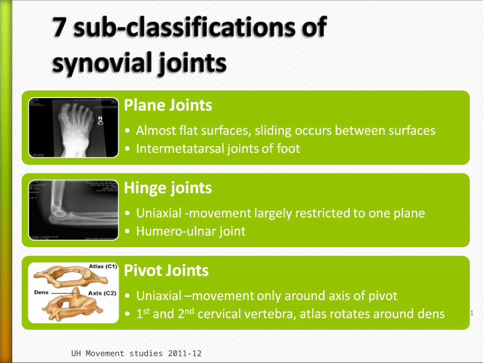
Joints can be divided in to those that have free movement
1. Synovialand those that have restricted movement2. Cartilaginous 3. Fibrous Within these three classifications there
are sub-classifications 18
UH Movement studies 2011-12

19
UH Movement studies 2011-12

Three Types:» Suture
Between bones of skull
» GomphosisPeg fits into socket e.g. roots of teeth
» SyndesmosisInferior tibiofibular joint
» Lack intervening cartilage between the two bones
» Articulation is fixed» Movement is very
restricted
UH Movement studies 2011-12
20

Primary cartilaginous Secondary cartilaginous
UH Movement studies 2011-12
21
» Occur at the epiphyseal growth plates
» Become obliterated as the two parts fuse(synostosis)
» Only one joint which in adulthood which allows some movement, sterno-clavicular joint
» Bone ends covered in hyaline cartilage but interposed between the bone ends is a pad of fibrocartilage
» In the midline of the body e.g joints between the vertebral bodies. Fibrocartilagenous pad is the disc

UH Movement studies 2011-12
22
» Free movement due to little friction
» Bone ends covered in hyaline cartilage, varying thicknesses
» Smooth movement facilitated by a viscous synovial fluid
VERY IMPORTANT TO MANUAL THERAPISTS

1. Articular cartilage2. Fibrous joint capsule3. Synovial membrane4. Synovial fluid5. Joint cavity6. Reinforcing ligaments
UH Movement studies 2011-12
23
UH Movement studies 2011-12
24

25
UH Movement studies 2011-12
Saddle Joints• Biaxial,concavoconvex surfaces- carpometacarpal jt• 1 surface concave and other convex at right angles

UH Movement studies 2011-12
26

UH Movement studies 2011-12
27

» Mobility!!!» Conflict between stability for load bearing and
mobility for functional activities» Dependant on primary function of joint either
stability or mobility is compromised to a certain extent˃ Hip is stable for weight bearing but lacks relative mobility˃ Shoulder is mobile in order to place the hand but lacks relative
stability ˃ http://www.youtube.com/watch?v=-ffpcRxWgsg&feature=related
28
UH Movement studies 2011-12
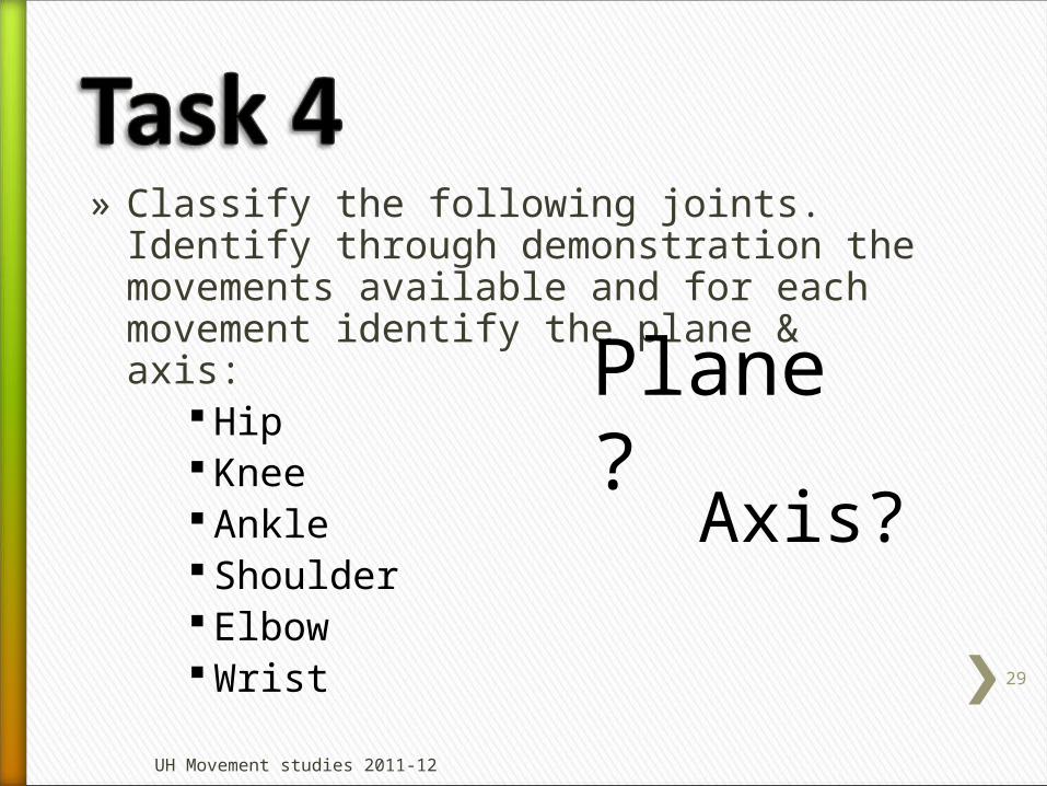
» Classify the following joints. Identify through demonstration the movements available and for each movement identify the plane & axis:
Hip Knee Ankle Shoulder Elbow Wrist
29
UH Movement studies 2011-12
Plane?
Axis?

» Recap on the learning outcomes from the beginning of the session˃ Do you feel you would be able to meet these?˃ If not go back over the material and check˃ Still unsure, ask a friend˃ Still unsure, come and speak to one of the tutors
» It is important to understand these elements before moving on to the next session on muscles and movement
30
UH Movement studies 2011-12

» Moore KL, Dalley AF (1999) Clinically Oriented Anatomy, 4th ed. Baltimore. Lippincott, Williams & Wilkens, pg 14-26 (up to ‘Muscular System’)
» Palastanga N, Field D, Soames R (2002) Anatomy and Human Movement – Structure and Function, 4th ed. Oxford, Butterworth Heinemann, pg 7-10 (up to ‘Muscular Tissue’)
» Sandring, S (2008) Grays Anatomy, 14th ed. London, Elsevier» Tyldesley, B and Grieve, J (2002) Muscles, Nerves and
Movement in Human Occupation, 3rd ed. Blackwell Publishing, Oxford, pg 3-8 (Framework and Support: The Connective Tissues)
31
UH Movement studies 2011-12