movements and joints. internal & external rotation
TRANSCRIPT

Movements and Joints


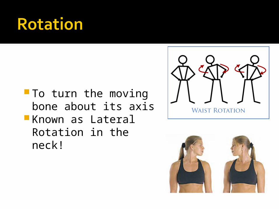
To turn the moving bone about its axis
Known as Lateral Rotation in the neck!

Horizontal Abduction Moving away from
the midline in the horizontal plane
Horizontal Adduction Moving toward the
midline in the horizontal plane
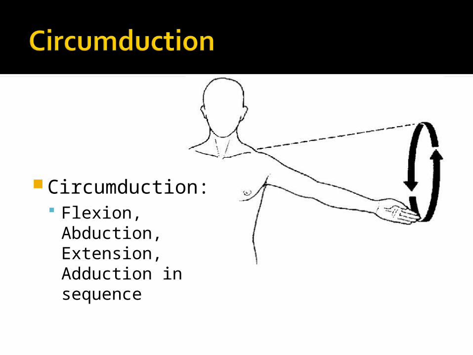
Circumduction: Flexion,
Abduction, Extension, Adduction in sequence

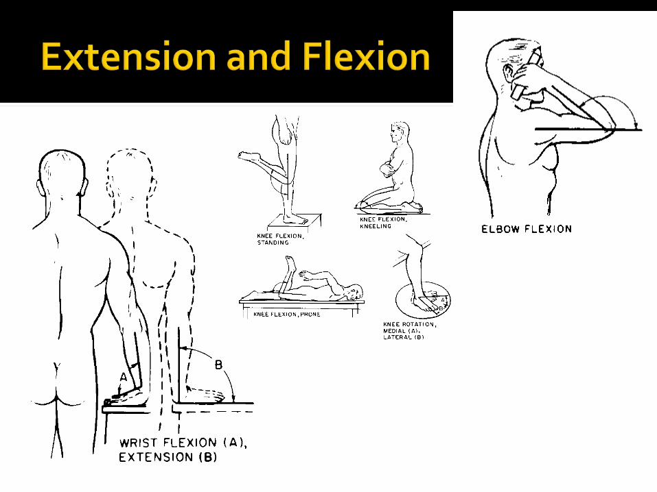
Flexion toward the outside of the body

Vary in size and shape.
Simple Joint: A joint with only two articulating surfaces Examples: Hip and Ankle (talotibal)
Compound Joint: A joint with three or more articulating surfaces Example: Wrist
Complex Joint: A joint with more than two articulating surfaces and
with a disc or fibrocartilage Example: Knee.

The hinge joint allows movement in one plane (flexion, extension)
Examples: Distal Interphalageal
(DIP) and Proximal Interphalageal (PIP) joints of the phalanges in the foot and hand
Ulnohumeral articulation at the elbow

Movement consists of two flat surfaces that slide over each other to allow movement.
In the hand the Carpals will slide over each other as the hand is moved to positions of flexion, extension, radial deviation, or ulnar deviation.
In the foot, the Tarsals shift during pronation and supination, sliding over each other in the process.


The ellipsoid joint allows movement in two planes (flexion, extension; abduction, adduction)
Examples: The radiocarpal
articulation at the wrist The metacarpophalangeal
articulation in the phalanges.

The saddle joint only found at the carpometacarpal articulation of the thumb
Allows two planes of motion (flexion, extension; abduction, adduction) with a small amount of rotation also allowed.
It is similar to the ellipsoid joint in function
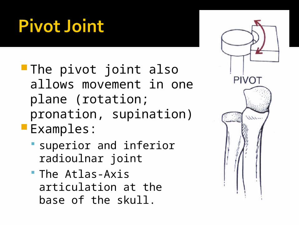
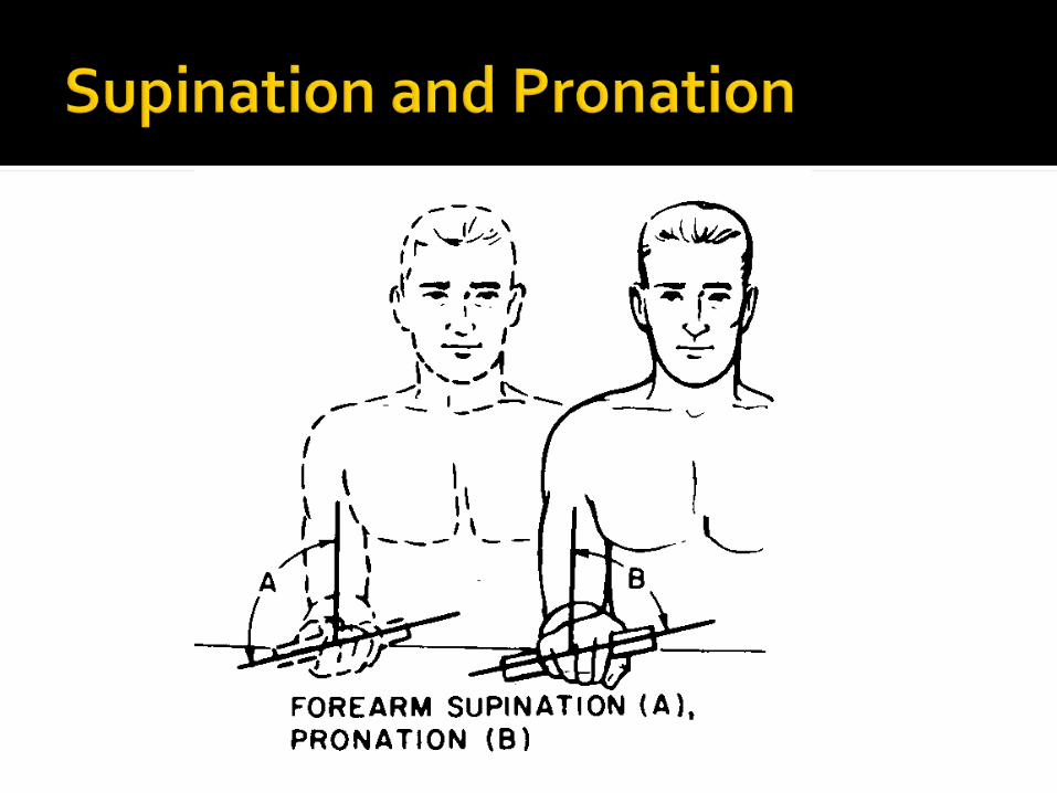
The pivot joint also allows movement in one plane (rotation; pronation, supination)
Examples: superior and inferior
radioulnar joint The Atlas-Axis articulation
at the base of the skull.
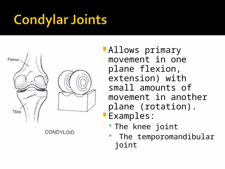
Allows primary movement in one plane flexion, extension) with small amounts of movement in another plane (rotation).
Examples: The knee joint The temporomandibular
joint
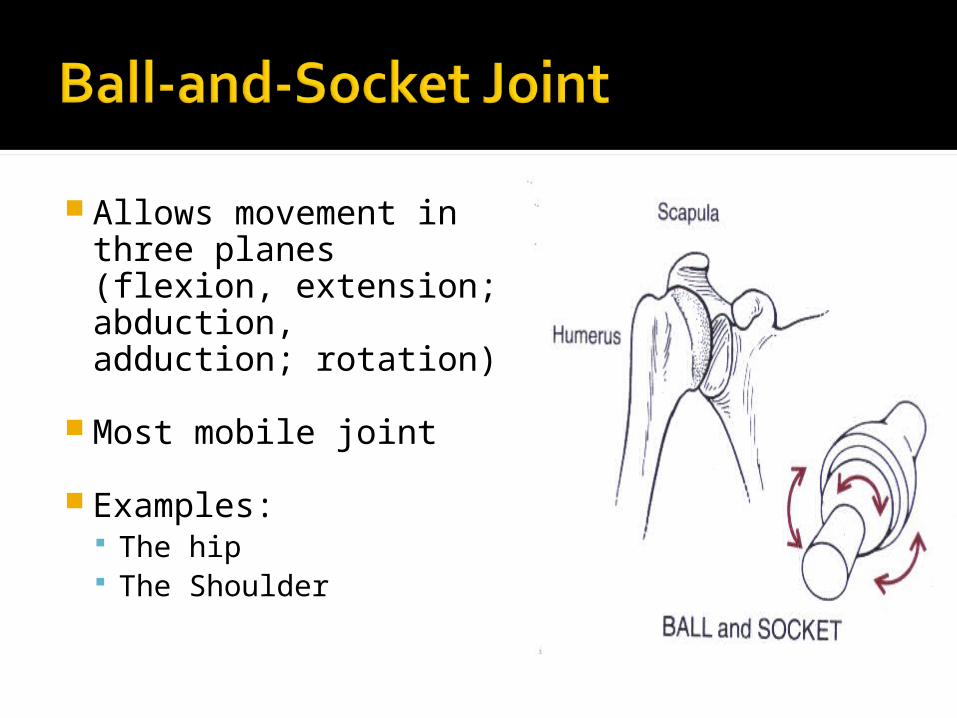
Allows movement in three planes (flexion, extension; abduction, adduction; rotation)
Most mobile joint
Examples: The hip The Shoulder

Bones held together by either hyaline cartilage Example: epiphyseal plates
Or by fibrocartilage Example: pubic symphysis and the intervertebral
discs
The movement is very limited, although not to the degree of the synarthodial joints.
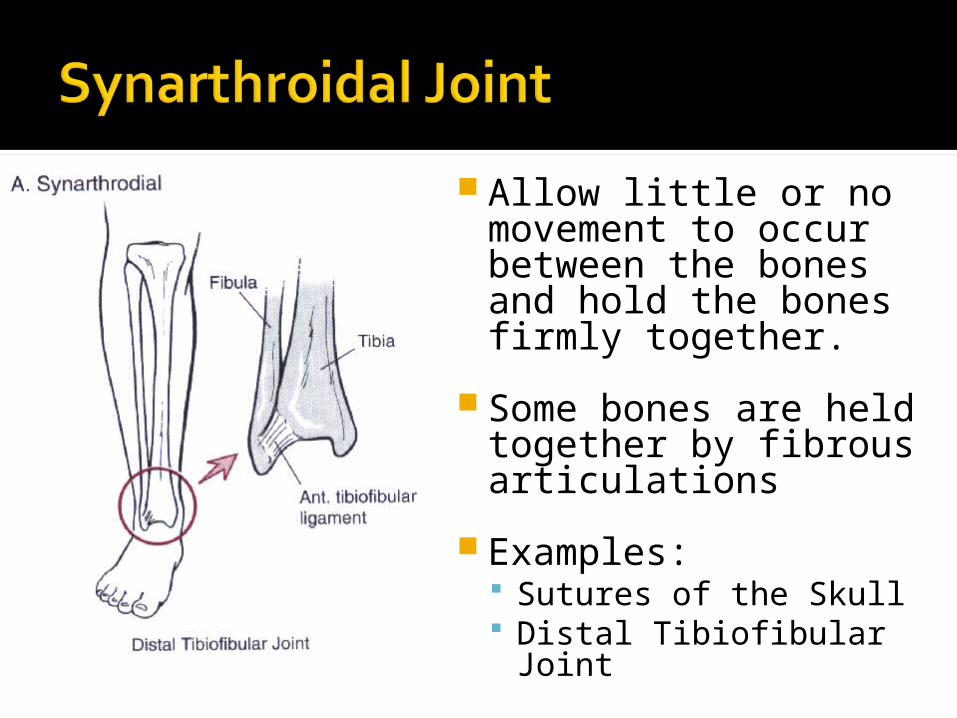
Allow little or no movement to occur between the bones and hold the bones firmly together.
Some bones are held together by fibrous articulations
Examples: Sutures of the Skull Distal Tibiofibular Joint
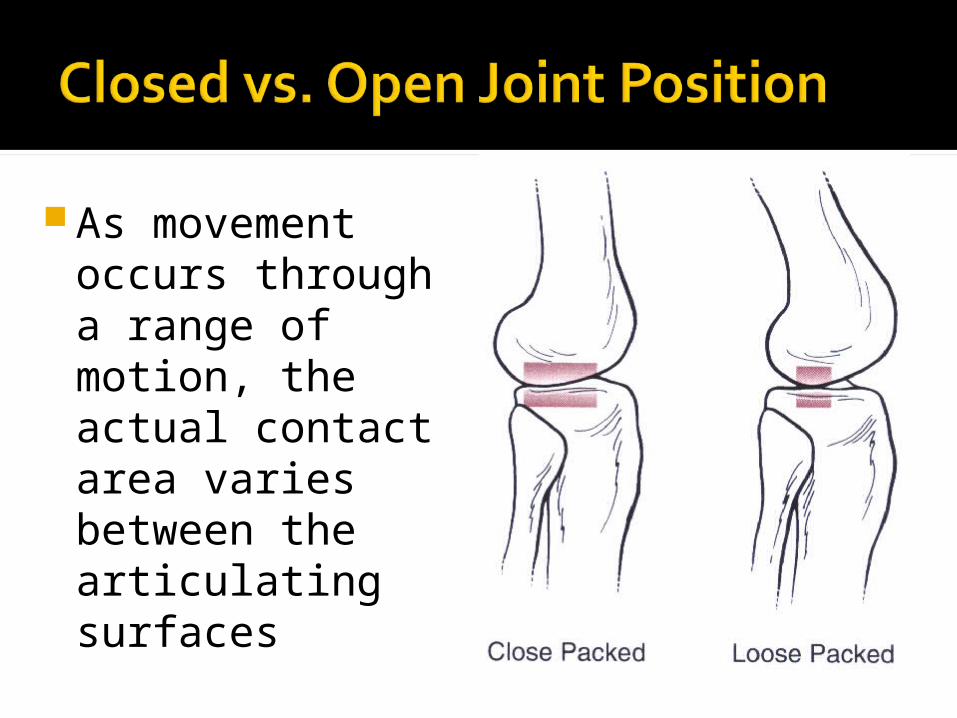
As movement occurs through a range of motion, the actual contact area varies between the articulating surfaces

Major Joints of the Body
Joint Type Degrees of Freedom
Vertebrae Amphiarthroidial 3
Hip Ball-and-Socket 3
Shoulder Ball-and-Socket 3
Knee Condyloid 2
Wrist Ellipsoid 2
Metacarpophalangeal (fingers)
Ellipsoid 2
Carpometacarpal (thumb)
Saddle 2
Elbow Hinge 1
Radioulnar Pivot 1
Atlantoaxial Pivot 1
Ankle Hinge 1
Interphalangeal Hinge 1

Supine: Face upward; on your back
Prone: Face downward; on your stomach


Cornonal (Frontal) Plane splits the body into anterior / posterior sections
Anterior: Front of the body or body part
Posterior: Back of the body or body part

When the body is split along the Mid Sagittal or Median
Sagittal Plane Lateral:
Away from the midline of the body
Anatomical position Medial:
Toward the midline of the body Anatomical position


Distal: Farthest from a
point of attachment to the body
Proximal: Used to describe
where the appendage joins the body
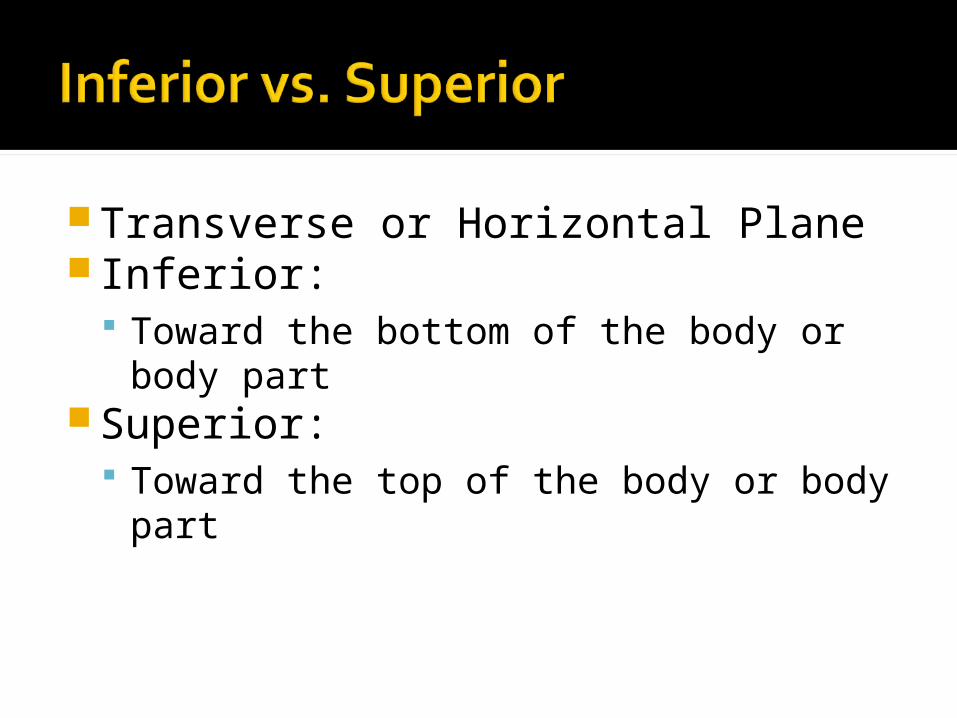
Transverse or Horizontal Plane Inferior:
Toward the bottom of the body or body part
Superior: Toward the top of the body or body part

Superficial: Toward the surface of the body
Deep (visceral): Deep inside the body

Mostly used in animalsDorsal (anterior):
Upper SurfaceVentral (posterior):
Bottom Surface

Internal: Deeper Inside Toward
External: Near the surface Outside Away

Volar Ventral aspect of the fingers
Palmar: Ventral aspect of the hand (palm of the
hand)Plantar:
Ventral aspect of the foot (sole of the foot)

Ipsilateral: One sided; pertaining to one side
Contralateral: Both sides; Bilateral