mr-guided parenchymal delivery of adeno-associated viral ...non-human... · open short...
TRANSCRIPT
OPEN
SHORT COMMUNICATION
MR-guided parenchymal delivery of adeno-associated viralvector serotype 5 in non-human primate brainL Samaranch1,3, B Blits2,3, W San Sebastian1, P Hadaczek1, J Bringas1, V Sudhakar1, M Macayan1, PJ Pivirotto1, H Petry2
and KS Bankiewicz1
The present study was designed to characterize transduction of non-human primate brain and spinal cord with AAV5 viral vectorafter parenchymal delivery. AAV5-CAG-GFP (1 × 10¹³ vector genomes per milliliter (vg ml− 1)) was bilaterally infused either intoputamen, thalamus or with the combination left putamen and right thalamus. Robust expression of GFP was seen throughoutinfusion sites and also in other distal nuclei. Interestingly, thalamic infusion of AAV5 resulted in the transduction of the entirecorticospinal axis, indicating transport of AAV5 over long distances. Regardless of site of injection, AAV5 transduced both neuronsand astrocytes equally. Our data demonstrate that AAV5 is a very powerful vector for the central nervous system and has potentialfor treatment of a wide range of neurological pathologies with cortical, subcortical and/or spinal cord affection.
Gene Therapy advance online publication, 16 March 2017; doi:10.1038/gt.2017.14
INTRODUCTIONNeurological gene therapy development with adeno-associatedvirus-based vectors (AAV), besides improvements in productionmethods leading to higher titers of vector, has been marked bythree seminal discoveries. First, pressurized parenchymal infusionsof different AAV serotypes by convection-enhanced delivery (CED)yield much more widespread distribution in the brain than simpleinjection does.1 Sequential innovations have steadily improvedCED, particularly with the advent of reflux-resistant cannulae andMR-guided visualization of infusions.2–5 Second, AAV serotypesexhibit distinct cellular specificities with some neuronallyrestricted, such as AAV2 or AAV6, and others with preference foreither neurons or astrocytes, such as AAV7 or AAV9.6–10 Thiscellular specificity has important immunological implications forexpression of non-self proteins over prolonged periods oftime.11,12 Finally, AAV serotypes are transported along axons andthereby direct expression in anatomical regions distal to theprimary vector infusion site. This latter discovery has obviousimplications to the clinical development of therapies forneurological disorders. Anatomical connectivity facilitates thespread of viral particles and enhances brain distribution of thetherapeutic gene. Interestingly, it has been found that thedirectionality of the transport is serotype dependent.6 The mostrepresentative and well-characterized serotype is AAV serotype 2.Direct infusion of AAV2 into the thalamus resulted in ananterograde transport of vector particles to cortical layers IV andV over a wide territory from prefrontal to occipital cortex13 andfrom putamen to substantia nigra pars reticulata (SNpr) in rats andnon-human primate (NHP).14,15 Recently, however, we studied inNHP the axonal transport of AAV2 prepared by methods thatdiffered sharply from the standard preparation technique.16,17 Toour surprise, this formulation was axonally transported in a
retrograde direction and was not completely neuron-specific.18
We surmise that anterograde axonal transport of AAV2 may be notbe an intrinsic property of AAV2 itself but perhaps may bedirected by the presence of strongly bound adventitious proteinsthat seem to be present in standard preparations and can beremoved by stringent washing.19 In contrast, the neurotropicAAV6 is axonally transported exclusively in a retrogradedirection.6,20 Infusion of AAV6 into putamen yielded abundanttransgene expression in cortical and thalamic projecting neuronsto striatum suggesting that nerve terminals took up viral particlesand transported to the neuronal cell bodies harbored in distalanatomically connected structures. Recently, axonal transport ofAAV serotype 9 has also been characterized.21–23 Putaminalinfusions revealed transport to thalamus and cortex as well assubstantia nigra pars compacta. This pattern indicates that AAV9underwent retrograde transport, however, SNpr, a region thatreceives projections from putamen, also showed transgeneexpression indicating anterograde transport of AAV9 as well.Further analysis demonstrated that the bidirectional transport ofserotype 9 is dose dependent.21 Dose range study with differentviral loads showed a dose requirement for effective transport,more pronounced on anterograde-linked structures to theinjection site than on distal structures that synapse with the targetstructure.21 Although the field is growing in knowledge on thespecifics of different serotypes, there is still a need to identify andcharacterize more new serotypes with different and more robustperformance to expand the current choices of viral vectors withgreat potential for clinical indication. In addition, the ability togenerate recombinant AAV using a reliable and scalablebaculovector-mediated technology will certainly aid vector devel-opment and boost its application in the gene therapy field.In that regards, the AAV serotype 5 has shown great potential
for different indications in small animal models and dogs24–26 but
1Department of Neurological Surgery, University of California, San Francisco, CA, USA and 2Neurobiology, Research and Development, UniQure NV, Amsterdam 1105BA,The Netherlands. Correspondence: Professor KS Bankiewicz, Department of Neurological Surgery, University of California, 1855 Folsom Street, MCB 226, San Francisco,CA 94103-0555, USA.E-mail: [email protected] authors contributed equally to this work.Received 9 September 2016; revised 19 January 2017; accepted 1 February 2017
Gene Therapy (2017), 1–9
www.nature.com/gt
the characterization of axonal transport in large animals is stillmissing. Deep understanding of AAV5 vector performanceregarding transport and distribution will allow a more precisetherapeutic indication. For this reason in the present study wecharacterized axonal transport of the AAV serotype 5 afterparenchymal delivery in primates. The study results suggest thatAAV5 has significant potential for indications where more globaltransduction of the brain and spinal cord is required.
RESULTS AND DISCUSSIONAnterograde transport of AAV5-GFP viral particlesAll putaminal infusions were performed with MR-guided CED.Each hemisphere received 51 μl of AAV5 encoding greenfluorescent protein gene (GFP) at 1.0 × 10¹³ vg ml− 1 (5.1 × 10¹¹
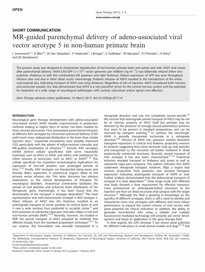
vg per hemisphere). Histological analysis of the brain showedidentically robust localized distribution of GFP within bothputamina and transduction that extended slightly laterallythrough the claustrum (arrowheads, Figure 1a). Direct proteinsequence and biochemistry comparison determined that AAV5serotype is markedly different than AAV2 in terms of capsidconformation.27 These differences among serotypes may affectviral particle dissemination due to variable features such as capsidstability, receptor binding, intercellular trafficking and intracellulargenome release.28 In that sense, three-dimensional (3D) recon-struction of the infusate gadolinium signal in the MRI (Figure 1b)revealed that after delivering 51 μl per side (volume of infusion;Vi), the volume of distribution (Vd) was ~ 3 times larger (Vdleft:155 μl; Vdright: 150 μl). This coefficient was higher compared withAAV2 that has a Vd/Vi ratio of about 2.5
Histological analysis revealed that putaminal medium spinyneurons were highly transduced in the area of injection along theprimary area of distribution (Figure 1c). Based on cellularmorphology, immunohistochemical analysis of representativesections indicated the presence of transduced neurons andastrocytes at the infusion site. No signal was found in caudatenucleus, thalamus (Figure 1d) or cortex (Figure 1e). Only distalstructures innervated by the putamen showed extensive GFPexpression (Figure 1f). In particular, SNpr, a structure that receivesprojections from the striatum through striato-nigral projections,showed high levels of GFP expression in the terminals (Figure 1g).Similarly, globus pallidus also contained GFP-positive fibers(Figure 1h). These results suggest robust anterograde transportof the AAV5 viral particles through striatal projections, eitherdirectly by striato-nigral or indirectly by striato-pallidal pathways.In addition, the lack of any signal in the cortex would indicate theabsence of retrograde transport via cortico-striatal projections. NoGFP was found in cervical, thoracic or lumbar levels of the spinalcord (Figure 1i). Based on these findings, AAV5 most closelyresembles AAV serotype 2 with respect to axonal transport andcell transduction, albeit more efficient.
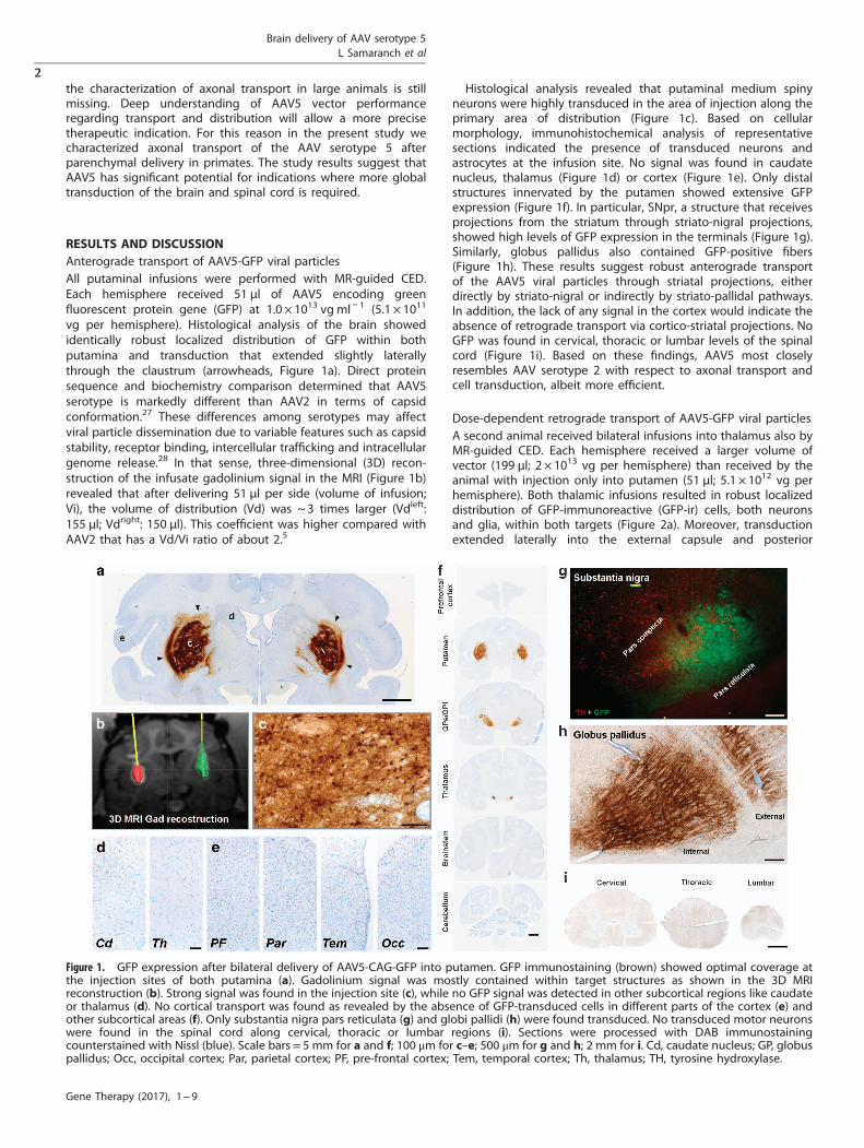
Dose-dependent retrograde transport of AAV5-GFP viral particlesA second animal received bilateral infusions into thalamus also byMR-guided CED. Each hemisphere received a larger volume ofvector (199 μl; 2 × 10¹³ vg per hemisphere) than received by theanimal with injection only into putamen (51 μl; 5.1 × 10¹² vg perhemisphere). Both thalamic infusions resulted in robust localizeddistribution of GFP-immunoreactive (GFP-ir) cells, both neuronsand glia, within both targets (Figure 2a). Moreover, transductionextended laterally into the external capsule and posterior
Figure 1. GFP expression after bilateral delivery of AAV5-CAG-GFP into putamen. GFP immunostaining (brown) showed optimal coverage atthe injection sites of both putamina (a). Gadolinium signal was mostly contained within target structures as shown in the 3D MRIreconstruction (b). Strong signal was found in the injection site (c), while no GFP signal was detected in other subcortical regions like caudateor thalamus (d). No cortical transport was found as revealed by the absence of GFP-transduced cells in different parts of the cortex (e) andother subcortical areas (f). Only substantia nigra pars reticulata (g) and globi pallidi (h) were found transduced. No transduced motor neuronswere found in the spinal cord along cervical, thoracic or lumbar regions (i). Sections were processed with DAB immunostainingcounterstained with Nissl (blue). Scale bars= 5 mm for a and f; 100 μm for c–e; 500 μm for g and h; 2 mm for i. Cd, caudate nucleus; GP, globuspallidus; Occ, occipital cortex; Par, parietal cortex; PF, pre-frontal cortex; Tem, temporal cortex; Th, thalamus; TH, tyrosine hydroxylase.
Brain delivery of AAV serotype 5L Samaranch et al
2
Gene Therapy (2017), 1 – 9
putamen (arrowheads, Figure 2a). During parenchymal infusion,reflux occurred up the outside of the cannula in the righthemisphere (Supplementary Figure 1a), which decreased theinternal pressure of the bolus and reduced Vi (o199 μl).Consequently, only the left hemisphere showed a Vd ~ 3 timeslarger than the Vi (Vdleft: 561 μl/Vileft: 199 μl) in the 3D analysis ofthe gadolinium signal from the thalamic infusate (Figure 2b). Theright hemisphere showed a Vd/Vi coefficient of ~ 2 (456 μl/199 μl),suggesting that pressurized delivery ensures the efficient disper-sion of the viral particles throughout the target structure. Moreimportantly, the suboptimal delivery also affected the axonaltransport. The backflow occurrence (Supplementary Figure 1b)became an excellent opportunity to evaluate back-to-back axonaltransport after different doses. Macroscopically, differences in thelevels of expression between hemispheres were found, mostly inpre-frontal and frontal cortices, whereas the levels of expression inthe injection sites were the same. This result suggests a linkbetween dose (total viral particles infused) and the directionalityof the axonal transport.Microscopic analysis showed that thalamic neurons were highly
transduced along the injection site and primary area of distribu-tion after infusion (Figure 2c), as well as in caudate, putamen andsubthalamic nucleus (Figure 2d). All these structures receiveprojections from the thalamus, suggesting anterograde transportof viral particles. Interestingly, this animal also showed high GFPexpression levels in distal structures (Figure 2e) thus SNpr, cortex(Figure 2f) and globus pallidus (Figure 2g), structures that projectneurons to the thalamus. GFP-ir neurons in these structures wouldindicate retrograde transport as well. This retrograde transportwas not seen in the animal that received putaminal injection
alone, which had no GFP signal in the cortex. Emborg andcolleagues recently described how vector titer could affect vectordistribution. In their experience, identical volumes with differentvector concentration revealed a positive direct correlationbetween high titers and large distribution pattern of proteinexpression.29 In the present study, thalamic infusion delivered atotal dose of viral capsids 3.9-fold higher that in the putaminalinfusion alone (2.0 × 10¹² vg vs 0.51 × 10¹² vg, respectively). Ourresults show that titer not only activates transport but alsoregulates direction, suggesting a dose-dependent retrogradetransport.A third animal received a combined putaminal and thalamic
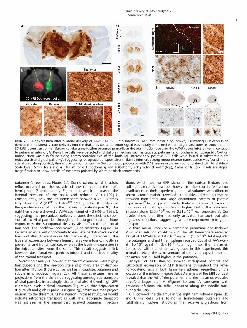
MR-guided infusion of AAV5-GFP. The left hemisphere received125 μl of AAV5-GFP at 1.0 × 10¹³ vg ml− 1 (1.3 × 10¹² total vg) intothe putamen, and right hemisphere received 200 μl of AAV5-GFPat 1 × 10¹³ vg ml− 1 (2. × 1012 total vg) into the thalamus.Compared with the other two groups in this experiment, thisanimal received the same amount of total viral capsids into thethalamus, but 2.5-fold higher in the putamen.Analysis of GFP staining showed widespread cortical and
subcortical expression of GFP transgene throughout the ante-rior–posterior axis in both brain hemispheres, regardless of thelocation of the infusion (Figure 3a). 3D analysis of the MRI contrastrevealed that the Vd of the putamen and the thalamus was alsothreefold larger than Vi (Figures 3b and c), consistent withprevious infusions. No reflux occurred along the needle trackduring delivery.GFP covered the thalamus in the right hemisphere (Figure 3b)
and GFP-ir cells were found in homolateral putamen andsubthalamic nucleus, structures that receive projections from
Figure 2. GFP expression after bilateral delivery of AAV5-CAG-GFP into thalamus. DAB immunostaining (brown) illustrating GFP expressionderived from bilateral vector delivery into the thalamus (a). Gadolinium signal was mostly contained within target structures as shown in the3D MRI reconstruction (b). Strong cellular transduction occurred primarily at the brain nuclei receiving the AAV5 vector infusion (c). In contrastto putaminal infusion, GFP-positive cells were detected in distal brain regions such as caudate, putamen and subthalamic nucleus (d). Corticaltransduction was also found along antero-posterior axis of the brain (e). Interestingly, positive GFP cells were found in substantia nigrareticulata (f) and globi pallidi (g), suggesting retrograde transport after thalamic infusion. Strong motor neuron transduction was found in thespinal cord along cervical, thoracic or lumbar regions (h). Sections were processed with DAB immunostaining counterstained with Nissl (blue).Scale bars= 5 mm for a and e; 100 μm for c, f (bottom), g and h (bottom); 200 μm for d and f (top); 2 mm for h (top). Insets are digitalmagnification to show details of the areas pointed by white or black arrowheads.
Brain delivery of AAV serotype 5L Samaranch et al
3
Gene Therapy (2017), 1 – 9
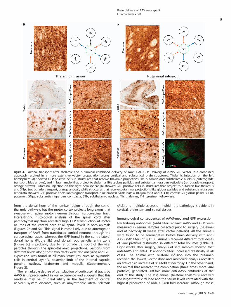
thalamus (Figure 4a), supporting our finding that AAV5 undergoesanterograde transport. Nevertheless, GFP-positive cells were alsofound in SNpr and GPi (Figure 4a), indicating that AAV5 is alsoretrogradely transported. Cerebral cortex, from frontal to occipitalregion, was highly transduced through cortico-thalamo-corticalloop (Figure 4a). This topological distribution of GFP-ir cell wasidentical to what was observed after the bilateral thalamicinfusion.On the other hand, histologic analysis of the putaminal injection
in the left hemisphere revealed considerable differences from theprevious putaminal infusion. High levels of GFP transduction werepresent at the injection site (Figure 3c). Distal structures were alsoanalyzed and GFP signal was found in the terminals of the striato-nigral projections, confirming the presence of anterogradetransport. But, unlike what was observed after the bilateralputaminal infusion, GFP-positive neurons were found in cerebralcortex, thalamus and substantia nigra pars compacta, regions that
project to the putamen, confirming that AAV5 viral particlesunderwent retrograde transport (Figure 4b), even after putaminalinjection. Similarly to the bilateral thalamic infusion, the combinednumber of particles for putaminal delivery was also higher thanthe previous one (2.5-fold higher). In our hands, high doses ofAAV5 in the range of 1.0–1.5 × 10¹² total vg, triggers retrogradetransport of the vector, if properly delivered into the brain, clearlysuggesting that retrograde axonal transport is dose dependent.
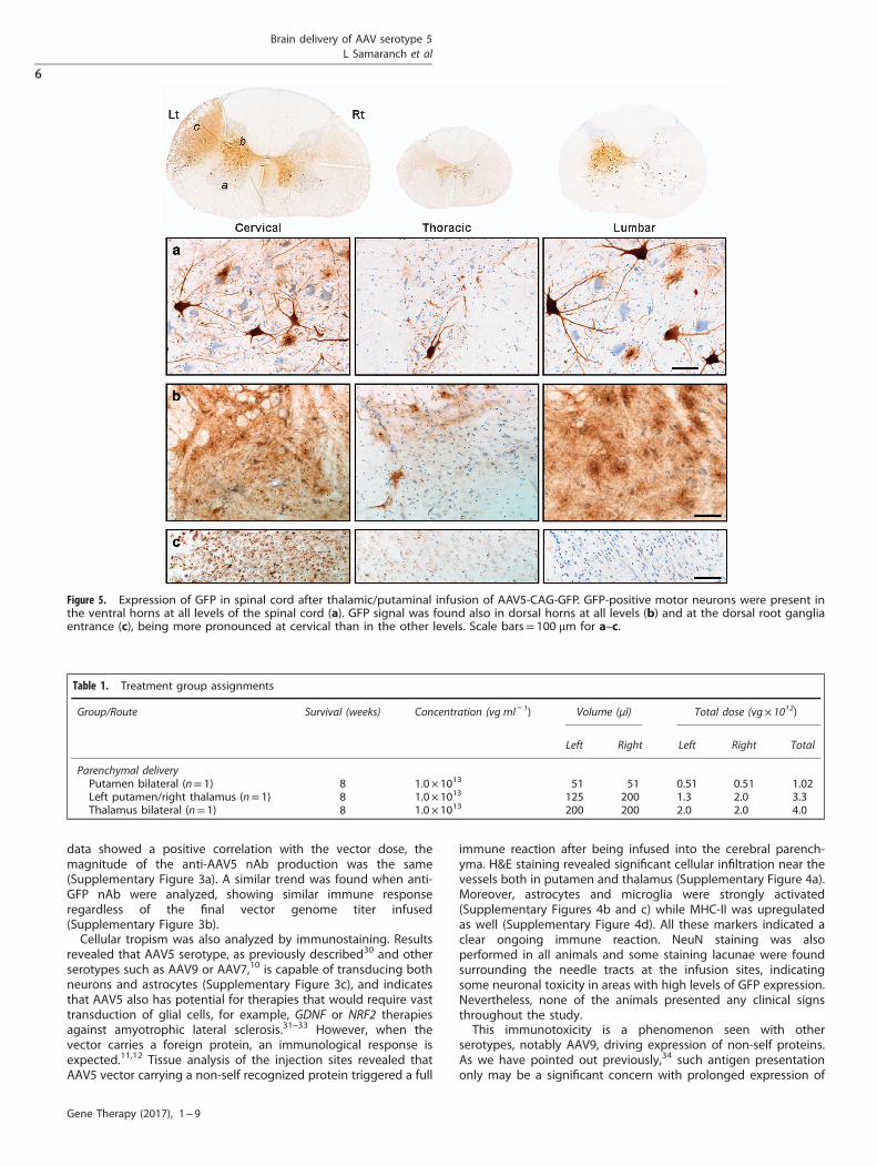
Spinal transduction after intracerebral infusion of AAV5-GFP viralparticlesAs in the putaminal infusion, spinal cord was also analyzed in theanimals that received AAV5 vector in the thalamus. Besides theascending and descending fibers that form the thalamo-cortico-thalamic and the striato-cortical projections, thalamus and cortexalso connect with spinal cord. Ventral posterior-lateral nucleus ofthe thalamus synapses with ascending axons that arise mainly
Figure 3. GFP expression after delivery of AAV5-CAG-GFP into left putamen and right thalamus. Infusion of AAV5-CAG-GFP vector into thebrain resulted in a widespread expression of the transgene throughout target structures for all animals. DAB immunostaining (brown) showedGFP expression into cortical and subcortical structures along prefrontal and occipital regions of the brain. Histology analysis of the anatomicaltargets showed a massive transduction both after thalamic (b) and putaminal infusions (c). Sections were processed with DABimmunostaining counterstained with Nissl (blue). Scale bars= 5 mm for a.
Brain delivery of AAV serotype 5L Samaranch et al
4
Gene Therapy (2017), 1 – 9
from the dorsal horn of the lumbar region through the spino-thalamic pathway, but the motor cortex projects long axons thatsynapse with spinal motor neurons through cortico-spinal tract.Interestingly, histological analysis of the spinal cord afterparenchymal injection revealed high GFP transduction of motorneurons of the ventral horn at all spinal levels in both animals(Figures 2h and 5a). This signal is most likely due to anterogradetransport of AAV5 from transduced cortical neurons through thecortico-spinal tracts, whereas the GFP found in the contra-lateraldorsal horns (Figure 5b) and dorsal root ganglia entry zone(Figure 5c) is probably due to retrograde transport of the viralparticles through the spino-thalamic projections. Sections fromdifferent levels along these two tracts were also analyzed and GFPexpression was found in all main structures, such as pyramidalcells in cortical layer V, posterior limb of the internal capsule,pontine nucleus, brainstem and medulla (SupplementaryFigure 2).The remarkable degree of transduction of corticospinal tracts by
AAV5 is unprecedented in our experience and suggests that thisserotype may be of great utility in the treatment of centralnervous system diseases, such as amyotrophic lateral sclerosis
(ALS) and multiple sclerosis, in which the pathology is evident incortical, brainstem and spinal tissues.
Immunological consequences of AAV5-mediated GFP expressionNeutralizing antibodies (nAb) titers against AAV5 and GFP weremeasured in serum samples collected prior to surgery (baseline)and at necropsy (8 weeks after vector delivery). All the animalswere found to be seronegative before brain delivery with anti-AAV5 nAb titers of ⩽ 1:100. Animals received different total dosesof viral particles distributed in different total volumes (Table 1).Eight weeks after surgery, analysis of sera samples showed thatanti-AAV5 and anti-GFP antibody titers increased drastically in allcases. The animal with bilateral infusion into the putamenreceived the lowest vector dose and molecular analysis revealedan anti-capsid increase of 851-fold at necropsy. On the other hand,the animal that received the combination (three times more viralparticles) generated 908-fold more anti-AAV5 antibodies at theend of the study. The last animal (bilateral thalamus) receivedthe largest total viral load and the serum levels correlated with thehighest production of nAb, a 1488-fold increase. Although these
Figure 4. Axonal transport after thalamic and putaminal combined delivery of AAV5-CAG-GFP. Delivery of AAV5-GFP vector in a combinedapproach resulted in a more extensive vector propagation along cortical and subcortical brain structures. Thalamic injection on the lefthemisphere (a) showed GFP-positive cells in structures that receive thalamic projections like putamen and subthalamic nucleus (anterogradetransport, blue arrows), and in brain nuclei that project to thalamus like globus pallidus and substantia nigra pars reticulate (retrograde transport,orange arrows). Putaminal injection on the right hemisphere (b) showed GFP-positive cells in structures that project to putamen like thalamusand SNpc (retrograde transport, orange arrows), while structures that receive putaminal projections like globus pallidus and substantia nigra parsreticulata showed GFP-positive fibers (anterograde transport, blue arrows). Scale bars=100 μm for a and b. Ctx, cortex; GP, globus pallidus; Put,putamen; SNpc, substantia nigra pars compacta; STN, subthalamic nucleus; Th, thalamus; TH, tyrosine hydroxylase.
Brain delivery of AAV serotype 5L Samaranch et al
5
Gene Therapy (2017), 1 – 9
data showed a positive correlation with the vector dose, themagnitude of the anti-AAV5 nAb production was the same(Supplementary Figure 3a). A similar trend was found when anti-GFP nAb were analyzed, showing similar immune responseregardless of the final vector genome titer infused(Supplementary Figure 3b).Cellular tropism was also analyzed by immunostaining. Results
revealed that AAV5 serotype, as previously described30 and otherserotypes such as AAV9 or AAV7,10 is capable of transducing bothneurons and astrocytes (Supplementary Figure 3c), and indicatesthat AAV5 also has potential for therapies that would require vasttransduction of glial cells, for example, GDNF or NRF2 therapiesagainst amyotrophic lateral sclerosis.31–33 However, when thevector carries a foreign protein, an immunological response isexpected.11,12 Tissue analysis of the injection sites revealed thatAAV5 vector carrying a non-self recognized protein triggered a full
immune reaction after being infused into the cerebral parench-yma. H&E staining revealed significant cellular infiltration near thevessels both in putamen and thalamus (Supplementary Figure 4a).Moreover, astrocytes and microglia were strongly activated(Supplementary Figures 4b and c) while MHC-II was upregulatedas well (Supplementary Figure 4d). All these markers indicated aclear ongoing immune reaction. NeuN staining was alsoperformed in all animals and some staining lacunae were foundsurrounding the needle tracts at the infusion sites, indicatingsome neuronal toxicity in areas with high levels of GFP expression.Nevertheless, none of the animals presented any clinical signsthroughout the study.This immunotoxicity is a phenomenon seen with other
serotypes, notably AAV9, driving expression of non-self proteins.As we have pointed out previously,34 such antigen presentationonly may be a significant concern with prolonged expression of
Figure 5. Expression of GFP in spinal cord after thalamic/putaminal infusion of AAV5-CAG-GFP. GFP-positive motor neurons were present inthe ventral horns at all levels of the spinal cord (a). GFP signal was found also in dorsal horns at all levels (b) and at the dorsal root gangliaentrance (c), being more pronounced at cervical than in the other levels. Scale bars= 100 μm for a–c.
Table 1. Treatment group assignments
Group/Route Survival (weeks) Concentration (vg ml− 1) Volume (μl) Total dose (vg× 1012)
Left Right Left Right Total
Parenchymal deliveryPutamen bilateral (n= 1) 8 1.0 × 10¹³ 51 51 0.51 0.51 1.02Left putamen/right thalamus (n= 1) 8 1.0 × 10¹³ 125 200 1.3 2.0 3.3Thalamus bilateral (n= 1) 8 1.0 × 10¹³ 200 200 2.0 2.0 4.0
Brain delivery of AAV serotype 5L Samaranch et al
6
Gene Therapy (2017), 1 – 9
foreign, or non-self-recognized transgenes. In clinical studies, inwhich self-proteins are being expressed, this type of immunotoxi-city should not be a problem, no matter whether the serotype isshowing astrocytic tropism or not.In summary, this exploratory study suggests that AAV5 has
considerable potential for the delivery of therapeutic candidategenes, where more global transduction of the brain is required,such as neurological pathologies ranging from Huntington diseaseto lysosomal storage disorders or spinal disorders. Even so, furtherexperiments are necessary to obtain a more comprehensivedescription of the dose-dependent axonal transport.
MATERIALS AND METHODSAnimalsThree male adult Cynomolgus macaques (Macaca fascicularis;6.45–6.61 kg) were included in this exploratory study (Table 1).Animals were screened for the presence of anti-AAV antibodiesprior to dosing and at termination as previously described.35 Allanimals were considered seronegative with neutralizing antibodytiters lower than 1:100 at baseline.
In-life observationsSince AAV5 encoded the gene for GFP, a foreign protein, animmune reaction was expected as previously described.12 There-fore, veterinary personnel monitored all animals after AAV deliveryuntil the end of the study to identify any possible adverse effects.Cage-side observations were performed twice daily throughoutthe study to evaluate general health, appearance, and appetite inall animals. Animals were weighed prior to surgery, weekly for thefirst 4 weeks after surgery, and biweekly thereafter until theconclusion of the study. None of the animals showed any adverseeffects.
Vector design and productionThe AAV5 vector encoding the cDNA of the enhanced GFP genewas packaged into AAV5 by a baculovirus-based method atAmsterdam Molecular Therapeutics (uniQure NV) as previouslydescribed,36,37 resulting in a single-stranded rAAV vector. Briefly,the GFP coding sequence was preceded at the 5′ end by a Kozaksequence and at the 3′ end by the bovine growth hormonepolyadenylation signal. The complete transcription unit wasflanked by two non-coding AAV-derived inverted terminal repeats,and expression was driven by a CAG promoter, a combination ofthe cytomegalovirus early enhancer element and chicken β-actinpromoter. For vector production, Sf9 insect cells were infected bythree recombinant baculoviruses; (i) encoding rep for replicationand packaging, (ii) cap-5 for the AAV5 capsid and (iii) with theexpression cassette. Cap-5 sequence was adapted and optimizedto the baculovirus-mediated assembly of rAAV. Although Cap-5sequence maintained its original amino acid sequence, theoptimization process (patent pending) improved its assemblyability and increase infectivity of the resulting AAV vector (patentpending). After viral particle assembled, prep purification wasperformed with AVB Sepharose high-performance affinity medium(GE Healthcare, Piscataway, NJ, USA). Vector concentration wasdetermined by quantitative PCR with primer-probe combinationsdirected against the transgene and the one with highestpackaging rate was selected for study. The final titer of the vectorpreparation was 1.4 × 10¹⁴ vg ml− 1.The single vector dilution assigned to parenchymal infusion
(1.0 × 10¹³ vg ml− 1) was spiked with gadolinium chelate (2 mM,Prohance, Bracco Diagnostics, Princeton, NJ, USA) to visualizedelivery during the MR-guided infusion.4 All drugs were preparedon the day of surgery by dilution of vector with phosphate-buffered saline (PBS) and 5% sucrose.
MR-guided vector deliveryAnimals were randomly assigned for the parenchymal infusions ofvirus into putamen (n= 1), thalamus (n= 1) or both (n= 1).Volumes and titers of different injections are described in Table 1.
Brain delivery. Briefly, animals were sedated (intramuscularketamine (10 mg kg− 1) and medetomidine (0.015 mg kg− 1)),intubated and maintained under anesthesia with isoflurane(1–3% v/v). Then, animals were placed supine in an MR-compatible stereotactic frame and, after craniotomy, underwentstereotactic placement of MR-compatible, skull-mounted, tempor-ary ball-joint cannula guides over each hemisphere.38 Animalswere then moved into the MRI (GE 1.5T MRI mobile unit, DMS,Imaging Reno, NV, USA) to determine the trajectory to target brainnuclei inside the brain. Once the trajectory was determined, aceramic custom-designed fused silica reflux-resistant cannula witha 3-mm stepped tip was used for the infusion.1,4,39,40 Infusion ratewas ramped up to a maximum of 3 μl min − 1. Once the infusionended, skull-mounted guide devices were removed and animalswere taken back to their home cages and monitored duringrecovery from anesthesia.
Tissue collection and section processingAnimals were transcardially perfused first with PBS and then with4% paraformaldehyde (PFA) in PBS. Brains were harvested, slicedinto 6-mm coronal blocks in a brain matrix, post-fixed byimmersion in 4% PFA/PBS overnight and then transferred to30% (w/v) sucrose. Representative segments of the spinal cords atcervical, thoracic and lumbar levels were collected and post-fixedas well. A sliding microtome was used to cut serial 40-μm sectionsfor histological processing.To assess transgene expression, we performed immunohisto-
chemistry with polyclonal antibodies against GFP (rabbit anti-GFP,1:1000, G10362, www.lifetechnologies.com). Briefly, tissue sectionswere washed in PBS, blocked in 1% H2O2/30% alcohol/PBS, andthen rinsed in PBST (PBS/1% Tween 20). Then incubated inBackground Sniper blocking solution (Cat. BS966G; www.biocare.net) followed by a 24-h incubation at 4 °C with primary antibodyagainst either GFP in Da Vinci Green diluent (PD900; www.biocare.net). The next day, sections were washed in PBST and incubated inRabbit Mach 2 HRP-polymer (RP531L; www.biocare.net) at roomtemperature for 1 h. All sections were chromogenically developedwith 3,3′-diaminobenzidine (DAB; DAB Peroxidase Substrate Kit,SK-4100, www.vectorlabs.com) according to the manufacturer’sinstructions. To study immune response, antibodies against glialfibrillary acidic protein (GFAP; mouse anti-GFAP, 1:100 000,MAB360 www.emdmillipore.com), ionized calcium-binding adap-tor molecule 1 (Iba1; rabbit anti-Iba1, 1:1000, CP290C www.biocare.net), the major histocompatibility complex class II (MHC-II;mouse anti-MHC-II, 1:300, M3887-30, www.usbio.net) and neuro-nal protein NeuN (mouse anti-NeuN, 1:5000, MAB377, www.merckmillipore.com) were used separately with the immunoper-oxidase staining protocol described above.Standard hematoxylin and eosin (H&E) staining was performed
on brain sections from all animals to detect pathological signs.Briefly, slide-mounted sections were washed in graded alcohol(100, 95 and 70% v/v; 3 min each), rehydrated in distilled waterand stained in Gill II hematoxylin (www.leica-microsystems.com)for 3 min. After sections were washed, they were differentiated in0.5% glacial acetic acid/70% alcohol, immersed in bluing solutionand counterstained for 2 min in eosin Y (www.leica-microsystems.com). Finally, after washing them a final time, sections weredehydrated in alcohol (95 and 100%; 3 min each), immersed infresh xylene (2 × 3 min), and coverslip and preserved withShandon mounting medium (www.thermofisher.com).
Neutralizing activity analysisNeutralizing antibodies against AAV5 capsid present in bloodwere analyzed before and after surgery. Typically, a cell-based
Brain delivery of AAV serotype 5L Samaranch et al
7
Gene Therapy (2017), 1 – 9
neutralizing antibody assay is used to determine the level of suchantibodies, but, since the AAV serotype 5 does not transduceefficiently cells in vitro, an in-house enzyme-linked immunosor-bent assay (ELISA) for total amount of IgG against AAV5 capsidwas developed. It is well known that the titer of nAb against thecapsid is proportional to the total amount of IgG against thiscapsid. Briefly, MaxiSorp 96-well plates (www.sigmaaldrich.com)were coated with AAV5-GFP vector at 3 × 10¹⁰ vg ml− 1. Afterblocking unspecific binding with Block & Sample Buffer (G3311;www.promega.com), serial dilutions of serum samples (from 1:50to 1:6400) were added to the wells and incubated for 2 h at roomtemperature. After washing plates in Tris-buffered saline withTween-20 (28360; www.thermofisher.com) goat anti-monkey IgG-HRP diluted with BS buffer at 1:20 000 (43R-IG020HRP; www.fitzgerald-fii.com) was added to all wells and the plates wereincubated for 2 h. Following five washes in TBST buffer, sampleswere incubated for 5 min by adding SuperSignal ELISA PicoChemiluminescent Substrate (37070; www.thermofisher.com) andthen luminescence signal was detected by SpectraMax i3 platereader (www.moleculardevices.com). The nAb production wasmeasured by comparison to the signal from serum samplescollected before the surgery. Fold differences were calculated bydividing normalized post-surgery and pre-surgery intensity valuesat 1:1600 dilution.
Study approvalAll procedures were carried out with the approval of theInstitutional Animal Care and Use Committee (IACUC), and inaccordance with the Standard Operating Procedures protocolat Valley Biosystems, West Sacramento, CA, USA (IACUC permit:14-10470).
CONFLICT OF INTERESTBas Blits and Harald Petry are employees and shareholders at UniQure. KrystofBankiewicz and UCSF have a collaborative agreement with UniQure.
ACKNOWLEDGEMENTSWe thank Foad Green for his technical assistance in histology. UniQure funded thepresent study and provided the AAV5 viral vector for the experiments. We thankJacek Lubelski for sharing his expertise on vector development.
REFERENCES1 Bobo RH, Laske DW, Akbasak A, Morrison PF, Dedrick RL, Oldfield EH. Convection-
enhanced delivery of macromolecules in the brain. Proc Natl Acad Sci USA 1994;91: 2076–2080.
2 Richardson RM, Kells AP, Rosenbluth KH, Salegio EA, Fiandaca MS, Larson PS et al.Interventional MRI-guided putaminal delivery of AAV2-GDNF for a planned clinicaltrial in Parkinson’s disease. Mol Ther 2011; 19: 1048–1057.
3 Richardson RM, Kells AP, Martin AJ, Larson PS, Starr PA, Piferi PG et al. Novelplatform for MRI-guided convection-enhanced delivery of therapeutics: preclinicalvalidation in nonhuman primate brain. Stereotact Funct Neurosurg 2011; 89:141–151.
4 Fiandaca MS, Forsayeth JR, Dickinson PJ, Bankiewicz KS. Image-guided convec-tion-enhanced delivery platform in the treatment of neurological diseases. Neu-rotherapeutics 2008; 5: 123–127.
5 Fiandaca MS, Varenika V, Eberling J, McKnight T, Bringas J, Pivirotto P et al. Real-time MR imaging of adeno-associated viral vector delivery to the primate brain.NeuroImage 2009; 47: T27–T35.
6 Salegio EA, Samaranch L, Kells AP, Mittermeyer G, San Sebastian W, Zhou S et al.Axonal transport of adeno-associated viral vectors is serotype-dependent. GeneTher 2013; 20: 348–352.
7 Foust KD, Nurre E, Montgomery CL, Hernandez A, Chan CM, Kaspar BK. Intra-vascular AAV9 preferentially targets neonatal neurons and adult astrocytes. NatBiotechnol 2009; 27: 59–65.
8 Gray SJ, Matagne V, Bachaboina L, Yadav S, Ojeda SR, Samulski RJ.Preclinical differences of intravascular AAV9 delivery to neurons and glia: acomparative study of adult mice and nonhuman primates. Mol Ther 2011; 19:1058–1069.
9 Samaranch L, Salegio EA, San Sebastian W, Kells AP, Foust KD, Bringas JR et al.Adeno-associated virus serotype 9 transduction in the central nervous system ofnonhuman primates. Hum Gene Ther 2012; 23: 382–389.
10 Samaranch L, Salegio EA, San Sebastian W, Kells AP, Bringas JR, Forsayeth J et al.Strong cortical and spinal cord transduction after AAV7 and AAV9 deliveryinto the cerebrospinal fluid of nonhuman primates. Hum Gene Ther 2013; 24:526–532.
11 Ciesielska A, Hadaczek P, Mittermeyer G, Zhou S, Wright JF, Bankiewicz KS et al.Cerebral infusion of AAV9 vector-encoding non-self proteins can elicit cell-mediated immune responses. Mol Ther 2013; 21: 158–166.
12 Samaranch L, San Sebastian W, Kells AP, Salegio EA, Heller G, Bringas JR et al.AAV9-mediated expression of a non-self protein in nonhuman primate centralnervous system triggers widespread neuroinflammation driven by antigen-presenting cell transduction. Mol Ther 2014; 22: 329–337.
13 Kells AP, Hadaczek P, Yin D, Bringas J, Varenika V, Forsayeth J et al. Efficient genetherapy-based method for the delivery of therapeutics to primate cortex. ProcNatl Acad Sci USA 2009; 106: 2407–2411.
14 Kells AP, Forsayeth J, Bankiewicz KS. Glial-derived neurotrophic factor genetransfer for Parkinson's disease: anterograde distribution of AAV2 vectors in theprimate brain. Neurobiol Dis 2012; 48: 228–235.
15 Ciesielska A, Mittermeyer G, Hadaczek P, Kells AP, Forsayeth J, Bankiewicz KS.Anterograde axonal transport of AAV2-GDNF in rat basal ganglia. Mol Ther 2011;19: 922–927.
16 Thorne BA, Takeya RK, Peluso RW. Manufacturing recombinant adeno-associatedviral vectors from producer cell clones. Hum Gene Ther 2009; 20: 707–714.
17 Martin J, Frederick A, Luo Y, Jackson R, Joubert M, Sol B et al. Generationand characterization of adeno-associated virus producer cell lines forresearch and preclinical vector production. Hum Gene Ther Methods 2013; 24:253–269.
18 Hadaczek P, Stanek L, Ciesielska A, Sudhakar V, Samaranch L, Pivirotto P et al.Widespread AAV1- and AAV2-mediated transgene expression in the nonhumanprimate brain: implications for Huntington's disease. Mol Ther Methods Clin Dev2016; 3: 16037.
19 Dong B, Duan X, Chow HY, Chen L, Lu H, Wu W et al. Proteomics analysis of co-purifying cellular proteins associated with rAAV vectors. PLoS One 2014; 9:e86453.
20 San Sebastian W, Samaranch L, Heller G, Kells AP, Bringas J, Pivirotto P et al.Adeno-associated virus type 6 is retrogradely transported in the non-humanprimate brain. Gene Ther 2013; 20: 1178–1183.
21 Green F, Samaranch L, Zhang HS, Manning-Bog A, Meyer K, Forsayeth J et al.Axonal transport of AAV9 in nonhuman primate brain. Gene Ther 2016; 23:520–526.
22 Matsuzaki Y, Konno A, Mukai R, Honda F, Hirato M, Yoshimoto Y et al.Transduction profile of the marmoset central nervous system using adeno-associated virus serotype 9 vectors. Mol Neurobiol 2016; doi:10.1007/s12035-016-9777-6.
23 Castle MJ, Perlson E, Holzbaur EL, Wolfe JH. Long-distance axonal transport ofAAV9 is driven by dynein and kinesin-2 and is trafficked in a highly motile Rab7-positive compartment. Mol Ther 2014; 22: 554–566.
24 Cressant A, Desmaris N, Vérot L, Bréjot T, Froissart R, Vanier M-T et al. Improvedbehavior and neuropathology in the mouse model of Sanfilippo type IIIB diseaseafter adeno-associated virus-mediated gene transfer in the striatum. J Neurosci2004; 24: 10229–10239.
25 Ellinwood NM, Ausseil J, Desmaris N, Bigou S, Liu S, Jens JK et al. Safe, efficient,and reproducible gene therapy of the brain in the dog models of Sanfilippo andHurler syndromes. Mol Ther 2011; 19: 251–259.
26 Keiser MS, Boudreau RL, Davidson BL. Broad therapeutic benefit after RNAiexpression vector delivery to deep cerebellar nuclei: implications for spinocer-ebellar ataxia type 1 therapy. Mol Ther 2013; 22: 588–595.
27 Zabner J, Seiler M, Walters R, Kotin RM, Fulgeras W, Davidson BL et al. Adeno-associated virus type 5 (AAV5) but not AAV2 binds to the apical surfaces of airwayepithelia and facilitates gene transfer. J Virol 2000; 74: 3852–3858.
28 Rayaprolu V, Kruse S, Kant R, Venkatakrishnan B, Movahed N, Brooke D et al.Comparative analysis of adeno-associated virus capsid stability and dynamics.J Virol 2013; 87: 13150–13160.
29 Emborg ME, Hurley SA, Joers V, Tromp DPM, Swanson CR, Ohshima-Hosoyama Set al. Titer and product affect the distribution of gene expression after intraputaminalconvection-enhanced delivery. Stereotact Funct Neurosurg 2014; 92: 182–194.
30 Davidson BL, Stein CS, Heth JA, Martins I, Kotin RM, Derksen TA et al. Recombinantadeno-associated virus type 2, 4, and 5 vectors: transduction of variant cell typesand regions in the mammalian central nervous system. Proc Natl Acad Sci 2000;97: 3428–3432.
31 Nanou A, Higginbottom A, Valori CF, Wyles M, Ning K, Shaw P et al. Viral deliveryof antioxidant genes as a therapeutic strategy in experimental models ofamyotrophic lateral sclerosis. Mol Ther 2013; 21: 1486–1496.
Brain delivery of AAV serotype 5L Samaranch et al
8
Gene Therapy (2017), 1 – 9
32 Merienne N, Douce JL, Faivre E, Déglon N, Bonvento G. Efficient gene delivery andselective transduction of astrocytes in the mammalian brain using viral vectors.Front Cell Neurosci 2013; 7: 106.
33 Jakobsson J, Lundberg C. Lentiviral vectors for use in the central nervous system.Mol Ther 2006; 13: 484–493.
34 Forsayeth J, Bankiewicz KS. Transduction of antigen-presenting cells in the brainby AAV9 warrants caution in preclinical studies. Mol Ther 2015; 23: 612.
35 San Sebastian W, Kells AP, Bringas J, Samaranch L, Hadaczek P, Ciesielska A et al.Safety and tolerability of MRI-guided infusion of AAV2-hAADC into the mid-brainof non-human primates. Mol Ther Methods Clin Dev 2014; 3. doi:10.1038/mtm.2014.49.
36 Urabe M, Ding C, Kotin RM. Insect cells as a factory to produce adeno-associatedvirus type 2 vectors. Hum Gene Ther 2002; 13: 1935–1943.
37 Unzu C, Hervás-Stubbs S, Sampedro A, Mauleón I, Mancheño U, Alfaro C et al.Transient and intensive pharmacological immunosuppression fails to improveAAV-based liver gene transfer in non-human primates. J Transl Med 2012; 10:122.
38 Salegio EA, Bringas J, Bankiewicz KS. MRI-guided delivery of viral vectors. MethodsMol Biol 2016; 1382: 217–230.
39 Bankiewicz KS, Eberling JL, Kohutnicka M, Jagust W, Pivirotto P, Bringas J et al.Convection-enhanced delivery of AAV vector in parkinsonian monkeys; in vivodetection of gene expression and restoration of dopaminergic function using pro-drug approach. Exp Neurol 2000; 164: 2–14.
40 Krauze MT, Saito R, Noble C, Tamas M, Bringas J, Park JW et al. Reflux-free cannulafor convection-enhanced high-speed delivery of therapeutic agents. J Neurosurg2005; 103: 923–929.
This work is licensed under a Creative Commons Attribution-NonCommercial-NoDerivs 4.0 International License. The images or
other third party material in this article are included in the article’s Creative Commonslicense, unless indicatedotherwise in the credit line; if thematerial is not included underthe Creative Commons license, users will need to obtain permission from the licenseholder to reproduce the material. To view a copy of this license, visit http://creativecommons.org/licenses/by-nc-nd/4.0/
© The Author(s) 2017
Supplementary Information accompanies this paper on Gene Therapy website (http://www.nature.com/gt)
Brain delivery of AAV serotype 5L Samaranch et al
9
Gene Therapy (2017), 1 – 9