mr. j. anand raj kumar (reg. no. 261226002) under the ...repository-tnmgrmu.ac.in/1954/1/anand raj...
TRANSCRIPT

'' Evaluation of Antiurolithiatic activity of ethanolic extract of Phyllanthus
urinaria against ethylene glycol induced urolithiasis in wistar albino rats.''
Dissertation submitted to
THE TAMILNADU Dr. M.G.R MEDICAL UNIVERSITY
CHENNAI
In partial fulfillment of the requirements for the degree of
MASTER OF PHARMACY in PHARMACOLOGY
BY
Mr. J. ANAND RAJ KUMAR (Reg. No. 261226002)
Under the guidance of
J.GUNASEKARAN M.Pharm
Associate Professor and Head
Department of Pharmacology
MOHAMED SATHAK A.J. COLLEGE OF PHARMACY,
SHOLINGANALLUR,CHENNAI - 600119.
April-2014

CERTIFICATE
This is to certify that the dissertation entitled “Evaluation of
Antiurolithiatic activity of ethanolic extract of Phyllanthus urinaria against
ethylene glycol induced urolithiasis in wistar albino rats.” submitted to The
Tamilnadu Dr. M.G.R. Medical university, Chennai, in partial fulfillment for the award of degree
of Master of Pharmacy in Pharmacology is a bonafide individual research work done by Mr.
J. ANAND RAJ KUMAR Mohamed Sathak A. J. College of Pharmacy, Chennai, under the
guidance and direct supervision of J.GUNASEKARAN M.Pharm, Associate Professor,
Department of Pharmacology during the academic year 2013-2014.
Place: Chennai
Date:
(Dr.R.SUNDARARAJAN,M.pharm.,Ph.D.,)
Principal,
J.GUNASEKARAN, M.Pharm

Associate Professor and Head
Department of Pharmacology.
CERTIFICATE
This is to certify that the dissertation entitled “Evaluation of Antiurolithiatic
activity of ethanolic extract of Phyllanthus urinaria against ethylene glycol
induced urolithiasis in wistar albino rats.” submitted to The Tamilnadu Dr. M.G.R.
Medical university, Chennai, in partial fulfillment for the award of degree of Master of
Pharmacy in Pharmacology is a bonafide individual research work done by Mr. J. ANAND
RAJ KUMAR (Reg.No.261226002), Mohamed Sathak A.J.College of Pharmacy, Chennai,
under the guidance and direct supervision of J.GUNASEKARAN M.Pharm, Associate
Professor, Department of Pharmacology during the academic year 2013-2014.
Place: Chennai
Date: (J.GUNASEKARAN, M.Pharm)
Head, Department of Pharmacology
J.GUNASEKARAN, M.Pharm
Associate Professor and Head

Department of Pharmacology
CERTIFICATE
This is to certify that the dissertation entitled “Evaluation of Antiurolithiatic
activity of ethanolic extract of Phyllanthus urinaria against ethylene glycol
induced urolithiasis in wistar albino rats.” submitted to The Tamilnadu Dr. M.G.R.
Medical university, Chennai, in partial fulfillment for the award of degree of Master of
Pharmacy in Pharmacology is a bonafide individual research work done by Mr.J.ANAND
RAJ KUMAR (Reg.No.261226002), Mohamed Sathak A.J.College of Pharmacy, Chennai,
under the guidance and direct supervision of J.GUNASEKARAN M.Pharm, Associate
Professor, Department of Pharmacology during the academic year 2013-2014.
Place: Chennai
Date:
(J.GUNASEKARAN, M.Pharm)
Guide and Supervisor


Mr. J. ANAND RAJ KUMAR
II year- M.Pharm, Pharmacology (Reg.no: 261226002),
Department of pharmacology,
Mohamed Sathak A. J. College of Pharmacy.
DECLARATION OF THE CANDIDATE
I hereby declare that the thesis titled “Evaluation of Antiurolithiatic activity of
ethanolic extract of Phyllanthus urinaria against ethylene glycol induced urolithiasis in
wistar albino rats.” submitted in partial fulfillment for the award of degree Master of Pharmacy
to The Tamilnadu Dr. M.G.R. Medical University and carried out at Mohamed Sathak
A.J.College of Pharmacy, Chennai, is my original and independent work done under the direct
supervision and guidance of J.GUNASEKARAN M.Pharm, Associate Professor,
Department of Pharmacology during the academic year 2013-2014 and this thesis contains no
material which has been accepted for the award of any degree or diploma of other Universities.
Place: Chennai
Date: [J.ANAND RAJ KUMAR]

Acknowledgment
ACKNOWLEDGEMENT
I take this opportunity to express my heartfelt thanks to all those, who knowingly
or unknowingly contributed to the success of my dissertation work.
I wish to express my gratitude towards “GOD – Almighty”, who gave me the
strength and courage to fulfill my dream and has showered upon me his choicest blessings.
My heartfelt thanks to my parents and my brother for their love affection and
constantly encouraging, guiding when I thought nothing is happening.
I wish to express my deepest gratitude to Management of Mohamed Sathak
trust, Chennai and Management of Mohamed Sathak A.J. College of Pharmacy, in
acknowledging all facilities provided to use at the institution enabling us to do work of this
magnitude.
I express my sincere thanks to Dr. R. Sundhararajan, M.Pharm., Ph.D.,
Principal, Mohamed Sathak A.J. College of Pharmacy, for his moral encouragement and
providing necessary facilities required for my dissertation work.
It is indeed a great pleasure to express my deep sense of gratitude and humble
thanks to my guide J.GUNASEKARAN M.Pharm, Associate Professor & Head of the
Department of Pharmacology,Mohamed Sathak A.J. College Of Pharmacy, Chennai, for his
invaluable guidance and constant encouragement that formed the foundation of this project. His
discipline, principle, simplicity, the profound knowledge and the subject understanding
influenced me a lot. I am proud to say that it has been a most fruitful and enjoyable experience to
work under his untiring and dynamic guidance.
I am deeply indebted to the teaching staff especially Dr. Deepa Sankar, M.
Pharm., Ph.D., Vice principle, Mohamed Sathak A.J. College of Pharmacy, Mrs. M. Komala,
M. Pharm, (Ph.D)., HOD and Mrs. N. B. Santha Sheela, M. Pharm., (Ph.D)., Assistant
professor, Department of Pharmaceutics and other teaching staff including Mr. S.
Ramachandran, Mr. Shakti Saravanan, M. Pharm., who was always a source of knowledge
and inspiration to me and also for their prompt assistance and cooperative attitude.

I thank Mr. A. Mohamad Jamaludeen, lab assistant, Department of Pharmacology
for his timely help.
I wish to express my special thanks to librarians Dr. M. Amudha, M.A.L.I.Sc.,
Ph.D., and Mrs. Kumari, M.A.L.I.Sc., for helping me in collecting my reference material.
I also wish to express my sincere thanks to Mr. C. Ramesh, M. Pharm., Ph.D.,
Director, SICRA labs, Hyderabad, for his technical support and advices given during the entire
course of my project work. With his dynamic approach he boosted my morale, which helped me
in completion of this dissertation.
I would like to thank Mr. Firasat Ali, M. Pharm., and Mr. Zuber Ali, M. Pharm.,
project officers for their valuable guidance, innovative advices, technical and moral support.
Friends are integral part of life, so I take this opportunity to thank my dearest friends
A.Pruthvidhar, Neelima, Saravanan, Dinesh Kumar, Jaya Kumar who always pushed my
confidence and creativity to the eventual extent of my mind and for their unflinching support and
co-operation during my dissertation.
Also I want to thank all teaching and non teaching staff, who directly or indirectly
helped me in completing this dissertation work successfully.
Thank you all…………..
J. Anand Raj Kumar.
LIST OF ABBREVATIONS
• AAP: American Academy of Pediatrics

•••• AHA: American Heart Association
•••• AMA: American Medical Association’s
•••• ATP III :Adult Treatment Panel III
• ACAT: Acyl coenzyme A cholesterol O-acyl transferase
• ACOA: Acetyl coenzyme A
• ANOVA: Analysis of variance
• ATP: Adenosine triphosphate
• BSCL2: Berardinelli-Seip congenital lipodystrophy, type 2
• BMI: Body mass index
• CAT: Catalase
• CETP: Cholesteryl ester transfer protein
•••• CHD: Coronary heart disease
• CHE: Cholesteryl ester
• CHOD POD: Cholesterol oxidase peroxidase
• CMC: Carboxy methyl cellulose
• CPCSEA: Committee for the purpose of control and supervision of experimental
animals
• CRP: C-reactive protein
• DMSO: Dimethyl sulfoxide
• FFA: Free fatty acid
• GPO: Glycerol phosphate oxidase
• GPx: Glutathione peroxidase
• GSH: S-glutathiolation
• HDL-C: High density lipoprotein cholesterol
• HFD: High fat diet HL: Hepatic lipase

• HMG-COA: 3-hydroxy 3-methyl glutaryl-CoA reductase
• HNE: 4-hydroxy-2-nonenal
• HOCL: Hypochlorous acid
• IDL: Intermediate density lipoprotein
•••• JNC7: Joint National Committee 7
• LCAT: Lecithin cholesterol acyl transferase
• LDL-C: Low density lipoprotein cholesterol
• LLD: Lipid lowering drugs
• LPL: Lipoprotein lipase
• LRP: LDL receptor related protein
• MDA: Malondialdehyde
• MI: Myocardial infraction
• MVA: Mevalonate
• NCEP: National Cholesterol Education Program
• NHLBI:National Heart Blood and Lung Institute
• NNT :number needed to treat
• NNH: number needed to harm
• OTC: over-the-counter
• OECD: Organization for economic co-operation and development
• P.O: Per oral
• PPARα: Paroxisome proliferator activated receptor α
• SEM: Standard error mean
• SOD: Superoxide dismutase
• SJM : Syzygium jambos(L) alston
• TBA: Trichloro butyric acid
• TC: Total cholesterol
• TCA: Trichloro acetic acid
•••• TG: Triglycerides
• TLC: Thin layer chromatography
• TLC: Therapeutic lifestyle change
•••• VLDL: Very low density lipoprotein cholesterol
• WHO: World health organization

CONTENTS
SL.
NOTITLE PAGE NO
I INTRODUCTION 1
II REVIEW OF LITERATURE 19
III AIM AND OBJECTIVES 22
IV PLANT PROFILE 24
V MATERIALS AND METHODS 28
VI RESULTS 44
VII DISCUSSION 72
VIII CONCLUSION 76
IX SUMMARY 78
X REFERENCES 80
1. INTRODUCTION
1.1 Definition
Urolithiasis: It is a consequence of complex physical processes. The major factors are
super saturation of urine with the offending salt and crystallization. Crystals retained
Department of pharmacology Page 1
in kidney can become nucleus for stone formation. This process is known as
Urolithiasis or nephrolithiasis.
1.2 Classification of renal stones
Urolithiasis or the renal stones which are formed in the kidneys of various forms
based on their chemical composition and their structures over 90 % of stones contain
uric acid, urates, oxalate or phosphates, the last two being most commonly found. Based
on the predominant chemical composition, Urolithiasis are classified as
1.2.1. Calcium containing stones
a. Calcium oxalate stones (CaOx)
b. Calcium phosphate stones (CaPh)
1.2.2. Uric acid stones
1.2.3. Struvite stones
1.2.4. Cystine stones and miscellaneous stones1-2
1.2.1 Calcium containing stones
Calcium stones are the most common type of kidney stones, accounting for about 70%
to 75% of stones. The highest incidence of calcium urolithiasis is between ages of 30
and 50 calcium stones either are composed entirely of CaOx or have a small core of
CaPh. The presence of a small amount of CaPh has no implications for pathogenesis
or management.
1.2.1.a Calcium oxalate stones
CaOx in pure form or in combination with CaPh is the most common component of
urinary stones. It generally varies from 4-8 mm in size. The commonest form of
oxalate is as calcium oxalate, mainly calcium oxalate monohydrate [Ca (COO)2, H2O],
but some calcium oxalate di hydrate crystals [Ca(COO)2, 2H2O] may also be present.
Oxalate stones are among the hardest found and so are not easily crushed.
Department of pharmacology Page 2

Idiopathic hypercalciuria: Hypercalciuria in the presence of normal serum calcium
is termed idiopathic hypercalciuria. The various causes of idiopathic hypercalciuria
are as follows:
� Absorptive hypercalciuria: The primary defect in absorptive hypercalciuria,
inherited as an autosomal dominant trait, is increased passive mucosal
absorption of calcium and oxalate in the jejunum.
� Renal hypercalciuria: In renal hypercalciuria, the underlying abnormality is a
primary renal wasting of calcium. Urinary losses of calcium reduce serum
calcium levels and cause a secondary elevation in parathyroid hormone (PTH).
This results in 1, 25-dihydroxy-vitamin D production and an increase in
intestinal calcium absorption.
Department of pharmacology Page 3
Fig 1: Stones in kidney (i) ureter and (ii) bladder

Resorptive hypercalciuria: The condition that results in hypercalciuria is increased
bone resorption, usually caused by subtle hyperparathyroidism. These patients have
parathyroid adenomas but do not have impressive hypercalcemia.
Primary hyperparathyroidism: Primary hyperparathyroidism is caused by a PTH-
secreting adenoma of the parathyroid glands. PTH increases serum calcium by the
following mechanisms:
1. PTH stimulates osteoclasts to demineralize bone by breaking down the bone crystal
apatite. The dissolution of apatite results in the release of calcium and phosphate into
the blood stream.
2. PTH causes calcium resorption by the kidneys and decreases renal absorption of
phosphates.
3. PTH stimulates production of 1, 25-dihydroxy vitamin D3 by the kidneys, which in
turn increases intestinal resorption of calcium. PTH does not seem to have an effect on
intestinal calcium absorption.
Hypercalcemia causes hypercalciuria, which predisposes to urinary calcium stone
formation. Decreased urinary citrate has been seen in hyper parathyroid patients.
Hypercalcemia of non parathyroid origin: Common causes of hypercalcemia-
causing urinary stone disease, include granulomatous diseases, hyperthyroidism,
glucocorticoid-induced hypercalcemia, pheochromocytoma, immobilization and
thiazide diuretics. These can be distinguished from hyperparathyroidism by serum
parathyroid hormone levels. Serum PTH is elevated in primary hyperparathyroidism,
whereas it is generally lower in other hyper calcemias.
Low urinary citrate: Citrate complexes urinary calcium and reduces its ionic
concentration. It inhibits spontaneous and heterogeneous nucleation of CaOx crystal.
Citrate restores the inhibitory activity of Tomm-Horsfall protein. The most important
cause of hypocitraturia is metabolic acidosis, which causes increased proximal tubular
reabsorption of citrate. Hypocitraturia is seen in 15% to 63% of patients with
urolithiasis.
Hyperoxaluria: Oxalate is the anion most frequently associated with calcium in the
precipitation of salts leading to crystal formation, growth, retention and stone
Department of pharmacology Page 4

formation. 80 % of urinary oxalate is endogenous in origin and 10% is dietary in
origin. Hyperoxaluria results from various factors like diet, genetic predisposure,
intestinal diseases and others.
� Dietary hyperoxaluria: Normal people excrete 20 – 40 mg (222 - 444 µmol)
of oxalate. A reasonable upper limit of excretion is 45 mg (500µmol) daily for
men and 40 mg for women. A simple dietary excess of oxalate of 50- 60 mg
(556-667µmol) is possible from foods such as spinach, rhubarb, Swiss chard,
cocoa, beets, peppers, wheat germ, pecans, peanuts, okra, chocolate and lime
peel. This type of hyperoxaluria is frequently observed in nephrolithiasis.
� Primary hyperoxaluria: Two genetic disorders lead to hyperoxaluria. Type I
primary oxaluria, an autosomal recessive trait is caused by molecular
abnormalities that reduce the activity of hepatic peroxisomal enzyme alanine:
glyoxylate aminotransferase, thereby increasing the availability of glyoxylate,
which is irreversibly converted to oxalic acid. Type II primary hyperoxaluria or
L-glyceric aciduria is a much rare variant caused by deficiencies of hepatic
enzymes D-glycerate dehydrogenase and glyoxylate reductase, which causes
an increase in urinary oxalate (135- 270 mg ) and glycerate excretion.
Enteric hyperoxaluria: Occurs in patients with short bowel syndrome or
malabsorption. The pathogenesis is exposure of the colonic mucosa to detergents in
the form of bile salts and fatty acids which nonselectively increases the permeability
to numerous molecules including oxalate. Hyperoxaluria causes oxalate crystal
formation, which combines with urinary calcium to form CaOx stones.
Hyperuricosuria: Excessive dietary intake of purine is the most common cause of
hyperuricosuria. An abnormality in the renal handling of urate is seen in such patients.
Uric acid promotes CaOx crystallization by facilitating the formation of nuclei.
Sodium hydrogen urate and uric acid crystals can initiate CaOx crystal formation in
seeded solution; however, sodium hydrogen urate crystals are not seen in fresh urine
or kidney stones.
1.2.1.b Calcium phosphate stones
Calcium phosphate stones occur only when the chemical pressure for crystallization is
high and thus they are usually seen in very active stone disease. Pure Calcium
Department of pharmacology Page 5

phosphate stones are almost always associated with the renal tubular acidification
defects. When the kidneys lose some of their ability to lower urinary pH, the resulting
higher pH increases the divalent and trivalent forms of phosphate, which causes
Calcium phosphate supersaturation3-4.
1.2.2 Uric acid stones
Uric acid calculi account for 5 to 10 % of all stones. The principal cause of uric acid
crystallization is the super saturation of urine with respect to un dissociated uric acid.
The most important risk factor is acid urine, with the pH persistently below 6.0 and
also infections of urinary tract.
1.2.3 Struvite stones
Struvite stones are composed of magnesium, ammonium and phosphate mixed with
carbonate. These stones are formed in urine with a pH of greater than 7.2 and
ammonia in the urine, produced by urease-producing bacteria. Urease hydrolyzes urea
into ammonia and carbon dioxide. The ammonia formed combines with hydrogen to
form ammonium. The carbon dioxide formed is hydrated to carbonic acid, which
dissociates to bicarbonate ion and a proton. The reaction is illustrated as follows:
NH2CONH2 + H2O ↔ 2 NH3 + CO2
2 NH3 + H2O ↔ 2 NH4+ + OH¯
CO2 + H2O ↔ H+ + HCO3 ↔ H+ + CO32¯
The responsible bacteria include Proteus species, Klebsiella, Serratia, enterobacteria,
Pseudomonas and Staphylococci. Amongst these, Proteus mirabilis is the most
common organism associated with struvite calculi. The other mechanism by which
bacterial infection can induce stone formation is by increased crystal adherence.
Ammonia damages the glycosaminoglycan layer lining of the bladder mucosa, which
in turn increases the adherence of struvite crystals in the bladder5.
Department of pharmacology Page 6
1.2.4 Cystine stones
Cystine stones account for less than 5 % of all calculi and occur only in patients with
cystinuria. Cystinuria is an autosomal recessive disorder of transmembrane cystine
transport manifested in the intestine and in the kidneys6.
1.3 Causes of renal stone formation
In majority of patients more than one abnormal stone risk factors are involved. It
include a mixture of metabolic and environmental factors like gout, renal tubular
acidosis, hypocitraturia, hyperoxaluria, hyperparathyroidism, urinary tract infections,
cystinuria and hyperuricosuric hypercalciuria are contributory factors for the
formation of urinary stones.
1.4 Mechanism of stone formation
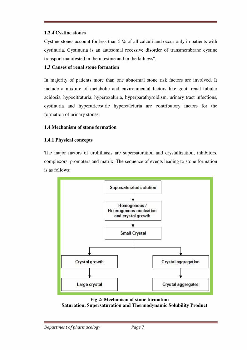
1.4.1 Physical concepts
The major factors of urolithiasis are supersaturation and crystallization, inhibitors,
complexors, promoters and matrix. The sequence of events leading to stone formation
is as follows:
Fig 2: Mechanism of stone formation
Saturation, Supersaturation and Thermodynamic Solubility Product
Department of pharmacology Page 7
1.4.2 Pathogenesis of stone formation
Renal stone formation requires that stone forming crystalloids in urine come out of
solution. Because crystalloids in solution are in equilibrium with crystalloids in the
solid phase, a minimum condition is that urine be supersaturated with relevant
crystalloids. This condition is often met: many healthy persons, probably the majority,
have concentrations of calcium and oxalate in urine such that their activity product
exceeds the solubility threshold (i.e. urine is supersaturated with these crystalloids).
But urine has a strong inhibitory action that prevents crystallization and other stone
forming processes. Three processes promote stone formation:
� Nucleation,
� Aggregation, and
� Crystal growth.
Nucleation:
Nucleation involves the association of crystalloids in solution (e.g. calcium and
oxalate) to form a sub microscopic particle of about 100 atoms. The process requires
energy and is facilitated when an external surface can serve as a lattice or anchor,
thereby lowering the free energy requirement. Such a surface is provided by
microscopic uric acid moieties, which function as promoters of CaOx stone formation.
� Homogeneous nucleation: The process by which the earliest crystal nuclei
form in pure solutions is called homogeneous nucleation. It occurs in the
absence of a surface or lattice.
� Heterogeneous nucleation: In this process the nuclei usually form on
existing surfaces. Epithelial cells, urinary casts, red blood cells and other
crystals can act as nucleating foci in urine.
Aggregation
Aggregation is the process by which nuclei or larger structures adhere to one another.
The initial nuclei can grow by the precipitation of additional salt on the lattice
framework. It takes between 5 and 7 min for urine to flow from the glomerulus to the
collecting duct. The earliest site of stone formation in human is the papillary duct or
the collecting tubule, where the diameter is 50 to 200 µm .Once nuclei are formed they
bounce apart from each other, float freely and become kinetically active. If they
Department of pharmacology Page 8
remain independent and float freely, they are washed away by urine flow. However,
under certain circumstances, these nuclei come in close contact and due to chemical or
electrical forces can bind to each other, a process called crystal aggregation.
Crystal growth
A third process is crystal growth, in which crystalloids come out of solution to
associate with the solid phase of growing crystal in a geometrically precise
arrangement. The combination of crystal aggregation and crystal growth can explain
the genesis of urinary calculi. Another process that may lead to CaOx stone formation
is crystal retention. In most instances, crystal aggregates are too fragile to occlude the
collecting duct long enough to give rise to a stone7-8.
1.4.3 Promoters, inhibitors and complexing agents:
Each process of stone formation has specific promoters and inhibitors.
Glycosaminoglycans promote crystal nucleation but inhibit crystal aggregation.
Tamm- Horsfall protein, a product of the thick ascending limb of Henle, may act as a
promoter or inhibitor of crystal formation depending on its state of aggregation.
Nucleation of CaOx is inhibited by magnesium and citrate, which forms soluble
complexes with calcium and so reduces its effective concentration. a highly negatively
charged molecule rich in aspartate and γ-carboxyglutamic acid residues, is potent
inhibitor of CaOx crystallization in simple solutions. Aggregation is inhibited by
uropontin is an aspartic acid-rich protein that shares the N-terminal amino acid
sequences with human osteopontin. It is an important inhibitor of CaOx crystal
growth. Citrate is a potent complexor of calcium and reduces the ionic calcium in the
urine with consequent reduction in the supersaturation of calcium salt. Citrate exerts
maximum effect at a pH of 6.5. Magnesium, a divalent cation, complexes oxalate in
the CaOx system7.
1.5 Signs and symptoms
Kidney stones on accumulation in the urinary tract exhibit specific symptoms and the
most specific one is severe pain in the. The common symptoms include, back pain on
one or both sides, Progressive severe colicky (spasm-like) may radiate or move to
lower in flank, pelvis, groin, genitals, Nausea, vomiting, Urinary frequency/urgency,
Department of pharmacology Page 9
increased (persistent urge to urinate), Blood in the urine , Abdominal pain, Testicle
pain , Fever, Chills and Abnormal urine color8
1.6 Diagnosis (Investigations)
A. Urine analysis: Urine analysis in most patients with ureteral calculi reveals the
presence of microscopic or gross hematuria. A urine analysis should be done promptly
after collection. The pH is best measured in a morning urine specimen after an
overnight fast. If the pH of urine is higher than 7.6, urea splitting organisms must be
present, for the kidneys cannot produce urine in this range of alkalinity. Such a finding
strongly suggests that the stones are composed of magnesium ammonium phosphate.
Fixation of the pH at 6 to 6.5 is compatible with renal tubular acidosis. Consistently
low pH is a common cause of the formation of uric acid calculi.
B. Blood investigations
���� Complete blood count: The white blood count may be increased as a result of
complicating infection. If renal function is not adequate, anaemia may be
found.
���� Renal function tests: Blood urea nitrogen and serum creatinine will give an
indication of the functional status of the kidney.
���� Serum blood chemistry: This investigation is usually done in recurrent stone
formers. Fasting serum calcium and phosphates should be determined on 3
occasions. Serum proteins should also be estimated, since almost half of the
calcium is normally unionized and bound to proteins. Hypercalcaemia is most
commonly seen in association with osteolytic or disseminated malignant
disease, especially cancers of the breast and lung, multiply myeloma, leukemia
and sarcoidosis, but serum phosphate is usually normal. Serum alkaline
phosphatase is increased in hyperparathyroidism only if bone disease is
present. Elevated serum uric acid is found in 50% of uric acid stone formers.
C. Plain X-ray: A plain film of the abdomen may show a calcific body in the region
of the kidney or in the ureter. This constitutes merely presumptive grounds for the
diagnosis. Ureteric calculi often looks like root of a tooth, sometime it may be
confused with pelvic phleboliths which are usually round.
Department of pharmacology Page 10

D. Excretory urography: Excretory urograms are useful in the diagnosis of ureteric
calculus. The ureterogram places the calcification in the ureter and also demonstrates
dilatation.
E. Ultrasonography: Ultrasonography is a noninvasive method of demonstrating
both ureteral calculi and the consequent hydronephrosis. Color dropper ultrasound
examination may demonstrate the increased resistive index in the obstructed kidney
and assymetry or absence of ureteral jets in urinary bladder.ons of the ureter above the
stone.
F. MRI: The study has potential usefulness when patients have renal impairment or
allergy to intravenous contrast agents and when X-rays are contraindicated. In contrast
to CT, not only MRI is unable to visualize most stones but invitro studies have
determined that this modality is not useful for characterizing the composition of
stones.
G. Radioisotope methods: Radioisotope renography and scanning have contributed
to the diagnosis of obstruction and location of calculus in a number of patients,
particularly those who are otherwise sensitive to contrast media. This technique is
helpful not only to the patients sensitive to the intravenous contrast and knowing the
location of the calculus but also the degree of urinary obstruction is clearly defined.
H. Instrumental examination: Cystoscope and ureteroscope is seldom needed for the
diagnosis of ureteral calculus.
I. Analysis of stone: Chemical analysis of renal calculi has been all but abandoned.
Significant error may occur because qualitative and semiquantitative chemical
methods are not accurate. Studies have shown that X-ray diffraction; Infrared
spectroscopy procedures are accurate in detecting components of urinary calculi9.
1.7 How are kidney stones treated?
Fortunately, surgery is not usually necessary. Most kidney stones can pass through the
urinary system with plenty of water (2 to 3 quarts a day) to help move the stone along.
Often, patient can stay home during this process, drinking fluids and taking pain
medication as needed.
1.7.1 The first step: Prevention
Department of pharmacology Page 11

If a patient has had more than one kidney stone, he is likely to form another; so
prevention is very important. To prevent stones from forming, doctor must determine
their cause and he or she will order laboratory tests, including urine and blood tests.
Doctor will also ask about patient’s medical history, occupation and eating habits. The
passed or removed stone has to be analyzed in the laboratory, because its composition
helps in planning treatment.
1.7.2 Lifestyle changes
A simple and most important lifestyle change to prevent stones is to drink more
liquids; water is the best. If patient tends to form stones, he or she should try to drink
enough liquids throughout the day to produce at least 2 quarts of urine in every 24 h
period. People who form calcium stones used to be told to avoid dairy products and
other foods with high calcium content. However, recent studies have shown that foods
high in calcium, including dairy foods, help prevent calcium stones. Taking calcium in
pill form, however, may increase the risk of developing stones.
1.7.3 Medical therapy
A 24-hour urine collection with measurement of the important analytes is usually
reserved for use in patients with recurrent stone formation. In these patients, the major
urinary risk factors include hypercalciuria, hyperoxaluria, hypocitraturia and
hyperuricosuria. Effective preventive and treatment measures include thiazides
therapy to lower the urinary calcium level, citrate supplementation to increase the
urinary citrate level and, sometimes, allopurinol therapy to lower uric acid excretion.
Uric acid stones are most often treated with citrate supplementation.
1.7.3 Surgical treatment
Surgery should be reserved as an option for cases where other approaches have failed
or should not be tried. Surgery may be needed to remove a kidney stone, if it
� Does not pass after a reasonable period of time and causes constant pain
� Is too large to pass on its own or is caught in a difficult place
� Blocks the flow of urine
� Causes ongoing urinary tract infection
� Damages kidney tissue or causes constant bleeding
Stone episode (resolved)
Department of pharmacology Page 12

Previous episode?
No Yes
Conservative management: Obtain history: Number of previous
episodes, Increase urine output to 2.1qt per day. onset of previous episodes,
bowel disease,
Cosider lower sodium intake. gout, diabetes, medications, family history
Consider lower meat intake.
Serum studies
24-hour urine studies
Uncomplicated calcium stone disease Other stone disease
(i.e., normocalcemia, no bowel
disease, no urinary tract infection)
Normo calciuria Hypercalciuria
Prescride potassium Treat with
Citrate. thiazide diuretics.
Add potassium chloride Add potassium citrate
if patient has normal if patient has low
urine citrate levels. urine citrate levels.
Cystinuria Uric acid calculi Hypercalcemia Hyperuricemia Struvite
calculi
Prescribe Prescribe Hyperparathyroid
Prescribe tiopronin potassium investigation Prescribe
antibiotics.
(Thiola) and citrate. allopurinol.
increase fluid
intake.
Treat relapses Treat based Add acetohydroxamic
With allopurinol on findings. acid (Lithostat) for
(Zyloprim). Severe infection stines.
Fig 3: Algorithm for the medical management of recurrent urinary calculi
Department of pharmacology Page 13

Until recently, surgery to remove a stone was very painful and required a recovery
time of 4 to 6 weeks. Today, treatment for these stones is greatly improved and many
options do not require major surgery.
A. Extracorporeal shockwave lithotripsy
Extracorporeal shockwave lithotripsy (ESWL) is the most frequently used procedure
for the treatment of kidney stones. In ESWL, shock waves that are created outside the
body travel through the skin and body tissues until they hit the denser stones.
The stones break down into sand-like particles and are easily passed through the
urinary tract in the urine10-11.
1.8 Natural products used in the treatments of urolithiasis.
Ammannia baccifera12, Blackcurrant (Ribes nigrum)13, Coleus aromaticus 14, Costus
spiralis Roscoe15, Cranberry (Vaccinium macrocarpon)16, Cyclea peltata17, Herniaria
hirsute18-19, Lupeol and Betulin20-21, Moringa oleifera22, Musa paradisiaca23-24,
Phyllanthus niruri25-26, Raphanus sativus27, Salix taxifolia28, Tribulus terrestris29-31,
Varuna (Crataeva nurvala)32-33, Crataeva adansonii34, Aerva lanata35-36, Bergenia
ligulata rhizome37, Nigella Sativa38, Eleusine Coracana39, Hibiscus sabdariffa40,
Quercus salicina Blume/Quercus stenophylla Makino41, Ammi visnaga42, Cynodon
dactylon43, Orthosiphon stamineus44, Rotula aquatica, Commiphora wightii and
Boerhaavia diffusa45, Herniaria hirsuta and Agropyron repens46Tephrosia purpurea47.
1.9 Ethylene glycol induced urolithiasis:
Ethylene glycol is present in many common substances such as antifreeze, de-icing
substances, detergents, lacquers, and polishes. Ethylene glycol itself is not toxic;
rather the metabolites, glycolic acid and oxalic acid exert their toxic effects. Three
stages of toxicity from ethylene glycol are classically identified: 1) CNS stage, 30 min
to 12 hours after ingestion with features of altered mental status, ataxia and slurred
speech; 2) cardiopulmonary stage, 12-24 hours after ingestion with hypertension,
tachycardia, congestive heart failure, and adult respiratory distress syndrome; 3) renal
stage, 24-72 hours after ingestion with flank pain, calcium-oxalate crystalluria, and
oliguria48 Of interesting note, two forms of oxalate crystals can be present in the urine
in ethylene glycol poisoning, one more specific than the other. A dumbbell shaped
monohydrate crystal is most common, however the dihydrate form is most specific, as
Department of pharmacology Page 14

the monohydrate form can be present in those who consume large quantities of
vitamin C as well as diets high in urate. The dihydrate form also requires a higher
concentration of oxalate to be present, and is thus more indicative of ethylene glycol
poisoning49.
Toxicity results from the depressant effects of ethylene glycol on the central nervous
system (CNS).Metabolic acidosis and renal failure are caused by the conversion of
ethylene glycol to noxious metabolites. Oxidative reactions convert ethylene glycol to
glycoaldehyde, and then to glycolic acid, which is the major cause of metabolic
acidosis.50-51 Both of these steps promote the production of lactate from pyruvate.52
The conversion of glycolic acid to glyoxylic acid proceeds slowly, further increasing
the serum concentration of glycolic acid.4 Glyoxylic acid is eventually converted to
oxalic acid and glycine. Oxalic acid does not contribute to the metabolic acidosis, but
it is deposited as calcium oxalate crystals in many tissues. Ethylene glycol is rapidly
absorbed by the stomach and small intestine, and is quickly redistributed throughout
the body. Metabolites of ethylene glycol remain in the body for several days,with
calcium oxalate present in tissues for much longer. The clinical syndrome of ethylene
glycol intoxication has traditionally been divided into three stages: progressive
involvement of the CNS, the cardiopulmonary systems, and the kidneys. However,
presentation is highly variable and dependent on the amount ingested, the combined
ingestion with ethanol, and the timing of medical intervention.53
1.9.1 Clinical Manifestations
Ethylene glycol produces CNS depression similar to that of ethanol. Symptoms of
ethyl-ene glycol toxicity include confusion, ataxia, hallucinations, slurred speech, and
coma. Symptoms are most severe six to 12 hours after ingestion, when the acidic
metabolites of ethylene glycol are at their maximal concentration The presentation
may be similar to ethanol intoxication, if the patient presents early or has consumed
small amounts of ethylene glycol. However, an ethanol odor will be absent, and serum
or respiratory ethanol levels will be too low to account for the degree of CNS
depression. The absence of a strong odor of alcohol in a patient who appears
intoxicated should raise the suspicion of ethylene glycol ingestion. Following a period
of CNS depression, metabolic acidosis and cardiopulmonary symptoms become
prominent, although co ingestion of ethanol will delay the metabolic acidosis. The
patient may experience nausea, vomiting, hyperventilation, and hypocalcemia with
Department of pharmacology Page 15
muscle tetany and seizures. Hypertension, tachycardia, and cardiac failure may ensue.
Pneumonitis, pulmonary edema, and adult respiratory distress syndrome have also
been reported.54 Renal involvement may become apparent within 24 to 72 hours after
ingestion. Urinary crystal formation requires a sufficient amount of time for ethylene
glycol to be metabolized into oxalate. Calcium oxalate formation depletes serum
calcium levels and deposits in intestinal mucosa, liver, brain, heart, lung, and kidney.
The excretion of calcium oxalate crystals in the urine is usually, but not always,
present. Oliguric or anuric renal failure is the result in the most severe cases and,
although permanent renal failure is rare, recovery of renal function may take up to two
months. If untreated, severe ethylene glycol toxicity is usually fatal within 24 to 36
hours.55
1.9.2 Treatment
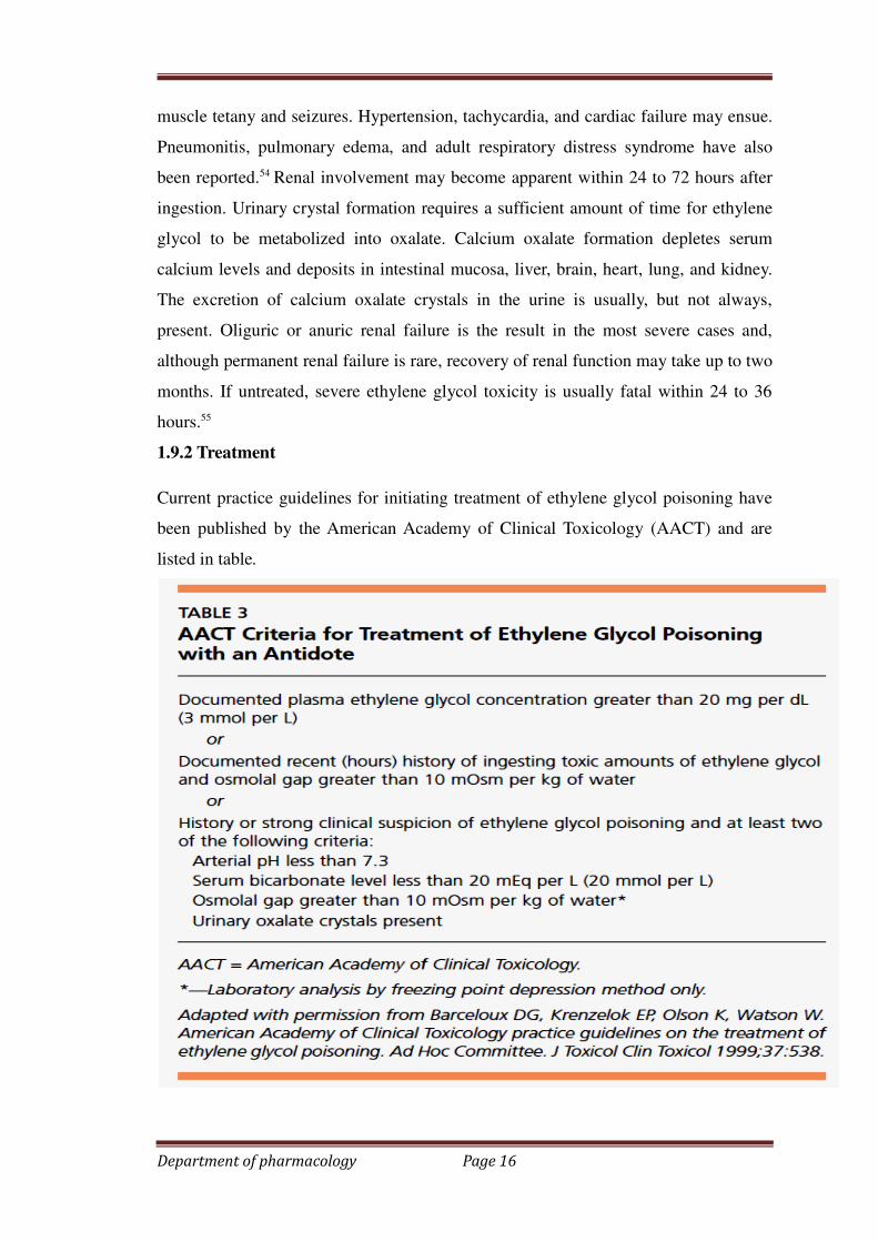
Current practice guidelines for initiating treatment of ethylene glycol poisoning have
been published by the American Academy of Clinical Toxicology (AACT) and are
listed in table.
Department of pharmacology Page 16
Ethylene glycol is rapidly absorbed from the stomach, making treatment with gastric
lavage and syrup of ipecac ineffective. Likewise, it requires large amounts of activated
charcoal to bind relatively small amounts of ethylene glycol, and the therapeutic
window for this action is less than an hour. Traditional treatment of ethylene glycol
poisoning consists of sodium bicarbonate, ethanol, and hemo dialysis. Fomepizole is a
new agent with a specific indication by the U.S. Food and Drug Administration for the
treatment of ethylene glycol poisoning.56 Ethanol and fomepizole are thought to act as
inhibitors of alcohol dehydrogenase and therefore prevent the formation of acidic
ethylene glycol metabolites, but only fomepizole has demonstrated this ability. If
patients are diagnosed and treated with these products early in the course of poisoning,
hemo dialysis may be avoided. Once severe acidosis and renal failure have occurred,
however, hemodialysis is necessary. Fomepizole treatment should be initiated
immediately when ethylene glycol poisoning is suspected. Within three hours of
initiating therapy with fomepizole, inhibition of metabolite production and resolution
of acidosis occurs, and the anion gap is normalized within four hours. If fomepizole
therapy is begun before a rise in the serum creatinine concentration, damage to the
kidney can be avoided. When compared with ethanol, the advantages of fomepizole
include a slower rate of excretion by the kidneys, lack of CNS depression or
hypoglycemia, and easier maintenance of effective plasma levels.
Ethanol may be administered orally or intravenously. The recommended therapeutic
blood ethanol level is 100 to 150 mg per dL (22 to 33 mmol per L). The AACT
provides specific dosage recommendations for ethanol in patients receiving standard
treatment and patients on hemodialysis. While oral ethanol can be simply
administered, it requires a conscious patient who is willing to drink the ethanol or
tolerate the placement of a nasogastric tube. Advantages of intravenous infusion
include greater absorption and no gastrointestinal upset. Disadvantages of treatment
with ethanol include variable metabolism of ethanol; inebriation and CNS depression;
frequent monitoring of serum concentrations (every one to two hours); difficulty
maintaining effective serum concentrations; and the need to administer ethanol in an
intensive care unit. Administration of intravenous sodium bicarbonate will correct the
metabolic acidosis, increase the elimination of renal glycolic acid, and inhibit the
precipitation of calcium oxalate crystals, although the latter benefit has not been
proved in clinical trials. Fifty to 100 mEq per L of intravenous fluid is usually
sufficient, with a goal of maintaining a urine pH greater than 7.0.3 If the diagnosis of
Department of pharmacology Page 17

ethylene glycol poisoning is not made, and the acidosis is treated only with
bicarbonate, organic acids will continue to be produced.10 Treatment with sodium
bicarbonate may worsen hypocalcemia initially because of the protein binding of
calcium. Hemodialysis is effective in removing ethylene glycol and glycolic acid, and
correcting the metabolic acidosis.57 A serum ethylene glycol concentration greater than
50 mg per dL (8 mmol per L) by itself is no longer considered a criterion for
hemodialysis. In the absence of renal dysfunction and significant metabolic acidosis,
the use of fomepizole should eliminate the need for hemodialysis in patients with high
serum ethylene glycol concentrations; in these patients, frequent monitoring of acid-
base balance is necessary. When ethanol or fomepizole is administered and renal
failure is present, dialysis is the only method for removal of ethylene glycol. If
metabolic acidosis persists, too little ethanol or fomepizole is being administered.
Traditionally, hemodialysis is continued until ethylene glycol and glycolic acid levels
cannot be detected in the blood, and there are no acid base disturbances. Prolonged
dialysis may not be necessary in patients treated with fomepizole or ethanol; the end
point for dialysis in these patients is correction of the anion and osmolar gaps. Serum
osmolality levels and electrolyte levels should be monitored closely every two to four
hours for 12 to 24 hours following the discontinuation of dialysis because
redistribution of ethylene glycol may result in an elevated serum concentration.
Pyridoxine (vitamin B6) and thiamine (vitamin B1) in dosages of 100 mg daily are
believed to promote the conversion of intermediate byproducts into nontoxic
metabolites, but clinical data supporting their effectiveness do not exist. Therapy with
100 mg of intravenous thiamine would be appropriate if ethanol withdrawal is
suspected. Parenteral calcium, given as gluconate or chloride salts, may be necessary
for treatment of tetany and seizures caused by hypocalcemia. 58
Department of pharmacology Page 18

Department of pharmacology Page 19

2. REVIEW OF LITERATURE
Lin SY et al reported Antioxidant, anti-semicarbazide-sensitive amine
oxidase, and anti-hypertensive activities of geraniin isolated from Phyllanthus
urinaria, Geraniin also showed dose-dependent inhibitory activities against
semicarbazide-sensitive amine oxidase (SSAO, IC50 were 6.58 microM) and against
angiotensin converting enzyme (ACE, IC50 were 13.22microM). For kinetic property
determinations, geraniin showed competitive inhibitions against SSAO (the apparent
inhibition constant, Ki, was 0.70microM) and mixed noncompetitive inhibitions
against ACE. The geraniin showed antihypertensive activity in lowering SBP and DBP
and showed a significant difference from the blank (distilled water) at 2, 4, 6, 8, and
24 h. Healthy food products could use geraniin for antioxidant protection and
therapeutic effects in the future.
Lee et al reported Hepatoprotective effect of Phyllanthus in Taiwan on acute
liver damage induced by carbon tetrachlorideThe effect of oral administration of
Phyllanthus methanolic extracts (PME) (P. urinaria L.s. urinaria is hepatoprotective
and antioxidant agents.
Chularojmontri L et.al reported Antioxidative and cardioprotective effects of
Phyllanthus urinaria L. on doxorubicin-induced cardiotoxicity.
Shen ZQ ,et al investigated the effects of PUW (a fraction containing 60%
corilagin from a Chinese herbal plant Phyllanthus urinaria) on thrombosis and
coagulation system.They showed that PUW administered intravenously significantly
decreased the mouse mortality, prolonged the occlusion time of rat carotid arteries,
and reduced the wet and dry thrombus weight of the inferior vena cava, respectively.
PUW markedly inhibited the binding of activated platelets to neutrophils, obtaining
39.7 mg/L of the medium inhibitory concentration. Intravenously administered PUW
significantly shortened ELT, prolonged KPTT while had no influence on PT; PUW
increased BT in rat tail tips but the BT caused by PUW was much shorter than that by
aspirin or urokinase.
Department of pharmacology Page 20

Zhang LZ et al reported Isolation and identification of a novel
ellagitannin from Phyllanthus urinaria L. Phyllanthusiin G is a new compound was
isolated. A novel ellagitannin named phyllanthusiin G was isolated, its structure was
established as 1-O-galloyl-2-phyllanthoyl-3,6-(R)-HHDP-beta-D-glucose.
Xu M, et al reported Phenolic antioxidants from the whole plant of
Phyllanthus urinaria The 1,1-diphenyl-2-picrydydrazyl (DPPH) assay on the extract of
Phyllanthus urinaria L. (Euphorbiaceae) displayed considerable radical-scavenging
activity (SC50 = 14.3 microg/ml). Further bioassay-guided purification of the extract
led to the isolation of a series of 15 phenolic compounds, including the ellagitannins
1-7, the flavonoids 8-10, and the simple hydroxylated (or glycosylated) aromatic acids
11-15. Their structures were identified by spectroscopic analyses and comparison with
authentic samples or literature data. The structure of repandinin B (1) was for the first
time fully assigned by 1D- and 2D-NMR experiments. The phenolic compounds 1, 3,
4, 6, 9, 11, and 15 have not been isolated before from the title plant.
Zhong Y et al reported Chemical constituents of Phyllanthus urinaria L. and
its antiviral activity against hepatitis B virus. Studies on the chemical constituents of
Phyllanthus urinaria and its antiviral activity against hepatitis B virus were completed.
Eleven compounds have been isolated. Two of them are new compounds methyl ester
dehydrochebulic acid and methyl brevifolin carboxylate. Antiviral experiments on
HBsAg in vitro and liver damage caused by CCl4 have shown that. Phyllanthus
urinaria possesses antiviral activities against HBV.
Paulino N, et al reported The relaxant effect of extract of Phyllanthus
urinaria in the guinea-pig isolated trachea. Evidence for involvement of ATP-sensitive
potassium channels. indicate that the ATP-activated potassium channels sensitive to
glibenclamide, but not the small conductance calcium-activated potassium channels
sensitive to apamin, largely contribute to the relaxation effect of the hydroalcoholic
extract of P. urinaria in GPT. In addition, both beta 2 and VIP-mediated responses
seem to account, at least in part, for the relaxation effect of the hydroalcoholic extract,
as its relaxant response was partially attenuated by both propranolol and VIP receptor
antagonist.
Department of pharmacology Page 21
3.1 Aims:
Urolithiasis is defined as the presence of one or more calculi in any location within the
urinary tract. Urolithiasis is the third most common disorder of the urinary tract, the
others being frequently occurring urinary tract infections and benign prostatic
hyperplasia. The worldwide incidence of urolithiasis is quite high and in spite of
tremendous advances in the field of medicine, there is no truly satisfactory drug for
the treatment of renal calculi. Most patients still have to undergo surgery to be rid of
this painful disease. Hyperoxaluria is the main initiating factor for urolithiasis.
Phyllanthus urinaria is said to be useful in the treating urinary calculi. Hence the
present study an effort has been made to establish the scientific validity for the anti-
urolithiatic activity.
3.2 OBJECTIVES OF THE STUDY:
1. To prepare ethanolic extract of Phyllanthus urinaria
Department of pharmacology Page 22

2. To study the phytochemical screening of ethanolic extract Phyllanthus
urinaria
3. To study the acute oral toxicity ethanolic extraction of Phyllanthus urinaria
4. To study the antiurolithiatic activity of ethanolic extract of Phyllanthus
urinaria in rats
5. To carry out histopathological studies.
Department of pharmacology Page 23

4. PLANT PROFILE
4.1 Plant profile of Phyllanthus urinaria:
Phyllanthus urinaria is an annual plant with the main stem erect, un branched or
sparsely branched and seldom more than a foot tall. The side branches with their two
rows of alternate leave resemble a compound leaf. The leaves themselves are finely
hairy, nearly sessile (stalkless), oblong to narrowly obovate.
Department of pharmacology Page 24
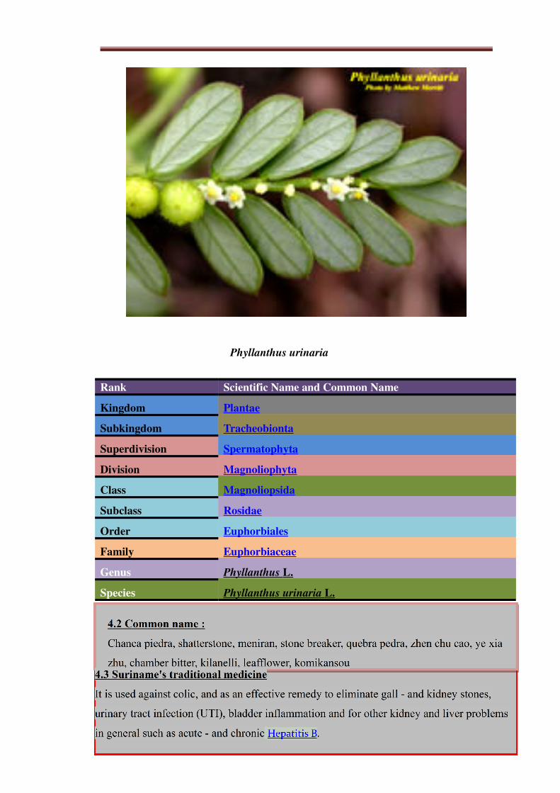
Phyllanthus urinaria
Rank Scientific Name and Common Name
Kingdom Plantae
Subkingdom Tracheobionta
Superdivision Spermatophyta
Division Magnoliophyta
Class Magnoliopsida
Subclass Rosidae
Order Euphorbiales
Family Euphorbiaceae
Genus Phyllanthus L.
Species Phyllanthus urinaria L.
Meniran has proven to be antihepatotoxic, antiviral, antibacterial and
hypoglycemic; also used for the elimination of kidney- and gallstones. It is excellent
in treating liver- and kidney ailments; used extensively for detoxification
Phyllanthus urinaria (P. urinaria), one of the herbal plants belonging to the genus
Phyllanthus (Euphorbiaceae), is widely distributed in China, Southern India and
Southern America. It has long been used in folk medicine for the treatment of several
diseases such as hepatitis B, neprolithiasis and in painful disorders59-61 7’-hydroxy-
3’,4’,5,9,9’-pentamethoxy-3,4- methylene dioxy lignan isolated from the ethyl acetate
extract of P. urinaria was shown to exhibit anticancer activity by inducing apoptosis
through the inhibition of telomerase activity and Bcl-2 expres- sion.62 previous studies
also demonstrated that the water extract prepared from P. urinaria has an anti- cancer
effect on Lewis lung carcinoma cells through a similar pathway.63 In addition, we
Department of pharmacology Page 26
4.4Overview;
Shatterstone is a small tropical annual herb growing up to 2 feet tall. Along the erect, red
stem are equally set small green, oblong feathered leaves. It has greenish white flowers. A
very small wart-like fruit, greenish-red, is underneath every pair of the feathered leaves.
When the plant is picked the feathery leaves fold in, completely closing themselves. The
plant is used for several conditions such as blennorrhagia (gonorrhea), diabetes, dysentery,
flu, tumors, jaundice (the yellow color of the skin and whites of the eyes caused by excess
bilirubin in the blood), vaginitis (swelling, itching, burning or infection in the vagina),
against headache, fever, conjuntivitis (pinkeye or bloodshot eyes), menstrual disorders and
dyspepsia (pain or an uncomfortable feeling in the upper middle part of the stomach).
4.5 Pharmacology
The primary action of shatterstone is on the liver; it acts by the inhibition of DNA
polymerase on the hepatitis B virus. The enzyme DNA polymerase is needed for the virus
to reproduce.Several studies suggest that Phyllanthus urinaria works better than the
related species p. amarus, p. debilis and p.niruri in the treatment of hepatitis B. An equally
important action is the use against kidney stones (renal calculi), urinary tract- and bladder
infections. In preliminary research in animals, extracts of Phyllanthus plants have shown
promising results in pain relief. The mechanism seems to be that this is reached by
decreasing inflammation.
demonstrated that the anti-tumor and anti-angiogenic effects of P. urinaria in mice
bearing Lewis lung carcinoma were due to interference with migration of vascular
endothelial cells but not viability.64 Therefore, P. urinaria became our study target and
we prepared the drug with a standardized protocol under regulation and used it for
further investigation.
Department of pharmacology Page 27
5. MATERIALS AND METHODS
Materials and methods
���� Materials-
Plant material - Plant material: The plant of Phyllanthus urinaria plants were
collected from the certified ayurvedic wholesaler. The plant was identified and
authenticated by Asst. Prof. Dr. K. Madhava chatty, MSc, Med, Department of Botany,
S.V. University, Tirupati
Drugs and Chemicals
Cystone (Himalaya Pharmaceutical, Banglore), Ethylene glycol (SRL Mumbai),
Tween 80 (Merck Pvt Ltd, B, Mumbai), Anaesthetic ether (SD Finechem Ltd.,
Mumbai), Chloroform (SD Finechem Ltd .Mumbai). Formaline (SD Finechem Ltd.,
Mumbai) and all other chemicals and reagents were of analytical grade.
Diagnostic kits:
Diagnostic kits used for estimation of Creatinine, Urea, Uric acid, Calcium,
Phosphorus, Calcium oxalate were procured from Robonik Diagnostic Ltd India.
Instruments:
Autoanalyzer (Robonik), Refrigerator centrifuge (MPW-350R),UV-Spectro-phot
ometer (UV-1601, Shimadzu Corporation, Kyoto, Japan), Mini Lyotrap (LTE
Scientific Ltd.), Research centrifuge (Remi industries, Mumbai) and homogenizer
(Remi Motors, Mumbai). Dhona balance (M/S Dhona instruments Pvt. Ltd., Kolkata,
India).
Experimental Animals:
Wistar albino male (180–220 g) was obtained from the central animal house of Sigma
Institute of Clinical Research and administration Pvt Ltd Hyderabad. The animals
Department of pharmacology Page 28
were housed at room temperature (22-28 ºC) for 12 hr dark and 12 hr light cycle and
given standard laboratory feed and water ad-libitum. The study was approved and
conducted as per the norms of the Institutional Animal Ethics Committee
(180/SICRA/IAEC).
Acute toxicity study
Procedure: Acute toxicity studies were performed according to OECD-423guidelines
category IV substance (acute toxic class method). Swiss albino mice (n=3) of either
sex selected by random sampling technique were employed in this study. The animals
were fasted for 4 hrs with free access to water only. The plant extracts of Phyllanthus
urinaria were administered orally with maximum dose of 2000 mg/kg body weight.
The mortality was observed for three days. If mortality was observed in 2/3 or 3/3 of
animals, then the dose administered was considered as a toxic dose. However, if the
mortality was observed only one mouse out of three animals then the same dose was
repeated again to confirm the toxic effect. If mortality was not observed, the procedure
was then repeated with higher dose (Organization for economic Co-operation and
development, 2001).68
Observations
Animals were observed individually at least once during the first 30 minutes
after dosing, periodically during the first 24 hours (with special attention given during
the first 4 hours) and daily thereafter, for a total of 14 days. All observations were
systematically recorded with individual records being maintained for each animal.
Observations included changes in skin, mortality and general behavioral pattern.
Attention was given for observations of tremors, convulsions, salivation, diarrhea,
lethargy, sleep and coma. No death was observed till the end of study.
Department of pharmacology Page 29

Department of pharmacology Page 30
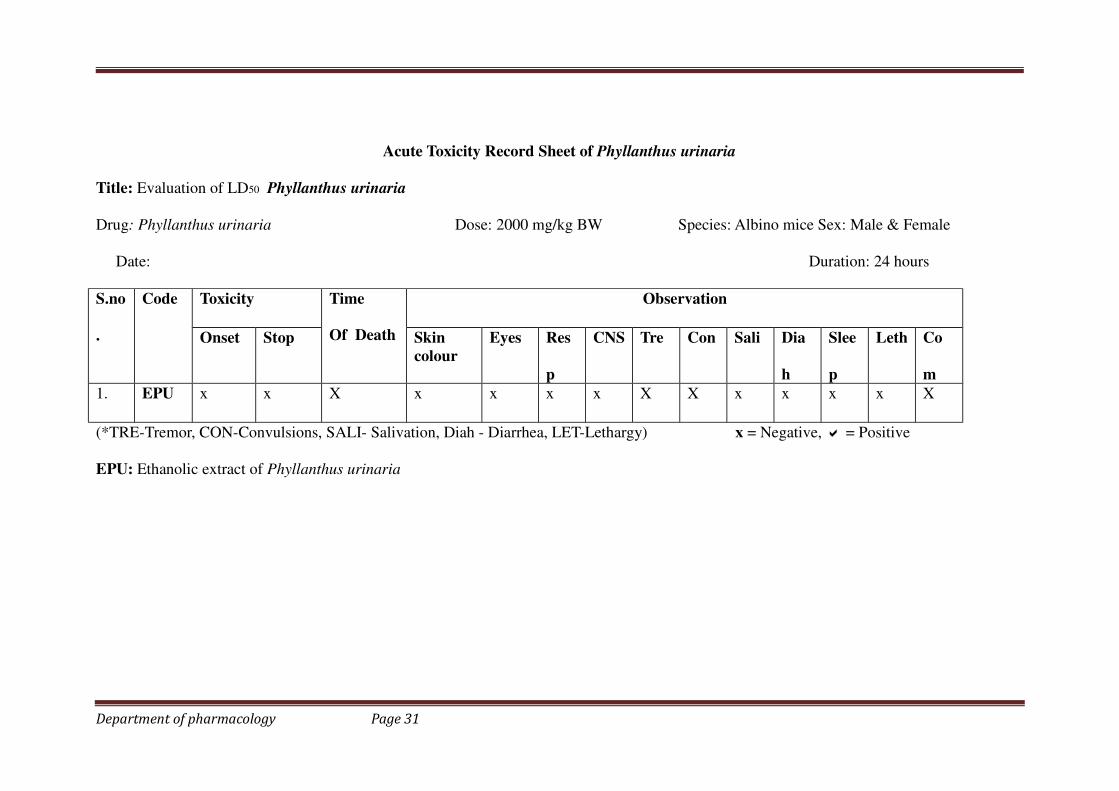
Acute Toxicity Record Sheet of Phyllanthus urinaria
Title: Evaluation of LD50 Phyllanthus urinaria
Drug: Phyllanthus urinaria Dose: 2000 mg/kg BW Species: Albino mice Sex: Male & Female
Date: Duration: 24 hours
S.no
.
Code Toxicity Time
Of Death
Observation
Onset Stop Skin
colour
Eyes Res
p
CNS Tre Con Sali Dia
h
Slee
p
Leth Co
m
1. EPU x x X x x x x X X x x x x X
(*TRE-Tremor, CON-Convulsions, SALI- Salivation, Diah - Diarrhea, LET-Lethargy) x = Negative, � = Positive
EPU: Ethanolic extract of Phyllanthus urinaria
Department of pharmacology Page 31
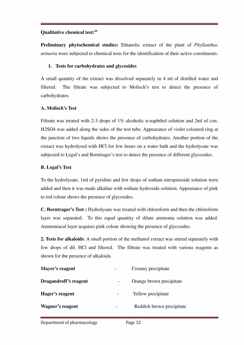
Qualitative chemical test:69
Preliminary phytochemical studies: Ethanolic extract of the plant of Phyllanthus
urinaria were subjected to chemical tests for the identification of their active constituents.
1. Tests for carbohydrates and glycosides
A small quantity of the extract was dissolved separately in 4 ml of distilled water and
filtered. The filtrate was subjected to Molisch’s test to detect the presence of
carbohydrates.
A. Molisch’s Test
Filtrate was treated with 2-3 drops of 1% alcoholic α-naphthol solution and 2ml of con.
H2SO4 was added along the sides of the test tube. Appearance of violet coloured ring at
the junction of two liquids shows the presence of carbohydrates. Another portion of the
extract was hydrolysed with HCl for few hours on a water bath and the hydrolysate was
subjected to Legal’s and Borntrager’s test to detect the presence of different glycosides.
B. Legal’s Test
To the hydrolysate, 1ml of pyridine and few drops of sodium nitroprusside solution were
added and then it was made alkaline with sodium hydroxide solution. Appearance of pink
to red colour shows the presence of glycosides.
C. Borntrager’s Test : Hydrolysate was treated with chloroform and then the chloroform
layer was separated. To this equal quantity of dilute ammonia solution was added.
Ammoniacal layer acquires pink colour showing the presence of glycosides.
2. Tests for alkaloids: A small portion of the methanol extract was stirred separately with
few drops of dil. HCl and filtered. The filtrate was treated with various reagents as
shown for the presence of alkaloids.
Mayer’s reagent - Creamy precipitate
Dragandroff’s reagent - Orange brown precipitate
Hager’s reagent - Yellow precipitate
Wagner’s reagent - Reddish brown precipitate
Department of pharmacology Page 32
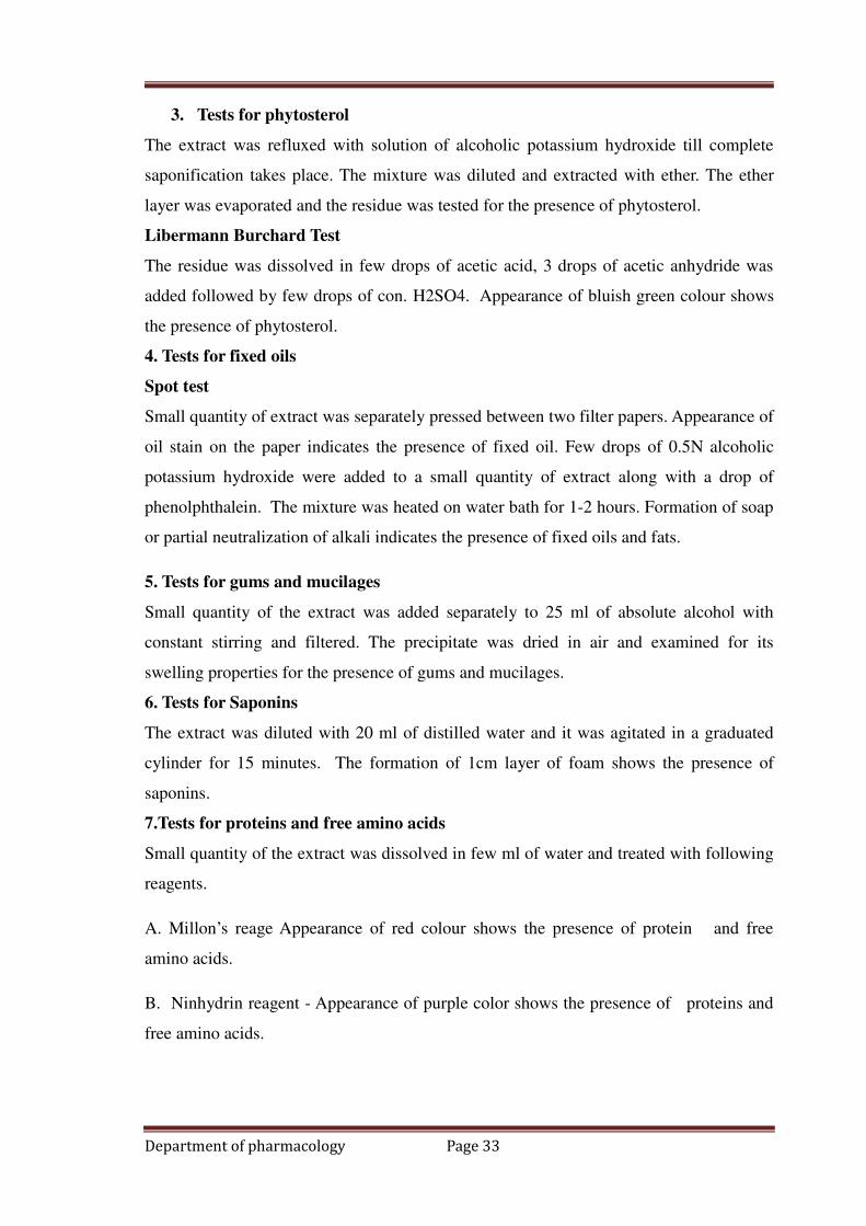
3. Tests for phytosterol
The extract was refluxed with solution of alcoholic potassium hydroxide till complete
saponification takes place. The mixture was diluted and extracted with ether. The ether
layer was evaporated and the residue was tested for the presence of phytosterol.
Libermann Burchard Test
The residue was dissolved in few drops of acetic acid, 3 drops of acetic anhydride was
added followed by few drops of con. H2SO4. Appearance of bluish green colour shows
the presence of phytosterol.
4. Tests for fixed oils
Spot test
Small quantity of extract was separately pressed between two filter papers. Appearance of
oil stain on the paper indicates the presence of fixed oil. Few drops of 0.5N alcoholic
potassium hydroxide were added to a small quantity of extract along with a drop of
phenolphthalein. The mixture was heated on water bath for 1-2 hours. Formation of soap
or partial neutralization of alkali indicates the presence of fixed oils and fats.
5. Tests for gums and mucilages
Small quantity of the extract was added separately to 25 ml of absolute alcohol with
constant stirring and filtered. The precipitate was dried in air and examined for its
swelling properties for the presence of gums and mucilages.
6. Tests for Saponins
The extract was diluted with 20 ml of distilled water and it was agitated in a graduated
cylinder for 15 minutes. The formation of 1cm layer of foam shows the presence of
saponins.
7.Tests for proteins and free amino acids
Small quantity of the extract was dissolved in few ml of water and treated with following
reagents.
A. Millon’s reage Appearance of red colour shows the presence of protein and free
amino acids.
B. Ninhydrin reagent - Appearance of purple color shows the presence of proteins and
free amino acids.
Department of pharmacology Page 33

C. Biuret test - Equal volumes of 5% NaOH solution and 1% copper sulphate solution
were added. Appearance of pink or purple colour shows the presence of proteins and free
amino acids.
8.Tests for phenolic compounds and tannins
Small quantity of the extract was taken separately in water and tested for the presence of
phenolic compounds and tannins using following reagents.
A. Dil.FeCl3 solution (5%) -violet colour
B. 1% solution of gelatin containing 10% NaCl - white precipitate
C. 10% lead acetate solution - white precipitate.
9. Tests for flavonoids
A. With aqueous Sodium hydroxide solution:
Blue to violet colour (anthocyanins), yellow colour (flavones), yellow to orange
(flavonones)
B. With Con. H2SO4:
Yellow orange colour (anthocyanins), yellow toorange colour (flavones), orange to
crimson (flavonones)
C. Shinoda’s test
Small quantity of the extract was dissolved in alcohol and to that a piece of magnesium
followed by Con. HCl drop wise was added and heated. Appearance of magenta colour
shows the presence of flavonoids.
The results of preliminary phytochemical studies of the plant extract are presented.
PHARMACOLOGICAL SCREENING MODEL
Ethylene glycol & ammonium chloride induced urolithiasis model12
Thirty healthy adult Wistar albino strain rats of either sex weighing 180-220g were
randomly divided into five groups. Each group consisted of 6 animals. The treatment
period was considered for 10 days.
Group 1: Normal rats were fed with standard rat chow diet and tap water ad libitum for
10 days.
Department of pharmacology Page 34

Group 2: EG and ammonium chloride intoxicated rats were given normal lab diet +
drinking water containing 0.75% [v/v] ethylene glycol (EG) and 2% [w/v] ammonium
chloride (AC) for 10 days to induce urolithiasis.
Group 3: Standard group were fed with normal lab diet + drinking water containing
0.75% [v/v] EG and 2% [w/v] AC + Cystone (5 ml/kg) for 10 days.
Group 4: the test groups treated with ethanolic extract Phyllanthus urinaria 200 mg/kg
with normal lab diet + drinking water containing 0.75% [v/v] EG and AC 2% [w/v].
Group 5: the test groups treated with ethanolic extract Phyllanthus urinaria 400 mg/kg
of body weight were fed with normal lab diet + drinking water containing 0.75% [v/v]
EG and AC 2% [w/v].
URINE AND BLOOD SAMPLING
The crystalluria and stone formation was verified by different biochemical marker
analysis of urine and serum. The urine samples of the test animals in different groups
were collected in their respective end day of the experiment (1%) EG model on 10th day
in (0.75%) EG + (2%) AC model. The collected urine sample volume and PH were
measured followed by centrifugation at 3000 rpm for 10 minutes. After centrifugation
Department of pharmacology Page 35
the urine samples were examined under light microscope (LAICA, DME Germany
400X) to ensure the presence of oxalate microcrystal followed by biochemical analysis
(urine oxalate, calcium and uric acid, creatinine, urea, magnesium and phosphorus). The
blood samples were collected from the animals under anaesthesia (ether) before
sacrificing. The collected blood samples were then centrifuged to obtain serum for the
analysis of serum creatinine and serum calcium, creatinine, urea, uric acid, magnesium
and phosphorus.
Estimation of serum and urine parameters:
SERUM CREATININE (Mod. Jaffe’s kinetic method)62
Principle
Picric acid in alkaline medium reacts with creatinine to form a orange coloured complex
with the alkaline picrate. Intensity of colour formed during the fixed time measured at
520 nm is directly proportional to the amount of creatinine present in sample.
Creatinine + alkaline picrate Creatinine picrate complex
Pipette in to test
tubes
Standard Sample
Working reagent 1000 µl 1000µl
Standard 100 µl ……..
Sample ……… 100 µl
Mix and read the variation of absorbance (∆A) between 30 seconds and 90 seconds
Calculation :
With standard or calibrator
Concentration in sample (mg/dl) = Concentration of Standard X ∆ A
Sample
∆ A Standard
URIC ACID63
Principle:
Uric acid is oxidized to allantoin by uricase. The generated hydrogen peroxide reacts with
4-aminoantipyrine and DHPS to quinoneimine.
Uricase
Department of pharmacology Page 36
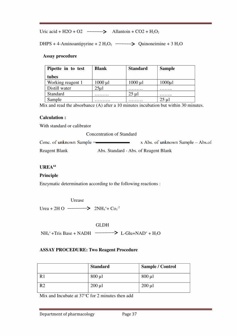
Uric acid + H2O + O2 Allantoin + CO2 + H2O2
DHPS + 4-Aminoantipyrine + 2 H2O2 Quinoneimine + 3 H2O
Assay procedure
Pipette in to test
tubes
Blank Standard Sample
Working reagent 1 1000 µl 1000 µl 1000µl
Distill water 25µl ……… ……..
Standard ……… 25 µl ……..
Sample ………. ……… 25 µl
Mix and read the absorbance (A) after a 10 minutes incubation but within 30 minutes.
Calculation :
With standard or calibrator
Concentration of Standard
Reagent Blank Abs. Standard - Abs. of Reagent Blank
UREA64
Principle
Enzymatic determination according to the following reactions :
Urease
Urea + 2H O 2NH4++ Co3
-2
GLDH
NH4+ +Tris Base + NADH L-Glu+NAD+ + H2O
ASSAY PROCEDURE: Two Reagent Procedure
Standard Sample / Control
R1 800 µl 800 µl
R2 200 µl 200 µl
Mix and Incubate at 37°C for 2 minutes then add
Department of pharmacology Page 37

Pipette in to test tubes Standard Sample
Working reagent 1000 µl 1000µl
Standard 10µl ……..
Sample ……… 10µl
Mix and read the variation of absorbance (∆A) between 30 seconds and 60 seconds.
Calculation :
With standard or calibrator
Concentration of Standard
Concentration in Sample (mg/dl) = X ∆ A Sample
∆ A Standard
CALCIUM (OCPC)
Principle :
Ortho-Cresolphthalein Complexone reacts with calcium ions in alkaline medium forming
a red-violet color. Interference by magnesium is eliminated by addition of 8-
hydroxyquinoline. The colour intensity is directly proportional to the serum total calcium
concentration.
Assay procedure:
Pipette in to test
tubes
Blank Standard Sample
Working reagent 1 1000 µl 1000 µl 1000µl
Distill water 10µl ……… ……..
Standard ……… 10µl ……..
Sample ………. ……… 10 µl
Mix and read the absorbance (A) after 5 minutes incubation, but within 30 minutes.
Calculation :
With standard or calibrator.
Concentration of Standard
Conc. of unknown Sample = X Abs. of unknown Sample –
Abs. of Reagent Blank
Abs. Standard – Abs. of Reagent Blank
PHOSPHORUS
Principle :
Ammonium molybdate + Sulfuric Acid Phosphomolybdate complex
Assay procedure :
Department of pharmacology Page 38

Pipette in to test
tubes
Blank Standard Sample
Working reagent 1 1000 µl 1000 µl 1000µl
Distill water 10µl ……… ……..
Standard ……… 10µl ……..
Sample ………. ……… 10 µl
Mix and read the absorbance (A) after a 5 minutes incubation.
Calculation :
With standard or calibrator.
Concentration of Standard
Conc. of unknown Sample = X Abs. of unknown Sample –
Abs. of Reagent Blank
Abs. Standard – Abs. of Reagent Blank
Oxalate
Principle:
Oxalate is co-precipitated with calcium sulphate, reduced to glycolic acid by boiling with
dilute sulphuric acid and a zinc pellet and estimated colorimetrically with chromotropic
acid.
Procedure:
Set the auto-analyzer instrument with the parameters given along with the kit. Prepare the
working, standard and test solutions as per the protocol. Incubate for 5 min at room
temperature mix and read on colorimeter.
Calculations:
Oxalate (mg/dl)= Absorbance of Test × Concemtration of Standard (mg/dl)
Absorbance of standard
MAGNESIUM
Principle:
Magnesium combines with calmagite in an alkaline medium to form a red coloured
complex. Interference of calcium and proteins is eliminated by the addition of specific
Department of pharmacology Page 39

chelating agent and detergents, Intensity of the colour formed is directly proportional to
the amount of magnesium present in the sample.
Magnesium+Calmagite Alkaline mdeium Red coloured complex
Procedure:
Set the auto-analyzer instrument with the parameters given along with the kit. Prepare the
working standard and test solutions as per the protocol. Incubate for 5 min at room temp.
Mix and read 510 nm within 30 min.
Calculations:
Magnesium mEq/L= Absorbance of Test × 2
Absorbance of standard
HISTOPATHOLOGY
The abdomen was cut open to remove both kidneys from each animal. Isolated kidneys
were rinsed in an ice-cold physiological solution, after the extraneous tissues were
removed. The right kidney was fixed in 10% neutral buffered formalin, processed in a
series of graded alcohol and xylene, embedded in paraffin wax, sectioned at 5 µm and
stained with hematoxylin and eosin (H and E) for histopathological examination. The
slides were examined under a light microscope to study the architecture of the kidney and
calcium oxalate deposits.
ENZYME ASSAY
A portion of kidney was taken from all the groups, and a 30% w/v homogenate was
prepared in 0.9% buffered KCl (pH 7.4) for the estimation of protein, superoxide
dismutase (SOD), catalase (CAT), glutathione (GSH) and malondialdehyde (MDA).
1. Lipid Peroxidation:
Malondialdehye, formed from the breakdown of polyunsaturated fatty acids,
serves as a convenient index for determining the extent of peroxidation reaction.
Malondialdehye reacts with thiobarbituric acid (TBA) to produce red colored species
which is measured at 532 nm.
Reagents:
1. TBA-TCA-HCl reagent.
Department of pharmacology Page 40
15% w/v TCA, 0.375 %w/v TBA and 0.25 N HCl. This solution was mildly heated to
assist the dissolution of TBA
1 ml of kidney homogenate was combined with 2 ml of TCA-TBA-HCl reagent and
mixed thoroughly. The solution was heated for 15 min. in a boiling water bath. After
cooling, the flocculent precipitate was removed by centrifugation at 1000 rpm for 10 min.
the absorbance of the supernatant was measured at 532 nm against a blank that contains
all the reagents minus the kidney homogenate. The malondialdehyde concentration of the
sample can be calculated using an extinction coefficient of 1.56x10⁵M-1cm-1
Malondialdehyde concentration (M) = Absorbance
1.56 x 10⁵
2. Estimation of SOD
The activity of superoxide dismutase was determined by the method of Misra and
Fridovich (1972) based upon the ability of SOD to inhibit the auto-oxidation of
epinephrine to adrenochrome at alkaline pH inhibition of the chromagen formation by
superoxide dismutase was linear with increase in enzyme concentration.
Reagents:
1. Sodium carbonate buffer 0.1M (pH 10.2)
2. Ephinephrine(1mM)
The entire supernatant 1 ml (S) was taken in 0.1 M carbonate buffer (pH 10.2). After
addition of epinephrine, the increase in absorbance was measured at 480 nm using a UV–
Visible double beam spectrophotometer. The activity of the enzyme has been expressed
as U/mg protein, where 1U of the enzyme is defined as the amount of enzyme required to
inhibit the rate of epinephrine auto-oxidation by 50% under the conditions of the assay.
3. Estimation of Catalase
In the presence of catalase, H2O2 shows a continual decrease in absorbane in UV
range. The decomposition of H2O2 can be followed directly by the decrease in absorbance
at 240nm (E240 = 0.00394 ±0.0002 ltr mmol-1 mm-1). The difference in absorbance (∆ A
240) per unit time is a measure of the catalase activity.
Reagents:
1. PBS 50 mM; pH7.0
Department of pharmacology Page 41
Dissolve (a)6.81 of KH2PO4 and (b) in the proportion 1:1.5(v/v).
2. H2O2 (0.17 mM): dilute 0.16 ml of (30%w/v) H2O2 with Phosphate buffer to 100
ml.
The catalase activity was determined spectrophotometrically according to the protocol of
Claiborne (1985).The reaction mixture (2 ml) contained 1.95 ml 10 mM H2O2 in 60 mM
phosphate buffer (pH 7.0).The reaction was started by adding 0.05 ml supernatant and the
absorbance was followed for 3 min at 240 nm. Phosphate buffer (60 mM, pH 7.0) was
used as a reference. The extinction coefficient of 0.04 mM−1cm−1 was used to determine
the specific activity of catalase. A unit of catalase is defined as the quantity, which
decomposes 1.0µmole of H2O2 per min at pH 7.0 at 250C, while this H2O2 concentration
falls from 10.3to 9.2mM.The data was expressed as U/mg protein
4. Estimation of glutathione (GSH):
Principle: GSH is a non protein compound containing sulphydryl group in its structure.
DTNB (5,5’ di thio bis (2-Nitrobenzoic acid) is a disulfide chromagen that is reduced by
sulphydryl compounds to an intensly yellow colored compound. The absorbance of the
reduced chromagen is measured at 412 nm and is directly proportional to the GSH
concentration.
Reagents:
1. 10% Trichloroacetic acid (TCA)
2. Phosphate buffer (0.2M) pH 8.0
0.218 g Sodium dihydrogen phosphate and 2.641 g disodium hydrogen
phosphate in 100 ml distilled water
3. DTNB (0.6 mM) (pH 8)
11.9 mg in 50 ml Phosphate buffer
GSH was measured by the method of Moran et al(1979).The kidney homogenate
proteins were precipitated by 10% TCA, centrifuged and the supernatant was collected. 1
ml of supernatant was mixed with 6 ml of 0.2 M Phosphate buffer pH 8.0 and 1 ml 0.6
mM DTNB and incubated for 10 min at room temperature. The absorbance of the
samples was recorded against the blank at 412 nm and the GSH concentration was
calculated from the standard curve by multiplying with the dilution factor
(mannervik1985; tetza 1969)
Statistical analysis
Department of pharmacology Page 42
Results were indicated in terms of mean ± SEM. Statistical significance of data were
assessed by analysis of variance (One way-ANOVA), followed by comparison between
different groups using ‘Dunnett’s multiple comparison test. The significance was
considered at the level of P<0.05.
Department of pharmacology Page 43
6.RESULTS
Preliminary phytochemical screening
The extract of drug were analysed for the presence of various constituents. The result of
this preliminary phytochemical examination is shown in Table no.
Table No. Qualitative chemical examination of ethanolic extract of Phyllanthus
urinaria.
Phytoconstituents Presence or Absence
Carbohydrates +
Glycosides +
Department of pharmacology Page 44

Fixed oils and fats +
Gums & mucilage -
Potein & amino acids -
Saponins ++
Tannins +
Phytosterols +
Flavonoids +++
Alkaloids +++
PHARMACOLOGICAL ACTIVITY
Acute oral toxicity
Acute oral toxicity was carried out according to OECD guideline. EPU was safe upto
2000mg/kg.
Effect of ethanolic extract of Phyllanthus urinaria on urinary volume and pH against
EG and AC induced urolithiasis.
EG and AC (0.75 and 2%.) administration showed significant (p<0.001) alteration of the
output of urine and pH as compared normal group. Administration of Cystone 5ml/kg,
EPU 200 and 400 mg/kg caused significamtly increased (p<0.01, p<0.05) urine output
and pH of the urine as compared to control (EG and AC) group.
Effect of ethanolic extract of Phyllanthus urinaria on serum biochemical parameters
against EG and AC induced urolithiasis.
Serum Creatinine
Administration of EG and AC (0.75% and 2%) for 10 days caused significant elevation
(p<0.01) in serum creatinine concentration compared to normal one. Standrad cystone
5ml/kg causes significant reduction (p<0.001) in serum creatinine concentration when
compared to EG and AC alone treated group. Pretreatment with EPU 200 and 400mg/kg
causes significant reduction (p<0.05 and p<0.001) in serum creatinine concentration
when compared to EG and AC alone treated group.
Serum Urea
Administration of EG and AC (0.75% and 2%) for 10 days caused significant elevation
(p<0.001) in serum urea concentration compared to normal one. Standrad cystone 5ml/kg
Department of pharmacology Page 45

causes significant reduction (p<0.001) in serum urea concentration when compared to EG
and AC alone treated group. Pretreatment with EPU 200 and 400mg/kg causes
significant reduction (p<0.01 and p<0.01) in serum urea concentration when compared to
EG and AC alone treated group.
Serum Uric Acid
Administration of EG and AC (0.75% and 2%) for 10 days caused significant elevation
(p<0.01) in serum uric acid concentration compared to normal one. Standrad cystone
5ml/kg causes significant reduction (p<0.001) in serum uric acid concentration when
compared to EG and AC alone treated group. Pretreatment with EPU 200 and 400mg/kg
causes significant reduction (p<0.05 and p<0.001) in serum uric acid concentration when
compared to EG and AC alone treated group.
Serum Calcium
Administration of EG and AC (0.75% and 2%) for 10 days caused significant elevation
(p<0.001) in serum calcium concentration compared to normal one. Standrad cystone
5ml/kg causes significant reduction (p<0.001) in serum calcium concentration when
compared to EG and AC alone treated group. Pretreatment with EPU 200 and 400mg/kg
causes significant reduction (p<0.001 and p<0.001) in serum calcium concentration when
compared to EG and AC alone treated group.
Serum Oxalate
Administration of EG and AC (0.75% and 2%) for 10 days caused significant increased
(p<0.001) in serum oxalate concentration compared to normal one. Standrad cystone
5ml/kg causes significant reduction (p<0.001) in serum oxalate concentration when
compared to EG and AC alone treated group. Pretreatment with EPU 200 and 400mg/kg
causes significant reduction (p<0.05 and p<0.001) in serum oxalate concentration when
compared to EG and AC alone treated group.
Serum Phosphorus
Administration of EG and AC (0.75% and 2%) for 10 days caused significant increased
(p<0.001) in serum phosphorus concentration compared to normal one. Standrad cystone
5ml/kg causes significant reduction (p<0.001) in serum phosphorus concentration when
compared to EG and AC alone treated group. Pretreatment with EPU 200 and 400mg/kg
causes significant reduction (p<0.05 and p<0.001) in serum phosphorus concentration
when compared to EG and AC alone treated group.
Serum Magnesium
Department of pharmacology Page 46

Administration of EG and AC (0.75% and 2%) for 10 days caused significant increased
(p<0.05) in serum magnesium concentration compared to normal one. Standrad cystone
5ml/kg causes significant reduction (p<0.01) in serum magnesium concentration when
compared to EG and AC alone treated group. Pretreatment with EPU 200 and 400mg/kg
causes significant reduction (p<0.01 and p<0.001) in serum magnesium concentration
when compared to EG and AC alone treated group.
Treatment group Urinary Volume
(ml/24hr)
Urine Ph
Normal 18.4±0.97 7.5±2.1
Control (EG+AC) 6.96±0.69a 4.5±1.39a
Standard Cystone (5ml/kg) 14.93±0.57*** 8.2±1.32**
EPU 200mg/kg 8.41±0.45* 5.9±1.21*
EPU 400mg/kg 9.9±0.64* 6.14±2.24**
Department of pharmacology Page 47
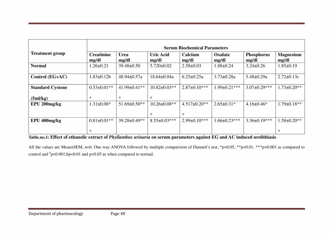
Treatment group
Serum Biochemical Parameters
Creatinine
mg/dl
Urea
mg/dl
Uric Acid
mg/dl
Calcium
mg/dl
Oxalate
mg/dl
Phosphorus
mg/dl
Magnesium
mg/dl
Normal 1.26±0.21 39.48±0.50 5.720±0.02 2.58±0.03 1.88±0.24 3.24±0.26 1.85±0.19
Control (EG+AC) 1.83±0.12b 48.94±0.57a 18.64±0.04a 6.25±0.25a 3.73±0.28a 5.48±0.29a 2.72±0.13c
Standard Cystone
(5ml/kg)
0.53±0.01**
*
41.99±0.41**
*
10.82±0.03**
*
2.87±0.10*** 1.99±0.21*** 3.07±0.29*** 1.73±0.20**
EPU 200mg/kg 1.31±0.08* 51.69±0.50** 10.26±0.08**
*
4.517±0.20**
*
2.65±0.31* 4.16±0.46* 1.79±0.18**
EPU 400mg/kg 0.81±0.01**
*
39.28±0.49** 8.55±0.03*** 2.99±0.10*** 1.66±0.23*** 3.36±0.19*** 1.58±0.20**
*
Table.no.1: Effect of ethanolic extract of Phyllanthus urinaria on serum parameters against EG and AC induced urolithiasis
All the values are Mean±SEM, n=6, One way ANOVA followed by multiple comparision of Dunnett’s test, *p<0.05, **p<0.01, ***p<0.001 as compared to
control and ap<0.001,bp<0.01 and p<0.05 as when compared to normal.
Department of pharmacology Page 48
Department of pharmacology Page 49
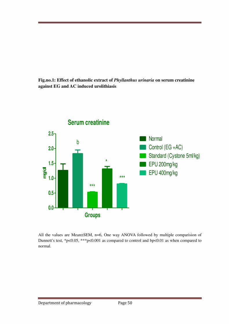
Fig.no.1: Effect of ethanolic extract of Phyllanthus urinaria on serum creatinine
against EG and AC induced urolithiasis
Serum creatinine
0.0
0.5
1.0
1.5
2.0
2.5Normal
Control (EG +AC)
Standard (Cystone 5ml/kg)
EPU 200mg/kg
EPU 400mg/kg
b
***
*
***
Groups
mg/d
l
All the values are Mean±SEM, n=6, One way ANOVA followed by multiple comparision of
Dunnett’s test, *p<0.05, ***p<0.001 as compared to control and bp<0.01 as when compared to
normal.
Department of pharmacology Page 50

Fig.no.2: Effect of ethanolic extract of Phyllanthus urinaria on serum urea against
EG and AC induced urolithiasis.
serum urea
0
20
40
60
a
***
**
***
Normal
Control (EG +AC)
Standard (Cystone 5ml/kg)
EPU 200mg/kg
EPU 400mg/kg
Groups
mg/d
l
All the values are Mean±SEM, n=6, One way ANOVA followed by multiple comparision of
Dunnett’s test, **p<0.01, ***p<0.001 as compared to control and ap<0.001as when compared
to normal.
Department of pharmacology Page 51
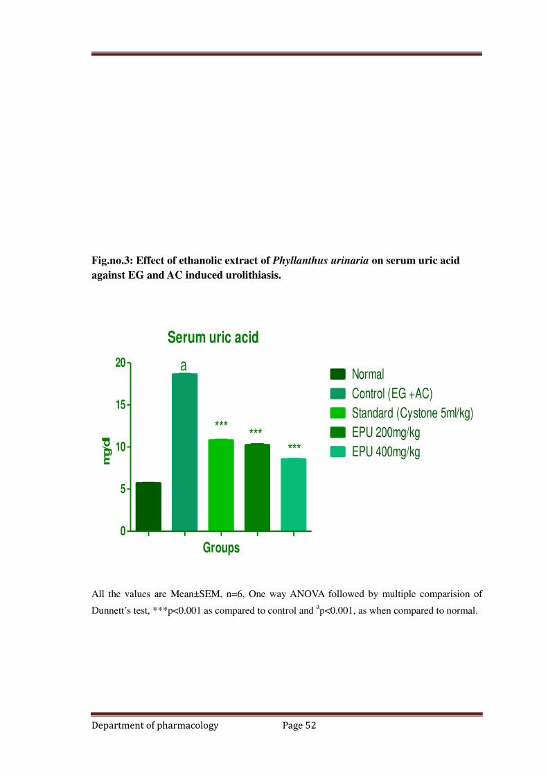
Fig.no.3: Effect of ethanolic extract of Phyllanthus urinaria on serum uric acid
against EG and AC induced urolithiasis.
Serum uric acid
0
5
10
15
20Normal
Control (EG +AC)
Standard (Cystone 5ml/kg)
EPU 200mg/kg
EPU 400mg/kg
a
*** ******
Groups
mg/d
l
All the values are Mean±SEM, n=6, One way ANOVA followed by multiple comparision of
Dunnett’s test, ***p<0.001 as compared to control and ap<0.001, as when compared to normal.
Department of pharmacology Page 52
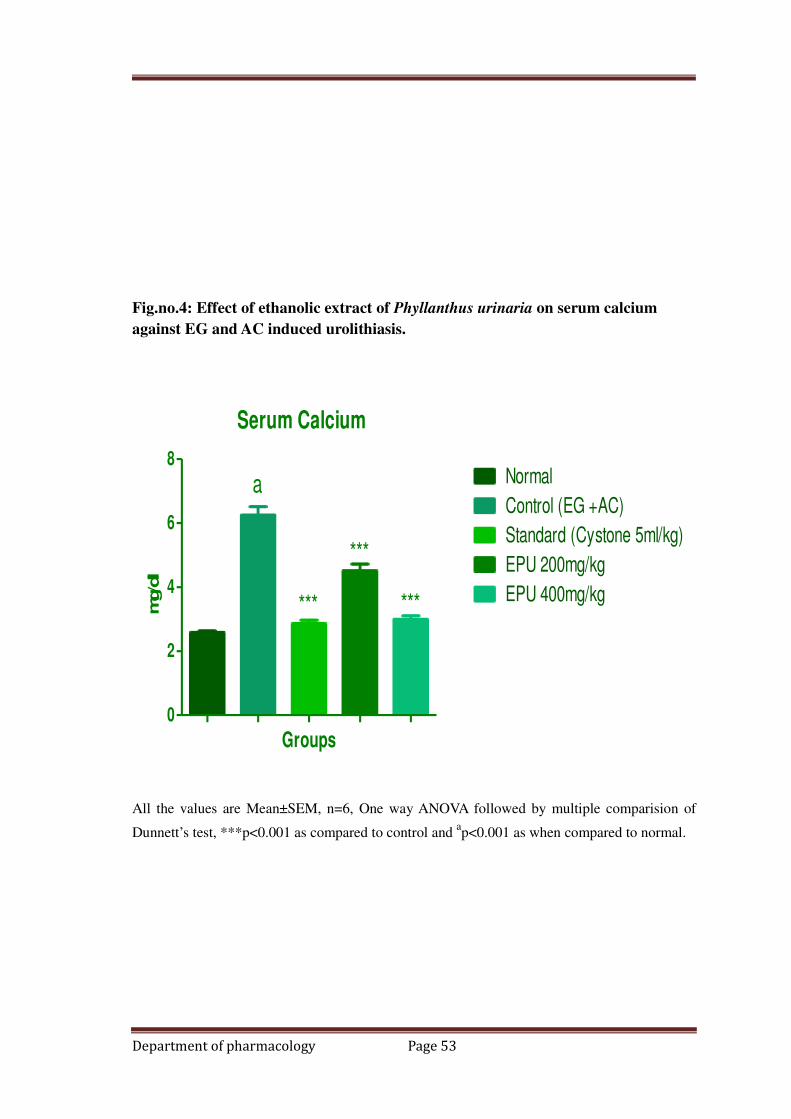
Fig.no.4: Effect of ethanolic extract of Phyllanthus urinaria on serum calcium
against EG and AC induced urolithiasis.
Serum Calcium
0
2
4
6
8Normal
Control (EG +AC)
Standard (Cystone 5ml/kg)
EPU 200mg/kg
EPU 400mg/kg
a
***
***
***
Groups
mg/d
l
All the values are Mean±SEM, n=6, One way ANOVA followed by multiple comparision of
Dunnett’s test, ***p<0.001 as compared to control and ap<0.001 as when compared to normal.
Department of pharmacology Page 53
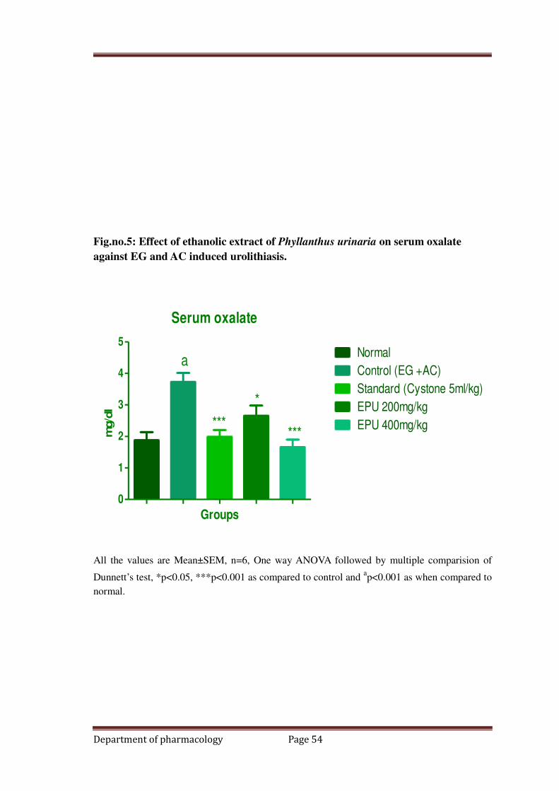
Fig.no.5: Effect of ethanolic extract of Phyllanthus urinaria on serum oxalate
against EG and AC induced urolithiasis.
Serum oxalate
0
1
2
3
4
5Normal
Control (EG +AC)
Standard (Cystone 5ml/kg)
EPU 200mg/kg
EPU 400mg/kg
a
***
*
***
Groups
mg/d
l
All the values are Mean±SEM, n=6, One way ANOVA followed by multiple comparision of
Dunnett’s test, *p<0.05, ***p<0.001 as compared to control and ap<0.001 as when compared to
normal.
Department of pharmacology Page 54

Fig.no.6: Effect of ethanolic extract of Phyllanthus urinaria on serum phosphorus
against EG and AC induced urolithiasis.
Serum phosphorus
0
2
4
6
8Normal
Control (EG +AC)
Standard (Cystone 5ml/kg)
EPU 200mg/kg
EPU 400mg/kg
a
***
*
***
Groups
mg/d
l
All the values are Mean±SEM, n=6, One way ANOVA followed by multiple comparision of
Dunnett’s test, *p<0.05, ***p<0.001 as compared to control and ap<0.001 as when compared to
normal.
Department of pharmacology Page 55
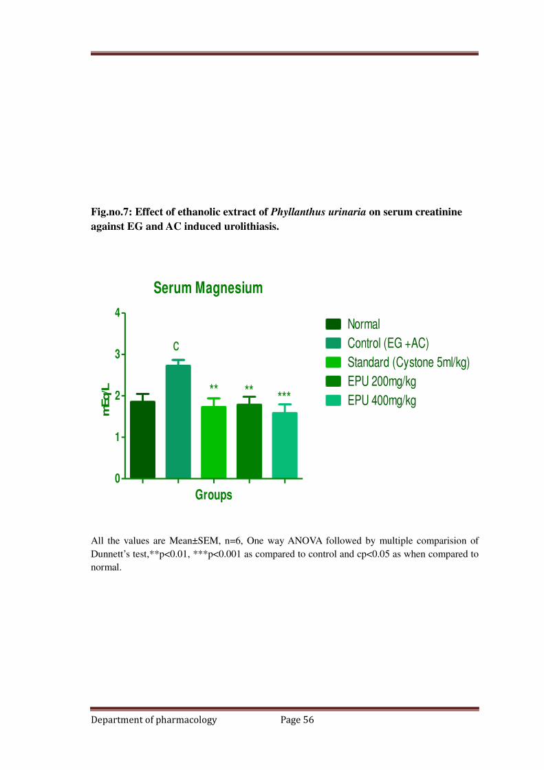
Fig.no.7: Effect of ethanolic extract of Phyllanthus urinaria on serum creatinine
against EG and AC induced urolithiasis.
Serum Magnesium
0
1
2
3
4Normal
Control (EG +AC)
Standard (Cystone 5ml/kg)
EPU 200mg/kg
EPU 400mg/kg
c
** ** ***
Groups
mEq/L
All the values are Mean±SEM, n=6, One way ANOVA followed by multiple comparision of
Dunnett’s test,**p<0.01, ***p<0.001 as compared to control and cp<0.05 as when compared to
normal.
Department of pharmacology Page 56
Effect of ethanolic extract of Phyllanthus urinaria on urine biochemical
parameters against EG and AC induced urolithiasis.
Urinary Creatinine:
Administration of EG and AC (0.75% and 2%) for 10 days caused significant increased
(p<0.001) in urine creatinine concentration compared to normal one. Standrad cystone
5ml/kg causes significant reduction (p<0.01) in urine creatinine concentration when
compared to EG and AC alone treated group. Pretreatment with EPU 200 and
400mg/kg causes significant reduction (not significant and p<0.001) in urine creatinine
concentration when compared to EG and AC alone treated group.
Urinary urea
Administration of EG and AC (0.75% and 2%) for 10 days caused significant increased
(p<0.001) in urine urea concentration compared to normal one. Standrad cystone
5ml/kg causes significant reduction (p<0.001) in urine urea concentration when
compared to EG and AC alone treated group. Pretreatment with EPU 200 and
400mg/kg causes significant reduction (p<0.01 and p<0.001) in urine urea
concentration when compared to EG and AC alone treated group.
Urinary Calcium
Administration of EG and AC (0.75% and 2%) for 10 days caused significant increased
(p<0.001) in urine calcium concentration compared to normal one. Standrad cystone
5ml/kg causes significant reduction (p<0.001) in urine calcium concentration when
compared to EG and AC alone treated group. Pretreatment with EPU 200 and
400mg/kg causes significant reduction (p<0.05 and p<0.001) in urine calcium
concentration when compared to EG and AC alone treated group.
Department of pharmacology Page 57
Urinary Oxalate
Administration of EG and AC (0.75% and 2%) for 10 days caused significant increased
(p<0.001) in urine oxalate concentration compared to normal one. Standrad cystone
5ml/kg causes significant reduction (p<0.001) in urine oxalate concentration when
compared to EG and AC alone treated group. Pretreatment with EPU 200 and
400mg/kg causes significant reduction (p<0.001 and p<0.001) in urine oxalate
concentration when compared to EG and AC alone treated group.
Urinary Phosphorus
Administration of EG and AC (0.75% and 2%) for 10 days caused significant increased
(p<0.001) in urine phosphorus concentration compared to normal one. Standrad cystone
5ml/kg causes significant reduction (p<0.001) in urine phosphorus concentration when
compared to EG and AC alone treated group. Pretreatment with EPU 200 and
400mg/kg causes significant reduction (p<0.05 and p<0.001) in urine phosphorus
concentration when compared to EG and AC alone treated group.
Urinary Magnesium
Administration of EG and AC (0.75% and 2%) for 10 days caused significant increased
(p<0.001) in urine magnesium concentration compared to normal one. Standrad cystone
5ml/kg causes significant reduction (p<0.001) in urine magnesium concentration when
compared to EG and AC alone treated group. Pretreatment with EPU 200 and
400mg/kg causes significant reduction (p<0.05 and p<0.001) in urine magnesium
concentration when compared to EG and AC alone treated group.
Department of pharmacology Page 58
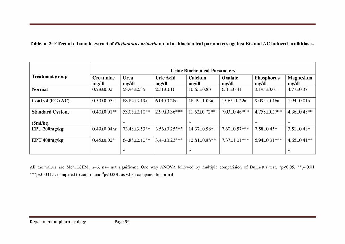
Table.no.2: Effect of ethanolic extract of Phyllanthus urinaria on urine biochemical parameters against EG and AC induced urolithiasis.
Treatment group
Urine Biochemical Parameters
Creatinine
mg/dl
Urea
mg/dl
Uric Acid
mg/dl
Calcium
mg/dl
Oxalate
mg/dl
Phosphorus
mg/dl
Magnesium
mg/dl
Normal 0.28±0.02 58.94±2.35 2.31±0.16 10.65±0.83 6.81±0.41 3.195±0.01 4.77±0.37
Control (EG+AC) 0.59±0.05a 88.82±3.19a 6.01±0.28a 18.49±1.03a 15.65±1.22a 9.093±0.46a 1.94±0.01a
Standard Cystone
(5ml/kg)
0.40±0.01** 53.05±2.10**
*
2.99±0.36*** 11.62±0.72**
*
7.03±0.46*** 4.758±0.27**
*
4.36±0.48**
*
EPU 200mg/kg 0.49±0.04ns 73.48±3.53** 3.56±0.25*** 14.37±0.98* 7.60±0.57*** 7.58±0.45* 3.51±0.48*
EPU 400mg/kg 0.45±0.02* 64.88±2.10**
*
3.44±0.23*** 12.81±0.88**
*
7.37±1.01*** 5.94±0.31*** 4.65±0.41**
*
All the values are Mean±SEM, n=6, ns= not significant, One way ANOVA followed by multiple comparision of Dunnett’s test, *p<0.05, **p<0.01,
***p<0.001 as compared to control and ap<0.001, as when compared to normal.
Department of pharmacology Page 59

Fig.no.8: Effect of ethanolic extract of Phyllanthus urinaria on urine creatinine
parameters against EG and AC induced urolithiasis.
Urine Creatinine
0.0
0.2
0.4
0.6
0.8Normal
Control (EG +AC)
Standard (Cystone 5ml/kg)
EPU 200mg/kg
EPU 400mg/kg
a
**
**
Groups
mg/d
l
All the values are Mean±SEM, n=6, One way ANOVA followed by multiple comparison of
Dunnett’s test, *p<0.05, **p<0.01 as compared to control and ap<0.001, as when compared to
normal.
Department of pharmacology Page 60

Fig.no.9: Effect of ethanolic extract of Phyllanthus urinaria on urine urea
parameters against EG and AC induced urolithiasis.
Urine Urea
0
20
40
60
80
100Normal
Control (EG +AC)
Standard (Cystone 5ml/kg)
EPU 200mg/kg
EPU 400mg/kg
a
***
*****
Groups
mg/d
l
All the values are Mean±SEM, n=6, One way ANOVA followed by multiple comparison of
Dunnett’s test, **p<0.01, ***p<0.001 as compared to control and ap<0.001, as when compared
to normal.
Department of pharmacology Page 61

Fig.no.10: Effect of ethanolic extract of Phyllanthus urinaria on urine uric aicd
parameters against EG and AC induced urolithiasis.
Urine Uric acid
0
2
4
6
8Normal
Control (EG +AC)
Standard (Cystone 5ml/kg)
EPU 200mg/kg
EPU 400mg/kg
a
*** *** ***
Groups
mg/d
l
All the values are Mean±SEM, n=6, One way ANOVA followed by multiple comparison of
Dunnett’s test, ***p<0.001 as compared to control and ap<0.001, as when compared to normal.
Department of pharmacology Page 62

Fig.no.11: Effect of ethanolic extract of Phyllanthus urinaria on urine calcium
parameters against EG and AC induced urolithiasis.
Urine Calcium
0
5
10
15
20
25Normal
Control (EG +AC)
Standard (Cystone 5ml/kg)
EPU 200mg/kg
EPU 400mg/kg
a
***
* ***
Groups
mg/d
l
All the values are Mean±SEM, n=6, One way ANOVA followed by multiple comparison of
Dunnett’s test, *p<0.05, ***p<0.001 as compared to control and ap<0.001, as when compared
to normal.
Department of pharmacology Page 63
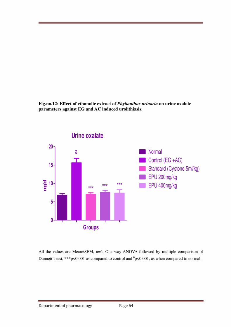
Fig.no.12: Effect of ethanolic extract of Phyllanthus urinaria on urine oxalate
parameters against EG and AC induced urolithiasis.
Urine oxalate
0
5
10
15
20Normal
Control (EG +AC)
Standard (Cystone 5ml/kg)
EPU 200mg/kg
EPU 400mg/kg
a
*** *** ***
Groups
mg/d
l
All the values are Mean±SEM, n=6, One way ANOVA followed by multiple comparison of
Dunnett’s test, ***p<0.001 as compared to control and ap<0.001, as when compared to normal.
Department of pharmacology Page 64

Fig.no.13: Effect of ethanolic extract of Phyllanthus urinaria on urine phosphorus
parameters against EG and AC induced urolithiasis.
Urine phosphorus
0
5
10
15Normal
Control (EG +AC)
Standard (Cystone 5ml/kg)
EPU 200mg/kg
EPU 400mg/kg
a
***
****
Groups
mg/d
l
All the values are Mean±SEM, n=6, One way ANOVA followed by multiple comparison of
Dunnett’s test, *p<0.05, ***p<0.001 as compared to control and ap<0.001, as when compared
to normal.
Department of pharmacology Page 65
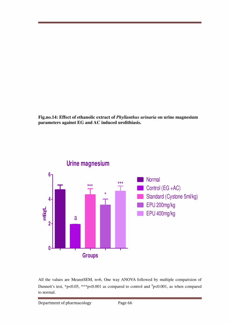
Fig.no.14: Effect of ethanolic extract of Phyllanthus urinaria on urine magnesium
parameters against EG and AC induced urolithiasis.
Urine magnesium
0
2
4
6Normal
Control (EG +AC)
Standard (Cystone 5ml/kg)
EPU 200mg/kg
EPU 400mg/kga
****
***
Groups
mEq/L
All the values are Mean±SEM, n=6, One way ANOVA followed by multiple comparision of
Dunnett’s test, *p<0.05, ***p<0.001 as compared to control and ap<0.001, as when compared
to normal.
Department of pharmacology Page 66
Effect of ethanolic extract of Phyllanthus urinaria on LPO (kidney enzyme)
parameters against EG and AC induced urolithiasis.
Oxidative stress
In-vivo LPO In control animals, EG/AC induced lithogenesis produced a significant
enhancement in the renal MDA levels (p<0.001) respectively, when compared to the
normal group. After treatment with standard cystone 5ml/kg and EPU 200 and
400mg/kg significant (P<0.001) reduction in the kidney MDA levels was observed in
the treated groups, when compared to their respective control groups.
Catalase levels of the kidney were significantly (p<0.001) decreased in the control
groups on EG/AC administration for 10 days, when compared to the normal group. On
treatment with Standrad cystone and EPU 200 and 400mg/kg (p<0.001), a significant
rise in the renal catalase levels was observed in treated groups (Table 3).
GSH levels of the kidney were significantly (p<0.001) decreased in the control groups
on EG/AC administration for 10 days, when compared to the normal group. On
treatment with Standrad cystone (p<0.01) and EPU 200 (p<0.05) and 400mg/kg
Department of pharmacology Page 67
(p<0.01), a significant rise in the renal catalase levels was observed in treated groups
(Table 3)
Department of pharmacology Page 68

Table.no.3: Effect of ethanolic extract of Phyllanthus urinaria on antioxidant (kidney enzyme) parameters against EG and AC induced
urolithiasis.
All the values are Mean±SEM, n=6, ns=
not significant, One way ANOVA followed by multiple comparision of Dunnett’s test, *p<0.05, **p<0.01, ***p<0.001 as compared to control and ap<0.001,
as when compared to normal.
Department of pharmacology Page 69
Treatment group
Invivo Antioxidant parameters
CATALASA
µm/mg tissue
GSH
µm/mg tissue
LPO
µm of /H2O2/mg
tissue
Normal 29.95±0.72 14.97±0.67 133.1±3.60
Control (EG+AC) 16.73±0.74a 7.970±0.83a 219.8±4.65a
Standard Cystone (5ml/kg) 26.19±1.25*** 12.80±0.68** 152.3±3.29***
EPU 200mg/kg 23.29±0.92*** 9.74±1.04* 176.7±5.05***
EPU 400mg/kg 26.00±0.56*** 12.68±0.85** 164.9±5.59***

Fig.no.16: Effect of ethanolic extract of Phyllanthus urinaria on Catalase (kidney
enzyme) parameters against EG and AC induced urolithiasis.
Catalase
0
10
20
30
40Normal
Control (EG +AC)
Standard (Cystone 5ml/kg)
EPU 200mg/kg
EPU 400mg/kga
******
***
Groups
µm
/mg tis
sue
All the values are Mean±SEM, n=6, One way ANOVA followed by multiple comparison of
Dunnett’s test, ***p<0.001 as compared to control and ap<0.001, as when compared to normal.
Dept. of Pharmacology. Page 70
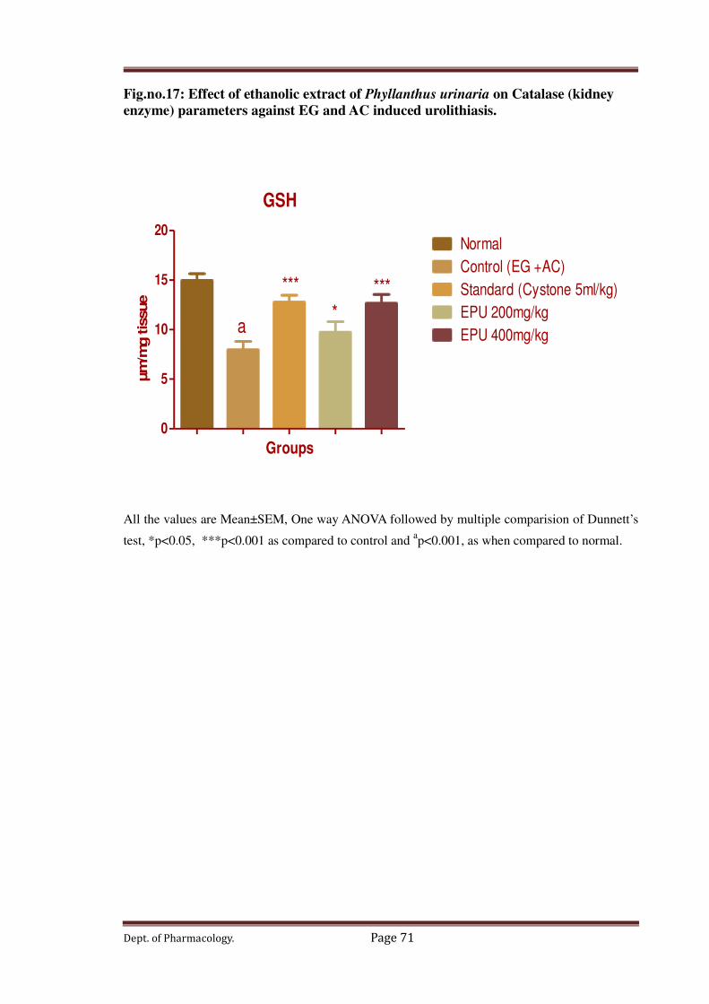
Fig.no.17: Effect of ethanolic extract of Phyllanthus urinaria on Catalase (kidney
enzyme) parameters against EG and AC induced urolithiasis.
GSH
0
5
10
15
20Normal
Control (EG +AC)
Standard (Cystone 5ml/kg)
EPU 200mg/kg
EPU 400mg/kga
***
*
***
Groups
µm
/mg tis
sue
All the values are Mean±SEM, One way ANOVA followed by multiple comparision of Dunnett’s
test, *p<0.05, ***p<0.001 as compared to control and ap<0.001, as when compared to normal.
Dept. of Pharmacology. Page 71
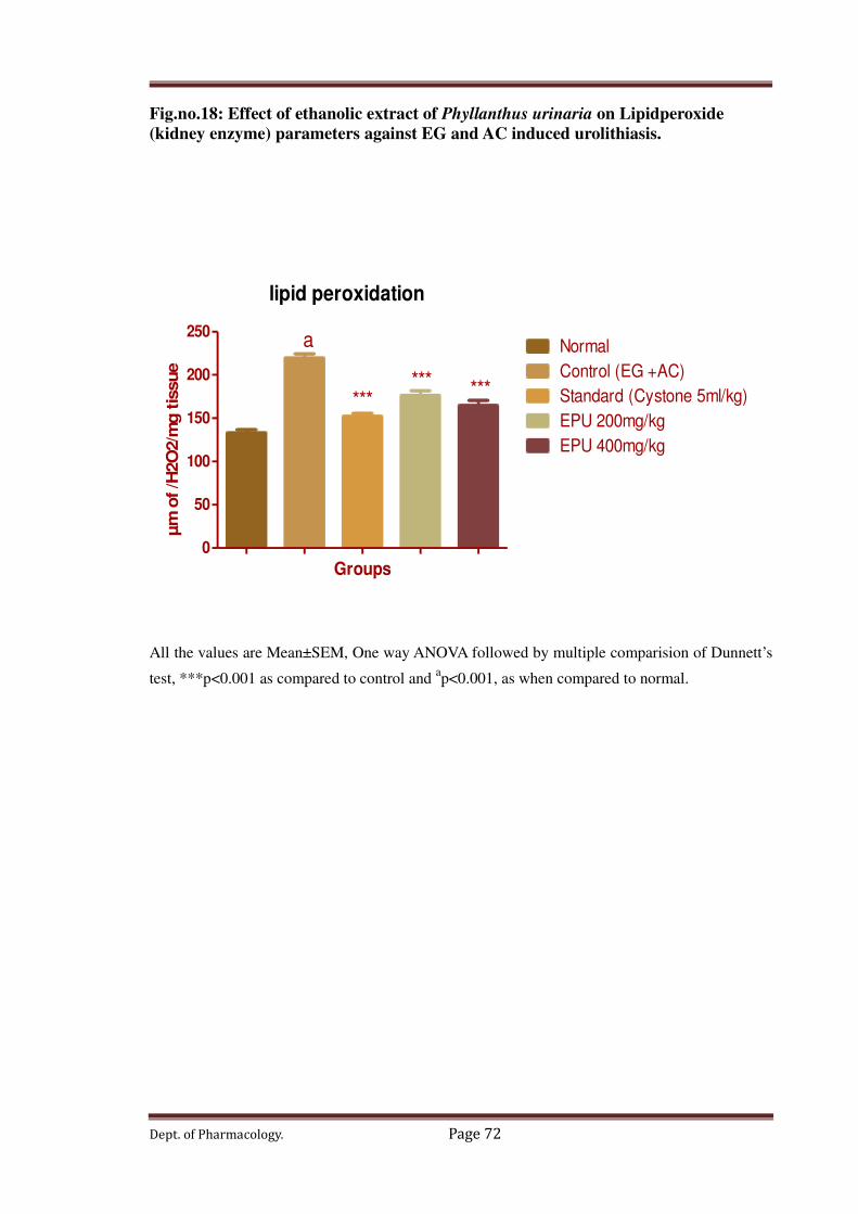
Fig.no.18: Effect of ethanolic extract of Phyllanthus urinaria on Lipidperoxide
(kidney enzyme) parameters against EG and AC induced urolithiasis.
lipid peroxidation
0
50
100
150
200
250 a
****** ***
Normal
Control (EG +AC)
Standard (Cystone 5ml/kg)
EPU 200mg/kg
EPU 400mg/kg
Groups
µm
of /H
2O
2/m
g t
issue
All the values are Mean±SEM, One way ANOVA followed by multiple comparision of Dunnett’s
test, ***p<0.001 as compared to control and ap<0.001, as when compared to normal.
Dept. of Pharmacology. Page 72
HISTOPATHOLOGY OF KIDNEY
Normal rats kidney showing normal cellular structure.
The histopathology of kidney samples of rats treated with EG+AC (0.75 and 2%) showed
loss of normal architecture with presence of crystalline structure in dilated collecting
tubules. The same section when viewed under polarising microscope revealed presence of
white chalky coloured calcium oxalate crystals in several tubules and glomeruli. These
groups also showed congestion of intersitum and inflammation of the pelvic calyceal
systems.
The histopathology of kidney samples of rats treated with standard drug cystone 5ml/kg
and EG+AC for 10 days showed normal architecture of the kidney.
The histopathology of kidney samples of rats treated with EUP 200mg/kg and EG+AC
for 10 days showed mild colloidal cast inside tubules and EUP 400mg/kg showed cloudy
changes and congestion of thes glomeruli. However the architecture of kidney approved
almost normal.
Fig.no.19: Histopathology of kidney
Dept. of Pharmacology. Page 73
A: Normal group, B: EG+AC group:, C Standard (Cystone 5ml/kg) group, D group EPU
200mg/kg, E group EPU 400mg/kg,
7.DISCUSSION
Urinary stone disease is a common, painful and expensive medical condition80. Though
extracorporeal shock wave lithotripsy has facilitated the stone removal and reduced the
morbidity associated with urinary stone, recurrence is common81. Several experimental
Dept. of Pharmacology. Page 74

and clinical studies on some of the plants used in the Indian traditional system of
medicine proved their efficacy in the management of renal stone disease. Therefore, it is
advisable to evaluate plants used in the traditional medicine to treat kidney stone disease
for Antiurolithiatic activity, which might be also useful in reducing stone recurrence rate.
Rats are commonly used to study the pathogenesis of human CaOx kidney stone disease,
as Ox metabolism is regarded almost similar in rats and humans82. Ingestion of EG/AC
has been found to be a reliable inducer of Ox lithiasis in rats. EG is converted to
endogenous oxalic acid by the liver enzyme glycolate oxidase [19] and AC induces
urinary acidification, is supposed to upset the enzyme sorting mechanism in the tubular
cells in the kidney83, thus favours adhesion and retention of CaOx particles within the
renal tubules.
Hence, in the present study, EG/AC in drinking water was employed to induce
hyperoxaluria in rats. Urinary supersaturation in relation to stone forming constituents,
mainly urinary oxalate is important in renal calculi formation84, as urinary oxalic acid
complexes with calcium and forms insoluble CaOx crystals85. Enhanced deposition and
urinary excretion of calcium and oxalate in the preventive and curative control group
animals indicate that administration of EG/AC induced hyperoxaluria. An increase in the
kidney weight and enhanced urinary creatinine excretion in the control group animals
also substantiated these results.
On administration of EPU, the dose-dependent reduction in calcium and oxalate
deposition in the kidneys and their urinary excretion in control groups implies the
potential of EPU. in preventing the formation and dissolving the preformed CaOx stones.
On treatment with the extract and standard cystone, the significant reduction in the
elevated urinary creatinine, urea, uric acid, calcium, phosphorus, oxalate and magnesium
in the treated groups reflects the improvement in hyperoxaluria induced renal
impairment. Dissolution of calculi can be achieved by alteration in urinary pH86. If the pH
is 5.0 or below, the stones likely to form are of uric acid type, if 5.0-6.5, calcium oxalate
type and if above 7 indicates crystals of magnesium ammonium phosphate. In the present
study, a decrease in the normal urine pH of 7.0-7.5 to 5.5-6.0 in the control groups,
indicates hyperoxaluria induced CaOx stone formation. In the treated groups, EPU and
Dept. of Pharmacology. Page 75

cystone 5ml/kg administration restored the pH to 6.5-7.5, supporting the decrease in the
deposition and excretion of calcium and oxalate.
Mucoproteins have significant affinity for CaOx surface and promote the growth of
crystals and cement them.87 Flavonoids act by disintegrating the mucoprotiens, thereby
prevent calcium and oxalate deposition and excretion88. In the present study also,
preliminary phytochemical screening of EPU revealed the presence of flavonoids. Thus,
in the EPU treated groups, flavonoids might have reduced calcium and oxalate deposition
by pre-coating CaOx crystals and disintegrating the mucoproteins. The stone forming
effects of EG are also ascribed to its hyperoxaluria induced oxidative damage. Oxalate
has been reported to induce LPO and to cause renal tissue damage. As kidney is rich in
polyunsaturated fatty acids, is susceptible to ROS attack. 89. Excessive generation of
ROS and/or a reduction in cellular antioxidant levels results in the development of OS.
MDA is one of the most common by products of ROS induced OS. In the present study,
increased levels of MDA, diminished levels of GSH and catalase in the control groups
indicate that EG/ AC administration promoted extensive generation of ROS. The resultant
ROS may have consumed GSH and catalase excessively and impaired antioxidant
protection. Thus, the unquenched ROS may have provoked cellular damage and resulted
in enhanced OS, which might have further favoured the accumulation and retention of
oxalate and subsequent deposition of CaOx. Studies show that treatment with
antioxidants prevents CaOx deposition in the kidney and reduce Ox excretion. 90 Daily
consumption of tea reduced the risk of kidney stone formation in women by 8%.
[Moreover, low concentration of renal cellular glutathione favours LPO and subsequent
retention of calcium and oxalate in the kidneys91.
Health benefits of tea are due to its antioxidant properties of flavonoids 92 which act by
quenching ROS and also by chelating metal ions like iron and copper. Lupeol and betulin
were proposed to act by scavenging oxalate promoted free radicals and enhancing body
antioxidant status, thus reducing oxalate induced renal peroxidative tissue damage. 93 In
the present study, lowered levels of MDA and enhanced levels of antioxidant enzymes,
GSH and catalase in the kidneys of the EPU treated animals indicate attenuation of
hyperoxaluria induced LPO and oxidative damage. Flavonoids may have minimized ROS
by free radical scavenging and prevented further generation, by metal chelating property,.
Thus, the flavonoid principles of phyllanthus urinaria might have been responsible for the
Dept. of Pharmacology. Page 76
inhibition of CaOx crystal aggregation and stone formation. The results support the use of
Phyllanthus urinaria plant as an effective alternative in treating CaOx urolithiasis.
Disintegration of the mucoproteins and pre-coating of CaOx crystals by antioxidant effect
of flavonoid principles may be responsible for the possible antiurolithiatic activity of
Phyllanthus urinaria. Further studies are necessary to find out the chemical components
responsible for the antiurolithiatic activity of Phyllanthus urinaria L.
Dept. of Pharmacology. Page 77
8. CONCLUSION
In conclusion, the presented data indicate that administration of the EPU plant
to rats with ethylene glycol/Ammonium chloride induced lithiasis and prevented the
formation of urinary stones, supporting folk information regarding antiurolithiatic activity
of the plant. The mechanism underlying this effect is still unknown, but is apparently
related to diuresis and lowering of urinary concentration of stone forming constituents.
The protective effect against oxalate induced lipid peroxidation may be contributing to
Dept. of Pharmacology. Page 78

the recovery of renal damage. These effects could conclude the antiurolithiatic property
of Phyllanthus urinaria.
Dept. of Pharmacology. Page 79
9. SUMMARY
In the present study, dried powder of Phyllanthus urinaria was subjected to
extraction using 90% ethanol for the extraction. Some extract was reserved for
preliminary phytochemical investigation and rest was utilized for pharmacological
screening.
The preliminary phytochemical investigation showed the presence of alkaloids,
carbohydrates, glycosides, saponins, and flavonoids.
The pharmacological screening included evaluation of antiurolithiatic activity
using 0.75% ethylene glycol/ammonium chloride induced urolithiasis model in male
Wistar albino rats. The kidney stone formation induced in rats, as a result of 10 days
chronic administration of 0.75% and 2% ammonium chloride, was significantly inhibited
by oral administration of EPU.
Administration of EPU and Cystone 5ml/kg caused significantly increased urine
output and pH of the urine as compared to control (EG and AC) group.
Dept. of Pharmacology. Page 80

Pretreatment with standard cystone 5ml/kg, EPU 200 and 400mg/kg causes
significant reduction in serum creatinine, urea, uric acid, calcium, oxalate, phosphorus
and magnesium concentration when compared to EG and AC alone treated group.
Pretreatment with standard cystone 5ml/kg, EPU 200 and 400mg/kg causes
significant reduction in urinary creatinine, urea, uric acid, calcium, oxalate, phosphorus
and magnesium concentration when compared to EG and AC alone treated group.
After treatment with standard cystone 5ml/kg and EPU 200 and 400mg/kg
significant reduction in the kidney MDA levels was observed in the treated groups, when
compared to their respective control groups.
Catalase and GSH levels of the kidney were significantly decreased in the
control groups on EG/AC administration for 10 days, when compared to the normal
group. On treatment with Standrad cystone and EPU 200 and 400mg/kg a significant rise
in the renal catalase and GSH levels was observed in treated groups.
Dept. of Pharmacology. Page 81

REFERENCES
1. Grases F, Costa Bauza A, Ramis M, Montesinos V, Conte A. Simple classification
of renal calculi closely related to their micromorphology and etiology. Clin Chim
Acta 2002; 322:29-36.
2. Bihl G, Meyers A. Recurrent renal stone disease-advances in pathogenesis and
clinical management. Lancet 2001;358:651-56.
3. Smith LH. Calcium-containing renal stones. Kidney int 1978;13:383-89.
4. Borsatti A. Calcium oxalate nephrolithiasis: Defective oxalate transport. Kidney Int
1991; 39:1283-98.
5. Coe FL. Uric acid and calcium oxalate nephrolithiasis. Kidney Int 1983;24:392-
403.
Dept. of Pharmacology. Page 82
6. Seyedzadeh A, Momtaz HE, Moradi MR, Moradi A. Pediatric Cystine Calculi in
West of Iran. Urol J 2006;3 (3):134-38.
7. Fleisch H. Inhibitors and promoters of stone formation. Kidney Int 1978;13:361-71.
8. Wells KA. Nephrolithiasis With Unusual Initial Symptoms. J Manipulative Physiol
Ther 2000;23 (3):196-01.
9. Portis AJ, Sundaram CP. Diagnosis and Initial Management of Kidney Stones. Am
Fam Physician 2001;63 (7):1329-38.
10. Delvecchio FC, Preminger GM. Medical management of stone diseases. Curr Opin
Urol 2003;13(3):229-33.
11. Siener R, Ebert D, Nicolay C, Hesse A. Dietary risk factors for hyperoxaluria in
calcium oxalate stone formers. Kidney Int 2003;63:1037-43.
12. Prasad KVSRG, Bharthi K, Srinivasan KK. Evalution of Ammania baccifera Linn.
For antiurolithiatic activity in albino rate. Ind J Expt Biol 1994;32 (5):311-13.
13. Keler T, Jansen B, Hesse A. Effect of blackcurrant, cranberry and plum juice
consumption on risk factors associated with kidney stone formation. Eur J Clin
Nutr 2002;56:1020-23.
14. Bhaskar R, Varalakshmi P, Amsaveni R. Changes in tissue enzymes produced by
Coleus aromaticus in experimental urolithiasis. Indian Drugs 1992;29:254-58.
15. Viel TAJ, Domingos CD, Monteiro APS, Lima-Landman MTR, Lapa AJ, Souccar
C. Evaluation of the antiurolithiatic activity of the extract of Costus spiralis Roscoe
in rats. J Ethnopharmacol 1999;66:193-98.
16. Harg TM, Rodgers A, Charlton K. Influence of cranberry juice on the urinary risk
factors for calcium oxalate kidney stone formation. Br J Urol 2003;92:765-68.
17. Christiana AJ, Lakshmi MP, Nagarajan M, Kurian S. Modulatory effect of Cyclea
pertata lam. On stone formation induced by ethylene glycol treatment in rats. Exp
Clin Pharmacol 2002;24:77-79.
18. Atmani F, Slimani Y, Mimouni M, Aziz M, Hacht B, Ziyyat A. Effect of aqueous
extract from Herniaria hirsute L. on experimentally nephrolithiasic rats. J
Ethnopharmacol 2004;95:87-93.
19. Grases F, Ramis M, Bauzi AC, March JG. Effect of Herniaria hirsuta and
Agropyron repens on calcium oxalate urolithiasis risk in rats. J Ethnopharmacol
1995;45:211-14.
Dept. of Pharmacology. Page 83
20. Vidya L, Varalakshmi P. Control of urinary risk factors of stones by betulin and
lupeol in experimental hyperoxaluria. Fitoterapia 2000;71:535-43.
21. Anand R, Patnaik GK, Roy K, Bhaduri AP. Antioxaluric and Anticalciuric activity
of lupeol derivatives. Ind J Pharmacol 1995;27:265-68.
22. Karadi RV, Gadge NB, Alagawadi KR, Savadi RV. Effect of Moringa oleifera Lam.
root-wood on ethylene glycol induced urolithiasis in rats. J Ethnopharmacol
2006;105:306-11.
23. Prasad KVSRG, Bharathi K, Srinivasan KK. Evaluation of Musa (paradisica
linn.cultivar) - “puttable” stem juice for antilithiatic activity in albino rats. Ind J
Phy Pharmacol 1993;33:337-41.
24. Poonguzhali PK, Chegu H. The influence of banana stems extract on urinary risk
factors for stones in normal and hyperoxaluric rats. Br J Urol 1994;74:23-25.
25. Barros ME, Lima R, Mercuri LP, Matos JR, Schor N, Boim MA. Effect of extract
of Phyllanthus niruri on crystal deposition in experimental urolithiasis. Urol Res
2006;34:351-57.
26. Murugaiyah V, Chan KL. Mechanisms of antihyperuricemic effect of Phyllanthus
niruri and its lignin constituents. J Ethnopharmacol 2009;124:233-39.
27. Vargas SR, Perez GRM, Perez GS, Zavala SMA, Perz GC. Antiurolithiatic activity
of Raphanus sativus extract on rats. J Ethnopharmacol 1999;68:335-38.
28. Vargas R, Perez RM. Effect of aqueous extract of Salix taxifolia in experimentally
induced urolithiatic rats. Pharm Biol 2002;40 (8):561-63.
29. Anand R, Patnaik GK, Kulshreshtha DK, Dhawan BN. Activity of certain fractions
of Tribulus terrestris fruits against experimentally induced urolithiasis in rats. Ind J
Exp Biol 1994;32:548-52.
30. Kambhoj J, Aggrwal M, Puri S, Singla SK. Effect of aqueous extract of Tribulus
terrestris on oxalate induced oxidative stress in rats. Indian journal of nephrology
2011;21 (5):154-59.
31. Ali MA, Wahbi S, Twaij H, Badr AA. Tribulus terrestris: preliminary study of its
diuretic and contractile effects and comparison with Zea mays. J Ethnopharmacol
2003;85:257-60.
32. Varalakshmi P, Shamila Y, Latha E. Effect of Crataeva nurvala in experimental
urolithiasis. J Ethnopharmacol 1990;28:313-21.
Dept. of Pharmacology. Page 84

33. Singh RG, Kapoor SU. Evaluation of antilithiatic properties of Varuna (Crataeva
nurvala). J Res Edu Indian med 1991;10:35-39.
34. Gupta P, Patel N, Bhatt L, Zambare GN, Bodhanka SLB. Nandakumar K. Anti-
urolithiatic effects of petroleum ether extract stem bark of Crataeva adansonii DC.
(Fam. Capparidaceae) in rats. Pharma Biol 2006;44(3):160-65.
35. Soundararajan P, Mahesh R, Ramesh T, Begum VH. Effect of Aerva lanata on
calcium oxalate urolithiasis in rats. Ind J Exp Biol 2006;44:981-86.
36. Selvam R, Kalaiselvi P, Govindaraj A, Murugan VB, Kumar ASS. Effect of Aerva
lanata leaf extract and vediuppu chunnam on the urinary risk factors of calcium
oxalate urolithiasis during experimental hyperoxaluria. Pharmacol Res 2001;43
(1):89-93.
37. Bashir S, Gilani AH. Antiurolithic effect of Bergenia ligulata rhizome: An
explanation of the underlying mechanisms. J Ethnopharmacol 2009;122:106-16.
38. Hadjzadeh MAR, Khoei A, Hadjzadeh Z, Parizady M. Ethanolic Extract of Nigella
sativa L Seeds on Ethylene Glycol-Induced Kidney Calculi in Rats. Urology 2007;4
(2):86-90.
39. Bahuguna YM, Rawat MSM, Juyal V, Gnanarajan G. Antilithiatic effect of grains
of Eleusine Coracana. Saudi Pharma J 2009;17 (2):182-87.
40. Prasongwatanaa V, Woottisin S, Sriboonlue P, Kukongviriyapan V. Uricosuric effect
of Roselle (Hibiscus sabdariffa) in normal and renal-stone former subjects. J
Ethnopharmacol 2008;117:491-95.
41. Moriyama MT, Suga K, Miyazawa K, Tanaka T, Higashioka M, Noda K, et al.
Inhibitions of urinary oxidative stress and renal calcium level by an extract of
Quercus salicina Blume/Quercus stenophylla Makino in a rat calcium oxalate
urolithiasis model. Int J Urol 2009;16:397-01.
42. Khan ZA, Assiri AM, Al-Afghani HMA, Maghrabi TMA. Inhibition of oxalate
nephrolithiasis with Ammi visnaga (AI-Khillah). Int Urol Nephrol 2001;33:605-08.
43. Atmani F, Sadki C, Aziz M, Mimouni M, Hacht B. Cynodon dactylon extract as a
preventive and curative agent in experimentally induced nephrolithiasis. Urol Res
2009;37:75-82.
44. Dharmaraj S, Hossain MA, Zhari S, Harn GL, Ismail Z. The use of principal
component analysis and self-organizing map to monitor inhibition of calcium
Dept. of Pharmacology. Page 85

oxalate crystal growth by Orthosiphon stamineus extract. Chemometrics and
Intelligent Lab Sys 2006;81:21-28.
45. Raut AA, Sunder S, Sarkar S, Pandita NS, Vaidya ADB. Preliminary study on
crystal dissolution activity of Rotula aquatica, Commiphora wightii and
Boerhaavia diffusa extracts. Fitoterapia 2008;79:544-47.
46. Grases F, Ramis M, Costa-Bauzi A, March JG. Effect of Herniaria hirsuta and
Agropyron repens on calcium oxalate urolithiasis risk in rats. J Ethnopharmacol
1995;45:211-14.
47. Swathi D, Sujatha D, Bharathi K, Prasad KVSRG. Antilithic activity of aqueous
extract of roots of Tephrosia purpurea Linn. Phcog mag 2008;4 (16):206-11.
48. Church AS, Witting MD (1997) Laboratory testing in ethanol, methanol, ethylene
glycol, and isopropanol toxicities. J Emerg Med 15: 687-692.
49. Abramson S, Singh AK (2000) Treatment of the alcohol intoxications: ethylene
glycol, methanol, and isopropanol. Curr Opin Nephrol Hypertens 9: 695-701
50. Frommer JP, Ayus JC. Acute ethylene glycol intoxication. Am J Nephrol 1982;2:1-
5.
51. Jacobsen D, McMartin KE. Antidotes for methanol and ethylene glycol poisoning. J
Toxicol Clin Toxicol 1997;35:127-43.
52. Goodman LS, Alfred G, Hardman JG, Limbird LE. Goodman & Gilman’s The
pharmacological basis of therapeutics. 9th ed. New York: McGraw-Hill, Health
Professions Division, 1996:1682-3.
53. Barceloux DG, Krenzelok EP, Olson K, Watson W. American Academy of Clinical
Toxicology practice guidelines on the treatment of ethylene glycol poisoning. Ad
Hoc Committee. J Toxicol Clin Toxicol 1999;37:537-60
54. Church AS, Witting MD. Laboratory testing in ethanol, methanol, ethylene glycol,
and isopropanol toxicities. J Emerg Med 1997;15:687-92.
55. Jacobsen D. New treatment for ethylene glycol poisoning. N Engl J Med
1999;340:879-81.
56. Antizol (fomepizole). Package insert. Minnetoka, Minn.: Orphan Medical, Inc.,
December 2000.
57. Sivilotti ML, Burns MJ, McMartin KE, Brent J. Toxicokinetics of ethylene glycol
during fomepizole therapy: implications for management. For the Methylpyrazole
for Toxic Alcohols Study Group. Ann Emerg Med 2000;36:114-25.
Dept. of Pharmacology. Page 86
58. Moreau CL, Kerns W II, Tomaszewski CA, McMartin KE, Rose SR, Ford MD, et
al. Glycolate kinetics and hemodialysis clearance in ethylene glycol poisoning.
META Study Group. J Toxicol Clin Toxicol 1998;36:659-66.
59. Wang M, Cheng H, Li Y, Meng L, Zhao G, Mai K. Herbs of the genus Phyllanthus
in the treatment of chronic hepatitis B: observations with three preparations from
different geographic sites. Journal of Laboratory and Clinical Medicine
1995;126(4):350–2.
60. Dias MA, Campos AH, Cechinel-Filho V, Yunes RA, Calixto JB. Analysis of the
mechanisms underlying the contractile responses induced by the hydroalcoholic
extract of Phyllanthus urinaria in the guinea pig urinary bladder in-vitro. Journal of
Pharmacy and Pharmacology 1995;47(10):846–51.
61. Santos AR, Cechinel-Filho V, Yunes RA, Calixto JB. Analysis of the mechanisms
underlying the antinociceptive effect of the extracts of plants from the genus
Phyllanthus. General Pharmacology 1995;26(7):1499–506.
62. Giridharan P, Somasundaram ST, Perumal K, Vishwakarma RA, Karthikeyan NP,
Velmurugan R, Balakrishnan A. Novel substituted methylenedioxy lignin
suppresses proliferation of cancer cells by inhibiting telomerase and activation of c-
myc and caspases leading to apoptosis. Br J Cancer 2002;87:98-105.
63. Huang ST, Yang RC, Yang LJ, Lee PN, Pang JHS. Phyllanthus urinaria triggers the
apoptosis and Bcl-2 down-regulation in Lewis lung carcinoma cells. Life Sci
2003;72:1705-16.
64. Huang ST, Yang RC, Lee PN, Yang SH, Liao SK, Chen TY, Pang JHS. Anti-tumor
and anti-angiogenic effects of Phyllanthus urinaria in mice bearing Lewis lung
carcinoma. Int Immunopharmacol 2006;6:870-9\
65. Lin SY1, Wang CC, Lu YL, Wu WC, Hou WC. Antioxidant, anti-semicarbazide-
sensitive amine oxidase, and anti-hypertensive activities of geraniin isolated from
Phyllanthus urinaria. Food Chem Toxicol. 2008 Jul;46(7):2485-92. doi:
10.1016/j.fct.2008.04.007. Epub 2008 Apr 13
66. Lee CY1, Peng WH, Cheng HY, Chen FN, Lai MT, Chiu TH. Hepatoprotective
effect of Phyllanthus in Taiwan on acute liver damage induced by carbon
tetrachloride. Am J Chin Med. 2006;34(3):471-82.
67. Chularojmontri L1, Wattanapitayakul SK, Herunsalee A, Charuchongkolwongse S,
Niumsakul S, Srichairat S.Antioxidative and cardioprotective effects of Phyllanthus
Dept. of Pharmacology. Page 87

urinaria L. on doxorubicin-induced cardiotoxicityBiol Pharm Bull. 2005
Jul;28(7):1165-71.
68. Shen ZQ1, Chen P, Duan L, Dong ZJ, Chen ZH, Liu JK.Effects of fraction from
Phyllanthus urinaria on thrombosis and coagulation system in animalsZhong Xi Yi
Jie He Xue Bao. 2004 Mar;2(2):106-10, 122.
69. Zhang LZ1, Guo YJ, Tu GZ, Guo WB, Miao F. Isolation and identification of a
novel ellagitannin from Phyllanthus urinaria L. Yao Xue Xue Bao. 2004
Feb;39(2):119-22.
70. Xu M1, Xu M1, Zha ZJ, Qin XL, Zhang XL, Yang CR, Zhang YJ. Phenolic
antioxidants from the whole plant of Phyllanthus urinariaChem Biodivers. 2007
Sep;4(9):2246-52
71. Zhong Y1, Zuo C, Li F, Ding X, Yao Q, Wu K, Zhang Q, Wang Z, Zhou LW, Lan J,
Wang X Chemical constituents of Phyllanthus urinaria L. and its antiviral activity
against hepatitis B virusZhongguo Zhong Yao Za Zhi. 1998 Jun;23(6):363-4, 384.
72. Paulino N1, Cechinel-Filho V, Yunes RA, Calixto JB. The relaxant effect of extract
of Phyllanthus urinaria in the guinea-pig isolated trachea. Evidence for involvement
of ATP-sensitive potassium channelsJ Pharm Pharmacol. 1996 Nov;48(11):1158-
63.
Dept. of Pharmacology. Page 88

PLANT PLANT PLANT PLANT
AUTHENTIFICATION AUTHENTIFICATION AUTHENTIFICATION AUTHENTIFICATION
Dept. of Pharmacology. Page 89
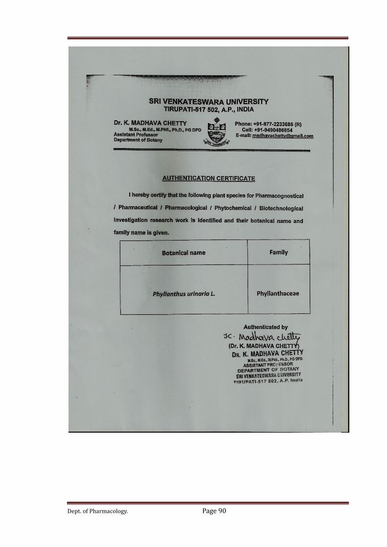
Dept. of Pharmacology. Page 90

APPROVAL
CERTIFICATE
Dept. of Pharmacology. Page 91
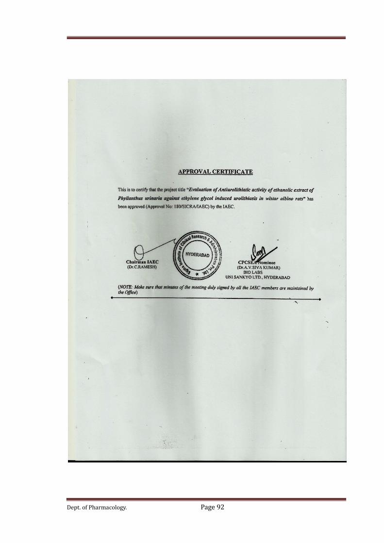
Dept. of Pharmacology. Page 92
ERRATA
S. No. Page No. Line No. Typed as Read as
1
2
3
4
5
6
7
8
9
10
11
12
13
14
Dept. of Pharmacology. Page 93