mr safety and the role of the mr safety expert
TRANSCRIPT

MR Safety and the Role of the MR Safety Expert
Heidi A. Edmonson PhD DABR, MRSO/MRSE (MRSCTM) April 26th, 2019
Financial Disclosure(s):Nothing to disclose
Other Disclosure(s):Chair, American Board of Magnetic Resonance Safety
Many committees and Councilor, ACRPresident-Elect MN Radiological Society (MN Chapter of the ACR)
Implantable medical devices described within this presentation are for illustrative purposes only and do not constitute endorsement
Disclaimer:

3
Outline1. Elements of an MR Safety Program2. Accreditation Requirements3. MR Safety Examples

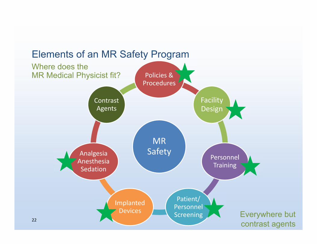
Elements of an MR Safety Program
4

Elements of an MR Safety Program
MR Safety
Policies & Procedures
Facility Design
Personnel Training
Patient/Personnel Screening
Implanted Devices
AnalgesiaAnesthesiaSedation
Contrast Agents
5

ISMRMSMRT
ESMRMBERFS
EFOMP

MR Safety Expert
Typically MR Physicist MR facility required to have ready access to services Not necessarily on site (not enough workforce)
Likely has no medical education Don’t expect advice on pharmaceuticals, anesthetics, etc

MR Safety Expert Provides high level advice on safe use of MR equipment
(engineering, scientific, administrative) Advice on safety framework for MR environment
Help with local rules and procedures
MR protocols –diagnostic effectiveness (artifacts, image contrast) Safety advice on risks associated with non-routine patients Safety audits Equipment selection Acceptance testing Maintain connections with local, national professional bodies Report back to MRMD

A Qualified Medical Physicist or a Qualified MR Scientist must be responsible for acceptance testing and monitoring of MRI equipment for the purposes of this practice parameter.
The Qualified Medical Physicist or MR scientist must maintain a thorough knowledge of the principles of MRI safety, physics, equipment, and relevant performance testing
Resolution 10, adopted 5/23/2017
9

10
American Board of Magnetic Resonance Safety
http://www.abmrs.org/
Grassroots effort to establish “internationally accepted standards for MR safety competencies” – test beyond the registry/board exam
MR Safety Certified (MRSC™) Magnetic Resonance Medical Director (MRMD)
Radiologist/Physician Magnetic Resonance Safety Officer (MRSO)
Executes MR safety practices under direction of MRMDCould be MR technologist – someone at the site
Magnetic Resonance Safety Expert (MRSE)Resource for MRMD and MRSO/AdvisoryCould be MR physicist

As a physicist, what am I thinking about?
11
B0
B1 (RF)B1 (RF)
dB/dt (Gradients)dB/dt (Gradients)

Three main systems used for MR image formation
12
B0 – Static magnetic fieldAligns proton magnetic moments
B1 (RF) – Tips magnetization into transverse plane (Flip angle)
B1 (RF) – Tips magnetization into transverse plane (Flip angle)
dB/dt (Gradients) –Encode spatial location dB/dt (Gradients) –Encode spatial location

Projectile Effect – location of max force
B0 Spatial Gradient of B0 (dB/dx)
Depends on B0, dB/dx, and amount of ferrous material

Projectile Effect – location of max force
B0 Spatial Gradient of B0 (dB/dx)
Depends on B0, dB/dx, and amount of ferrous material

TorqueStrongest at uniform region within MRI bore
Depends on • B0, geometry/magnetic moment of ferrous object

Aneurysm clips: Many safe, some unsafe

Depends on • change in magnetic field, geometry of conductive object
Lenz’s forces
Strongest near mouth of bore, force tends to oppose direction of motion

Static Magnetic Field Interactions
Magnetic reed switches Set to default programming Pacemakers (magnet mode)
Magnetic adjustments Programmable shunts
Mechanical failure Pumps
Projectiles

Radiofrequency (B1) Effects - MHzHeating and potential for thermal injury
Normal Mode operation –allows warming up to 0.5°C
Proximity to RF transmit coil
Patient skin-to-skin contact(Electric field loops within the body)
Resonant circuitry (loops, antenna)
Electrically conductive circuits/wires can get hot – thermal burns
10‐15 cm for 3T20‐25 cm for 1.5T

Gradient Effects
KHz range – audiofrequencies – Hearing protectionConductive objects: Eddy currents (case heating, Lenz’s forces) Pathway to enhance Peripheral Nerve Stimulation
Circuitry
Current loops can have signals induced by changing magnetic field “Power on reset” when device gets confused and resets to base valuesGradient effects are typically pretty minor. Use normal mode for active implants.

Cochlear implants (with magnets)
B0• Magnets! Pull and torque
B0• Magnets! Pull and torque
B1 (RF)• Leads! Potential for heating
B1 (RF)• Leads! Potential for heating
dB/dt (Gradients)• Conductive, small surface area
dB/dt (Gradients)• Conductive, small surface area
Active Device• Clicks and odd noises during scanning
Active Device• Clicks and odd noises during scanning
*www.Cochlear.com
Elements of an MR Safety Program
MR Safety
Policies & Procedures
Facility Design
Personnel Training
Patient/Personnel Screening
Implanted Devices
AnalgesiaAnesthesiaSedation
Contrast Agents
22
Where does the MR Medical Physicist fit?
Everywhere butcontrast agents

23
Outline1. Elements of an MR Safety Program2. Accreditation Requirements3. MR Safety Examples

24
2015 ACR Quality Control Manual for MRI
Requirements went into effect July 1, 2016

MR Safety Program Assessment (pgs 111-113)
25

MR Safety Program Assessment (pgs 111-113)
Divides safety concerns for MRI into three categories:
1. Facility Design
2. Operational
3. Clinical
26
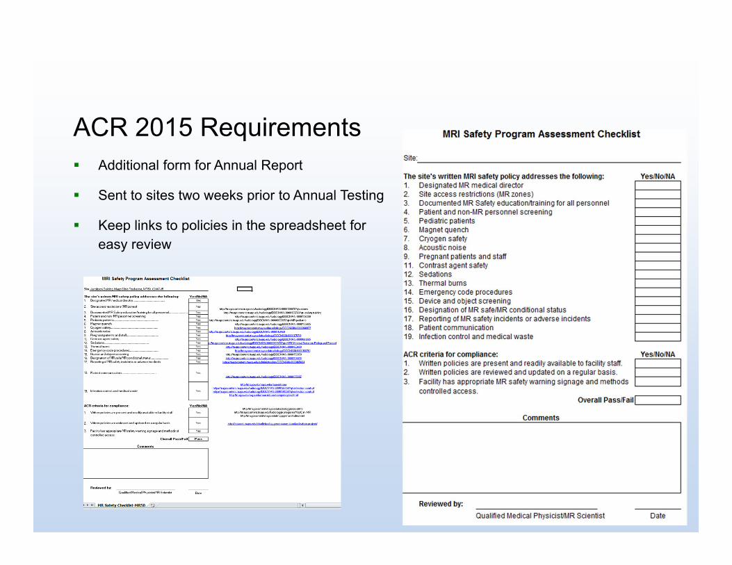
Additional form for Annual Report
Sent to sites two weeks prior to Annual Testing
Keep links to policies in the spreadsheet for easy review
ACR 2015 Requirements

Findings from initial MRI Safety Program Assessments RST – Policy regarding pregnant staff had vanished
(not specifically requested for MRI in “Toolkit for Practice Sites”)
Helpful for obtaining approvals for construction
Acquired hospital came Zone III-free
Sign off on “Facility has appropriate…methods controlled access”?
Review with Mobile MRI Technologist:
Patients were changing in scan room (Zone IV) for privacy
Patient screening challengeslack of access to medical records

The Joint Commission –Diagnostic Imaging Requirements
Effective July 1, 2015 Both outpatient (ambulatory) and hospital programs
Managing MRI safety risks Data collection on MRI incidents Annual education requirement for MR Technologists
29

The Joint Commission –Diagnostic Imaging Requirements
The organization collects data on: patient thermal injuries that occur during MRI exams
Incidents where ferromagnetic objects unintentionally entered the MRI scanner room
Injuries resulting from the presence of ferromagnetic items in the MRI scanner room
Process improvement chapter
30
31
Outline1. Elements of an MR Safety Program2. Accreditation Requirements3. MR Safety Examples

ICD - Artifacts
“cannot exclude the possibility of artifact from nearby intracardiac CRT‐D leads”
FIESTA/SSFP techniques – banding artifacts• Use localized shim• Switch to gradient echo sequences
MDE – artifact near lead tip• Interferes with B1‐field • Flip angle not correct

What can an MR Physicist do? Especially working with the Radiologist!Myocarditis/? Subsequent fibrosis (ICD)
Local Shim Parameter changes
Phase Sensitive? No!
Type in L/R ShimMade it worse!
Type in L/R Shimopposite direction
Interactive troubleshooting for these exams
• Can’t anticipate location of implant
• Can’t anticipate disease state or scan protocol
• Geometry within the scanner very important
• Can’t protocol ahead of timeSubepicardial delayed enhancement
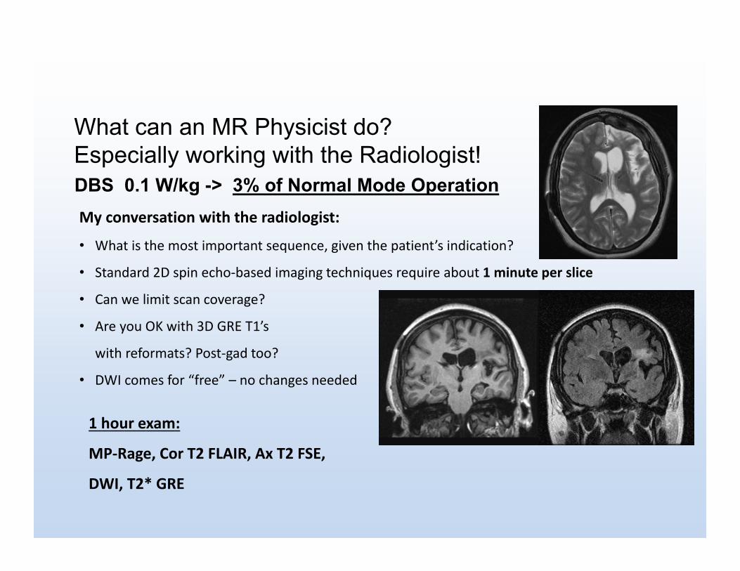
What can an MR Physicist do? Especially working with the Radiologist!DBS 0.1 W/kg -> 3% of Normal Mode OperationMy conversation with the radiologist:
• What is the most important sequence, given the patient’s indication?
• Standard 2D spin echo‐based imaging techniques require about 1 minute per slice
• Can we limit scan coverage?
• Are you OK with 3D GRE T1’s
with reformats? Post‐gad too?
• DWI comes for “free” – no changes needed
1 hour exam:
MP‐Rage, Cor T2 FLAIR, Ax T2 FSE,
DWI, T2* GRE

MRI Safety Training
Examples from your own institution make a powerful impact during training!
Oxygen tank
Screwdriver
Needle

“Scary” Equation
Translational force equation for magnetic object
So, this is a scary equation, because the force is so hard to predict!
Depends on the object, its magnetic properties, its shape, and properties of the MRI scanner we can’t see with our eyes
Unrestrained force results in acceleration, and the acceleration might be faster than your reflexes!

Removed Spinal Cord Stimulator
2008 2017

38
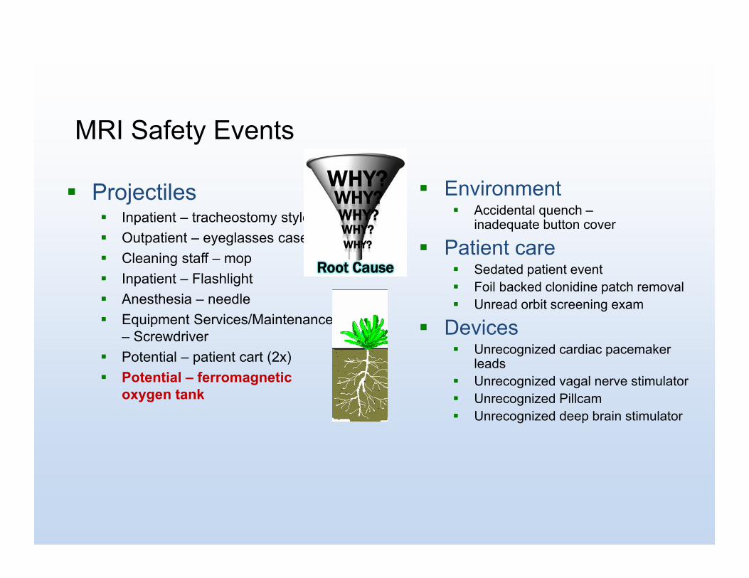
MRI Safety Events
Projectiles Inpatient – tracheostomy stylet Outpatient – eyeglasses case Cleaning staff – mop Inpatient – Flashlight Anesthesia – needle Equipment Services/Maintenance
– Screwdriver Potential – patient cart (2x) Potential – ferromagnetic
oxygen tank
Environment Accidental quench –
inadequate button cover
Patient care Sedated patient event Foil backed clonidine patch removal Unread orbit screening exam
Devices Unrecognized cardiac pacemaker
leads Unrecognized vagal nerve stimulator Unrecognized Pillcam Unrecognized deep brain stimulator

Unsecured ferromagnetic O2 tank discovered Monday morning
• Anesthesia case over weekend• “regular” personnel not present
Unsecured ferromagnetic oxygen tank in Zone III

Abandoned O2 tank responses
• Improved education for Anesthesia colleagues
• Empower “stop the line”
• Eliminate ferromagnetic O2 tanks • Carts must be adapted
• Different diameters – 3/8 inch• Ergonomic benefits

Replacement of ferromagnetic oxygen tanks completed MCR

Other parts on the Aluminum O2 Tanks
Required to be in cart (prevent tip hazard, “the other missile effect”) Ferrous Free carts often get repaired with ferrous parts May be replaced with incorrect
regulator Quarterly QC for any carts in the MRI
areas, signature card and pink info tag Removal of ferrous O2 tanks significantly
decreases attraction to magnet More time to react even if in a
ferrous cart
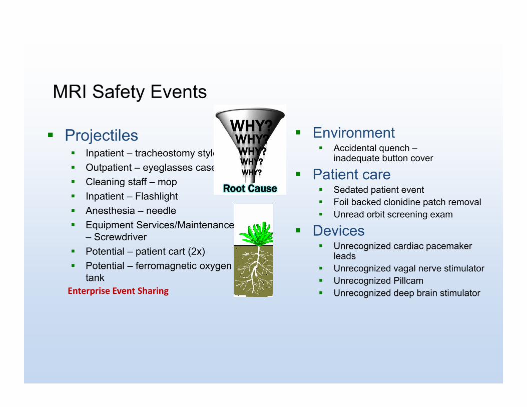
MRI Safety Events
Projectiles Inpatient – tracheostomy stylet Outpatient – eyeglasses case Cleaning staff – mop Inpatient – Flashlight Anesthesia – needle Equipment Services/Maintenance
– Screwdriver Potential – patient cart (2x) Potential – ferromagnetic oxygen
tank
Environment Accidental quench –
inadequate button cover
Patient care Sedated patient event Foil backed clonidine patch removal Unread orbit screening exam
Devices Unrecognized cardiac pacemaker
leads Unrecognized vagal nerve stimulator Unrecognized Pillcam Unrecognized deep brain stimulatorEnterprise Event Sharing

Enterprise Event Sharing
Pad QC ProgramTechnologist requests anytime
Biannual QC Program

FDA poster on MRI Burn Prevention (Partnership with SMRT)
Quick reference for how to avoid burnsMentions several suggestions from
Ensure that no items (such as leads) are formed into a loop, since magnetic induction can occur and cause burns.
If the patient’s body touches the bore of the MRI scanner, use non-conductive foam padding to insulate the patient’s skin and tissues.

Conclusion
MR Safety is collaboration across entire team
Medical Physicist/MRI Scientist integral to safe facility design
Alternate in-depth training and credentialing helps you know-what-they-know (MRMD/MRSO/MRSE)
Patients benefit from solid knowledge of MR physics
47