mr thermometry in cerebrovascular disease: physiologic
TRANSCRIPT
REVIEW ARTICLEADULT BRAIN
MR Thermometry in Cerebrovascular Disease: PhysiologicBasis, Hemodynamic Dependence, and a New Frontier in
Stroke ImagingS. Dehkharghani and D. Qiu
ABSTRACT
SUMMARY: The remarkable temperature sensitivity of the brain is widely recognized and has been studied for its role in thepotentiation of ischemic and other neurologic injuries. Pyrexia frequently complicates large-vessel acute ischemic stroke and devel-ops commonly in critically ill neurologic patients; the profound sensitivity of the brain even to minor intraischemic temperaturechanges, together with the discovery of brain-to-systemic as well as intracerebral temperature gradients, has thus compelled theexploration of cerebral thermoregulation and uncovered its immutable dependence on cerebral blood flow. A lack of pragmaticand noninvasive tools for spatially and temporally resolved brain thermometry has historically restricted empiric study of cerebraltemperature homeostasis; however, MR thermometry (MRT) leveraging temperature-sensitive nuclear magnetic resonance phenom-ena is well-suited to bridging this long-standing gap. This review aims to introduce the reader to the following: 1) fundamentalaspects of cerebral thermoregulation, 2) the physical basis of noninvasive MRT, and 3) the physiologic interdependence of cerebraltemperature, perfusion, metabolism, and viability.
ABBREVIATIONS: BOLD = blood oxygen level–dependent; BTR = brain thermal response; NMR = nuclear magnetic resonance; OEF = oxygen extraction frac-tion; PRESS = point-resolved spectroscopy; PRF = proton resonance frequency
Cerebral thermoregulation is a critical, but enigmatic aspect ofbrain physiology at the intersection of cerebral perfusion
and metabolism. The brain exhibits exquisite sensitivity todisruptions in temperature homeostasis, with even small eleva-tions rapidly potentiating ischemic or other neurologic injuries.1-5
Nevertheless, formalized and experimentally tested theories ofcerebral temperature control are lacking, due to the absence ofpragmatic tools to measure spatiotemporally-resolved thermalgradients under physiologic and pathologic conditions. Thisreview aims to introduce the reader to fundamental principles ofcerebral temperature (dys)regulation and homeothermy, withan emphasis on temperature as a potential biomarker of
cerebrovascular disease and the use of noninvasive approachesto measuring brain temperatures using MR thermometry.
THERMOREGULATION AND TEMPERATUREHOMEOSTASISBody temperature is tightly controlled in humans at approxi-mately 37°C and regulated through the balance of heat-conservingand heat-dissipating mechanisms mediated by the hypothalamus.6
Most heat production arises from so-called obligatory thermogen-esis—that is, the thermal energy of metabolically active organs,including the brain, with the remainder arising from voluntaryand involuntary muscle activity or acquired passively from ambi-ent sources.6 While the body efficiently dissipates heat primarilythrough radiative as well as conductive, convective, and evapora-tive mechanisms, cerebral thermoregulation proves more com-plex. Throughout much of the highly metabolic brain (consuming20% of the oxygen and 25% of the glucose at only 2%–3% of thetotal body weight),7 the temperature exceeds that of the arterialinflow. While superficial cortical brain regions may dissipateheat to a greater extent through convention/conduction andradiation, cerebral temperature homeostasis largely depends onsteady CBF to sustain countercurrent heat exchange mecha-nisms that prevent cerebral hyperthermia and prove critical tonormal brain physiology.6,8-10 The specific influence of systemictemperatures on cerebral thermoregulation, however, remains
Received November 13, 2019; accepted after revision January 2, 2020.
From the Department of Radiology (S.D.), New York University Langone Health,New York, New York; and Department of Radiology (D.Q.), Emory UniversityHospital, Atlanta, Georgia.
Portions of this work were supported by the American Society of NeuroradiologyScholar Award in Neuroradiology Research (2012–2013, 2013–2014), and an EmoryUniversity Research Committee (2013–2014) grant awarded to the lead author. Thiswork was supported, in part, by National Center for Research Resources and theOffice of Research Infrastructure Programs of the National Institutes of Health(P51RR000165, OD P51OD011132).
Please address correspondence to Seena Dehkharghani, MD, Center for BiomedicalImaging, Department of Radiology, New York University Langone Medical Center,660 First Ave, 2nd Floor, Attention Hillary Gumiel, New York, NY 10016; e-mail:[email protected]
Indicates open access to non-subscribers at www.ajnr.org
http://dx.doi.org/10.3174/ajnr.A6455
AJNR Am J Neuroradiol 41:555–65 Apr 2020 www.ajnr.org 555
poorly understood. Existing studies have produced discrepantand sometimes contradictory conclusions regarding: 1) thedirection and magnitude of the brain-to-systemic temperatureoffset in certain brain regions, 2) the spatial distribution andmagnitude of intracerebral temperature gradients, and 3) thespecific mechanisms by which potentially mild pyrexia potenti-ates myriad forms of neurologic injury, including ischemia.
The implications of cerebral thermosensitivity, in fact, reachbeyond a manifest susceptibility to injury and may even havesteered the course of human evolution. Specifically, the develop-ment of efficient mechanisms of heat dissipation are proposed tohave released human ancestors from fundamental thermal con-straints to brain development and growth. Simply, the energeticcost and therefore the metabolic output of the large human brainposes considerable demands on such mechanisms of temperaturecontrol, the development of which facilitated the remarkableencephalization of early humans.11-13 Thus, by comparison withother key constraints of human evolution, brain temperature wasemphasized by Baker (1979)13 to be the “single most importantfactor limiting the survival of man and other animals.”Neuroprotective adaptations such as selective brain cooling—themaintenance of brain temperatures selectively despite rising pe-ripheral temperatures by rerouting cooler, valveless facial/ocularand emissary venous supply—were therefore critical for thrivinghumans under thermal stress, exercise, and so forth.8,13-15 Suchadaptations are traceable to recognized changes in the humancalvarial fossil record, developing in tandem with epochs ofimmense advancement, and may have compensated for the lossin humans of the rete caroticum (ie, the rete mirabilis) first pro-posed by Herophilus �2000 years ago and documented later byGalen in other mammals including dogs, cats, and sheep.14,16-20
From these insights, a coherent picture of the evolving humanbrain emerged, linking intelligence, the growing brain, bipedal-ism, and efficient cerebral heat exchange in a seminal paradigmknown as the radiator hypothesis, which aptly underscores theimportance of steady blood flow at the nexus of temperature ho-meostasis.11-13,15,16 This point is easily contextualized when con-sidering the thermogenicity of the metabolically active brain:Approximately 0.66 J/min/g are produced by the brain, primarilyby the consumption of oxygen through chemical reaction withglucose which, absent heat dissipation, would produce a rise of0.16°C/min.18,21,22
The study of cerebral thermoregulation, nevertheless, remainsdifficult. Past investigations have consisted mostly of surrogatemeasures of brain temperature (eg, tympanic, sublingual, andetc.) and sporadic human and animal studies using directlyimplanted temperature probes. Surrogate measures, however, arelimited by the presence of temporally varying offsets relative toactual brain temperatures.23-26 Implantable probes are similarlysuboptimal for large-scale experimentation due to their requisiteinvasiveness and cost,27-30 which limit their widespread place-ment throughout the brain and in turn, their suitability for inter-rogation of spatially distributed temperature gradients.27,29,31,32
Notwithstanding their limitations, a small number of animaland human studies have confirmed the presence of measurablespatial temperature gradients that can be altered during braininjury.2,9,21,29,33-41 A theoretic framework for understanding
temperature homeostasis has also been established throughnumeric simulations often based in the seminal bioheat equa-tions proposed by Pennes (1948),42 and the reader is referred toexcellent resources on the topic.8-10,43-47
CENTRAL FEVER AND CEREBRAL TEMPERATUREDYSREGULATION DURING HYPOPERFUSIONSystemic and cerebral temperature dysregulation during and fol-lowing neurovascular ischemia is well-described, yet the mecha-nisms underpinning this relationship remain unclear.
The sensitivity of neuronal substrate to hyperthermia hasbeen the focus of multiple human and animal stroke studies, andidentification of this potentially modifiable biomarker has natu-rally promoted investigation into therapeutic cooling protocolsfollowing different forms of neurologic injury.1,2,4-6,48 Indeed,therapeutic cooling remains among the most potent neuroprotec-tants following ischemia and cardiac arrest, dating to landmarkstudies of the mid-20th century.49,50 Early canine studies byHosler (1953)49 found that at a body temperature of 20°C, noneurologic injury was observed following resuscitation from 11–13minutes of cardiac fibrillation. Similarly, Donnelly (1956)51
reported that brain anoxia can be tolerated for only 3 minutes atphysiologic temperatures, but remarkably with full resistance for9 minutes when cooling to 26.5°C. In part through depression ofmetabolic demand—a 2.2-fold reduction in oxygen consumptionfor each 10°C decrease in temperature—as well as protectionagainst immediate and programmed cytotoxicity and vascularpermeability, brisk hypothermia (� 20°–32°C) permits prolongedcirculatory arrest potentially with little neurologic injury.7
Decades of subsequent investigation have highlighted the protec-tive attributes of corporeal and brain cooling following braininjury, yet a number of large-scale prospective trials have failed tocapture a population-level benefit from cooling, owing perhapsto systemic adverse effects or, we posit, the use of non-neurologictemperature benchmarks. This latter point, we believe, holds tre-mendous potential promise for noninvasive brain thermometry,with which the known variability between systemic surrogatesand brain temperatures can be disentangled toward more guidedtherapeutic hypothermia protocols.
In contradistinction, hyperthermia proves a strong potentia-tor of neurologic injury.1,2,5,52 Fever, an adaptation intended toraise host defenses, has been shown to influence clinical out-comes negatively following stroke, potentially even whenmild.1,2,4-6,53-59 Fever following neurologic injury is unfortunatelycommon, even without direct hypothalamic injury.60 While themechanisms of central fever following ischemic stroke are incom-pletely understood, the aforementioned impairment in radiativeheat exchange may act in concert with cytokine-mediated influ-ences involving the hypothalamus, as well as more regional andzonal inflammatory trafficking as described by the inflammatorypenumbra paradigm.6,7,41,53,61,62 These inflammatory cascadesmay be moderated through hypothermia which, together withreduced expression of pro-apoptotic genes, may provide a power-ful potential target for intervention.63-66
Radiologic progression of ischemia due to hyperthermiaremains comparatively less explored. We previously reported onthe tendency for greater consumption of penumbral (ie, at risk)
556 Dehkharghani Apr 2020 www.ajnr.org
tissues in fully reperfused patients with acute ischemic strokewhen fevers exceeded 37.5°C in the early stroke aftermath. Whileinfarction expansion following reperfusion comprises an array ofreported causes, identification of a potentially modifiable andreadily attainable biomarker may significantly influence imagingand clinical outcomes.53,67
Larger studies, including a series of 4295 patients, have firmlyestablished the detrimental effects of fever as producing longerintensive care unit and hospital stays, higher mortality, and over-all worse outcomes, even when controlling for disease severity,complications, and age.68 We propose that the frequency withwhich pyrexia occurs in neurologically injured patients—up to47% of neurologic patients in intensive care units (increasing to93% after 2 weeks69)—itself motivates investigation into the useof noninvasive, direct brain thermography in diagnosis, prognos-tication, and perhaps treatment selection or response to therapy.
MR THERMOMETRYPrinciples and Techniques2D MRI thermometry dates to initial experimentation by Parkeret al,70 who studied the temperature sensitivity of proton longitu-dinal relaxation (T1) times in human blood specimens, decadesafter the far earlier discovery of temperature-sensitive nuclearmagnetic resonance (NMR) phenomena in the mid-20th cen-tury. While a strong linear relationship and sensitivity of �2°Cwas reported from a 5-minute scan, the speed and accuracy ofT1 thermometry have limited its application in neuroscience.Subsequently, other temperature-sensitive MRI contrast mecha-nisms were explored, including transverse relaxation time (T2),equilibrium magnetization/susceptibility, spin density, micro-scopic diffusion, and the proton resonance frequency (PRF), thelatter now most commonly used, particularly for thermal abla-tive procedures (eg, laser interstitial thermal therapy, high-inten-sity focused ultrasound, and radiofrequency ablation).71 Whileeach approach may offer relative, application-specific advan-tages, PRF thermometry, which exploits either of 2 attributes ofthe proton resonance frequency, is the most accurate, versatile,and widely used technique and will be the subject of the remain-der of this review.72 Specifically, this review will emphasize clini-cally pragmatic approaches allowing the sufficient spatial,temporal, and temperature resolution to probe dynamic thermalgradients in the healthy brain and during cerebrovascularimpairment. Emerging strategies, including potentially promis-ing and accurate temperature-sensitive contrast agents, remainunfit for translational human or clinical imaging and are beyondthe intended scope of this review; however, the interested readeris referred to thorough topical reviews on the subject.73-75
PRF ThermometryPRF thermometry stems from the temperature dependence ofthe water proton chemical shift. Due to the shielding effects oftheir surrounding electron currents, the magnetic field experi-enced by hydrogen protons varies slightly relative to that of theexternally applied field (B0). The effect manifests in a tempera-ture-dependent chemical shift (s) as follows:
1) v Tð Þ ¼ g � ½1� s Tð Þ� � Blocal;
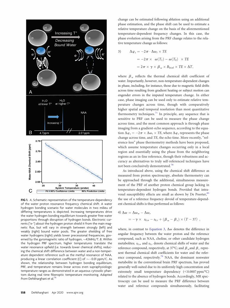
where vðTÞ represents the angular frequency of proton preces-sion; g ; the gyromagnetic ratio (�42 MHz/T for the hydrogennucleus); and s Tð Þ; the temperature-dependent chemical shiftmodulated by the electron-shielding effect on the local magneticfield (BlocalÞ, itself a function of both the externally applied ðB0Þfield and spatially varying magnetic susceptibility. The shieldingeffect of the water proton is influenced heavily by the strength ofhydrogen bonds, which is a function of temperature. Hydrogenbonds are formed between the hydrogen of 1 water molecule andthe electronegative oxygen atom of another water molecule(Fig 1A). Hydrogen bonds among water molecules, therefore,reduce the opposing electron currents about the bound hydrogenproton, exposing the proton to less attenuated Blocal fields,thereby inducing higher precessional frequencies. (Fig 1B). Athigher temperatures, however, the hydrogen bonds betweenwater molecules become less stable and/or disrupted, resulting ingreater electronic shielding and therefore reduced magnetic fluxdensity and a lower frequency of precession of the hydrogen pro-ton. It has been shown in multiple studies, including our own,that the water proton chemical shift s(T) is largely a linear func-tion of temperature T, (Fig 1C):
2) s Tð Þ � s0 þ b � T � 37ð Þ;
where S0 denotes the chemical shift at 37°C and b denoteschange in chemical shift per unit increase in temperature. b hasbeen measured to be between �0.009 and �0.011 ppm perdegree Celsius,29,72,76,77 dating to its initial description in NMRby Hindman (1966).77
While most MR thermometry applications have sought tomeasure relative temperature changes (eg, during and followingthermal ablation), they are poorly suited for the study of base-line temperatures and pathophysiologic temperature gradientsbetween brain regions, comparisons between subjects, or longi-tudinal study, together motivating the development of quanti-tative techniques approximating absolute temperatures. Asapparent from Equation 1, an accurate measure of the chemicalshift difference would require precise measurement of Blocal
down to 0.01 ppm. To resolve this issue, a stable (ie, non-tem-perature-dependent) reference frequency is necessary to nor-malize the water frequency, thereby allowing nearly absolutethermometry based on the temperature-dependent chemicalshift difference between the 2 (or sometimes more) metabolites.Such self-referenced thermometry has been the subject of con-siderable study using the chemical shift difference between thetemperature-sensitive water resonance and a non-temperature-sensitive reference metabolite, such as the methyl resonance ofneuronal NAA at �1.98 ppm.
By comparison, when the primary interest is simply thecollection of relative changes in temperature across time, forexample during ablative procedures, explicit measurement of ei-ther water or reference metabolite frequency is unnecessary, anda baseline water resonance phase can instead be measuredbefore treatment begins. Thereafter, a relative temperature
AJNR Am J Neuroradiol 41:555–65 Apr 2020 www.ajnr.org 557
change can be estimated following ablation using an additionalphase estimation, and the phase shift can be used to estimate arelative temperature change on the basis of the aforementionedtemperature-dependent frequency changes. In this case, thephase evolution arising from the PRF change relates to the rela-tive temperature change as follows:
3) Dw t ¼ �2p � Dv t � TE
¼ �2p � v T1ð Þ � v T0ð Þ� �� TE
¼ 2p � g � b w � Blocal � TE� DT;
where b w reflects the thermal chemical shift coefficient ofwater. Importantly, however, non-temperature-dependent changesin phase, including, for instance, those due to magnetic field driftsacross time resulting from gradient heating or subject motion canengender errors in the imputed temperature change. In eithercase, phase imaging can be used only to estimate relative tem-perature changes across time, though with comparativelyhigher spatial and temporal resolution than most quantitativethermometry techniques.71 In principle, any sequence that issensitive to PRF can be used to measure the phase changeacross time, and the most common approach is through phaseimaging from a gradient-echo sequence, according to the equa-tion Dw t ¼ �2p � Dv t � TE, where Dw t represents the phasechange across time, and TE, the echo time. More recently, “ref-erence-less” phase thermometry methods have been proposed,which assume temperature changes occurring only in a localregion and essentially using the phase from the neighboringregions as an in-line reference, though their robustness and ac-curacy as alternatives to truly self-referenced techniques havenot been conclusively demonstrated.78
As introduced above, using the chemical shift difference asmeasured from proton spectroscopy, absolute thermometry canbe approached through the additional, simultaneous measure-ment of the PRF of another proton chemical group lacking intemperature-dependent hydrogen bonds. Provided that intra-voxel susceptibility effects are small as shown by De Poorter,85
the use of a reference frequency devoid of temperature-depend-ent chemical shifts is thus performed as follows:
4) Dv ¼ Dvw � Dv r
¼ �g � s0;w � s0;r þ b w � b rð Þ � T � 37ð Þ� �;
where, in contrast to Equation 3, Dv denotes the difference inangular frequency between the water proton and the referencecompound, such as NAA, choline, or other candidate hydrogenmetabolites; s0;w and s0;r denote chemical shifts of water and thereference compound, respectively, at 37°C; and b wand b r repre-sent thermal chemical shift coefficients for water and the refer-ence compound, respectively.79 NAA, the dominant nonwatermetabolite in the conventional brain PRF spectrum, has provedgenerally well-suited due to its uniformly high concentration andextremely small temperature dependency (,0.0007 ppm/°C)related to the absence of hydrogen bonds. Accordingly, MR spec-troscopy can be used to measure the PRF difference betweenwater and reference compounds simultaneously, facilitating
FIG 1. A, Schematic representation of the temperature-dependencyof the water proton resonance frequency chemical shift. A waterhydrogen bonding scenario for water molecules in two milieu ofdiffering temperatures is depicted. Increasing temperatures drivethe water hydrogen bonding equilibrium towards greater free waterproportions through disruption of hydrogen bonds. Electronic cur-rents (“e-”) about the hydrogen proton shield it from the main mag-netic flux, but will vary in strength between strongly (left) andweakly (right) bound water pools. The greater shielding of freewater hydrogens (right) yields lower precessional frequencies, gov-erned by the gyromagnetic ratio of hydrogen, �42MHz/T. B, Withinthe hydrogen PRF spectrum, higher temperatures translate thewater resonance upfield (i.e. towards lower chemical shifts), reduc-ing the chemical shift difference between water and a non-temper-ature dependent reference such as the methyl resonance of NAA,producing a linear correlation coefficient (C) of ��0.01 ppm/C. Asshown, the relationship between hydrogen bonding equilibrium,PRF, and temperature remains linear across even supraphysiologictemperature ranges as demonstrated in an aqueous cytosolic phan-tom during real time fiberoptic temperature monitoring. Adaptedfrom Dehkharghani et al.76
558 Dehkharghani Apr 2020 www.ajnr.org
quantitative temperatures at single time points or longitudinally,either between or among individual subjects and across experi-mental conditions.29,41,59,76,80
MR Thermometry in Stroke and Cerebrovascular DiseaseAcute Ischemic Stroke. Noninvasive cerebral MRT has beenexplored in small-but-growing numbers of human and animalstudies, aiming to capture empirically the link between brain tem-perature and hemodynamic compromise. While such mechanis-tic insights are valuable motivations for noninvasive MRT, thedevelopment of biomarkers of tissue viability and outcome argu-ably remain the most compelling long-term objective of ther-mometry in this setting.
Corbett et al29 first described the use of single-voxel protonMR spectroscopy for in vivo brain thermometry in a piglet modelof ischemic stroke using a 4.7T NMR spectroscopy system. Usingwater-NAA chemical shift thermometry, their in vivo experi-ments recapitulated the linearly changing water-NAA chemicalshift difference coefficient of approximately 0.01 ppm/°C acrossboth physiologic and ischemic conditions (slope, 1.00 6 0.03,r2 = 0.96), importantly, with little impact by either pH or proteinconcentration in physiologically relevant ranges. The authors fur-thermore reported that even with falling NAA concentrations inischemic and infarcted tissues, sufficient concentrations remainedto allow peak assignment of the NAA resonance. The potentialfor alternative candidate reference frequencies such as choline ortrimethylamines was, nevertheless, tested and confirmed by theauthors in subsequent studies, further supporting the feasibilityof in vivo brain thermometry in scenarios of falling or undetect-able NAA.31,32 Of particular interest, the authors reproducedcentripetal temperature gradients as measured from directlyimplanted thermocouples at varying depths, on order of 1°Ctemperature drop from a 1-cm depth to the brain surface.
Later work by Corbett et al31 in healthy human adults againused individual, single-voxel point-resolved spectroscopy spectra(PRESS). By interrogating temperatures in both superficial anddeep brain loci, the authors demonstrated, for the first time, thatcentripetal temperature gradients within the brain itself are ame-nable to noninvasive detection under clinically practicable condi-tions. The preceding, however, left the need for noninvasive,real-time detection of fully spatially-resolved temperaturegradients unmet, motivating the expansion of existing single-voxel spectroscopy techniques to multivoxel thermographs in2D or 3D. Such multivoxel MR spectroscopic imaging orchemical shift imaging is not, however, without inherent chal-lenges.72,81,82 While Ishihara et al83 had reported on the pre-liminary development of relative cerebral thermal maps usingphase-contrast thermography, the dependence of phase-basedPRF thermometry on baseline phase mapping (see above) andits potential vulnerability to poorly compensated effects, suchas from susceptibility changes, limited its applicability for thereasons detailed above.72,81,84,85 Successful extension of single-voxel spectroscopy to MR spectroscopic imaging–based ther-mometry was, thereafter, reported by Kuroda et al81,84 in invitro and in vivo animal studies, though long acquisition timesand analytic errors related to chemical shift misregistrationand data corruption by lipid contamination are well-recognized
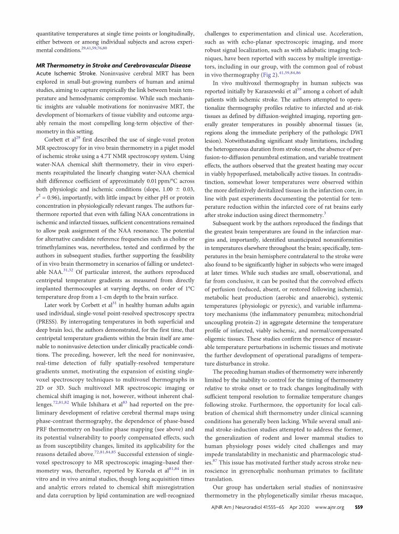
challenges to experimentation and clinical use. Acceleration,such as with echo-planar spectroscopic imaging, and morerobust signal localization, such as with adiabatic imaging tech-niques, have been reported with success by multiple investiga-tors, including in our group, with the common goal of robustin vivo thermography (Fig 2).41,59,84,86
In vivo multivoxel thermography in human subjects wasreported initially by Karaszewski et al39 among a cohort of adultpatients with ischemic stroke. The authors attempted to opera-tionalize thermography profiles relative to infarcted and at-risktissues as defined by diffusion-weighted imaging, reporting gen-erally greater temperatures in possibly abnormal tissues (ie,regions along the immediate periphery of the pathologic DWIlesion). Notwithstanding significant study limitations, includingthe heterogeneous duration from stroke onset, the absence of per-fusion-to-diffusion penumbral estimation, and variable treatmenteffects, the authors observed that the greatest heating may occurin viably hypoperfused, metabolically active tissues. In contradis-tinction, somewhat lower temperatures were observed withinthe more definitively devitalized tissues in the infarction core, inline with past experiments documenting the potential for tem-perature reduction within the infarcted core of rat brains earlyafter stroke induction using direct thermometry.3
Subsequent work by the authors reproduced the findings thatthe greatest brain temperatures are found in the infarction mar-gins and, importantly, identified unanticipated nonuniformitiesin temperatures elsewhere throughout the brain; specifically, tem-peratures in the brain hemisphere contralateral to the stroke werealso found to be significantly higher in subjects who were imagedat later times. While such studies are small, observational, andfar from conclusive, it can be posited that the convolved effectsof perfusion (reduced, absent, or restored following ischemia),metabolic heat production (aerobic and anaerobic), systemictemperatures (physiologic or pyrexic), and variable inflamma-tory mechanisms (the inflammatory penumbra; mitochondrialuncoupling protein-2) in aggregate determine the temperatureprofile of infarcted, viably ischemic, and normal/compensatedoligemic tissues. These studies confirm the presence of measur-able temperature perturbations in ischemic tissues and motivatethe further development of operational paradigms of tempera-ture disturbance in stroke.
The preceding human studies of thermometry were inherentlylimited by the inability to control for the timing of thermometryrelative to stroke onset or to track changes longitudinally withsufficient temporal resolution to formalize temperature changesfollowing stroke. Furthermore, the opportunity for local cali-bration of chemical shift thermometry under clinical scanningconditions has generally been lacking. While several small ani-mal stroke-induction studies attempted to address the former,the generalization of rodent and lower mammal studies tohuman physiology poses widely cited challenges and mayimpede translatability in mechanistic and pharmacologic stud-ies.87 This issue has motivated further study across stroke neu-roscience in gyrencephalic nonhuman primates to facilitatetranslation.
Our group has undertaken serial studies of noninvasivethermometry in the phylogenetically similar rhesus macaque,
AJNR Am J Neuroradiol 41:555–65 Apr 2020 www.ajnr.org 559
specifically in efforts to bridge this gap.41,76 Using robust adia-batic multivoxel chemical shift imaging strategies (sLASER,Siemens Healthineers, Erlangen, Germany) with greater perform-ance against chemical shift misregistration, together with higherorder magnetic field shimming, we recently reported on dynamicbrain temperature changes following highly-controlled endovas-cular stroke induction in NHP.41 This experimental design per-mits repeated production of thermographs under physiologic andpostischemic conditions without the confounding effects of sur-gery and ambient heat loss, the constraints of focally implantedtemperature probes, or the limitations of unknown disease onset.The preoperative calibration of water-NAA chemical shift ther-mometry under essentially identical imaging conditions usingclinical 3T scanning conditions also facilitates future translation.While the potentially nontrivial effect of anesthesia induction oncerebral blood flow, metabolism, and temperature regulation can-not be overlooked in such models, dynamic imaging immediately
following anesthesia induction in control and ischemic experi-mental sessions fortuitously produces an additional form of phys-iologic contrast during which temperatures can be measured andcompared between conditions.
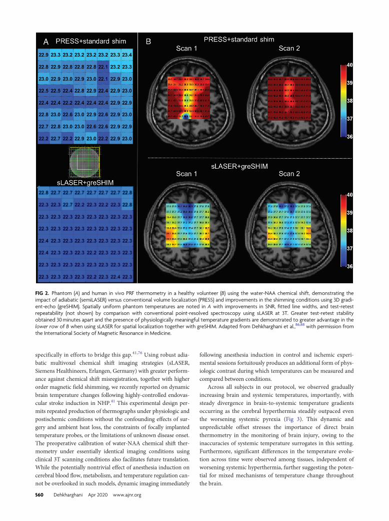
Across all subjects in our protocol, we observed graduallyincreasing brain and systemic temperatures, importantly, withsteady divergence in brain-to-systemic temperature gradientsoccurring as the cerebral hyperthermia steadily outpaced eventhe worsening systemic pyrexia (Fig 3). This dynamic andunpredictable offset stresses the importance of direct brainthermometry in the monitoring of brain injury, owing to theinaccuracies of systemic temperature surrogates in this setting.Furthermore, significant differences in the temperature evolu-tion across time were observed among tissues, independent ofworsening systemic hyperthermia, further suggesting the poten-tial for mixed mechanisms of temperature change throughoutthe brain.
FIG 2. Phantom (A) and human in vivo PRF thermometry in a healthy volunteer (B) using the water-NAA chemical shift, demonstrating theimpact of adiabatic (semiLASER) versus conventional volume localization (PRESS) and improvements in the shimming conditions using 3D gradi-ent-echo (greSHIM). Spatially uniform phantom temperatures are noted in A with improvements in SNR, fitted line widths, and test-retestrepeatability (not shown) by comparison with conventional point-resolved spectroscopy using sLASER at 3T. Greater test-retest stabilityobtained 30minutes apart and the presence of physiologically meaningful temperature gradients are demonstrated to greater advantage in thelower row of B when using sLASER for spatial localization together with greSHIM. Adapted from Dehkharghani et al.,86,88 with permission fromthe International Society of Magnetic Resonance in Medicine.
560 Dehkharghani Apr 2020 www.ajnr.org
Chronic Cerebrovascular Ischemia. Hemodynamic and thermo-regulatory theory predict a strong dependency of brain tempera-ture on blood flow, which has largely been confirmed duringexperimentation in low-flow states such as ischemic stroke asdescribed above. The theoretic, converse cooling effects of hemo-dynamic augmentation are, however, more difficult to test experi-mentally. A brief review of the dynamic autoregulatory processunderlying hemodynamic failure is worthwhile at this stage. Theresponse of the cerebrovascular system to falling perfusion pres-sures was expounded in initial work by Powers and further byDerdeyn et al.89,90 using 15O-PET.89-93 A sequential, quasi-step-wise response to incremental hemodynamic failure is commonlyencountered, culminating in the up-regulation of the oxygenextraction fraction (OEF) from the heme moiety of hemoglo-bin to sustain the cerebral metabolic rate of oxygen when fall-ing perfusion pressures outstrip the cerebrovascular reserve.This tenuous state of so-called misery perfusion would, on thebasis of cerebral thermoregulatory theory, seem conduciveto heating. Specifically, one would anticipate that viable
(ie, with continued but potentiallyreduced metabolic activity) tissue bedsdownstream from chronic steno-occlusive lesions could exhibit a par-ticular propensity for heating throughthe combination of increased ther-mogenic oxygen cleavage and re-duced perfusion.
Cerebral oximetry and the identifi-cation of misery perfusion by MRimaging remain elusive aims and areasof active study by many groups in-cluding our own.94-98 The unambigu-ous demonstration of misery perfu-sion proves challenging without directMR oximetry, which remains difficultunder clinically pragmatic condi-tions.99,100 The characterization of cer-ebral temperatures could, however,reflect a measurable epiphenomenonof the hemodynamic and metabolicderangements inherent to misery per-fusion. In a study of contemporane-ous 15O-PET and single-voxel point-resolved spectroscopy by Ishigakiet al,101 local temperatures and theOEF were estimated in the deep whitematter of healthy subjects and pa-tients with unilateral, anterior circu-lation steno-occlusive disease. Asexpected, uniformly normal distribu-tions of OEF were measured inhealthy subjects, while elevations ofOEF in the diseased territories ofpatients with steno-occlusive diseasewere observed as compensations forreduced blood flow. The authorsalso confirmed the hypothesized inter-
actions between interhemispheric temperature offset and OEF.While the findings are compelling as potential evidence for braintemperatures as an imaging biomarker in chronic ischemia, thecomprehensive characterization of tissue thermal signatures inhemodynamic failure would require acquisition of thermographsto assess the spatiotemporal relationship between perfusion andtemperature.
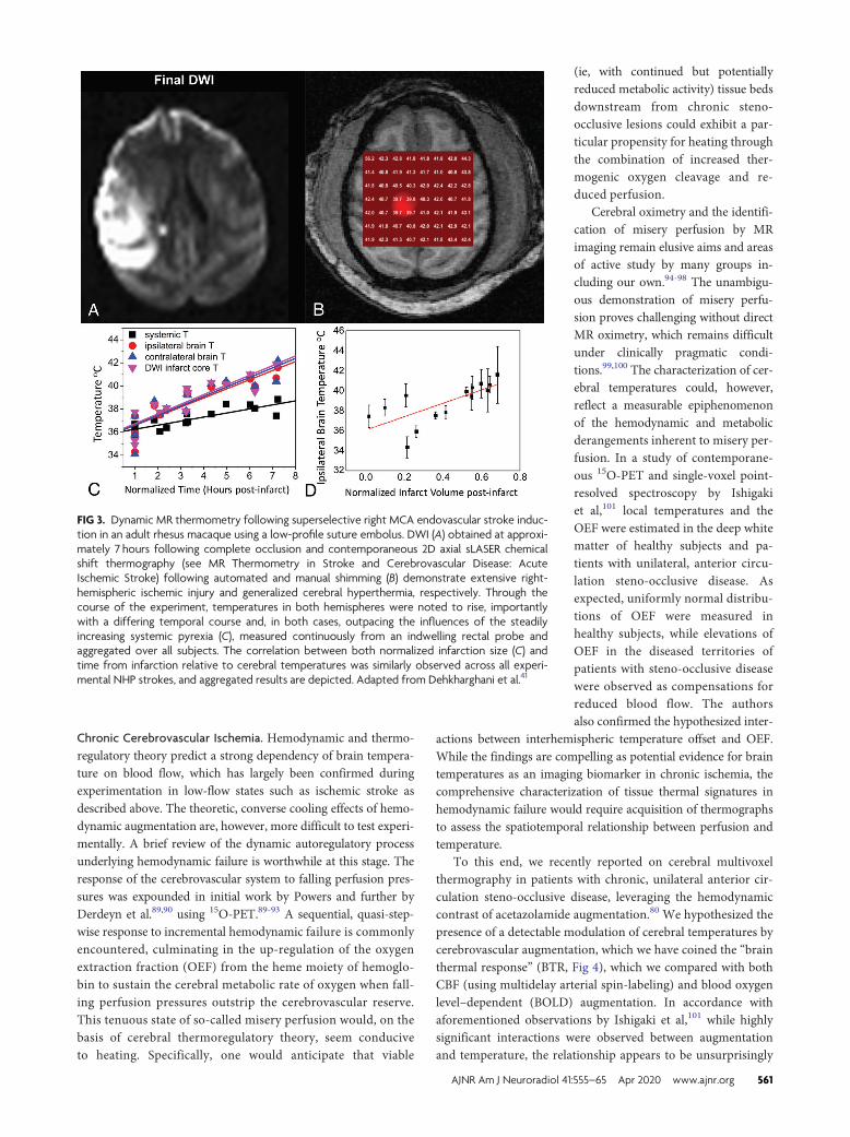
To this end, we recently reported on cerebral multivoxelthermography in patients with chronic, unilateral anterior cir-culation steno-occlusive disease, leveraging the hemodynamiccontrast of acetazolamide augmentation.80 We hypothesized thepresence of a detectable modulation of cerebral temperatures bycerebrovascular augmentation, which we have coined the “brainthermal response” (BTR, Fig 4), which we compared with bothCBF (using multidelay arterial spin-labeling) and blood oxygenlevel–dependent (BOLD) augmentation. In accordance withaforementioned observations by Ishigaki et al,101 while highlysignificant interactions were observed between augmentationand temperature, the relationship appears to be unsurprisingly
FIG 3. Dynamic MR thermometry following superselective right MCA endovascular stroke induc-tion in an adult rhesus macaque using a low-profile suture embolus. DWI (A) obtained at approxi-mately 7 hours following complete occlusion and contemporaneous 2D axial sLASER chemicalshift thermography (see MR Thermometry in Stroke and Cerebrovascular Disease: AcuteIschemic Stroke) following automated and manual shimming (B) demonstrate extensive right-hemispheric ischemic injury and generalized cerebral hyperthermia, respectively. Through thecourse of the experiment, temperatures in both hemispheres were noted to rise, importantlywith a differing temporal course and, in both cases, outpacing the influences of the steadilyincreasing systemic pyrexia (C), measured continuously from an indwelling rectal probe andaggregated over all subjects. The correlation between both normalized infarction size (C) andtime from infarction relative to cerebral temperatures was similarly observed across all experi-mental NHP strokes, and aggregated results are depicted. Adapted from Dehkharghani et al.41
AJNR Am J Neuroradiol 41:555–65 Apr 2020 www.ajnr.org 561
complex and nonlinear. In particular, BOLD augmentationreflects a convolved effect of flow and volume and also, crit-ically, of blood oxygenation/oxygen extraction. This latter effect,which may be variably up-regulated at baseline, depending onthe metabolic need and also on vasoreactivity and disease
chronicity, is difficult to measuredirectly. However, in concert, thesevariables likely determine the direc-tion of the baseline arterial blood-to-brain temperature offset and,therefore, the magnitude and direc-tion of the BTR. While furtherinvestigation into the diagnostic andprognostic merits of BTR are under-way, we anticipate that the overarch-ing mechanisms are governed byalready well-recognized principles ofcerebral hemodynamic control andthermoregulation and reflect a tend-ency for tissue heating arising fromimpaired perfusion and up-regulatedOEF.80,101
An interesting potential implica-tion of such thermal disturbance incerebrovascular disease was describedrecently by Murakami et al,103 whoexplored the potential for preopera-tive hyperthermia as a predictor ofpost-carotid endarterectomy hyper-perfusion syndrome. A strong andhighly significant correlation wasobserved between baseline tempera-ture elevation and potentially danger-ous post-carotid endarterectomyblood flow augmentation (r= 0.763,P, .001). Similarly, our experience
with the BTR demonstrated a significant negative relationshipbetween baseline temperatures and cerebrovascular reservecapacity, in line with past reports of impaired cerebrovascularreserve capacity as a strong predictor of hyperperfusionsyndrome.104
FIG 4. Cerebrovascular reserve (CVR) percentage augmentation maps calculated with BOLD and arterial spin-labeling (ASL), as well as a BTR mapoverlaid on a T1-weighted anatomic image. Images are all from the same subject (a 32-year-old woman with unilateral left MCA stenosis andmultiple TIAs). The white grid overlay represents the MR thermometry VOI derived from multivoxel spectroscopy analysis using the water-NAAchemical shift difference. Images are displayed in the radiologic convention. Impaired cerebrovascular reserve in the left hemisphere is presentin both BOLD and ASL, with a greater severity of impairment in arterial spin-labeling, likely related to tag decay and residual delay sensitivity de-spite the use of 10 separate, in-line postlabel delays of varying duration. The BTR map demonstrates an asymmetric thermal response, with lessbrain cooling following vasodilatory stimulus in the diseased left hemisphere, indicated by reduced (ie, less negative) BTR values and correspond-ing primarily to the areas of greatest impairment in the anterior and posterior MCA borderzone territories. Maximal (most negative) BTRs arenoted in the regions spatially concordant with the greatest hemodynamic augmentation (blue regions) in the right parietal lobe. Adapted fromFleischer et al.80
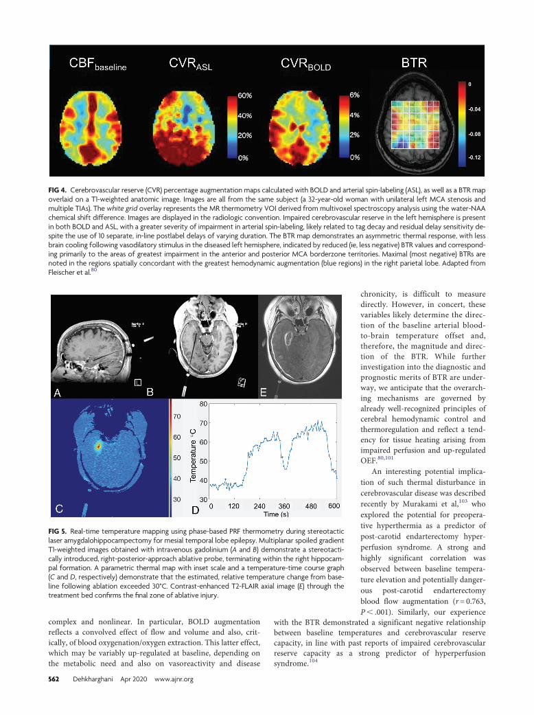
FIG 5. Real-time temperature mapping using phase-based PRF thermometry during stereotacticlaser amygdalohippocampectomy for mesial temporal lobe epilepsy. Multiplanar spoiled gradientT1-weighted images obtained with intravenous gadolinium (A and B) demonstrate a stereotacti-cally introduced, right-posterior-approach ablative probe, terminating within the right hippocam-pal formation. A parametric thermal map with inset scale and a temperature-time course graph(C and D, respectively) demonstrate that the estimated, relative temperature change from base-line following ablation exceeded 30°C. Contrast-enhanced T2-FLAIR axial image (E) through thetreatment bed confirms the final zone of ablative injury.
562 Dehkharghani Apr 2020 www.ajnr.org
MR Thermometry in Thermal Therapy. MR thermometry hasfound broad applications in the real-time monitoring of thermaltherapies, and despite obvious differences from the preceding sec-tions, for completeness, we introduce the reader to this emergentapplication of cerebral thermometry.
The coagulative response of thermally injured tissue is a func-tion of both maximal temperature and the duration over which itis sustained beyond the ablative threshold. In such applications,MRT can be used to ensure that temperatures in target regionsare sustained at prespecified levels to achieve coagulation andfatal thermal dose. In combination with sophisticated dose andthermal models, the use of real-time monitoring affords greatersafety in such procedures by ensuring that ablative doses notexceed cavitation thresholds and by confirming that temperaturesin vulnerable and eloquent nearby regions remain below injuri-ous ranges (Fig 5). The scale of temperature change in theseapplications is on the order of tens of degrees Celsius and, hence,much larger than that described either in basal cerebral tempera-ture gradients or occurring in the pathophysiologic states dis-cussed elsewhere in this review. During thermal therapy, thebaseline cerebral temperature gradient can therefore be neglectedand the temperatures assumed to be spatially homogeneous andequal to the systemic temperature. By freeing the methodologyfrom the demands for absolute, baseline temperature estimation,many alternative approaches to thermometry can be used in suchapplications beyond those discussed for the study of cerebralthermoregulation described herein.71
CONCLUSIONSThis review introduces the reader to fundamental aspects of cere-bral and systemic thermoregulation, emphasizing the importanceof tight cerebral thermoregulation in homeotherms and specifi-cally the vulnerability of the ischemic neurovascular unit to theeffects of hyperthermia. Temperature represents a powerful bio-marker of brain function and a potentially valuable target forinterrogation through noninvasive means as permitted by emerg-ing cerebral MR thermometry techniques. We believe that cere-bral temperatures are, therefore, well-suited for diagnostic andprognostic purposes and mechanistic study and hope to see themfind their way deeper into the scientific agenda of the neuroimag-ing community.
Disclosures: Seena Dehkharghani—RELATED: Grant: Foundation of the ASNR,Emory University Research Council, Comments: Scholar Award in Neuroradio-logy Research in the name of the lead author in successive years, EmoryUniversity Research Council Grant to the lead author*; Support for Travel toMeetings for the Study or Other Purposes: Foundation of the ASNR*;UNRELATED: Travel/Accommodations/Meeting Expenses Unrelated to ActivitiesListed: ISchemaView, Comments: travel costs. Deqiang Qiu—UNRELATED: Grants/Grants Pending: Siemens.* *Money paid to the institution.
REFERENCES1. Busto R, Dietrich WD, Globus MY, et al. The importance of brain
temperature in cerebral ischemic injury. Stroke 1989;20:1113–14CrossRef Medline
2. Busto R, Dietrich WD, Globus MY, et al. Small differences inintraischemic brain temperature critically determine the extentof ischemic neuronal injury. J Blood Flow Metab 1987;7:729–38CrossRef Medline
3. Minamisawa H, Mellergard P, Smith ML, et al. Preservation ofbrain temperature during ischemia in rats. Stroke 1990;21:758–64CrossRef CrossRef Medline
4. Minamisawa H, Nordstrom CH, Smith ML, et al. The influence ofmild body and brain hypothermia on ischemic brain damage. JCereb Blood Flow Metab 1990;10:365–74 CrossRef Medline
5. Wass CT, Lanier WL, Hofer RE, et al. Temperature changes of >or = 1 degree C alter functional neurologic outcome and histo-pathology in a canine model of complete cerebral ischemia.Anesthesiology 1995;83:325–35 CrossRef Medline
6. Axelrod YK, Diringer MN. Temperature management in acuteneurologic disorders. Neurol Clin 2008;26:585–603, xi CrossRefMedline
7. Saad H, Aladawy M. Temperature management in cardiac sur-gery. Glob Cardiol Sci Pract 2013;2013:44–62 CrossRef Medline
8. Baker MA. Brain cooling in endotherms in heat and exercise.Annu Rev Physiol 1982;44:85–96 CrossRef Medline
9. Zhu M, Ackerman JJ, Sukstanskii AL, et al. How the body controlsbrain temperature: the temperature shielding effect of cerebralblood flow. J Appl Physiol 2006;101:1481–88 CrossRef Medline
10. Zhu M, Ackerman JJ, Yablonskiy DA. Body and brain temperaturecoupling: the critical role of cerebral blood flow. J Comp Physiol B2009;179:701–10 CrossRef Medline
11. Falk D. Brain evolution in homo: the “radiator” theory. BehavBrain Sci 1990;13:333–44 CrossRef
12. Baker MA, Hayward JN. Intracranial heat exchange and regula-tion of brain temperature in sheep. Life SCI 1968;7:349–57CrossRef Medline
13. Baker MA. A brain-cooling system in mammals. Sci Am 1979;240:130–39 CrossRef Medline
14. Cabanac M, Caputa M. Natural selective cooling of the humanbrain: evidence of its occurrence and magnitude. J Physiol 1979;286:255–64 CrossRef Medline
15. Hayward JN, Baker MA. Role of cerebral arterial blood in the reg-ulation of brain temperature in the monkey. Am J Physiol 1968;215:389–403 CrossRef Medline
16. Cabanac M. Selective brain cooling in humans: “fancy” or fact?FASEB J 1993;7:1143–46; discussion 1146–47 Medline
17. Caputa M. Selective brain cooling: a multiple regulatory mecha-nism. J Therm Biol 2004;29:691–702 CrossRef
18. Falk D. Brain evolution in homo: the radiator theory. Behavioraland Brain Sciences 1990;13:333–34 CrossRef
19. Falk D. Evolution of the primate brain. In: Handbook of Paleoan-thropology. Berlin: Springer-Verlag; 2007:1133–62
20. Kuhnen G, Jessen C. Thermal signals in control of selective braincooling. Am J Physiol 1994;267:R355–59 CrossRef Medline
21. Yablonskiy DA, Ackerman JJ, Raichle ME. Coupling betweenchanges in human brain temperature and oxidative metabolismduring prolonged visual stimulation. Proc Natl Acad Sci U S A2000;97:7603–08 CrossRef Medline
22. Zhu L, Diao C. Theoretical simulation of temperature distribu-tion in the brain during mild hypothermia treatment for braininjury.Med Biol Eng Comput 2001;39:681–87 CrossRef Medline
23. Stone JG, Young WL, Smith CR, et al. Do standard monitoringsites reflect true brain temperature when profound hypothermiais rapidly induced and reversed? Anesthesiology 1995;82:344–51CrossRef Medline
24. Simon E. Tympanic temperature is not suited to indicate selectivebrain cooling in humans: a re-evaluation of the thermophysiolog-ical basics. Eur J Appl Physiol 2007;101:19–30 CrossRef Medline
25. Baker MA, Stocking RA, Meehan JP. Thermal relationship betweentympanic membrane and hypothalamus in conscious cat andmonkey. J Appl Physiol 1972;32:739–42 CrossRef Medline
26. McCaffrey TV, McCook RD, Wurster RD. Effect of head skin tem-perature on tympanic and oral temperature in man. J Appl Physiol1975;39:114–18 CrossRef Medline
AJNR Am J Neuroradiol 41:555–65 Apr 2020 www.ajnr.org 563
27. Cady EB, D’Souza PC, Penrice J, et al. The estimation of local braintemperature by in vivo 1H magnetic resonance spectroscopy.Magn Reson Med 1995;33:862–67 CrossRef Medline
28. Childs C, Hiltunen Y, Vidyasagar R, et al. Determination of re-gional brain temperature using proton magnetic resonance spec-troscopy to assess brain-body temperature differences in healthyhuman subjects.Magn Reson Med 2007;57:59–66 CrossRef Medline
29. Corbett RJ, Laptook AR, Tollefsbol G, et al.Validation of a noninva-sive method to measure brain temperature in vivo using 1H NMRspectroscopy. J Neurochem 1995;64:1224–30 CrossRef Medline
30. Schwab S, Spranger M, Aschoff A, et al. Brain temperature moni-toring and modulation in patients with severe MCA infarction.Neurology 1997;48:762–67 CrossRef Medline
31. Corbett R, Laptook A, Weatherall P. Noninvasive measurements ofhuman brain temperature using volume-localized proton mag-netic resonance spectroscopy. J Cereb Blood Flow Metab 1997;17:363–69 CrossRef Medline
32. Corbett RJ, Purdy PD, Laptook AR, et al. Noninvasive measure-ment of brain temperature after stroke. AJNR Am J Neuroradiol1999;20:1851–57 Medline
33. Verlooy J, Heytens L, Veeckmans G, et al. Intracerebral tempera-ture monitoring in severely head injured patients. Acta Neurochir(Wien) 1995;134:76–78 CrossRef Medline
34. Sun Z, Zhang J, Chen Y, et al. Differential temporal evolutionpatterns in brain temperature in different ischemic tissues in amonkey model of middle cerebral artery occlusion. J BiomedBiotechnol 2012;2012:980961 CrossRef Medline
35. Sukstanskii AL, Yablonskiy DA. Theoretical limits on brain cool-ing by external head cooling devices. Eur J Appl Physiol 2007;101:41–49 CrossRef Medline
36. Soukup J, Rieger A, Holz C, et al. Temperature gradient betweenbrain tissue and arterial blood mirrors the flow-metabolismrelationship in uninjured brain: an experimental study. ActaAnaesthesiol Scand 2007;51:872–79 CrossRef Medline
37. Otawara Y, Ogasawara K, Kubo Y, et al. Brain and systemic tem-perature in patients with severe subarachnoid hemorrhage. SurgNeurol 2003;60:159–64; discussion 164 CrossRef Medline
38. Mellergard P. Changes in human intracerebral temperature inresponse to different methods of brain cooling. Neurosurgery1992;31:671–77; discussion 677 Medline
39. Karaszewski B, Wardlaw JM, Marshall I, et al. Measurement ofbrain temperature with magnetic resonance spectroscopy in acuteischemic stroke. Ann Neurol 2006;60:438–46 CrossRef Medline
40. Kalmbach AS, Waters J. Brain surface temperature under a crani-otomy. J Neurophysiol 2012;108:3138–46 CrossRef Medline
41. Dehkharghani S, Fleischer CC, Qiu D, et al. Cerebral temperaturedysregulation: MR thermographic monitoring in a nonhumanprimate study of acute ischemic stroke. AJNR Am J Neuroradiol2017;38:712–20 CrossRef Medline
42. Pennes HH. Analysis of tissue and arterial blood temperatures inthe resting human forearm. J Appl Physiol 1948;1:93–22 CrossRefMedline
43. Nelson DA, Nunneley SA. Brain temperature and limits on trans-cranial cooling in humans: quantitative modeling results. Eur JAppl Physiol Occup Physiol 1998;78:353–59 CrossRef Medline
44. Sukstanskii AL, Yablonskiy DA. An analytical model of temper-ature regulation in human head. J Therm Biol 2004;29:583–87CrossRef
45. Sukstanskii AL, Yablonskiy DA. Theoretical model of temperatureregulation in the brain during changes in functional activity. ProcNatl Acad Sci USA 2006;103:12144–49 CrossRef Medline
46. Wissler EH. Pennes’ 1948 paper revisited. J Appl Physiol 1998;85:35–41 CrossRef Medline
47. Collins CM, Smith MB, Turner R. Model of local temperaturechanges in brain upon functional activation. J Appl Physiol 2004;97:2051–55 CrossRef Medline
48. Minamisawa H, Smith ML, Siesjo BK. The effect of mild hyper-thermia and hypothermia on brain damage following 5, 10, and
15 minutes of forebrain ischemia. Ann Neurol 1990;28:26–33CrossRef Medline
49. Hosler RM. The emergency treatment of cardiac arrest. J Int CollSurg 1953;19:336–40 Medline
50. Wolfe KB. Effect of hypothermia on cerebral damage resultingfrom cardiac arrest. Am J Cardiol 1960;6:809–12 CrossRef Medline
51. Donnelly CJ, Frobese AS, Stone HH. The effect of lowered bodytemperature on the cerebral hemodynamics and metabolism ofman. Surg Gynecol Obstet 1956;103:313–7 Medline
52. Ginsberg MD, Sternau LL, Globus MY, et al. Therapeutic modula-tion of brain temperature: relevance to ischemic brain injury.Cerebrovasc Brain Metab Rev 1992;4:189–25 Medline
53. Dehkharghani S, Bowen M, Haussen DC, et al. Body temperaturemodulates infarction growth following endovascular reperfusion.AJNR Am J Neuroradiol 2017;38:46–51 CrossRef Medline
54. Dietrich WD, Busto R, Halley M, et al. The importance of braintemperature in alterations of the blood-brain barrier followingcerebral ischemia. J Neuropathol Exp Neurol 1990;49:486–97CrossRef Medline
55. Dietrich WD, Busto R, Valdes I, et al. Effects of normothermic ver-sus mild hyperthermic forebrain ischemia in rats. Stroke 1990;21:1318–25 CrossRef Medline
56. Ginsberg MD, Busto R. Combating hyperthermia in acute stroke:a significant clinical concern. Stroke 1998;29:529–34 CrossRefMedline
57. Karaszewski B, Thomas RG, Dennis MS, et al. Temporal profile ofbody temperature in acute ischemic stroke: relation to stroke se-verity and outcome. BMC Neurol 2012;12:123 CrossRef Medline
58. Karaszewski B, Wardlaw JM, Marshall I, et al. Early brain tempera-ture elevation and anaerobic metabolism in human acute ischae-mic stroke. Brain 2009;132:955–64 CrossRef Medline
59. Maudsley AA, Goryawala MZ, Sheriff S. Effects of tissue suscepti-bility on brain temperature mapping. Neuroimage 2017;146:1093–1101 CrossRef Medline
60. Wrotek SE, Kozak WE, Hess DC, et al. Treatment of fever afterstroke: conflicting evidence. Pharmacotherapy 2011;31:1085–91CrossRef Medline
61. Gauberti M, De Lizarrondo SM, Vivien D. The “inflammatory pe-numbra” in ischemic stroke: from clinical data to experimentalevidence. Eur Stroke J 2016;1:20–27 CrossRef Medline
62. Gauberti M, Montagne A, Marcos-Contreras OA, et al. Ultra-sensi-tive molecular MRI of vascular cell adhesion molecule-1 reveals adynamic inflammatory penumbra after strokes. Stroke 2013;44:1988–96 CrossRef Medline
63. Han HS, Karabiyikoglu M, Kelly S, et al.Mild hypothermia inhibitsnuclear factor-kappa B translocation in experimental stroke. JCereb Blood Flow Metab 2003;23:589–98 CrossRef Medline
64. Inamasu J, Suga S, Sato S, et al. Postischemic hypothermia attenu-ates apoptotic cell death in transient focal ischemia in rats. ActaNeurochir Suppl 2000;76:525–27 CrossRef Medline
65. Ohmura A, Nakajima W, Ishida A, et al. Prolonged hypothermiaprotects neonatal rat brain against hypoxic-ischemia by reducingboth apoptosis and necrosis. Brain Dev 2005;27:517–26 CrossRefMedline
66. Wang GJ, Deng HY, Maier CM, et al. Mild hypothermia reducesICAM-1 expression, neutrophil infiltration and microglia/mono-cyte accumulation following experimental stroke. Neuroscience2002;114:1081–90 CrossRef Medline
67. Haussen DC, Nogueira RG, Elhammady MS, et al. Infarct growthdespite full reperfusion in endovascular therapy for acute ische-mic stroke. J Neurointerv Surg 2016;8:117–21 CrossRef Medline
68. Diringer MN, Reaven NL, Funk SE, et al. Elevated body tempera-ture independently contributes to increased length of stay in neu-rologic intensive care unit patients. Crit Care Med 2004;32:1489–95 CrossRef Medline
69. Kilpatrick MM, Lowry DW, Firlik AD, et al. Hyperthermia in theneurosurgical intensive care unit. Neurosurgery 2000;47:850–55;discussion 855–56 CrossRef Medline
564 Dehkharghani Apr 2020 www.ajnr.org
70. Parker DL, Smith V, Sheldon P, et al. Temperature distributionmeasurements in two-dimensional NMR imaging.Med Phys 1983;10:321–25 CrossRef Medline
71. Rieke V, Butts Pauly K. MR thermometry. J Magn Reson Imaging2008;27:376–90 CrossRef Medline
72. Kuroda K. Non-invasive MR thermography using the water pro-ton chemical shift. Int J Hyperthermia 2005;21:547–60 CrossRefMedline
73. Coman D, Trubel HK, Rycyna RE, et al. Brain temperature and pHmeasured by (1)H chemical shift imaging of a thulium agent.NMR Biomed 2009;22:229–39 CrossRef Medline
74. Hekmatyar SK, Kerkhoff RM, Pakin SK, et al. Noninvasive ther-mometry using hyperfine-shifted MR signals from paramagneticlanthanide complexes. Int J Hyperthermia 2005;21:561–74 CrossRefMedline
75. Lindner LH, Reinl HM, Schlemmer M, et al. Paramagnetic thermo-sensitive liposomes for MR-thermometry. Int J Hyperthermia2005;21:575–88 CrossRef Medline
76. Dehkharghani S, Mao H, Howell L, et al. Proton resonance fre-quency chemical shift thermometry: experimental design andvalidation toward high-resolution noninvasive temperature moni-toring and in vivo experience in a nonhuman primate model ofacute ischemic stroke. AJNR Am J Neuroradiol 2015;36:1128–35CrossRef Medline
77. Hindman J. Proton resonance shift of water in the gas and liquidstates. The Journal of Chemical Physics 1966;44:4582–92 CrossRef
78. Zou C, Tie C, Pan M, et al. Referenceless MR thermometry-a com-parison of five methods. Phys Med Biol 2017;62:1–16 CrossRefMedline
79. Cady EB, Penrice J, Robertson NJ. Improved reproducibility ofMRS regional brain thermometry by “amplitude-weighted com-bination.” NMR Biomed 2011;24:865–72 CrossRef Medline
80. Fleischer CC, Wu J, Qiu D, et al. The brain thermal response as apotential neuroimaging biomarker of cerebrovascular impair-ment. AJNR Am J Neuroradiol 2017;38:2044–51 CrossRef Medline
81. Kuroda K, Suzuki Y, Ishihara Y, et al. Temperature mapping usingwater proton chemical shift obtained with 3D-MRSI: feasibilityin vivo.Magn Reson Med 1996;35:20–29 CrossRef Medline
82. Kuroda K, Oshio K, Chung AH, et al. Temperature mapping usingthe water proton chemical shift: a chemical shift selective phasemapping method. Magn Reson Med 1997;38:845–51 CrossRefMedline
83. Ishihara Y, Calderon A, Watanabe H, et al. A precise and fast tem-perature mapping using water proton chemical shift.Magn ResonMed 1995;34:814–23 CrossRef Medline
84. Kuroda K, Mulkern RV, Oshio K, et al. Temperature mappingusing the water proton chemical shift: self-referenced methodwith echo-planar spectroscopic imaging. Magn Reson Med 2000;43:220–25 CrossRef Medline
85. De Poorter J. Noninvasive MRI thermometry with the proton res-onance frequency method: study of susceptibility effects. MagnReson Med 1995;34:359–67 CrossRef Medline
86. Dehkharghani S, Wei L, Mao H, et al. Multivoxel proton spectros-copy for non-invasive MR thermometry: phantom comparison ofPRESS and semiLASER-localized chemical shift imaging for tem-perature monitoring. In: Proceedings of the Joint Meeting of theInternational Society of Magnetic Resonance in Medicine and theEuropean Society of Magnetic Resonance in Medicine and Biology,Milan, Italy. May 10–16, 2014; 2828
87. Cook DJ, Tymianski M. Nonhuman primate models of stroke fortranslational neuroprotection research. Neurotherapeutics 2012;9:371–79 CrossRef Medline
88. Dehkharghan S, Fleischer CC, Qiu D, et al. Non-Invasive Brain MRThermometry in a Nonhuman Primate Model of Acute IschemicStroke, ISMRM Annual Meeting 2017; Honolulu, HI
89. Derdeyn CP, Videen TO, Fritsch SM, et al. Compensatory mecha-nisms for chronic cerebral hypoperfusion in patients with carotidocclusion. Stroke 1999;30:1019–24 CrossRef Medline
90. Derdeyn CP, Videen TO, Yundt KD, et al. Variability of cerebralblood volume and oxygen extraction: stages of cerebral haemody-namic impairment revisited. Brain 2002;125:595–607 CrossRefMedline
91. Powers WJ. Cerebral hemodynamics in ischemic cerebrovasculardisease. Ann Neurol 1991;29:231–40 CrossRef Medline
92. Powers WJ. Stroke: misery perfusion in cerebrovascular disease—is it important? Nat Rev Neurol 2012;8:479–80 CrossRef Medline
93. Powers WJ, Press GA, Grubb RL Jr, et al. The effect of hemody-namically significant carotid artery disease on the hemodynamicstatus of the cerebral circulation. Ann Intern Med 1987;106:27–34CrossRef Medline
94. Yablonskiy DA, Sukstanskii AL, He X. Blood oxygenation level-de-pendent (BOLD)-based techniques for the quantification of brainhemodynamic and metabolic properties: theoretical models andexperimental approaches. NMR Biomed 2013;26:963–86 CrossRefMedline
95. Wu J, Dehkharghani S, Nahab F, et al. Acetazolamide-augmenteddynamic BOLD (aczBOLD) imaging for assessing cerebrovascu-lar reactivity in chronic steno-occlusive disease of the anterior cir-culation: an initial experience. Neuroimage Clin 2017;13:116–22CrossRef Medline
96. Wu J, Dehkharghani S, Nahab F, et al. The effects of acetazolamideon the evaluation of cerebral hemodynamics and functional con-nectivity using blood oxygen level-dependent MR imaging inpatients with chronic steno-occlusive disease of the anterior cir-culation. AJNR Am J Neuroradiol 2017;38:139–45 CrossRef Medline
97. Leatherday C, Dehkharghani S, Nahab F, et al. Cerebral MR oxime-try during acetazolamide augmentation: beyond cerebrovascularreactivity in hemodynamic failure. J Magn Reson Imaging 2019;50:175–82 CrossRef Medline
98. Christen T, Bolar DS, Zaharchuk G. Imaging brain oxygenationwith MRI using blood oxygenation approaches: methods, valida-tion, and clinical applications. AJNR Am J Neuroradiol 2013;34:1113–23 CrossRef Medline
99. Atkinson IC, Thulborn KR. Feasibility of mapping the tissue masscorrected bioscale of cerebral metabolic rate of oxygen consump-tion using 17-oxygen and 23-sodium MR imaging in a humanbrain at 9.4 T.Neuroimage 2010;51:723–33 CrossRef Medline
100. Lakshmanan K, Dehkharghani S, Madelin G, et al. A dual-tuned(17) O/(1) H head array for direct brain oximetry at 3 Tesla.Magn Reson Med 2020;83:1512–18 CrossRef Medline
101. Ishigaki D, Ogasawara K, Yoshioka Y, et al. Brain temperaturemeasured using proton MR spectroscopy detects cerebral hemo-dynamic impairment in patients with unilateral chronic majorcerebral artery steno-occlusive disease: comparison with positronemission tomography. Stroke 2009;40:3012–16 CrossRef Medline
102. Nanba T, Nishimoto H, Yoshioka Y, et al.Apparent brain tempera-ture imaging with multi-voxel proton magnetic resonance spec-troscopy compared with cerebral blood flow and metabolismimaging on positron emission tomography in patients with uni-lateral chronic major cerebral artery steno-occlusive disease.Neuroradiology 2017;59:923–35 CrossRef Medline
103. Murakami T, Ogasawara K, Yoshioka Y, et al. Brain temperaturemeasured by using proton MR spectroscopy predicts cerebralhyperperfusion after carotid endarterectomy. Radiology 2010;256:924–31 CrossRef Medline
104. Buczek J, Karliński M, Kobayashi A, et al. Hyperperfusion syn-drome after carotid endarterectomy and carotid stenting.Cerebrovasc Dis 2013;35:531–37 CrossRef Medline
AJNR Am J Neuroradiol 41:555–65 Apr 2020 www.ajnr.org 565