mri-directed cognitive fusion-guided biopsy of the … cognitive fusion-guided biopsy of the...
TRANSCRIPT

MRI-directed cognitive fusion-guided biopsy of the anterior prostate tumors
Ian G. Murphy Elaine NiMhurchuRobert G. Gibney Colm J. McMahon
Patients with clinical suspicion of malignant prostate neoplasm (i.e., elevated pros-tate-specific antigen [PSA], suspicious nodule on digital rectal examination) typically undergo systematic transrectal ultrasonography (TRUS)-guided sectoral biopsy; how-
ever, the overall yield of initial biopsy is 22%–29% (1, 2). Potential reasons for false negative TRUS biopsy include sampling error or technical limitation due to the location of tumor. Anteriorly located tumors, where the dominant tumor mass is anterior to the urethra repre-sent a particular diagnostic challenge, as they are not sampled in standard systematic 12-core needle biopsy, and it is estimated that 21% of malignant prostate tumors occur in the anterior prostate (3, 4). Furthermore, when TRUS biopsy detects minimal volume/low-grade carcinoma, failure to sample a coexistent more aggressive anterior tumor may lead to an underestimation of disease burden and aggressiveness (5) and inappropriate management by enrollment in an active surveillance program.
In addition to an established role in the staging of prostate cancer (6–8), magnetic reso-nance imaging (MRI) has an increasing role for the localization of prostate tumors which can then be targeted for biopsy (9–11). For lesion localization, anatomic T2-weighted sequences are combined with one or more functional technique including diffusion-weighted imaging (DWI) (12), magnetic resonance spectroscopy (MRS) or dynamic contrast-enhanced (DCE) MRI (9–11). Based on suspicion of prostate cancer, targeted biopsy of the suspicious area can then be performed with either MRI guidance (10) or TRUS guidance. The level of suspicion of prostate cancer on MRI can be quantified using the Prostate Imaging Reporting and Data
87
From the Department of Radiology ( ( I.G.M., E.N., R.G., C.J.M. [email protected]) St. Vincent’s University Hospital, Dublin Ireland; the Department of Radiology (C.J.M.), Beth Israel Deaconess Medical Center, Boston, USA.
Received 23 September 2015; revision requested 13 April 2016; revision received 10 May 2016; accepted 25 May 2016.
Published online 11 January 2017.DOI 10.5152/dir.2016.15445
Diagn Interv Radiol 2017; 23:87–93
© Turkish Society of Radiology 2017
A B D O M I N A L I M AG I N GO R I G I N A L A R T I C L E
PURPOSE We aimed to evaluate the efficacy of magnetic resonance imaging (MRI)-directed cognitive fu-sion transrectal ultrasonography (TRUS)-guided anterior prostate biopsy for diagnosis of anteri-or prostate tumors and to illustrate this technique.
METHODSA total of 39 patients with previous negative TRUS biopsy, but high clinical suspicion of occult prostate cancer, prospectively underwent prostate MRI including diffusion-weighted imag-ing (DWI). Patients with a suspicious anterior lesion on MRI underwent targeted anterior gland TRUS-guided biopsy with cognitive fusion technique using sagittal probe orientation. PIRADS version 1 scores (T2, DWI, and overall), lesion size, prostate-specific antigen (PSA), PSA density, and prostate gland volume were compared between positive and negative biopsy groups and between clinically significant cancer and remaining cases. Logistic regression analysis of imaging parameters and prostate cancer diagnosis was performed.
RESULTSAnterior gland prostate adenocarcinoma was diagnosed in 18 patients (46.2%) on targeted anterior gland TRUS-guided biopsy. Clinically significant prostate cancer was diagnosed in 13 patients (33.3%). MRI lesion size, T2, DWI, and overall PIRADS scores were significantly higher in patients with positive targeted biopsies and those with clinically significant cancer (P < 0.05). Biopsies were positive in 90%, 33%, and 29% of patients with overall PIRADS scores of 5, 4, and 3 respectively. Overall PIRADS score was an independent predictor of all prostate cancer diagnosis and of clinically significant prostate cancer diagnosis.
CONCLUSIONTargeted anterior gland TRUS-guided biopsy with MRI-directed cognitive fusion enables accu-rate sampling and may improve tumor detection yield of anterior prostate cancer.
88 • March–April 2017 • Diagnostic and Interventional Radiology Murphy et al.
System (PIRADS) (13). Where TRUS is used to guide targeted biopsy, electronic fusion of magnetic resonance images with TRUS images can be performed to guide biopsy (14, 15), or “cognitive fusion” can be used (16), by which the operator prospectively reviews the MRI appearances and uses TRUS to guide targeted sampling of the area of suspected tumor in the prostate gland.
Limited literature is available regarding the technique for TRUS-guided biopsy of the anterior apex of the prostate (17, 18). The goal of the current study is to evaluate the effectiveness of MRI-directed cognitive fusion TRUS-guided anterior prostate biop-sy for diagnosis of anterior prostate tumors and to illustrate this technique.
MethodsStudy design
The study was reviewed by the institu-tional ethics committee and approved as a clinical audit. In accordance with local policy, informed consent was not required. Patients were included if they had undergone MRI-di-rected targeted TRUS-guided anterior pros-tate biopsy in 2011–2014 because of suspi-cion of anterior tumor on MRI (Chart 1). All patients had undergone previous nontarget-ed TRUS-guided sectoral biopsy which was negative for carcinoma, but had undergone MRI for further evaluation due to persistent clinical concern for occult neoplasm.
MRI technique All patients were scanned using 1.5 T
MRI whole body scanner (GE Signa HDx or Siemens Avanto), with phased array ex-ternal surface coil with biparametric tech-nique (T2-weighted imaging and DWI) (19). MRI scan technique included multiplanar T2-weighted imaging including high-reso-lution axial T2-weighted images (TR, 3720 ms; TE, 107 ms; NEX, 4; slice thickness, 3 mm; interslice gap, 0 mm) and diffusion-weighted images (Siemens: TR, 4100; TE, 84 ms; NEX, 4; slice thickness, 4 mm; interslice gap, 0 mm; b
values, 50, 400, 800 s/mm2; GE: TR, 5000 ms; TE, minimum; NEX, 8; slice thickness, 3 mm; interslice gap, 1 mm; b value, 800 s/mm2).
MRI interpretationFor the purpose of this analysis, MRI
findings of each patient were reviewed ret-rospectively by two radiologists with five and three years of experience in pelvic MRI interpretation. The studies were reviewed in a randomized order, with the radiologists blinded to the biopsy results. For each pa-tient the anterior lesion suspected on MRI, which had prompted targeted biopsy, was scored using the PIRADS system (version 1) (13), with scores ranging 1–5 for T2 and DWI appearances. Interpretation of DWI data in-cluded both diffusion-weighted images and apparent diffusion coefficient (ADC) map, which were analyzed qualitatively. An over-all score was also applied based on overall interpretation of MRI findings and scored 1–5. Additionally, the maximum dimension of the lesion on MRI was measured on the axial T2-weighted images, while visually referencing the diffusion-weighted images and ADC map. Two radiologists in consensus carried out all scoring and measurements. The overall prostate volume was calculated by multiplying the maximum dimensions of the gland in three planes (anterior-to-poste-rior × transverse × craniocaudal) × 0.52.
Anterior TRUS biopsy techniqueAll biopsies were performed using either
a BK ProFocus 2202 ultrasound machine (BK Medical) with the end-fire convex array of a multi-frequency 8818 transducer or a Phillips iU22 ultrasound machine (Philips Health-care), with an end-fire curved array C9-5ec transducer. All biopsies were performed by one of two radiologists with seven and 30 years of experience in TRUS prostate biop-sy. At our institution, TRUS biopsies (both standard systematic and targeted) are typ-ically performed with conscious sedation, peri-prostatic local anesthetic (10 mL 1% lidocaine) and a transverse plane of US and biopsy guidance. This limits effective sam-pling of the anterior gland. In a modification of technique described for apical anterior horn biopsy (17, 18), a sagittal plane of TRUS imaging and biopsy guidance was used. The endorectal ultrasound probe was positioned in the sagittal plane, such that the needle guide was positioned anteriorly with respect to the probe (Fig. 2). Cognitive fusion was employed to determine the exact location for biopsy sampling, following review of the
patient’s MRI. The probe was then angled as needed in the sagittal plane such that the needle trajectory included the area of con-cern (Fig. 1). For lesions located more crani-ally, the biopsy needle was advanced 1–2 cm into the prostate gland prior to deploying the biopsy device. For more caudally located lesions, the biopsy device was activated from the prostate surface or with minimal ad-vancement into the gland prior to triggering the device. Medial-to-lateral localization was performed by identifying the urethra in the midline as a landmark, then scanning lateral-ly to the lateral margin of the gland to define the lateral border. Samples were taken more medially or laterally as needed based on the preprocedure MRI. Finally, if a concordant hypoechoic nodule was identified in a simi-lar position to a suspicious lesion on MRI, the biopsy needle was directed into this area. At least two cores of tissue were obtained from the target area.
Clinical and biopsy dataPatient age and PSA levels prior to biopsy
were recorded. PSA density was calculated by dividing serum PSA by prostate gland volume as determined on MRI. Gleason grade of biopsy samples positive for car-cinoma were recorded in addition to the percentage of core biopsy sample involved by tumor. Clinically significant cancers were defined based on Epstein criteria: Gleason grade of ≥7; or percentage core biopsy in-volvement by tumor ≥50% (20, 21).
Statistical analysisAll analysis was performed using STATA
Version 13.1 (Statacorp). The primary hypothesis was that targeted
anterior prostate biopsy would have a sim-ilar yield to that established in the existing literature for targeted prostate biopsy not limited to the anterior prostate gland. The yield of targeted TRUS biopsy based on MRI findings for the diagnosis of occult pros-tate cancer ranges from 39%–56% (10, 12, 16, 22, 23). For the purpose of sample size calculation, the null hypothesis was that the yield of anterior biopsy would be low (20%) and the alternative hypothesis was that the yield would be similar to the literature for targeted prostate biopsy not limited to the anterior prostate gland (45%). Sample size estimation was carried out using pow-er analysis for a one sample Wald test with probability (power) of 0.9, and type I error probability of 0.05. This resulted in sample size estimation of 42.
Main points
• Transrectal targeted ultrasound-guided biopsy with cognitive fusion allows accurate sampling of clinically significant anterior prostate tumors suspected on MRI.
• In this setting, overall PIRADS score was an independent predictor of positive biopsy.
• PSA density was higher in those with positive biopsies; however, PSA was not.

The mean (±standard deviation) patient age, PSA, prostate volume, PSA density, le-sion size, and PIRADS scores for all patients were calculated.
PIRADS scores (overall score, T2 score, DWI score) were compared between pos-
itive and negative biopsy groups using a Mann-Whitney U test. In a similar way, PI-RADS scores were also compared between cases where clinically significant cancer was diagnosed and the remaining cases (nega-tive and clinically insignificant cancer).
Continuous data (age, lesion size, PSA, prostate volume, PSA density) were com-pared between biopsy positive and nega-tive groups using an independent samples t-test. In a similar way, continuous data were compared between clinically signifi-cant cancer cases and the remaining cases.
The positive predictive value of PIRADS overall score for prediction of a positive targeted biopsy and for the prediction of clinically significant cancer was calculated. Forward stepwise logistic regression analy-sis was performed for prediction of positive biopsy and for prediction of clinically signif-icant cancer diagnosis.
ResultsDuring the study period, 1984 TRUS-guided
prostate biopsies were performed at this insti-tution. Of these, 110 were targeted biopsies with MRI-directed cognitive fusion technique. Of these, 47 cases had biopsy targeted to an anterior prostate lesion. Excluding patients with previous positive biopsy (active surveil-lance candidates), 39 patients were eligible for the study. Mean age was 67.1±7.6 years. Case examples are shown in Figs. 3 and 4. Patient demographics, imaging and biochemical pa-rameters are presented in Table 1.
Comparison of overall biopsy yield with PIRADS score, lesion size, PSA, prostate volume, and PSA density is presented in Table 2. Comparison of clinically significant prostate cancer yield with PIRADS score, lesion size, PSA, prostate volume, and PSA density is presented in Table 3. The over-all PIRADS score, T2 score and DWI score were all significantly higher in the positive biopsy group compared with the negative biopsy group, and were also higher in cases of clinically significant cancer diagnosis. Se-rum PSA was not significantly different be-tween positive and negative biopsy groups or between clinically significant cancer and remaining cases. However, PSA density was higher in patients with positive biopsies. Forward stepwise logistic regression analy-sis demonstrated that overall PIRADS score was an independent predictive factor for positive biopsy (odds ratio, 3.77; 95% con-fidence interval [CI], 1.40–10.16; P = 0.009) and for the diagnosis of clinically significant cancer (odds ratio, 3.91; 95% CI, 1.38–11.09; P = 0.011).
The positive predictive values of overall PIRADS 3, 4, and 5 score for positive anterior targeted biopsy were 29%, 33%, and 90%, respectively, and for clinically significant cancer were 21%, 13%, and 80%, respective-
MRI-directed cognitive fusion-guided biopsy of the anterior prostate tumors • 89
Table 1. Baseline characteristics of study population
Patients (n) 39
Age (years), mean±SD 67.1±7.6
PSA (ng/mL), mean±SD 15.2±9.3
Prostate volume (cc), mean±SD 71.4±36.2
PSA density (ng/mL per cc of prostate volume), mean ±SD 0.25±0.17
Anterior lesion size on MRI (mm), mean±SD 16.5±7.7
Overall PIRADS score, n/N (%)
3 14/39 (35.9)
4 15/39 (38.5)
5 10/39 (25.6)
SD, standard deviation; PSA, prostate-specific antigen; MRI, magnetic resonance imaging; PIRADS, prostate imaging reporting and data system.
Figure 1. Flowchart demonstrating subject selection TRUS, transrectal ultrasonography; MRI, magnetic resonance imaging.
1984Total number of TRUS-guided
prostate biopsies during the studyperiod
110MRI-directed TRUS-guided
prostate biopsies
47MRI-directed TRUS-guided
biopsies of the anterior prostate
39Eligible patients for study
Active surveillancecandidates excluded
Target biopsies withoutanterior prostate biopsy
excluded
Nontargeted biopsiesexcluded

90 • March–April 2017 • Diagnostic and Interventional Radiology Murphy et al.
ly. Biopsies were more likely to be positive in larger lesions; the mean maximum diameter of lesions on MRI with subsequent positive biopsy was 20.2±8.7 mm vs. 13.4±5.3 mm for cases with negative biopsy (P = 0.005).
In 18 of 39 patients (46.2%), adenocarci-noma was identified in the anterior targeted biopsy samples. Clinically significant cancer was identified in 13 of 39 patients (33.3%). The mean age was 68.1±8.6 years in patients with positive targeted biopsy, and 66.1±6.7 years in those with negative biopsy.
On histology, the overall median tumor Gleason grade was 7 (range, 6–9). Six cas-es had Gleason 6 (3+3) tumors. Nine cases had Gleason 7 tumors, of which there were eight (3+4) and two (4+3) cases. There were three Gleason 9 (4+5) tumors. The mean percentage of involvement by tumor of each core sample was 30.6%±21.2%.
Review of microbiology records for posi-tive urine cultures within one week follow-ing the biopsy date, and the radiology infor-mation system for complications revealed one case of postprocedural sepsis. The pa-tient was successfully treated with intrave-nous antibiotics for five days (gentamycin and amoxicillin-clavulanate), with a subse-quent course of oral antibiotics and sepsis resolved without long-term consequences.
DiscussionIn this study, a technically feasible tran-
srectal approach to targeting anterior pros-tate lesions identified with MRI for biopsy is described and validated by correlating results with MRI PIRADS score. The results in-dicate a close correlation of targeted biopsy results with PIRADS overall score, with 90% of biopsies positive in cases with the highest prebiopsy suspicion based on MRI PIRADS 5 score. The yield was moderate in cases with PIRADS 4 (33%) and PIRADS 3 (29%) lesions.
These targeted anterior biopsy results, with an overall yield of 46.2%, compare favorably with the existing literature for MRI-directed TRUS prostate biopsies for biopsy-occult tumors in other parts of the prostate (not confined to the anterior pros-tate), where cancer diagnosis ranges from 39% to 56% (10, 12, 16, 22, 23). For example, using a cognitive fusion technique, Park et al. (12) demonstrated an overall yield of 39.5% of prostate cancer in a study of 43 men with prior negative biopsy, persistent elevation of PSA, and suspicious lesion on MRI. MRI has been shown to correlate with anterior biop-sy findings, but previous studies have used positive anterior biopsy as a starting point,
Table 3. Comparison of MRI and PSA parameters with clinically significant cancer results
Clinically Negative biopsy significant or clinically cancer insignificant cancer P
Patients, n/N (%) 13/39 (33.3) 26/39 (66.7)
Target lesion size (mm)a 20.5 (14.4–26.5) 14.6 (12.3–16.8) 0.023
Overall PIRADS scoreb 5 (4–5) 4 (3–4) 0.009
T2 PIRADS scoreb 4 (4–4) 3 (3–4) 0.039
DWI PIRADS scoreb 5 (5–5) 4.5 (4–5) 0.095
PSA (ng/mL)a 18.3 (10.8–25.8) 13.6 (10.7–16.5) 0.139
Prostate volume (cc)a 59.2 (44.2–74.1) 77.5 (31.4–93.6) 0.138
PSA density (ng/mL per cc of prostate volume)a 0.309 (0.211–0.408) 0.217 (0.148–0.285) 0.112
P < 0.05 was used as threshold for significance.MRI, magnetic resonance imaging; PSA, prostate-specific antigen; PIRADS, prostate imaging reporting and data system; DWI, diffusion-weighted imaging.aMean (95% confidence interval); bMedian (interquartile range).
Table 2. Comparison of MRI and PSA parameters with biopsy results
Positive biopsy Negative biopsy P
Patients, n/N (%) 18/39 (46.15) 21/39 (53.85)
Target lesion size (mm)a 20.2 (15.9–24.5) 13.4 (11.0–15.8) 0.005
Overall PIRADS scoreb 4.5 (4–5) 4 (3–4) 0.007
T2 PIRADS scoreb 4 (3–4) 3 (3–4) 0.027
DWI PIRADS scoreb 5 (5–5) 4 (4–5) 0.023
PSA (ng/mL)a 16.3 (10.6–22.0) 14.1 (10.9–17.4) 0.470
Prostate volume (cc)a 54.8 (43.5–66.1) 85.6 (67.5–103.8) 0.006
PSA density (ng/mL per cc of prostate volume)a 0.313 (0.211–0.414) 0.191 (0.139–0.244) 0.025
P < 0.05 was used as threshold for significance.MRI, magnetic resonance imaging; PSA, prostate-specific antigen; PIRADS, prostate imaging reporting and data system; DWI, diffusion-weighted imaging.aMean (95% confidence interval); bMedian (interquartile range).
Figure 2. Schematic illustration of targeted anterior biopsy technique. The transducer is positioned in sagittal orientation with needle path to anterior gland tumor shown as a dashed line.
with MRI findings analyzed retrospectively (5, 24). The current study, however, includes all cases where an anterior targeted biopsy was performed based on MRI suspicion of prostate cancer (PIRADS 3 or greater).
The advantage of the technique utilized in this study is the use of widely available TRUS
equipment and procedure performance with conscious sedation and local anes-thetic. The results of the current study also compare favorably with the efficacy of tran-sperineal template biopsy. For example, Tai-ra et al. (25) demonstrated a yield of 47% of prostate cancer in men with previous nega-
tive biopsy, with a high prevalence of occult anterior tumors diagnosed in that study, al-though results were not limited to anterior lesions. Additional alternative approaches include MRI-guided (in-bore) biopsies (26); however, the hardware and expertise for this approach is less widely available than
MRI-directed cognitive fusion-guided biopsy of the anterior prostate tumors • 91
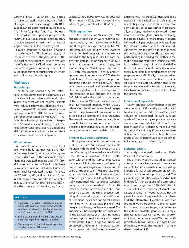
Figure 3. a–f. A 63-year-old man considering active surveillance for minimal volume low-grade prostate cancer. Initial sectoral biopsy was positive for Gleason grade 6 carcinoma, in two cores with 5% and <5% of core length involved in these samples. Axial T2-weighted image (a) demonstrates an anterior lesion centered in the right mid anterior peripheral zone (arrow); T2 PIRADS score was 4. Axial b=800 s/mm² DWI (b) demonstrates isointensity of the lesion (arrow). Axial ADC map (c) shows an area of restricted diffusion correlating with T2 appearance; DWI PIRADS score was 4. Sagittal T2-weighted image (d) demonstrates the same lesion as a wedge-shaped anterior low-signal lesion (arrow). Note rectum posteriorly (asterisk). TRUS (e) with sagittal probe orientation; in this case a matching hypoechoic lesion is seen anteriorly (arrow). Anterior, posterior, caudal, and cranial orientations are denoted by A, P, Cau, and Cra, respectively. Targeted anterior TRUS-guided biopsy image (f) demonstrates an 18 G biopsy needle (arrowheads) in the mass (arrow) and yielded diagnostic samples. Overall PIRADS score was 4. Targeted anterior biopsy yielded 50% of core length samples positive for Gleason grade 7 prostate carcinoma. Radical prostatectomy was performed, and the anterior gland lesion was confirmed as a gland-confined Gleason grade 7 carcinoma.
d
a
e
b
f
c
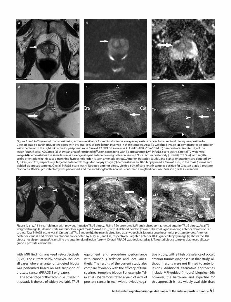
Figure 4. a–c. A 51-year-old man with previous negative TRUS biopsy. Rising PSA prompted MRI and subsequent targeted anterior TRUS biopsy. Axial T2-weighted image (a) demonstrates anterior low signal mass (arrowheads), with ill-defined borders (“erased charcoal sign”) invading anterior fibromuscular stroma; T2W PIRADS score was 5. On sagittal TRUS image (b), the mass is visualized as a hypoechoic lesion along the anterior prostate (arrow). Anterior, posterior, caudal, and cranial orientations are denoted by A, P, Cau, and Cra, respectively. Targeted anterior TRUS-guided biopsy image (c) shows the 18 G biopsy needle (arrowheads) sampling the anterior gland lesion (arrow). Overall PIRADS was designated as 5. Targeted biopsy samples diagnosed Gleason grade 7 prostate carcinoma.
a b c

92 • March–April 2017 • Diagnostic and Interventional Radiology Murphy et al.
for TRUS biopsy. Saturation biopsies can also be performed in the setting of clinical sus-picion of biopsy-occult prostate cancer (27) but require a high number of biopsy passes, without specifically targeting the areas of potentially highest yield. Another promising technique is electronic fusion of magnetic resonance and TRUS images to guide biop-sy. With this technique, software is used to coregister prior MRI data and real-time TRUS images (14). This makes use of spatial posi-tioning sensors attached to the endorectal ultrasound probe. Images from the MRI are reconstructed to the real-time plane of TRUS imaging thereby providing increased certainty that the TRUS-guided biopsy is effectively sampling the area of concern on MRI (15).
Our study has some limitations. The study is a retrospective review. The MRI protocol included DWI in addition to anatomic imag-ing, a strategy that has been correlated with a high cancer detection rate particularly for high Gleason grade disease (28). Howev-er, the addition of a further functional se-quence (DCE or MRS) may have refined the overall PIRADS score, with potential effect on correlation with biopsy result. It is not-ed that a b value of 800 s/mm2 was used in this study. Higher b value imaging may be advantageous, where adequate signal-to-noise ratio (SNR) permits, as recommended by in the PIRADS version 2 guidelines (29). Additionally, the use of a cognitive target-ing technique to direct the biopsy makes it less certain that the exact location of the abnormality characterized on MRI has been sampled. The finding that the average size of lesions, which were positive for carcinoma on targeted biopsy was greater than those which were negative (20.2 mm vs. 13.4 mm) highlights this potential sampling limitation. However, it is interesting that such large lesions were present in patients with prior negative biopsies, highlighting the impor-tance of strategies for diagnosis of occult an-terior prostate tumors. Since the study group did not all have radical prostatectomy, we cannot determine the false negative biopsy rate in this study. It is also worth noting that the study does not include patients who had no target for biopsy in the anterior prostate gland, so the proportion of men with occult anterior tumors in the clinical settings de-scribed is not determined. A further consid-eration is the impact of operator experience on the efficacy of this technique. In the cur-rent study, the operators had experience in both prostate MRI and TRUS-guided biopsies
and an understanding of both modalities is needed to perform the procedure effective-ly. However, the transition from performing sectoral nontargeted biopsies to manipu-lating the biopsy needle into the portion of the gland deemed suspicious for neoplasm on MRI proved straightforward for both op-erators. It is noted that the targeted lesions were sometimes visible on ultrasound. How-ever, this was not routinely documented, and the yield of visible lesions versus lesions that were sampled purely based on anatom-ic location could not be evaluated by this ret-rospective study.
All of the targeted anterior biopsies in this study were performed with a sagit-tal approach, described in detail above. It is hoped that this will be useful to the reader, as the exact biopsy technique for TRUS-guided targeted biopsy is often not specifically described in existing literature and this can be a barrier to implementing this approach in local clinical practice. This biopsy technique was well tolerated by all patients. The specific yield of targeted an-terior biopsy with sagittal approach com-pared with axial approach was not directly compared in this study. In smaller volume prostate glands, based on local experience, some anterior tumors can be sampled us-ing a transaxial approach. However, it is difficult to direct the biopsy needle into the anterior-most aspect of the prostate with axial TRUS imaging guidance in larger glands. We did not routinely perform both techniques, as performing both in all cases would have resulted in unnecessary addi-tional biopsy passes.
In general terms, targeted prostate biopsy has a number of potential advantages. The morbidity associated with prostate biopsy, particularly procedure related sepsis has been shown to be related to the number of biopsy samples performed (30). In a study of 5802 biopsies in 2002, 50% had hemato-spermia at three days, and 3.5% developed sepsis (31). Targeted TRUS biopsy of the pros-tate has the potential to reduce the number of cores required by reducing the need for repeated extended (12-core) biopsies and could potentially reduce the yield of clinically insignificant prostate cancers (32).
In conclusion, MRI-directed targeted TRUS-guided prostate biopsy with cogni-tive fusion enables accurate sampling of clinically significant prostate cancer in the anterior prostate, enabling improved tumor detection yield of lesions occult to routine sectoral biopsy.
Conflict of interest disclosureThe authors declared no conflicts of interest.
References1. Roehl KA, Antenor JAV, Catalona WJ. Serial bi-
opsy results in prostate cancer screening study. J Urol 2002; 167:2435–2439. [Crossref]
2. Djavan B, Ravery V, Zlotta A, et al. Prospective evaluation of prostate cancer detected on bi-opsies 1, 2, 3 and 4: when should we stop? J Urol 2001; 166:1679–1683. [Crossref]
3. Al-Ahmadie HA, Tickoo SK, Olgac S, et al. Anteri-or-predominant prostatic tumors: zone of origin and pathologic outcomes at radical prostatecto-my. Am J Surg Pathol 2008; 32:229–235. [Crossref]
4. Bott SRJ, Young MPA, Kellett MJ, Parkinson MC, Contributors to the UCL Hospitals’ Trust Radi-cal Prostatectomy Database. Anterior prostate cancer: is it more difficult to diagnose? BJU In-ternational 2002; 89:886–889. [Crossref]
5. Lawrentschuk N, Haider MA, Daljeet N, et al. “Prostatic evasive anterior tumours”: the role of magnetic resonance imaging. BJU Int 2010; 105:1231–1236. [Crossref]
6. Hoeks CMA, Barentsz JO, Hambrock T, et al. Prostate cancer: multiparametric MR imaging for detection, localization, and staging. Radiol-ogy 2011; 261:46–66. [Crossref]
7. Bloch BN, Furman-Haran E, Helbich TH, et al. Prostate cancer: accurate determination of extracapsular extension with high-spatial-res-olution dynamic contrast-enhanced and T2-weighted MR imaging—initial results 1. Ra-diology 2007; 245:176–185. [Crossref]
8. Graser A, Heuck A, Sommer B, et al. Per-sextant localization and staging of prostate cancer: correlation of imaging findings with whole-mount step section histopathology. AJR Am J Roentgenol 2007; 188:84–90. [Crossref]
9. Prando A, Kurhanewicz J, Borges AP, Oliveira EM Jr, Figueiredo E. Prostatic biopsy directed with en-dorectal MR spectroscopic imaging findings in pa-tients with elevated prostate specific antigen levels and prior negative biopsy findings: early experi-ence 1. Radiology 2005; 236:903–910. [Crossref]
10. Franiel T, Stephan C, Erbersdobler A, et al. Areas suspicious for prostate cancer: MR-guided biopsy in patients with at least one transrectal US-guided biopsy with a negative finding--multiparametric MR imaging for detection and biopsy planning. Radiology 2011; 259:162–172. [Crossref]
11. Sciarra A, Panebianco V, Ciccariello M, et al. Value of magnetic resonance spectroscopy imaging and dynamic contrast-enhanced imaging for detecting prostate cancer foci in men with prior negative biop-sy. Clin Cancer Res 2010; 16:1875–1883. [Crossref]
12. Park BK, Lee HM, Kim CK, Choi HY, Park JW. Lesion localization in patients with a previous negative transrectal ultrasound biopsy and persistently elevated prostate specific anti-gen level using diffusion-weighted imaging at three Tesla before rebiopsy. Invest Radiol 2008; 43:789–793. [Crossref]
13. Barentsz JO, Richenberg J, Clements R, et al. ESUR prostate MR guidelines 2012. Eur Radiol 2012; 22:746–757. [Crossref]
14. Singh AK, Kruecker J, Xu S, et al. Initial clinical experience with real‐time transrectal ultra-sonography‐magnetic resonance imaging fusion‐guided prostate biopsy. BJU Int 2008; 101:841–845. [Crossref]

15. Cool DW, Zhang X, Romagnoli C, Izawa JI, Ro-mano WM, Fenster A. Evaluation of MRI-TRUS fusion versus cognitive registration accuracy for MRI-targeted, TRUS-guided prostate bi-opsy. AJR Am J Roentgenol 2015; 204:83–91. [Crossref]
16. Testa C, Schiavina R, Lodi R, et al. Accuracy of MRI/MRSI-based transrectal ultrasound biopsy in peripheral and transition zones of the pros-tate gland in patients with prior negative biopsy. NMR Biomed 2010; 23:1017–1026. [Crossref]
17. Moussa AS, Meshref A, Schoenfield L, et al. Im-portance of additional “extreme” anterior api-cal needle biopsies in the initial detection of prostate cancer. Urology 2010; 75:1034–1039. [Crossref]
18. Meng MV, Franks JH, Presti JC Jr, Shinohara K. The utility of apical anterior horn biopsies in prostate cancer detection. Urol Oncol 2003; 21:361–365. [Crossref]
19. Radtke JP, Boxler S, Kuru TH, et al. Improved de-tection of anterior fibromuscular stroma and transition zone prostate cancer using bipara-metric and multiparametric MRI with MRI-tar-geted biopsy and MRI-US fusion guidance. Prostate Cancer Prostatic Dis 2015; 18:288–296. [Crossref]
20. Epstein JI, Walsh PC, Carmichael M, Brendler CB. Pathologic and clinical findings to predict tumor extent of nonpalpable (stage T1c) prostate can-cer. JAMA 1994; 271:368–374. [Crossref]
21. Murphy G, Haider M, Ghai S, Sreeharsha B. The expanding role of MRI in prostate cancer. AJR Am J Radiol 2013; 201:1229–1238. [Crossref]
22. Portalez D, Rollin G, Leandri P, et al. Prospective comparison of T2w-MRI and dynamic-con-trast-enhanced MRI, 3D-MR spectroscopic imaging or diffusion-weighted MRI in re-peat TRUS-guided biopsies. Eur Radiol 2010; 20:2781–2790. [Crossref]
23. Anastasiadis AG, Lichy MP, Nagele U, et al. MRI-guided biopsy of the prostate increases diagnostic performance in men with elevated or increasing PSA levels after previous nega-tive TRUS biopsies. Eur Urol 2006; 50:738–749. [Crossref]
24. Ouzzane A, Puech P, Lemaitre L, Leroy X, Nev-oux P. Combined multiparametric MRI and targeted biopsies improve anterior prostate cancer detection, staging, and grading. Urolo-gy 2011; 78:1356–1362. [Crossref]
25. Taira AV, Merrick GS, Galbreath RW, et al. Perfor-mance of transperineal template-guided map-ping biopsy in detecting prostate cancer in the initial and repeat biopsy setting. Pros Cancer Prostatic Dis 2009; 13:71–77. [Crossref]
26. Pondman KM, Fütterer JJ, Haken ten B, et al. MR-guided biopsy of the prostate: an overview of techniques and a systematic review. Eur Urol 2008; 54:517–527. [Crossref]
27. Delongchamps NB, Haas GP. Saturation biop-sies for prostate cancer: current uses and fu-ture prospects. Nat Rev Urol 2009; 6:645–652. [Crossref]
28. Watanabe Y, Terai A, Araki T, et al. Detection and localization of prostate cancer with the targeted biopsy strategy based on ADC map: a prospective large-scale cohort study. J Magn Reson Imaging 2012; 35:1414–1421. [Crossref]
29. American College of Radiology. MR Prostate Imaging Reporting and Data System version 2.0. Available at http://www.acr.org/~/media/ACR/Documents/PDF/QualitySafety/Resourc-es/PIRADS/PIRADS%20V2.pdf.
30. Rodríguez LV, Terris MK. Risks and complica-tions of transrectal ultrasound guided prostate needle biopsy: a prospective study and review of the literature. J Urol 1998; 160:2115–2120. [Crossref]
31. Raaijmakers R, Kirkels WJ, Roobol MJ, Wildha-gen MF, Schrder FH. Complication rates and risk factors of 5802 transrectal ultrasound-guided sextant biopsies of the prostate within a pop-ulation-based screening program. Urology 2002; 60:826–830. [Crossref]
32. Moore CM, Robertson NL, Arsanious N, et al. Image-guided prostate biopsy using magnetic resonance imaging–derived targets: a systemat-ic review. Eur Urol 2013; 63:125–140. [Crossref]
MRI-directed cognitive fusion-guided biopsy of the anterior prostate tumors • 93