mri of soft tissue masses of the hand and wrist · pdf filemri of soft tissue masses of the...
TRANSCRIPT

PICTORIAL REVIEW
MRI of soft tissue masses of the hand and wrist
J TEH, BSc, MRCP, FRCR and G WHITELEY, FRCR
Radiology, Nuffield Orthopaedic Centre, Windmill Road, Headington, Oxford OX3 7LD, UK
Received 24 January 2005Revised 2 June 2005Accepted 10 August 2005
DOI: 10.1259/bjr/53596176
’ 2006 The British Institute of
Radiology
The vast majority of soft tissue mass lesions of thewrist and hand are benign [1, 2]. In practice, the mostcommon lesions encountered are ganglia. The mostfrequently seen solid masses include giant cell tumoursof tendon sheath (GCTTS), lipomas, Dupuytrens con-tractures, nerve sheath tumours, glomus tumours,haemangioma/vascular malformations and synovialpathology. In general MRI is unable to differentiatebetween benignity and malignancy, but in manycircumstances a specific diagnosis may be achieved bytaking into account the location of the lesion within thehand or wrist and its signal characteristics [2–4]. Plainfilms and CT may detect calcification and allow assess-ment of adjacent bony structures but unlike MRI do notoffer much in the way of tissue characterization.Ultrasound has an extremely useful role in localizinglesions and determining if the lesion is cystic or solid [5,6], but further tissue characterization is limited.
This pictorial review covers the MRI appearances ofthe most commonly encountered soft tissue masses of thewrist and hand, and describes the main features,specifically the signal characteristics and location, thathelp differentiate them.
Imaging technique
Ideally the patient should be scanned supine with thearm by the side and the dorsum of the hand parallel tothe coronal plane of the magnet. However some patientsmay need to be scanned prone with the arm above theirhead in the so-called ‘‘Superman’’ position. The dis-advantage of this position is that it may be uncomfor-table and therefore prone to increased motion artefact.
A dedicated wrist coil is advised and to achieve highresolution a small field of view (FOV) in the order of 8–12 cm, with a matrix of at least 256 by 512 and slicethickness of 1.5–3 mm. A number of pulse sequences andimage planes can be used. At the authors’ institution aroutine examination for a mass would include coronal orsagittal T1 and T2 weighted sequences with axial short
tau inversion recovery (STIR) and T1 weighted images.Intravenous gadolinium may be administered to helpdifferentiate solid from cystic lesions.
Ganglion
Ganglia are the most common cause for a palpablemass in the wrist and hand. They most commonly occurin young women. Approximately 10% of patients have ahistory of trauma. Ganglia were described byHippocrates as ‘‘knots of tissue containing mucoidflesh’’. Histologically ganglia have a thin connectivetissue capsule, but no true synovial lining, and containmucinous material. Synovial cysts, which have a syno-vial lining, are histologically distinct from ganglia but areindistinguishable on imaging [7]. The terms ‘‘ganglion’’and ‘‘synovial cyst’’ are therefore often used inter-changeably. The aetiology of ganglia is unclear; theymay represent a synovial herniation or coalescence ofsmall degenerative cysts arising from the tendon sheath,joint capsule or bursae [8].
On MRI a unilocular or multilocular rounded orlobular fluid signal mass is seen adjacent to a joint ortendon sheath. Very small cysts may simulate a smalleffusion but a clue to the diagnosis is the paucity of fluidin the remainder of the joint and the focal nature of thefluid. Typically ganglia are low signal on T1 weightedimages and high signal on T2 weighted images, but highproteinaceous content or haemorrhage can result inlesions appearing isointense or hyperintense on T1
weighted images.Mild enhancement of the capsule or of septae may be
seen following post intravenous gadolinium (Figure 1)[9]. A ganglion is considered occult when it is notclinically palpable, usually because it lies deep totendons. Ganglia occur in four main areas (Figure 2) [8,10]:
1. Dorsum of the wrist (around 60%).These usuallyoriginate from the scapholunate joint or ligament. A
The British Journal of Radiology
The British Journal of Radiology, Month 2006 1 of 18
The British Institute of Radiology, doi: 10.1259/bjr/53596176Published online before print May 10, 2006

small synovial pedicle frequently extends throughthe fibres of the scapholunate ligament and dissectsthrough overlying structures proximally or distally.Erosion of the dorsal lunate is occasionallyassociated.
2. Volar aspect of the wrist (20%). These arise from theradio-scaphoid, scapho-trapezial, or metacarpo-trapezial joint. They can extend around the flexor
carpi radialis tendon and/or radial artery. Ulnarganglia are associated with tears in the triangularfibrocartilage complex.
3. Flexor tendon sheath (10%). Typically these occur atthe metacarpo-phalangeal joint.
4. Distal interphalangeal joint, located on the dorsumof the fingers between the nail and distal inter-phalangeal joint (10%). These are commonly
(a) (b)
Figure 1. (a) Dorsal ganglion. Axial T2 weighted image demonstrating a well defined fluid signal lobulated mass arising fromthe region of the dorsal scapho-lunate ligament (arrow). The lunate (Lu) and scaphoid (Sc) are labelled. (b) Axial T1 weightedimage demonstrating an intermediate signal mass.
(a) (b)
Figure 2. (a) Occult dorsal ganglion. Coronal short tau inversion recovery (STIR) image demonstrating a fluid signal mass(arrows) arising from the dorsum of the wrist. (b) Axial T2 weighted image demonstrating a high signal mass (arrows) lying deepto the extensor tendons.
J Teh and G Whiteley
2 of 18 The British Journal of Radiology

referred to as ‘‘mucous cysts’’ and are usuallyassociated with osteoarthritis [11]. They may causepain and nail distortion and may discharge.
Giant cell tumour of the tendon sheath
Giant cell tumours of the tendon sheath (GCTTS) arecommon benign synovial masses of unknown aetiologyarising from the tendon sheath, often referred to as focalpigmented villonodular synovitis (PVNS). There is aslight female predominance. These lesions usually affectthe volar aspect of the first three digits [12], much lesscommonly affecting the wrist.
MRI typically shows a well defined mass adjacent to orenveloping a tendon [13]. Characteristically the mass ishypointense on T1 weighted images, approximatelyequal to skeletal muscle. On T2 weighted images thereis usually low signal due to chronic haemorrhage withhaemosiderin deposition [14]. Blooming artefact mayoccur with gradient echo sequences. There may be areasof low signal and high signal on T2 weighted images dueto the presence of haemosiderin and fluid respectively.Uniform enhancement can be seen post intravenousgadolinium (Figure 3) [13].
Fibromatosis
Fibromatosis refers to group of benign but sometimeslocally aggressive proliferative lesions comprised ofmyofibroblasts [15]. These are characterized by infiltra-tive growth, and may therefore mimic a malignantlesion. Lesions may be superficial, resulting inDupuytren’s contracture or deep.
Dupuytren’s contracture
Proliferation of fibrous tissue within the palmaraponeurosis of the hand results in Dupuytren’s contrac-ture [16]. Patients present with subcutaneous nodules onthe palmar surface of the distal crease of the hand whichprogresses to cords and bands and finally the character-istic flexion contracture secondary to fibrous attachmentsto the underlying tendon sheath. Up to 25% of patientsover 65 years are affected with a male predominance.Most commonly the fourth ray is involved.
MRI demonstrates focal nodules or cords arising fromthe palmar aponeurosis extending distally and super-ficially parallel to the flexor tendons. The lesionsterminate in fine strands extending into the subcuta-neous tissues at the level of the distal metacarpals [17].Lesions of high cellularity tend to be of higher signal onT2 weighted images than those with a large collagenouscomponent [15, 16] (Figure 4).
Deep musculoaponeurotic fibromatosis
Deep musculoaponeurotic fibromatosis typically occursbetween the ages of 10 years and 40 years. There is equalsex predominance. Usually solitary lesions occur butsynchronous multicentric lesions have been reported[18]. Plain films may reveal evidence of bone erosionwithout invasion or destruction. The lesions may havepoorly defined margins and appear infiltrative. Greatvariability in appearance is seen on MRI depending on thedegree of cellularity and collagen content [19]. Generallylesions are of low signal on T1 weighted images, with areasof low and high signal on T2 weighted images representingcollagen and cellularity, respectively.
(a) (b)
Figure 3. (a) Giant cell tumours of tendon sheath (GCTTS) of index finger. Axial T1 weighted image demonstrating a welldefined intermediate/low signal mass (arrow) adjacent to the flexor tendon (arrowhead). (b) Axial T2 weighted imagedemonstrating a low signal mass indicating the presence of haemosiderin, surrounded by a rim of high signal (arrowheads).
Pictorial review: MRI of the hand and wrist
The British Journal of Radiology, Month 2006 3 of 18

Fibroma of the tendon sheath
This is a rare benign tumour of the tendon sheathwhich may be confused with GCTTS. It is usually a firmwell defined mass attached to the tendon sheath. TheMRI appearance is variable but usually the presence offibrous tissue results in low signal on both T1 and T2
weighted images [20].
Lipoma
Although simple benign lipomata are the mostcommon soft tissue tumour, they are uncommon in thehand [21]. They are lesions of mature adipose tissuewhich may occur in a subcutaneous or deep location,presenting as a slow growing painless mass. 80% oflesions measure less than 5 cm [17]. Pressure effects canresult on neighbouring structures such as nerves andvessels in locations where there is confined space, e.g.Carpal tunnel and Guyon’s canal. Typically they occur atthe thenar or hypothenar eminence.
Superficial lipomata may be inconspicuous on MRIparticularly when they blend in with normal subcuta-neous fat. They demonstrate typical fatty signal withhomogeneous high signal on T1 weighted and low signalon STIR or T2 fat saturated images [22] (Figure 5).Septations may be seen but nodules or solid componentssuggest a liposarcoma. Complications such as haemor-rhage or infarction can result in a more complexappearance which may simulate liposarcoma.Intramuscular lipomata may infiltrate between musclefibres resulting in a more heterogeneous appearance onMRI.
Fibrolipomatous hamartoma
The aetiology of fibrolipomatous hamartoma may berelated to hypertrophy of mature fat and fibroblasts inthe epineurium [23]. It presents in young adulthood witha slow growing mass on the volar aspect of the hand or
wrist or forearm and may be associated with macro-dactyly. It has a marked predilection for the mediannerve with up to 85% occurring at this location and maygive rise to symptoms of pain or paresthesia [24]. TheMRI appearances are pathognomonic with longitudin-ally orientated cylindrical foci of low signal intensitysurrounded by fatty signal intensity representing nervefascicles, giving a spaghetti-like appearance on coronalplanes and co-axial cable appearance on axial images [25,26] (Figure 6). On MRI areas of low and high signal areseen on both T1 and T2 weighted images representing thefatty and fibrous contributions of the tumour.
Haemangioma/vascular malformations
Mulliken and Golwacki’s classification of vascularanomalies defines a haemangioma as a tumour character-ized by increased cell turnover of endothelium, mast cells,fibroblasts and macrophages [27]. This tumour is notusually present at birth (except in the congenital haeman-gioma) but becomes apparent during the first few weeks oflife as a firm non-compressible mass within the soft tissuesafter which there is rapid growth (the proliferative phase)followed 6–10 months later by an involuting phase.Complete resolution occurs in 70% by 7 years and theremainder continue to diminish until the age of 12 years.Some lesions persist into adulthood, and may presentincidentally or following trauma. In the proliferative phasethese appear as a soft tissue mass isointense or hypoin-tense to muscle on T1 weighted images and high signal onT2 weighted images. Serpentine vascular flow voids maybe evident on both sequences and there is uniformenhancement with intravenous gadolinium. With involu-tion there is decrease in size of the mass with replacementby variable and increasing amounts of fat, loss of the highflow signal voids and absence of enhancement(Figure 7a,b).
Vascular malformations on the other hand are notneoplastic lesions but are errors of vascular morpho-genesis with a normal rate of endothelial turnover, andhence grow commensurately with the child [27].
(a) (b)
Figure 4. (a) Dupuytren’s contracture. Axial T1 weighted image demonstrating an ill-defined intermediate/low signal mass(arrow) in the subcutaneous tissues overlying the flexor tendons (arrowhead). (b) Axial T2 fat saturated image demonstrating alow signal subcutaneous mass (arrow).
J Teh and G Whiteley
4 of 18 The British Journal of Radiology

(a) (b)
Figure 5. (a) Lipoma. Coronal T1 weighted image demonstrating a well defined homogeneously high signal mass (arrowheads)in the thenar eminence. (b) Coronal short tau inversion recovery (STIR) image demonstrating a low signal mass (arrowheads) inthe thenar eminence.
(a) (b)
Figure 6. (a) Fibrolipomatous hamartoma of the median nerve. Axial T1 weighted image demonstrating a mass (arrows)comprised of low signal foci surrounded by fatty signal, the ‘‘co-axial cable’’ sign. (b) Coronal T1 weighted image demonstratingseparation of nerve fascicles by fatty tissue (arrows), the ‘‘spaghetti’’ sign.
Pictorial review: MRI of the hand and wrist
The British Journal of Radiology, Month 2006 5 of 18

(a)
(d)
(e)
(b)
(c)
J Teh and G Whiteley
6 of 18 The British Journal of Radiology

Occasionally they can suddenly enlarge due to haemor-rhage, infection or hormonal influence at puberty. Theyare present at birth although they may not becomeapparent until adolescence or early adulthood. Thelesions can be subdivided according to vessel type intothe following groups: capillary, venous, arterial andlymphatic. Often these occur in combination. It isclinically important to separate them in to low flow(capillary, venous, lymphatic or a combination) or highflow (arteriovenous) [28]:
Capillary malformations are the equivalent of the portwine stain. A clinical diagnosis may be made in mostinstances. On imaging skin thickening is seen with novascular channels apparent.
Venous malformations are comprised of dilated slowflowing vascular spaces and channels with no solidtissue component aside from septations. They appear onMRI as septated high signal soft tissue masses on T2
weighted images with no evidence of high flow velocitysignal voids. On T1 weighted images they are isointenseto muscle. The presence of phleboliths results in foci ofsignal void. Gradient echo imaging is useful to document
slow flow and exclude the presence of any high flow.With contrast, there is uniform or inhomogeneousenhancement of the vascular spaces.
Lymphatic malformations consist of multiple lympha-tic fluid containing spaces with intervening septa. OnMRI they are comprised of septated cysts of variable size.Fluid–fluid levels may be seen. Stranding of the adjacentsubcutaneous tissue may be seen due to associatedlymphatic obstruction and may simulate cellulites [29](Figure 7c–e).
Arteriovenous malformations (AVMs) are high flowvascular anomalies with abnormal connections betweenarteries and veins. AVMs have an intervening centralnidus whereas arteriovenous fistulae do not. On MRIthey appear as enlarged vascular channels without adiscrete soft tissue mass which on spin echo imagingappear as signal voids with corresponding bright signalon flow-enhanced GE sequences [30]. MR angiographyor ultrasound can be used to confirm the high flow stateand assess arterial supply. Skin thickening and fatdeposition may be seen in association as well asperilesional oedema [29] (Figure 8).
Figure 7. (a) Haemangioma. Coronal T1 weighted image demonstrating a lobulated mass (arrowheads) with extension into thethumb (arrow), which is slightly hyperintense to skeletal muscle. (b) Haemangioma. Coronal short tau inversion recovery (STIR)image demonstrating a lobulated hyperintense mass. (c) Venous malformation. Axial T2 fat-saturated image demonstratinga lobulated, serpentine lesion in the thenar eminence. (d) Venous malformation. Coronal T1 weighted image demonstrating amass with well defined, low signal margin (arrowheads). (e) Venous malformation. Coronal STIR image demonstrating alobulated high signal mass.
(a) (b)
Figure 8. (a) Small arteriovenous malformation. Coronal short tau inversion recovery (STIR) image demonstrating a smallserpiginous high signal mass on the radial aspect of the wrist (arrow), confirmed as a vascular structure on ultrasound. (b) AxialT2 fat-saturated image demonstrating a high signal serpiginous mass (arrow) on the radial aspect of the wrist. There is also aganglion (arrowhead) on the ulnar aspect of the wrist.
Pictorial review: MRI of the hand and wrist
The British Journal of Radiology, Month 2006 7 of 18

Benign peripheral nerve sheath tumours
Benign peripheral nerve sheath tumours (PNST) arecommon masses of the forearm and hand. Schwannomasarise from the schwann cells surrounding the nervewhereas neurofibromas arise from the central nervefascicles [31]. In the hand and wrist, schwannomas arisefrom deeper and larger nerves (particularly the ulnarnerve) and often occur along the flexor surfaces whereasneurofibromas tend to involve smaller cutaneous nerves.Both tend to present in young adulthood and are small,solitary and slow growing. The vast majority of these arenot associated with neurofibromatosis [21].
The most important imaging feature of neurogenictumours is recognition of a fusiform mass with a ‘‘dural-tail’’, representing the entering and exiting nerve at atypical nerve location [31]. This is usually straightfor-ward where the nerve is large or deep but withsuperficial small PNSTs this feature may not be seen(Figure 9a,b). The eccentric nature of the schwannoma inrelation to the nerve may allow its differentiation from aneurofibroma [32] (Figure 9c).
The signal intensity of these tumours is fairly non-specific demonstrating isointensity or slight hyperinten-sity to muscle on T1 weighted images and markedhyperintensity on T2 weighted images. There may beareas of heterogeneity but this finding is more commonlyseen with malignant PNST. A target sign with hyper-intense periphery and central hypointensity on T2
weighted is most commonly seen with neurofibromabut occurs with other PNST. This corresponds to centralfibromatosis and surrounding myxomatosis tissue [33]Figure 10a,b). The ‘‘split fat’’ sign is often seen aroundneurogenic tumours and relates to the fact that theneurovascular bundle is surrounded by fat so massesarising from this location maintain a rim of fat aroundthem. The ‘‘fascicular’’ sign describes small ring likestructures on T2 weighted images corresponding to thefascicular bundles within the nerve [31] (Figure 10c).Muscular atrophy with concomitant increased fat contentmay be appreciated but can be quite subtle particularlywith the small muscles of the forearm and hand. This isbest appreciated on T1 weighted images and may requirethe contralateral side for comparison [32]. Most PSNTsenhance vividly with contrast but the pattern ofenhancement is variable with foci of heterogeneity notuncommonly seen. Features suggestive of malignancyinclude large size (greater than 5 cm), prominentenhancement, infiltrative margins, marked heterogeneityand rapid growth [31].
Glomus tumour
Glomus bodies are responsible for thermoregulationand are present throughout the dermis of the body butare particularly concentrated in the digits of the handand foot. Glomus tumours are small hamartomas of theneuromyoarterial apparatus within the glomus body andare responsible for up to 5% of soft tissue tumours of thehand [34]. They are most commonly found at the fingertip, either in the pulp or beneath the fingernail, andtypically present in the fourth and fifth decades asexquisitely painful lesions exacerbated by temperature
changes [35]. Clinically, disappearance of pain afterapplication of a tourniquet proximally on the arm ispathognomonic of the tumour and is known as theHildreth sign [36]. On plain films, smooth extrinsic boneerosions adjacent to the lesion may be seen if the lesion islarge enough.
With the use of surface coils lesions as small as 2 mmmay be detected with MRI. They are typically of low orintermediate signal on T1 weighted images and homo-genously high intensity on T2 weighted images. Theyenhance uniformly following gadolinium [34, 37](Figure 11).
Soft tissue chondroma
Soft tissue chondromas are small nodules of cartilagenot attached to bone. They represent 6% of all hand andwrist soft tissue tumours [31]. There is a wide range ofages at presentation. Patients present with a slowgrowing mass less than 2 cm in size. They may beattached or associated with tendons or tendon sheaths,joint capsule or periosteum [17]. Plain film shows a welldefined soft tissue mass with most lesions showing fociof calcification which may be central or peripherallylocated. Typical rings and arcs of mineralization may beseen and adjacent bony remodelling or extrinsic erosionmaybe evident (Figure 12a).
On MRI the lesions are intermediate signal on T1
weighted images and high on T2 weighted images. Ifcalcification is present foci of low signal may be seen onboth sequences (Figure 12b,c).
Malignant masses
Malignant soft tissue tumours of the hand areuncommon [1, 2]. The lesions most often encounteredare malignant fibrous histiocytoma in the older popula-tion, synovial sarcoma, rhabdomyosarcoma, malignantnerve sheath tumours, liposarcomas and extraskeletalchondrosarcomas. Because of non-specific morpho-logical features, these tumours can be confused withbenign lesions such as aggressive fibromatosis or gang-lion cysts, particularly when they are small. Thepossibility of a malignant lesion needs to be consideredwhen the mass does not have an unequivocal benigndiagnosis on MRI [38].
The MRI features that should raise the possibility ofmalignancy are a large lesion with poorly definedmargins, inhomogeneity on T2 weighted images, irregu-lar enhancement following intravenous contrast and thepresence of necrosis (Figure 13).
Pseudomasses
Synovial pathologySynovial hyperplasia and tenosynovitis can present as
a focal masses around joints and tendons. This mayoccur as an isolated abnormality or in the setting of anarthropathy, particularly rheumatoid arthritis.
MRI is extremely useful for defining the extent ofdisease. Inflamed, hypertrophied pannus demonstrates
J Teh and G Whiteley
8 of 18 The British Journal of Radiology

(a)
(c)
(b)
Figure 9. (a) Schwannoma. Sagittal T1 weighted image demonstrating an ovoid intermediate signal mass (arrowheads) on thevolar aspect of the wrist. (b) Sagittal T2 fat saturated image demonstrating dural tail at both ends of a high signal mass (arrows),strongly suggesting a neurogenic lesion. (c) Schwannoma. Coronal short tau inversion recovery (STIR) sequence demonstratingan ovoid high signal mass in the palm with a clearly defined dural tail (arrow).
Pictorial review: MRI of the hand and wrist
The British Journal of Radiology, Month 2006 9 of 18

(a)
(b)
(c)
Figure 10. (a) Neurofibroma. Coronal T1 weighted image demonstrating an ovoid intermediate signal mass (arrowheads). (b)Axial T2 fat saturated image demonstrating the ‘‘target’’ sign, with a mass of central low signal intensity (arrowheads) withsurrounding high signal. (c) Fascicular sign. Sagittal T2 weighted image demonstrating a small low signal ring like structurewithin a small neurofibroma (arrows). Note the presence of a dural tail (arrowhead).
J Teh and G Whiteley
10 of 18 The British Journal of Radiology

(a) (b)
Figure 11. (a) Nail-bed glomus tumour. Coronal T1 weighted image demonstrating a small intermediate signal mass (arrow) inthe nail-bed. (b) Coronal short tau inversion recovery (STIR) image demonstrating a high signal mass in the nail-bed (arrow).
Pictorial review: MRI of the hand and wrist
The British Journal of Radiology, Month 2006 11 of 18

(a) (b)
Figure 12. (a) Periosteal chondroma. Lateral plain radiograph of the ring finger showing a soft tissue mass with scalloping ofthe underlying bone (arrowheads) and an arc of peripheral calcification (arrow). (b) Sagittal T1 weighted image showing anintermediate signal mass (arrowheads). The underlying bone is scalloped. (Continued)
J Teh and G Whiteley
12 of 18 The British Journal of Radiology

intermediate signal on T1 weighted images and highsignal on T2 weighted and STIR images [39] (Figure 14).Intravenous gadolinium can help differentiate pannusfrom fluid because pannus demonstrates diffuseenhancement on T1 weighted images whereas the fluidremains low signal. With tenosynovitis, fluid is seenaround tendons as high signal on T2 weighted images.
The tendon may appear normal but can be swollen withincreased signal on T2 weighted images.
Anomalous muscles
Anatomic variants of the muscles of the wrist andhand are common [40, 41]. These can be clinicallysignificant when they cause compressive symptoms orappear mass-like. Often lesions are not recognized onimaging as the signal characteristics are identical tonormal muscle.
The accessory abductor digiti minimi muscle is presentin up to 24% of all wrists [40].On axial images a fusiformmass is present with the signal characteristics of musclelateral and anterior to the pisiform bone at the level ofthe origin of the abductor digiti minimi muscle. This maycause an ulnar or median neuropathy [42] (Figure 15).
The extensor digitorum brevis manus muscle occurs in1–3% of the population [43]. Clinically this may bemistaken for a dorsal ganglion. This arises from the distalradius and inserts on the index finger. Normally theextensor tendons are unaccompanied by their musclebellies at the level of the carpus. However, in thepresence of an extensor digitorum brevis manus muscle,it is noted that there is a muscle medial to the tendonof the index finger at or distal to the level of the carpus[44].
Palmaris longus muscle variants may compress theulnar and median nerves. The normal muscle arises fromthe medial epicondyle and inserts into the palmaraponeurosis. MRI may demonstrate a midline masssuperficial to the flexor retinaculum at the wrist butdiagnosis may require more proximal imaging of theforearm [45] (Figure 16).
Proximal origin of the lumbrical muscles occurs whenthe origin of these muscles is within the carpal tunnelrather than just distal to it (diagnosed with the fingers inextension) and is seen in up to 22% of individuals [40]. Itmay be the cause of carpal tunnel syndrome [46].
Soft tissue infection
Soft tissue infection with abscess formation may mimica soft tissue mass. MRI not only allows demonstration ofthe extent of soft tissue involvement but can helpdetermine the presence of osteomyelitis [47]. On MRIan abscess is typically demonstrates fluid signal, withintermediate/low signal on T1 weighted images andhigh signal on T2 weighted and STIR images, and anenhancing wall following intravenous gadolinium(Figure 17).
Conclusion
The vast majority of soft tissue mass lesions of thewrist and hand are benign. By noting the signalcharacteristics and determining lesion location, a specificdiagnosis of the mass can often be made (Figure 18).Certain lesions such as lipomata, ganglia, fibrolipoma-tous hamartoma and GCTTS have characteristic appear-ances. Unfortunately, where the lesion does not exhibit
(c)
Figure 12. (Cont.) (c) Sagittal T2 fat saturated imagedemonstrating a slightly heterogeneous high signal mass(arrowheads).
Pictorial review: MRI of the hand and wrist
The British Journal of Radiology, Month 2006 13 of 18

(a) (b)
Figure 13. (a) Synovial sarcoma. Axial T1 weighted image demonstrating an intermediate signal mass on the palmar aspect ofthe hand. (b) Axial T2 fat-saturated image demonstrating a predominantly hyperintense mass with central hypointense areas.The features are non-specific.
(a) (b)
Figure 14. (a) Seronegative arthropathy with synovitis. Coronal short tau inversion recovery (STIR) image demonstrating diffusesynovitis of the carpus with fluid at the distal radioulnar joint (arrowhead) and at the scapho-trapezoid joint (arrow). There isdiffuse peri-articular bone marrow oedema. (b) Axial T2 weighted image demonstrating synovial fluid and hypertrophy(arrowheads) arising from between the scaphoid and capitate (arrow).
J Teh and G Whiteley
14 of 18 The British Journal of Radiology
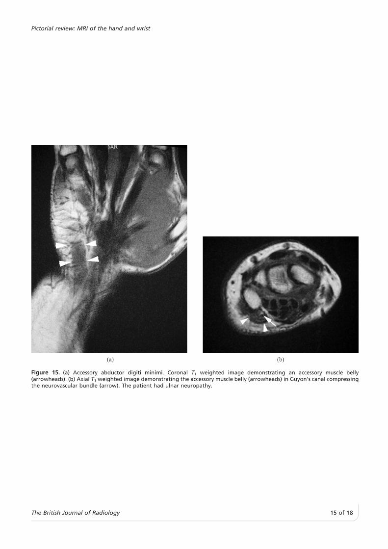
(a) (b)
Figure 15. (a) Accessory abductor digiti minimi. Coronal T1 weighted image demonstrating an accessory muscle belly(arrowheads). (b) Axial T1 weighted image demonstrating the accessory muscle belly (arrowheads) in Guyon’s canal compressingthe neurovascular bundle (arrow). The patient had ulnar neuropathy.
Pictorial review: MRI of the hand and wrist
The British Journal of Radiology, Month 2006 15 of 18

Figure 16. Accessory palmaris longus muscle. Sagittal T1 weighted image demonstrating an accessory muscle belly at the level ofthe wrist (arrows).
J Teh and G Whiteley
16 of 18 The British Journal of Radiology

typical features, differentiation from malignancy cannotbe categorically made.
References
1. Johnson J, Kilgore E, Newmeyer W. Tumorous lesions ofthe hand. J Hand Surg [Am] 1985;10:284–6.
2. Capelastegui A, Astigarraga E, Fernandez-Canton G,Saralegui I, Larena JA, Merino A. Masses and pseudo-masses of the hand and wrist: MR findings in 134 cases.Skeletal Radiol 1999;28:498–507.
3. Miller TT, Potter HG, McCormack RR Jr. Benign soft tissuemasses of the wrist and hand: MRI appearances. SkeletalRadiol 1994;23:327–32.
4. Binkovitz LA, Berquist TH, McLeod RA. Masses of thehand and wrist: detection and characterization with MRimaging. AJR Am J Roentgenol 1990;154:323–6.
5. Hoglund M, Muren C, Brattstrom G. A statistical model forultrasound diagnosis of soft-tissue tumours in the hand andforearm. Acta Radiol 1997;38:355–8.
6. Teefey SA, Middleton WD, Boyer MI. Sonography of thehand and wrist. Semin Ultrasound CT MR 2000;21:192–204.
7. Steiner E, Steinbach LS, Schnarkowski P, Tirman PF, GenantHK. Ganglia and cysts around joints. Radiol Clin North Am1996;34:395–425, xi–xii.
8. el-Noueam KI, Schweitzer ME, Blasbalg R, et al. Is a subsetof wrist ganglia the sequela of internal derangements of the
Figure 18. Figure illustrating the typical location of com-monly encountered soft tissue masses of the hand and wrist.GCTTS, giant cell tumours of tendon sheath; PNST, peripheralnerve sheath tumours.
(a) (b)
Figure 17. (a) Joint infection, osteomyelitis and abscess. Sagittal short tau inversion recovery (STIR) image demonstrating adestructive lesion of the metacarpal head (arrow), with a fluid signal abscess pointing to the skin (arrowhead). (b) Sagittal T1
weighted image demonstrating an intermediate signal mass (arrows).
Pictorial review: MRI of the hand and wrist
The British Journal of Radiology, Month 2006 17 of 18

wrist joint? MR imaging findings. Radiology 1999;212:537–40.
9. Blam O, Bindra R, Middleton W, Gelberman R. The occultdorsal carpal ganglion: usefulness of magnetic resonanceimaging and ultrasound in diagnosis. Am J Orthop1998;27:107–10.
10. Thornburg LE. Ganglions of the hand and wrist. J Am AcadOrthop Surg 1999;7:231–8.
11. Drape JL, Idy-Peretti I, Goettmann S, et al. MR imaging ofdigital mucoid cysts. Radiology 1996;200:531–6.
12. Ushijima M, Hashimoto H, Tsuneyoshi M, Enjoji M. Giantcell tumor of the tendon sheath (nodular tenosynovitis). Astudy of 207 cases to compare the large joint group with thecommon digit group. Cancer 1986;57:875–84.
13. De Beuckeleer L, De Schepper A, De Belder F, et al.Magnetic resonance imaging of localized giant cell tumourof the tendon sheath (MRI of localized GCTTS). Eur Radiol1997;7:198–201.
14. Peh WC, Wong Y, Shek TW, Ip WY. Giant cell tumour of thetendon sheath of the hand: a pictorial essay. AustralasRadiol 2001;45:274–80.
15. Liu P, Thorner P. MRI of fibromatosis: with pathologiccorrelation. Pediatr Radiol 1992;22:587–9.
16. Yacoe ME, Bergman AG, Ladd AL, Hellman BH.Dupuytren’s contracture: MR imaging findings and correla-tion between MR signal intensity and cellularity of lesions.AJR Am J Roentgenol 1993;160:813–7.
17. Kransdorf MJ, Murphey MD. MR imaging of musculoske-letal tumors of the hand and wrist. Magn Reson ImagingClin N Am 1995;3:327–44.
18. Disler DG, Alexander AA, Mankin HJ, O’Connell JX,Rosenberg AE, Rosenthal DI. Multicentric fibromatosiswith metaphyseal dysplasia. Radiology 1993;187:489–92.
19. Quinn SF, Erickson SJ, Dee PM, et al. MR imaging infibromatosis: results in 26 patients with pathologic correla-tion. AJR Am J Roentgenol 1991;156:539–42.
20. Horcajadas AB, Lafuente JL, de la Cruz Burgos R, et al.Ultrasound and MR findings in tumor and tumor-likelesions of the fingers. Eur Radiol 2003;13:672–85.
21. Kransdorf MJ. Benign soft-tissue tumors in a large referralpopulation: distribution of specific diagnoses by age, sex,and location. AJR Am J Roentgenol 1995;164:395–402.
22. Laorr A, Greenspan A. Hand lipomas: detection andcharacterization by magnetic resonance imaging. CanAssoc Radiol J 1993;44:14–8.
23. Silverman TA, Enzinger FM. Fibrolipomatous hamartomaof nerve. A clinicopathologic analysis of 26 cases. Am J SurgPathol 1985;9:7–14.
24. Kransdorf MJ, Moser RP Jr, Meis JM, Meyer CA. Fat-containing soft-tissue masses of the extremities.Radiographics 1991;11:81–106.
25. Cavallaro MC, Taylor JA, Gorman JD, Haghighi P, ResnickD. Imaging findings in a patient with fibrolipomatoushamartoma of the median nerve. AJR Am J Roentgenol1993;161:837–8.
26. Marom EM, Helms CA. Fibrolipomatous hamartoma:pathognomonic on MR imaging. Skeletal Radiol1999;28:260–4.
27. Mulliken JB, Glowacki J. Classification of pediatric vascularlesions. Plast Reconstr Surg 1982;70:120–1.
28. Fishman SJ, Mulliken JB. Hemangiomas and vascularmalformations of infancy and childhood. Pediatr ClinNorth Am 1993;40:1177–200.
29. Robertson RL, Robson CD, Barnes PD, Burrows PE. Headand neck vascular anomalies of childhood. NeuroimagingClin N Am 1999;9:115–32.
30. Meyer JS, Hoffer FA, Barnes PD, Mulliken JB. Biologicalclassification of soft-tissue vascular anomalies: MR correla-tion. AJR Am J Roentgenol 1991;157:559–64.
31. Murphey MD, Smith WS, Smith SE, Kransdorf MJ, TempleHT. From the archives of the AFIP. Imaging of musculos-keletal neurogenic tumors: radiologic-pathologic correla-tion. Radiographics 1999;19:1253–80.
32. Cerofolini E, Landi A, DeSantis G, Maiorana A, Canossi G,Romagnoli R. MR of benign peripheral nerve sheathtumors. J Comput Assist Tomogr 1991;15:593–7.
33. Anderson MW, Kaplan PA, Dussault RG, Degnan GG.Magnetic resonance imaging of the wrist. Curr Probl DiagnRadiol 1998;27:187–229.
34. Dalrymple NC, Hayes J, Bessinger VJ, Wolfe SW, Katz LD.MRI of multiple glomus tumors of the finger. SkeletalRadiol 1997;26:664–6.
35. Drape JL, Idy-Peretti I, Goettmann S, Guerin-Surville H,Bittoun J. Standard and high resolution magnetic resonanceimaging of glomus tumors of toes and fingertips. J AmAcad Dermatol 1996;35:550–5.
36. Hildreth DH. The ischemia test for glomus tumor: a newdiagnostic test. Rev Surg 1970;27:147–8.
37. Drape JL, Idy-Peretti I, Goettmann S, et al. Subungualglomus tumors: evaluation with MR imaging. Radiology1995;195:507–15.
38. Nakajima H, Matsushita K, Shimizu H, et al. Synovialsarcoma of the hand. Skeletal Radiol 1997;26:674–6.
39. Bergman AG. Synovial lesions of the hand and wrist. MagnReson Imaging Clin N Am 1995;3:265–79.
40. Timins ME. Muscular anatomic variants of the wrist andhand: findings on MR imaging. AJR Am J Roentgenol1999;172:1397–401.
41. Timins ME, O’Connell SE, Erickson SJ, Oneson SR. MRimaging of the wrist: normal findings that may simulatedisease. Radiographics 1996;16:987–95.
42. Harvie P, Patel N, Ostlere SJ. Ulnar nerve compression atGuyon’s canal by an anomalous abductor digiti minimimuscle: the role of ultrasound in clinical diagnosis. HandSurg 2003;8:271–5.
43. Ogura T, Inoue H, Tanabe G. Anatomic and clinical studiesof the extensor digitorum brevis manus. J Hand Surg [Am]1987;12:100–7.
44. Anderson MW, Benedetti P, Walter J, Steinberg DR. MRappearance of the extensor digitorum manus brevis muscle:a pseudotumor of the hand. AJR Am J Roentgenol1995;164:1477–9.
45. Polesuk BS, Helms CA. Hypertrophied palmaris longusmuscle, a pseudomass of the forearm: MR appearance--casereport and review of the literature. Radiology 1998;207:361–2.
46. Middleton WD, Kneeland JB, Kellman GM, et al. MRimaging of the carpal tunnel: normal anatomy andpreliminary findings in the carpal tunnel syndrome. AJRAm J Roentgenol 1987;148:307–16.
47. Hsu CY, Lu HC, Shih TT. Tuberculous infection of thewrist: MRI features. AJR Am J Roentgenol 2004;183:623–8.
J Teh and G Whiteley
18 of 18 The British Journal of Radiology