mri of the hip - scbtmr.org · of the femur and/or acetabulum . femoroacetabular impingement •...
TRANSCRIPT

MRI of the Hip:MRI of the Hip:Femoroacetabular ImpingementFemoroacetabular Impingement (FAI)(FAI)
Lynne S. Steinbach, M.D.
Professor of Radiology and Orthopaedic SurgeryDepartment of RadiologyUniversity of California San Francisco

Femoroacetabular Impingement
• Major cause of hip labral tears and cartilage damage
• Leads to OA of the hip• Related to morphologic
variants/abnormalities of the femur and/or acetabulum
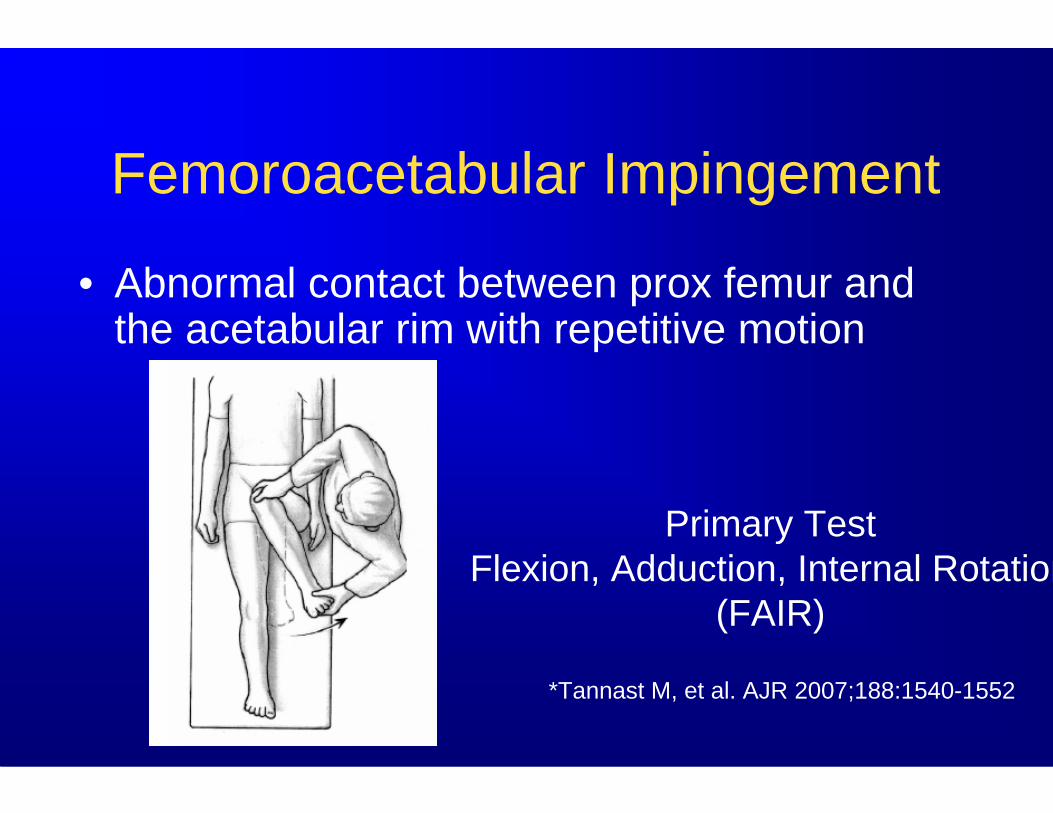
Femoroacetabular Impingement
• Abnormal contact between prox femur and the acetabular rim with repetitive motion
Primary TestFlexion, Adduction, Internal Rotation
(FAIR)
*Tannast M, et al. AJR 2007;188:1540-1552

Femoroacetabular Impingement• Cam- younger males
– Femoral cause– Asphericity
anterior/lateral femoral head-neck junction
• Pincer-middle aged female– Acetabular cause– Overcoverage contacts
femoral head-neck jct• Retroversion • Coxa profunda or
protrusio acetabulum *Lavigne M, et al. Clin Orth 2004

Femoroacetabular Impingement
• Beck et al 302 FAI analysed hips–26 isolated cam–16 isolated pincer–14% pure FAI form–Remainder mixed cam-pincer FAI -86%
Beck M et al JBJS, B 2005

Femoroacetabular ImpingementCam Type

Femur: Cam Impingement
Changes rotary motion
to linear motion
CAM
Nonspherical femoral head

Cam ImpingementCam Impingement

Femoroacetabular ImpingementPincer Type
• Acetab overcoverage– Retroversion
• Figure of 8-anterior overcoverage
– Coxa profunda/protrusio• Leads to
– Labral tears & degeneration cartilage
– Small rim chondral lesions posteroinferior acetabulum

Pincer ImpingementPincer Impingement

Femoral Head DefectFemoral Head Defect

Femoroacetabular ImpingementPincer Type
• Acetab overcoverage– Retroversion
• Figure of 8-anterior overcoverage
– Coxa profunda/protrusio• Leads to
– Labral tears & degeneration
– Small rim chondrallesions posteroinferioracetabulum Reynolds et al, JBJS-B 1999

Normal Retroversion
Local Excessive Coverage

General OvercoverageNormal Hip Jt Space Model
Normal Coxa profunda Protrusio acetabuliFemoral headIlioischial line
Acetabular fossa Beck M et al, Clin Orthop, 2004

Acetabular Depth Axial Cross Section
•• AbnormalAbnormal–– Can be associated Can be associated
with pincer FAIwith pincer FAI•• Point in center of Point in center of
femoral head lies femoral head lies medial to a line drawn medial to a line drawn between front and back between front and back of of acetabulumacetabulum Deep
femoral headNegative number

Pincer & Cam: Acetabular DepthPincer & Cam: Acetabular DepthPincer (protrusio acetabuli):acetabular depth deeper
Oblique transverse
Cam:acetabular depth shallower
Pfirrmann C et al Radiology2006;240(3):778-785
Acetabular protrusionChen L et al Skeletal Radiol 2008

Femoroacetabular ImpingementFemoral Cause = Cam Type
• Aspherical femoral head/neck junction– ↓Concavity at the anterior or
lateral femoral head-neck junction
• Premature contact b/t femur and acetabular rim
• Triad– Anterior loss of head-neck
junction offset (bump)– Anterosuperior labral tear– Adjacent chondrosis

Femoroacetabular ImpingementAlpha Angle
•• Method to quantify femoral Method to quantify femoral head neck offsethead neck offset
•• Measured on oblique axial Measured on oblique axial section parallel to fem neck section parallel to fem neck and passing through and passing through narrowest portion of narrowest portion of femoral neckfemoral neck
•• > 55 degrees predisposes > 55 degrees predisposes to cam impingementto cam impingement
Leunig M, et al. Radiology 2005;236:237*Kassarjian etl al. Radiology 2005;236:588 Normal Alpha Angle <55 degrees
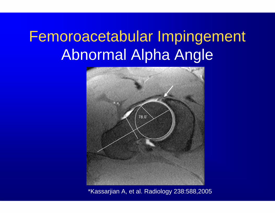
Femoroacetabular ImpingementAbnormal Alpha Angle
*Kassarjian A, et al. Radiology 238:588,2005

Hip LabrumHip Labrum
•• Static stabilizerStatic stabilizer–– Increases depth of Increases depth of
acetabulumacetabulum–– Maintains intraMaintains intra--
articulararticular pressurepressure•• Has pain receptorsHas pain receptors•• Tears do not healTears do not heal•• Tears lead to Tears lead to
cartilage stress and cartilage stress and injuryinjury
Acetabular cartilage
Femoral cartilage
Labrum

•• Fibrocartilaginous Fibrocartilaginous labrum rims outer labrum rims outer acetabulum acetabulum
•• Triangular (66%)Triangular (66%)–– Decreases in older Decreases in older
populationpopulation•• Heterogeneous SIHeterogeneous SI
–– With agingWith aging•• Absence (1Absence (1--14%) and 14%) and
sublabral sulcussublabral sulcus–– Nl variant vs. Nl variant vs.
degenerativedegenerative
Hip Labrum
Acetabular cartilag
Femoral cartilage
Labrum

Czerny C et al. AJR 1999;173:345Toomayan GA et al. AJR 2006;186:449
Labral Tear MR Arthrography
• Study of 35 patients• MR arthrography• Surgical correlation• Sensitivity
– 91%-92%• Specificity
– 71%-100%
Anterior labraltear
SagT1FS

Labral TearsLabral Tears•• Four quadrantsFour quadrants
–– AnteriorAnterior–– AnterosuperiorAnterosuperior–– PosterosuperiorPosterosuperior–– PosteriorPosterior
•• Most tears in anterior or Most tears in anterior or anterosuperior portion (3anterosuperior portion (3--11 o11 o’’clock)clock)
•• Two types of morphologyTwo types of morphology–– Detachment or avulsion Detachment or avulsion
from cartilagefrom cartilage•• Most commonMost common
–– IntrasubstanceIntrasubstance*Siebenrock KA, et al. JBJS 2003;85-A:278

MR Arthrography Technique
• 20-22 g spinal needle
• Needle tip-bone contact at femoral head-neck junction
• Aspirate, then inject small amt of iodinated contrast
• 10 cc dilute gad– .1 cc gad– 5 cc
Ropivacaine– 15 cc saline

Direct Hip MR Arthrography Technical Considerations
Image Single HipProtocol
– Coronal T1 + T1 FS– Coronal FSE PD FS
or STIR– Oblique axial T1FS
(parallel to long axis of femoral neck)
– Sagittal T1 FS Cor T1 Scout For Obl Axial Plane

Hip LabrumCoronal Plane-Ant and Post Sup
Anterosuperior Posterosuperior
Perilabral recess

Hip LabrumHip LabrumOblique Axial PlaneOblique Axial Plane--Ant & PostAnt & Post
Anterior
Posterior

*Studler U et al. Radiology 2008;249:947-954
Seen in 18% of hips
SublabralSublabral RecessRecess

Anterior and Posterior Labral Anterior and Posterior Labral Cleft Cleft
Sublabral Recess vs.TearSublabral Recess vs.Tear
Anterior Cleft Posterior Cleft

Hip LabrumHip LabrumSagittal PlaneSagittal Plane
Normal Tear
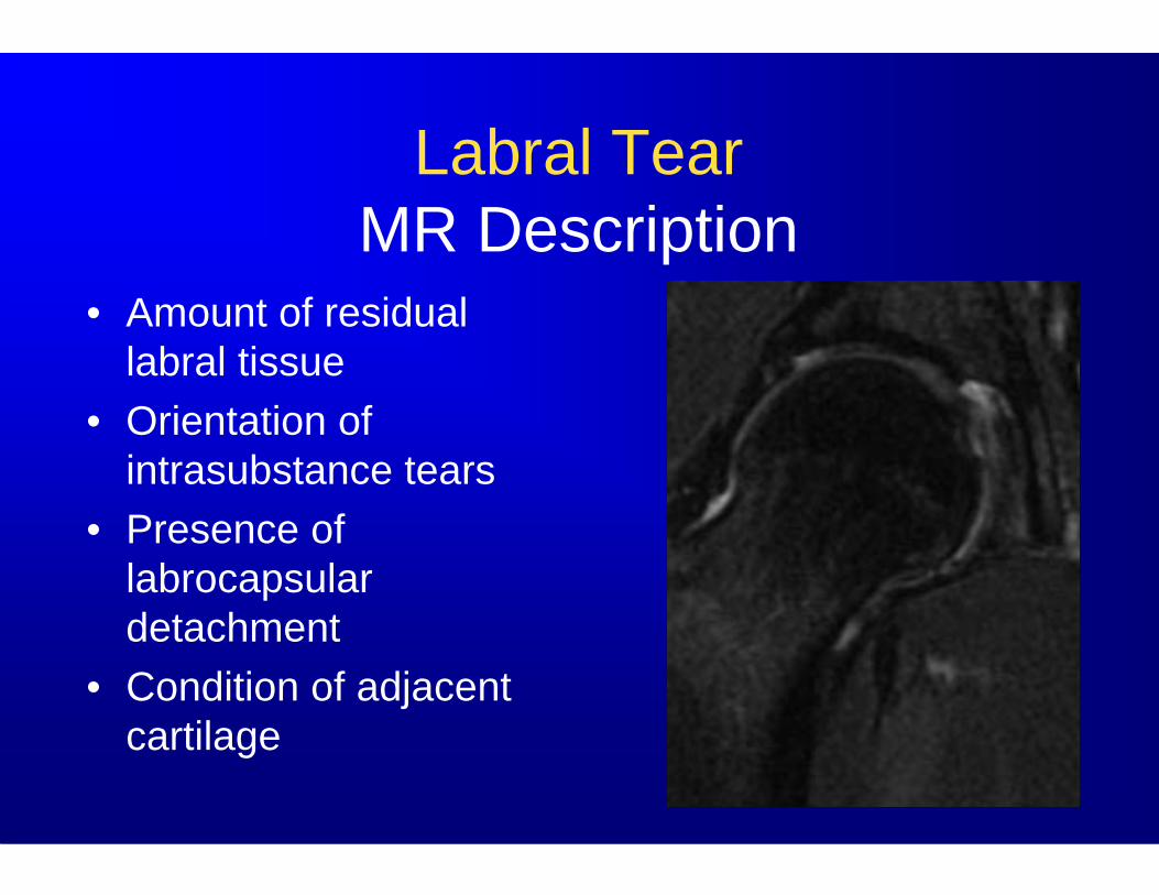
Labral Tear MR Description
• Amount of residual labral tissue
• Orientation of intrasubstance tears
• Presence of labrocapsular detachment
• Condition of adjacent cartilage

Cartilage DamageCartilage Damage
•• Underestimated by MRI Underestimated by MRI –– Sensitivity Sensitivity 4747--79%79%–– Specificity Specificity 7777--89%89%
•• DescriptionsDescriptions–– SizeSize–– LocationLocation–– Defect thicknessDefect thickness–– Subchondral bone interfaceSubchondral bone interface–– Subjacent marrow signalSubjacent marrow signal

Other Findings with FAI• Herniation pit
– 1-2 cm– Round or oval lytic
lesion– Anterior femoral neck– Some are related to
acetabular impingement• Labral ossification
– Os acetabulum– Double contour sign– Stress fracture acet rim

Other Findings with FAI• Herniation pit
– 1-2 cm– Round or oval lytic
lesion– Anterior femoral neck– Some are related to
acetabular impingement• Labral ossification
– Os Acetabulum– Double contour sign– Stress fracture acet rim
Leunig M, et al. Radiology 236;237-246

Other Findings with FAI• Herniation pit
– 1-2 cm– Round or oval lytic
lesion– Anterior femoral neck– Some are related to
acetabular impingement• Labral ossification
– Os acetabulum– Double contour sign– Stress fracture acet rim
*Ganz R et al CORR 2003

Other Findings with FAI• Herniation pit
– 1-2 cm– Round or oval lytic
lesion– Anterior femoral neck– Some are related to
acetabular impingement• Labral ossification
– Os acetabulum– Double contour sign– Stress fracture acet rim

•• Benign cystic lesion in Benign cystic lesion in acetabular roof or acetabular roof or posterolateral soft posterolateral soft tissuestissues
•• Association with Association with labral tearlabral tear
•• May erode bone, May erode bone, contain gascontain gas
•• More common with More common with hip dysplasia than FAIhip dysplasia than FAI
Paralabral Cyst

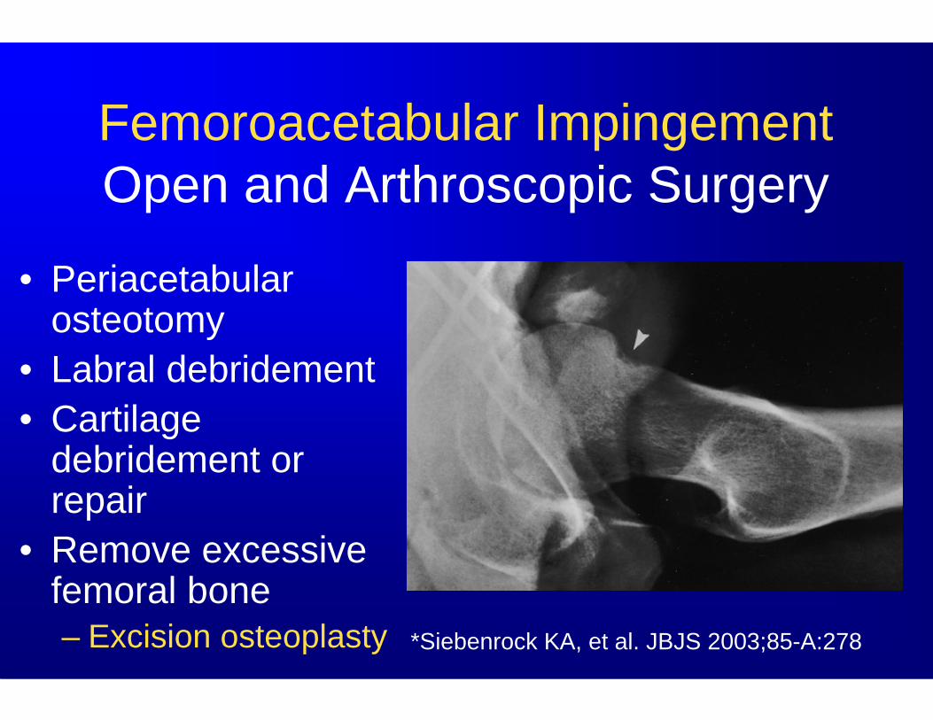
Femoroacetabular ImpingementTreatments
• Periacetabularosteotomy
• Labral debridement• Cartilage
debridement or repair
• Remove excessive femoral bone– Excision osteoplasty *Siebenrock KA, et al. JBJS 2003;85-A:278
Anterior rim
Iliac osteotomy
Correction of acetabular retroversion
Femoroacetabular ImpingementOpen and Arthroscopic Surgery
• Periacetabular osteotomy
• Labral debridement• Cartilage
debridement or repair
• Remove excessive femoral bone– Excision osteoplasty *Siebenrock KA, et al. JBJS 2003;85-A:278
Iliac osteotomy

FAI Pre-Op and Post-OpFemoral Osteoplasty
Pre-op Post-op

Post Op FAIFemoral Osteoplasty
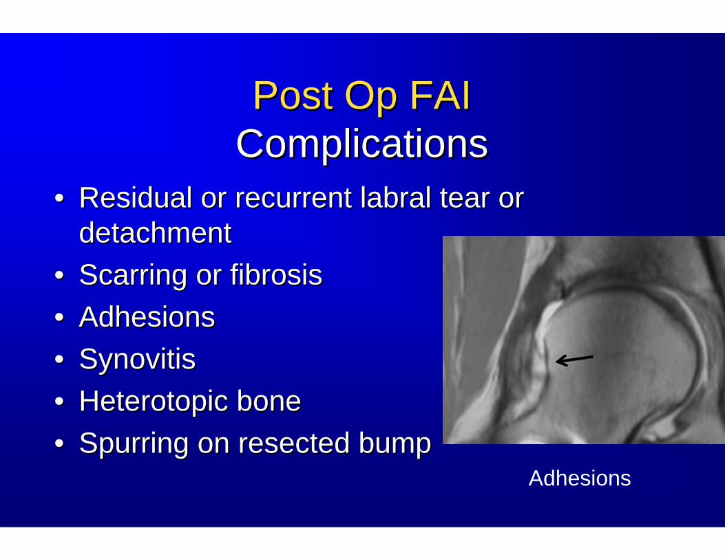
Post Op FAIPost Op FAIComplicationsComplications
•• Residual or recurrent labral tear or Residual or recurrent labral tear or detachmentdetachment
•• Scarring or fibrosisScarring or fibrosis•• AdhesionsAdhesions•• SynovitisSynovitis•• Heterotopic boneHeterotopic bone•• Spurring on resected bumpSpurring on resected bump
Adhesions

Post Op FAIPost Op FAIComplicationsComplications
•• Residual or recurrent labral tear or Residual or recurrent labral tear or detachmentdetachment
•• Scarring or fibrosisScarring or fibrosis•• AdhesionsAdhesions•• SynovitisSynovitis•• Heterotopic boneHeterotopic bone•• Spurring on resected bumpSpurring on resected bump Adhesions
Spurring

FAI Surgery ➙ THR


MRI of the HipMRI of the HipFemoroacetabular ImpingementFemoroacetabular Impingement
•• It is important for the radiologist to be aware of the features It is important for the radiologist to be aware of the features of FAIof FAI
•• FAI can cause hip pain and predisposes patients to later FAI can cause hip pain and predisposes patients to later osteoarthritisosteoarthritis
•• FAI has certain characteristics related to the femoral head FAI has certain characteristics related to the femoral head neck junction and acetabulum that are best seen on MRIneck junction and acetabulum that are best seen on MRI
•• MR arthrography aids in evaluation of the labrum and MR arthrography aids in evaluation of the labrum and cartilagecartilage
•• Open and arthroscopic surgical treatment is being widely Open and arthroscopic surgical treatment is being widely performed in the orthopedic communityperformed in the orthopedic community