mtor inhibitors control the growth of egfr mutant … inhibitors control the growth of egfr mutant...
TRANSCRIPT
mTOR Inhibitors Control the Growth of EGFR MutantLung Cancer Even after Acquiring Resistance by HGFDaisuke Ishikawa1,3, Shinji Takeuchi1, Takayuki Nakagawa1, Takako Sano1, Junya Nakade1,
Shigeki Nanjo1, Tadaaki Yamada1, Hiromichi Ebi1, Lu Zhao1, Kazuo Yasumoto1, Takahiro Nakamura2,
Kunio Matsumoto2, Hiroshi Kagamu3, Hirohisa Yoshizawa3, Seiji Yano1*
1 Division of Medical Oncology, Cancer Research Institute, Kanazawa University, Kanazawa, Japan, 2 Division of Tumor Dynamics and Regulation, Cancer Research
Institute, Kanazawa University, Kanazawa, Japan, 3 Department of Medicine (II), Niigata University Medical and Dental Hospital, Niigata City, Japan
Abstract
Resistance to epidermal growth factor receptor tyrosine kinase inhibitors (EGFR-TKIs), gefitinib and erlotinib, is a criticalproblem in the treatment of EGFR mutant lung cancer. Several mechanisms, including bypass signaling by hepatocytegrowth factor (HGF)-triggered Met activation, are implicated as mediators of resistance. The mammalian target of rapamycin(mTOR), is a downstream conduit of EGFR and MET signaling, and is thus considered a therapeutically attractive target in thetreatment of various types of cancers. The purpose of this study was to examine whether 2 clinically approved mTORinhibitors, temsirolimus and everolimus, overcome HGF-dependent resistance to EGFR-TKIs in EGFR mutant lung cancercells. Both temsirolimus and everolimus inhibited the phosphorylation of p70S6K and 4E-BP1, which are downstreamtargets of the mTOR pathway, and reduced the viability of EGFR mutant lung cancer cells, PC-9, and HCC827, even in thepresence of HGF in vitro. In a xenograft model, temsirolimus suppressed the growth of PC-9 cells overexpressing the HGF-gene; this was associated with suppression of the mTOR signaling pathway and tumor angiogenesis. In contrast, erlotinibdid not suppress this signaling pathway or tumor growth. Multiple mechanisms, including the inhibition of vascularendothelial growth factor production by tumor cells and suppression of endothelial cell viability, contribute to the anti-angiogenic effect of temsirolimus. These findings indicate that mTOR inhibitors may be useful for controlling HGF-triggeredEGFR-TKI resistance in EGFR mutant lung cancer, and they provide the rationale for clinical trials of mTOR inhibitors inpatients stratified by EGFR mutation and HGF expression status.
Citation: Ishikawa D, Takeuchi S, Nakagawa T, Sano T, Nakade J, et al. (2013) mTOR Inhibitors Control the Growth of EGFR Mutant Lung Cancer Even afterAcquiring Resistance by HGF. PLoS ONE 8(5): e62104. doi:10.1371/journal.pone.0062104
Editor: Jung Weon Lee, Seoul National University, Korea
Received November 29, 2012; Accepted March 18, 2013; Published May 14, 2013
Copyright: � 2013 Ishikawa et al. This is an open-access article distributed under the terms of the Creative Commons Attribution License, which permitsunrestricted use, distribution, and reproduction in any medium, provided the original author and source are credited.
Funding: This study was supported by KAKENHI (Grants-in-Aid for Scientific Research on Innovative Areas ‘Integrative Research on Cancer MicroenvironmentNetwork’ (to SY) and Grant-in-Aid for Young Scientists (to TY and ST)), and Grants-in-Aid for Project for Development of Innovative Research on CancerTherapeutics (P-Direct) from the Ministry of Education, Culture, Sports, Science and Technology (MEXT). The funders had no role in study design, data collectionand analysis, decision to publish, or preparation of the manuscript.
Competing Interests: Seiji Yano received honoraria from Chugai Pharmaceutical Co., Ltd. and AstraZeneca. Seiji Yano received research funding from ChugaiPharmaceutical Co., Ltd., Kyowa Hakko Kirin Co., Ltd. and Eisai Co., Ltd. This does not alter the authors’ adherence to all the PLOS ONE policies on sharing data andmaterials.
* E-mail: [email protected]
Introduction
Lung cancer is the leading cause of malignancy-related death
worldwide, and more than 80% of cases are classified as non-small
cell lung cancer (NSCLC). Epidermal growth factor receptor
(EGFR) activating mutations, such as exon 19 deletion and exon
21 L858R point mutation, are found in a population of NSCLC,
and are associated with a clinical response to the EGFR tyrosine
kinase inhibitors (EGF-TKIs), gefitinib and erlotinib [1]–[3].
However, almost all responders acquire resistance and develop
recurrence after varying periods of time (acquired resistance) [4].
In addition, 20–30% of the patients show unfavorable responses,
although their tumors have target mutations (intrinsic resistance)
[5]. Many studies have been performed in order to delineate
strategies that may overcome acquired and intrinsic resistance.
These studies have identified several mechanisms of acquired
resistance, including EGFR T790M mutation [6], [7], MET
amplification [8], [9], hepatocyte growth factor (HGF) overex-
pression [10], loss of PTEN [11], transformation to a small cell
lung cancer (SCLC) phenotype [12]–[][14], epithelial-to-mesen-
chymal transition (EMT) [15]–[][17], activation of the NFkB
pathway [18], alteration of microRNA [19], and Gas6-Axl axis
activation [20]. The complexity of NSCLC is reflected by the co-
occurrence of various combinations of these resistance mecha-
nisms in different individuals. We previously discovered that HGF
triggers EGFR-TKI resistance by activating the MET/PI3K/
AKT axis [10]. Furthermore, we showed that HGF overexpres-
sion is present in tumors from Japanese patients with acquired and
intrinsic tumor resistance to EGFR-TKI at frequencies of about
60% and 30%, respectively [21]. This indicates that HGF is an
ideal target for overcoming EGFR-TKI resistance in EGFR
mutant lung cancer patients. To overcome HGF-triggered
resistance, 2 signals from EGFR and HGF-MET should be
blocked simultaneously. We already reported that HGF-depen-
dent resistance can be controlled by an anti-HGF neutralizing
antibody [22], the HGF antagonist NK4 [22], MET-TKI [23]–
[][25], and phosphatidylinositol 3-kinase (PI3K) inhibitors [26] in
combination with EGFR-TKI. However, these inhibitors are not
PLOS ONE | www.plosone.org 1 May 2013 | Volume 8 | Issue 5 | e62104
clinically approved and therefore cannot be used for treatment of
cancer patients.
The mammalian target of rapamycin (mTOR), a serine/
threonine kinase, is a downstream target of the PI3K and AKT
pathways, and it plays a critical role in cell survival and
proliferation [27]–[][29]. Activation of PI3K/AKT and subse-
quent phosphorylation of mTOR initiates the phosphorylation of
important downstream targets, including ribosomal p70S6 serine/
threonine kinase (S6K1) and eukaryotic initiation factor (EIF)-4E
binding protein (4E-BP1), resulting in an increase in mRNA
translation and cap-dependent protein synthesis, respectively.
Thus, mTOR kinase is a key node of the PI3K/AKT signaling
pathway [27]–[][30]. To date, several mTOR inhibitor rapamycin
analogs have been developed, including temsirolimus and ever-
olimus, which have been used to treat renal cell carcinomas and
pancreatic neuroendocrine tumors. Rapamycin and its analogs
bind FK506-binding protein-12 (FKBP12) and interact with
mTOR, inhibiting its kinase activities and halting the translation
of proteins critical for cell proliferation and survival.
Because mTOR is downstream of both EGFR and MET, we
hypothesized that mTOR inhibition, even as a monotherapy
agent, may block EGFR- and MET-mediated signaling simulta-
neously and overcome HGF-triggered EGFR-TKI resistance. In
the present study, we examined whether the clinically approved
mTOR inhibitors, temsirolimus and everolimus, circumvent
HGF-triggered EGFR-TKI resistance in EGFR mutant lung
cancer cells using in vitro and in vivo models, and assessed
underlying mechanisms.
Materials and Methods
Cell cultures and reagentsEGFR mutant human lung adenocarcinoma cell lines PC-9 (del
E746_A750) and HCC827, with deletions in EGFR exon 19, were
purchased from Immuno-Biological Laboratories Co. (Takasaki,
Gunma, Japan) and the American Type Culture Collection
(Manassas, VA), respectively. Human HGF-gene transfectants (PC-
9/HGF) and vector control (PC-9/Vec) cells were established as
previously described [10]. All cell lines were maintained in RPMI
1640 medium supplemented with 10% FBS and antibiotics. All
cells were passaged for less than 3 months before renewal from
frozen, early-passage stocks. Cells were regularly screened for
mycoplasma by using MycoAlert Mycoplasma Detection Kits
(Lonza, Rockland, ME). The cell lines were authenticated at the
laboratory of the National Institute of Biomedical Innovation
(Osaka, Japan) by short tandem repeat analysis. Erlotinib,
everolimus, and temsirolimus were obtained from Selleck Chem-
icals (Houston, TX). Human recombinant HGF was prepared as
previously described [10].
Production of HGF and VEGF in cell culture supernatantsCells (26105) were cultured in 2 mL medium with 10% FBS for
24 h, washed with PBS and incubated for 48 h in medium with
10% FBS. In some experiments, HGF was added to the medium.
The culture media were harvested and centrifuged, and the
supernatants were stored at 270uC until analysis. The concen-
trations of HGF and VEGF were determined by IMMUNIS HGF
EIA (Institute of Immunology, Tokyo, Japan) and Quantikine
VEGF ELISA (R&D Systems, Minneapolis, MN), respectively,
according to the manufacturers’ protocols. All samples were run in
duplicate. Color intensity was measured at 450 nm using a
spectrophotometric plate reader. Growth factor concentrations
were determined by comparison with standard curves. The
detection limits for HGF and VEGF were 100 pg/mL and
31 pg/mL, respectively.
Cell viability assayCell viability was measured by the MTT dye reduction method.
Tumor cells, plated at 26103/100 mL RPMI 1640 medium plus
10% FBS/well in 96-well plates, were incubated for 24 h. Then,
erlotinib, temsirolimus, everolimus, and/or HGF were added to
each well, and incubation was continued for an additional 72 h.
Cell viability was measured with MTT solution (2 mg/mL; Sigma,
St. Louis, MO), as previously described [10]. Each experiment was
performed with triplicate samples.
Antibodies and western blottingProtein aliquots (25 mg each) were resolved by SDS polyacryl-
amide gel electrophoresis (Bio-Rad, Hercules, CA) and transferred
to polyvinylidene difluoride membranes (Bio-Rad). After washing
4 times, the membranes were incubated with Blocking One
(Nacalai Tesque, Inc., Kyoto, Japan) for 1 h at room temperature
(RT) and overnight at 4uC with primary antibodies to b-actin
(13E5), Met (25H2), phospho-MET (anti-p-MET, Y1234/Y1235;
3D7), p-EGFR (Y1068), AKT or p-AKT (S473), mTOR, p-
mTOR (S2448), p70S6K, pp70S6K (T389), 4E-BP1, p4E-
BP1(T37/46) (Cell Signaling Technology, Beverly, MA), human
EGFR (1 mg/mL), human/mouse/rat ERK1/ERK2 (0.2 mg/
mL), and p-ERK1/ERK2 (T202/Y204; 0.1 mg/mL) (R&D
Systems). After 3 washes, the membranes were incubated for 1 h
at RT with species-specific horseradish peroxidase–conjugated
secondary antibodies. Immunoreactive bands were visualized with
SuperSignal West Dura Extended Duration Substrate, an
enhanced chemiluminescent substrate (Pierce Biotechnology,
Rockford, IL).
Xenograft studies in SCID miceSuspensions of PC-9/Vec and PC-9/HGF cells (56106) were
subcutaneously injected into the back of 5-week-old male mice
with severe combined immunodeficiency (SCID) (Clea, Tokyo,
Japan). After 7 days, the mice were randomized to no treatment
(control group), oral erlotinib (20 mg/kg/day), or intravenous
temsirolimus (50 mg/kg/week in water). Tumor size was mea-
sured twice a week, and tumor volume was calculated (mm3) as
[(width)2 6 length]/2. All animal experiments complied with the
guidelines for the Institute for Experimental Animals, Advanced
Science Research Center, Kanazawa University, Kanazawa,
Japan (approval no. AP-081088).
Histological analysesFor the detection of proliferating tumor cells (Ki-67), 5-mm-thick
sections were deparaffinized in xylene, rehydrated in decreasing
concentrations of ethanol, and antigen-retrieved. For detection of
endothelial cells (CD31), 5-mm-thick frozen sections of xenograft
tumors were fixed with cold acetone and washed with PBS. Then,
endogenous peroxidase activity was blocked by incubation in 3%
aqueous H2O2 for 10 min. Following treatment with 5% normal
horse serum, the sections were incubated with primary antibodies
to Ki-67 (MIB-1) (Dako Cytomation, Glostrup, Denmark), and
mouse CD31 (MEC13.3) (BD Pharmingen, Franklin Lakes, NJ).
After probing with species-specific biotinylated secondary anti-
bodies, the sections were incubated for 30 min with avidin–
biotinylated peroxidase complex (ABC) using a Vectastain ABC kit
(Vector Laboratories, Burlingame, CA). The DAB (3,39-diamino-
benzidine tetrahydrochloride) Liquid System (Dako Cytomation)
was used to detect immunostaining. Omission of primary
mTOR Inhibitors in Erlotinib-Resistant Lung Cancer
PLOS ONE | www.plosone.org 2 May 2013 | Volume 8 | Issue 5 | e62104
antibodies served as a negative control. The 5 areas containing the
highest numbers of stained cells within a section were selected for
histologic quantitation by light or fluorescent microscopy at 400-
fold magnification. All results were independently evaluated by 2
two investigators (D.I. and T.N.).
Statistical analysisBetween group comparisons were assessed by two-tailed
Student’s t-tests. All analyses were performed using GraphPad
Software. A P value,0.01 was considered statistically significant.
Results
mTOR inhibitors suppress viability of EGFR mutant lungcancer cells in the presence of HGF
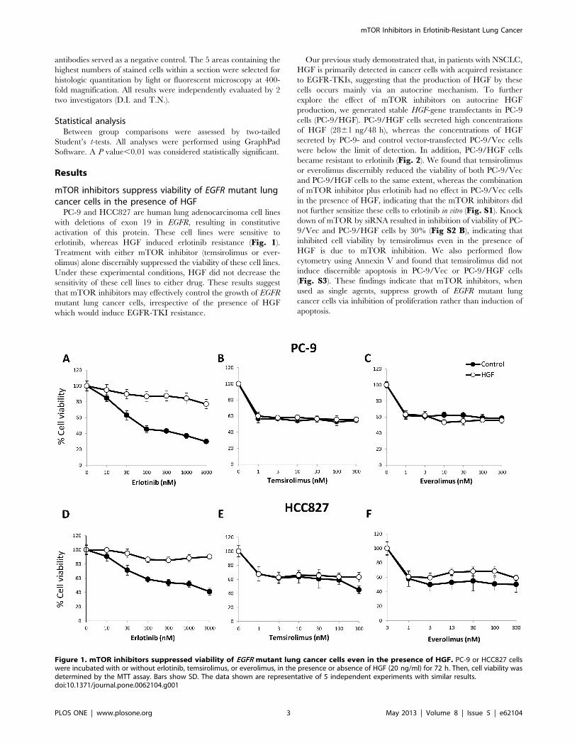
PC-9 and HCC827 are human lung adenocarcinoma cell lines
with deletions of exon 19 in EGFR, resulting in constitutive
activation of this protein. These cell lines were sensitive to
erlotinib, whereas HGF induced erlotinib resistance (Fig. 1).
Treatment with either mTOR inhibitor (temsirolimus or ever-
olimus) alone discernibly suppressed the viability of these cell lines.
Under these experimental conditions, HGF did not decrease the
sensitivity of these cell lines to either drug. These results suggest
that mTOR inhibitors may effectively control the growth of EGFR
mutant lung cancer cells, irrespective of the presence of HGF
which would induce EGFR-TKI resistance.
Our previous study demonstrated that, in patients with NSCLC,
HGF is primarily detected in cancer cells with acquired resistance
to EGFR-TKIs, suggesting that the production of HGF by these
cells occurs mainly via an autocrine mechanism. To further
explore the effect of mTOR inhibitors on autocrine HGF
production, we generated stable HGF-gene transfectants in PC-9
cells (PC-9/HGF). PC-9/HGF cells secreted high concentrations
of HGF (2861 ng/48 h), whereas the concentrations of HGF
secreted by PC-9- and control vector-transfected PC-9/Vec cells
were below the limit of detection. In addition, PC-9/HGF cells
became resistant to erlotinib (Fig. 2). We found that temsirolimus
or everolimus discernibly reduced the viability of both PC-9/Vec
and PC-9/HGF cells to the same extent, whereas the combination
of mTOR inhibitor plus erlotinib had no effect in PC-9/Vec cells
in the presence of HGF, indicating that the mTOR inhibitors did
not further sensitize these cells to erlotinib in vitro (Fig. S1). Knock
down of mTOR by siRNA resulted in inhibition of viability of PC-
9/Vec and PC-9/HGF cells by 30% (Fig S2 B), indicating that
inhibited cell viability by temsirolimus even in the presence of
HGF is due to mTOR inhibition. We also performed flow
cytometry using Annexin V and found that temsirolimus did not
induce discernible apoptosis in PC-9/Vec or PC-9/HGF cells
(Fig. S3). These findings indicate that mTOR inhibitors, when
used as single agents, suppress growth of EGFR mutant lung
cancer cells via inhibition of proliferation rather than induction of
apoptosis.
Figure 1. mTOR inhibitors suppressed viability of EGFR mutant lung cancer cells even in the presence of HGF. PC-9 or HCC827 cellswere incubated with or without erlotinib, temsirolimus, or everolimus, in the presence or absence of HGF (20 ng/ml) for 72 h. Then, cell viability wasdetermined by the MTT assay. Bars show SD. The data shown are representative of 5 independent experiments with similar results.doi:10.1371/journal.pone.0062104.g001
mTOR Inhibitors in Erlotinib-Resistant Lung Cancer
PLOS ONE | www.plosone.org 3 May 2013 | Volume 8 | Issue 5 | e62104
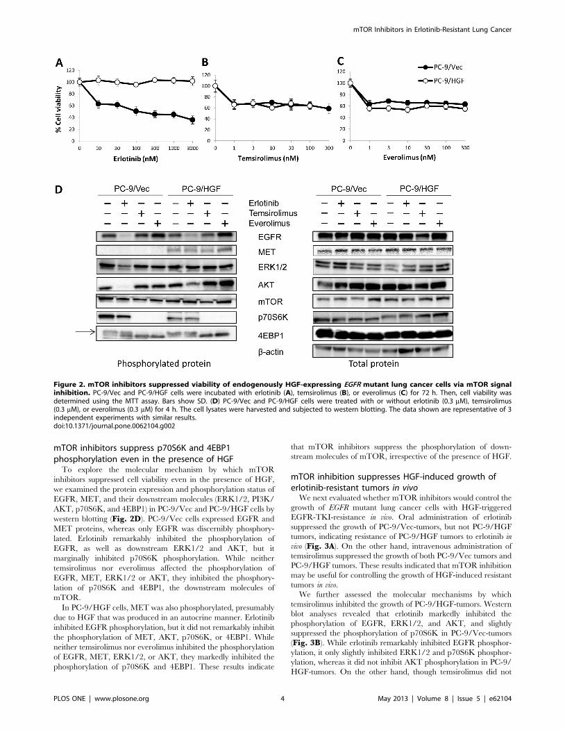
mTOR inhibitors suppress p70S6K and 4EBP1phosphorylation even in the presence of HGF
To explore the molecular mechanism by which mTOR
inhibitors suppressed cell viability even in the presence of HGF,
we examined the protein expression and phosphorylation status of
EGFR, MET, and their downstream molecules (ERK1/2, PI3K/
AKT, p70S6K, and 4EBP1) in PC-9/Vec and PC-9/HGF cells by
western blotting (Fig. 2D). PC-9/Vec cells expressed EGFR and
MET proteins, whereas only EGFR was discernibly phosphory-
lated. Erlotinib remarkably inhibited the phosphorylation of
EGFR, as well as downstream ERK1/2 and AKT, but it
marginally inhibited p70S6K phosphorylation. While neither
temsirolimus nor everolimus affected the phosphorylation of
EGFR, MET, ERK1/2 or AKT, they inhibited the phosphory-
lation of p70S6K and 4EBP1, the downstream molecules of
mTOR.
In PC-9/HGF cells, MET was also phosphorylated, presumably
due to HGF that was produced in an autocrine manner. Erlotinib
inhibited EGFR phosphorylation, but it did not remarkably inhibit
the phosphorylation of MET, AKT, p70S6K, or 4EBP1. While
neither temsirolimus nor everolimus inhibited the phosphorylation
of EGFR, MET, ERK1/2, or AKT, they markedly inhibited the
phosphorylation of p70S6K and 4EBP1. These results indicate
that mTOR inhibitors suppress the phosphorylation of down-
stream molecules of mTOR, irrespective of the presence of HGF.
mTOR inhibition suppresses HGF-induced growth oferlotinib-resistant tumors in vivo
We next evaluated whether mTOR inhibitors would control the
growth of EGFR mutant lung cancer cells with HGF-triggered
EGFR-TKI-resistance in vivo. Oral administration of erlotinib
suppressed the growth of PC-9/Vec-tumors, but not PC-9/HGF
tumors, indicating resistance of PC-9/HGF tumors to erlotinib in
vivo (Fig. 3A). On the other hand, intravenous administration of
temsirolimus suppressed the growth of both PC-9/Vec tumors and
PC-9/HGF tumors. These results indicated that mTOR inhibition
may be useful for controlling the growth of HGF-induced resistant
tumors in vivo.
We further assessed the molecular mechanisms by which
temsirolimus inhibited the growth of PC-9/HGF-tumors. Western
blot analyses revealed that erlotinib markedly inhibited the
phosphorylation of EGFR, ERK1/2, and AKT, and slightly
suppressed the phosphorylation of p70S6K in PC-9/Vec-tumors
(Fig. 3B). While erlotinib remarkably inhibited EGFR phosphor-
ylation, it only slightly inhibited ERK1/2 and p70S6K phosphor-
ylation, whereas it did not inhibit AKT phosphorylation in PC-9/
HGF-tumors. On the other hand, though temsirolimus did not
Figure 2. mTOR inhibitors suppressed viability of endogenously HGF-expressing EGFR mutant lung cancer cells via mTOR signalinhibition. PC-9/Vec and PC-9/HGF cells were incubated with erlotinib (A), temsirolimus (B), or everolimus (C) for 72 h. Then, cell viability wasdetermined using the MTT assay. Bars show SD. (D) PC-9/Vec and PC-9/HGF cells were treated with or without erlotinib (0.3 mM), temsirolimus(0.3 mM), or everolimus (0.3 mM) for 4 h. The cell lysates were harvested and subjected to western blotting. The data shown are representative of 3independent experiments with similar results.doi:10.1371/journal.pone.0062104.g002
mTOR Inhibitors in Erlotinib-Resistant Lung Cancer
PLOS ONE | www.plosone.org 4 May 2013 | Volume 8 | Issue 5 | e62104
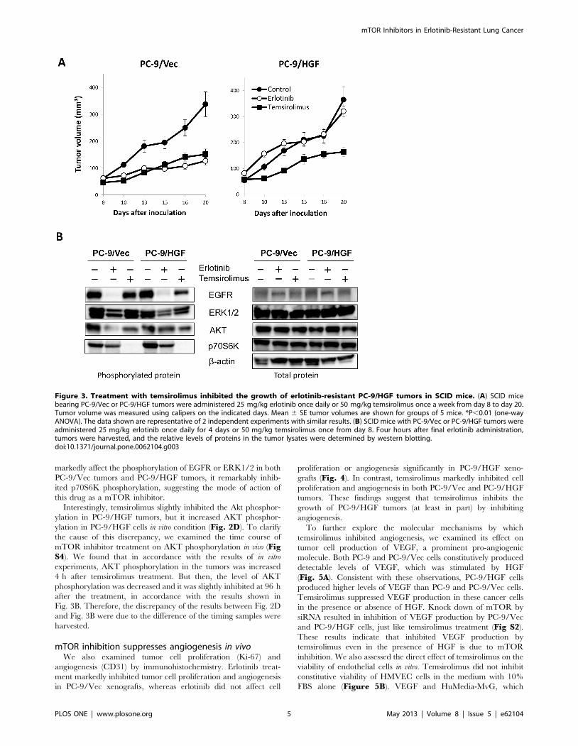
markedly affect the phosphorylation of EGFR or ERK1/2 in both
PC-9/Vec tumors and PC-9/HGF tumors, it remarkably inhib-
ited p70S6K phosphorylation, suggesting the mode of action of
this drug as a mTOR inhibitor.
Interestingly, temsirolimus slightly inhibited the Akt phosphor-
ylation in PC-9/HGF tumors, but it increased AKT phosphor-
ylation in PC-9/HGF cells in vitro condition (Fig. 2D). To clarify
the cause of this discrepancy, we examined the time course of
mTOR inhibitor treatment on AKT phosphorylation in vivo (FigS4). We found that in accordance with the results of in vitro
experiments, AKT phosphorylation in the tumors was increased
4 h after temsirolimus treatment. But then, the level of AKT
phosphorylation was decreased and it was slightly inhibited at 96 h
after the treatment, in accordance with the results shown in
Fig. 3B. Therefore, the discrepancy of the results between Fig. 2D
and Fig. 3B were due to the difference of the timing samples were
harvested.
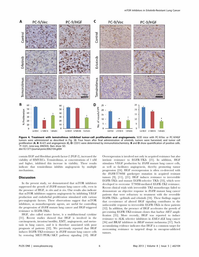
mTOR inhibition suppresses angiogenesis in vivoWe also examined tumor cell proliferation (Ki-67) and
angiogenesis (CD31) by immunohistochemistry. Erlotinib treat-
ment markedly inhibited tumor cell proliferation and angiogenesis
in PC-9/Vec xenografts, whereas erlotinib did not affect cell
proliferation or angiogenesis significantly in PC-9/HGF xeno-
grafts (Fig. 4). In contrast, temsirolimus markedly inhibited cell
proliferation and angiogenesis in both PC-9/Vec and PC-9/HGF
tumors. These findings suggest that temsirolimus inhibits the
growth of PC-9/HGF tumors (at least in part) by inhibiting
angiogenesis.
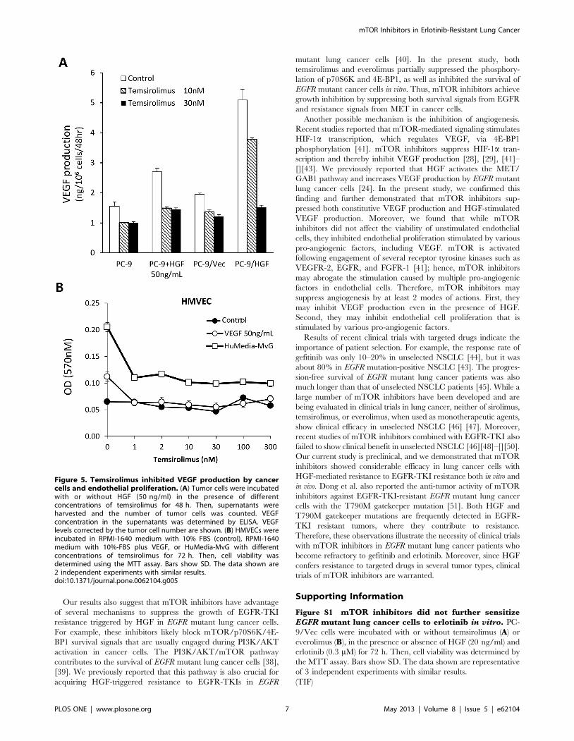
To further explore the molecular mechanisms by which
temsirolimus inhibited angiogenesis, we examined its effect on
tumor cell production of VEGF, a prominent pro-angiogenic
molecule. Both PC-9 and PC-9/Vec cells constitutively produced
detectable levels of VEGF, which was stimulated by HGF
(Fig. 5A). Consistent with these observations, PC-9/HGF cells
produced higher levels of VEGF than PC-9 and PC-9/Vec cells.
Temsirolimus suppressed VEGF production in these cancer cells
in the presence or absence of HGF. Knock down of mTOR by
siRNA resulted in inhibition of VEGF production by PC-9/Vec
and PC-9/HGF cells, just like temsirolimus treatment (Fig S2).
These results indicate that inhibited VEGF production by
temsirolimus even in the presence of HGF is due to mTOR
inhibition. We also assessed the direct effect of temsirolimus on the
viability of endothelial cells in vitro. Temsirolimus did not inhibit
constitutive viability of HMVEC cells in the medium with 10%
FBS alone (Figure 5B). VEGF and HuMedia-MvG, which
Figure 3. Treatment with temsirolimus inhibited the growth of erlotinib-resistant PC-9/HGF tumors in SCID mice. (A) SCID micebearing PC-9/Vec or PC-9/HGF tumors were administered 25 mg/kg erlotinib once daily or 50 mg/kg temsirolimus once a week from day 8 to day 20.Tumor volume was measured using calipers on the indicated days. Mean 6 SE tumor volumes are shown for groups of 5 mice. *P,0.01 (one-wayANOVA). The data shown are representative of 2 independent experiments with similar results. (B) SCID mice with PC-9/Vec or PC-9/HGF tumors wereadministered 25 mg/kg erlotinib once daily for 4 days or 50 mg/kg temsirolimus once from day 8. Four hours after final erlotinib administration,tumors were harvested, and the relative levels of proteins in the tumor lysates were determined by western blotting.doi:10.1371/journal.pone.0062104.g003
mTOR Inhibitors in Erlotinib-Resistant Lung Cancer
PLOS ONE | www.plosone.org 5 May 2013 | Volume 8 | Issue 5 | e62104
contain EGF and fibroblast growth factor-2 (FGF-2), increased the
viability of HMVECs. Temsirolimus, at concentrations of 1 nM
and higher, inhibited this increase in viability. These results
indicate that temsirolimus inhibits angiogenesis by multiple
mechanisms.
Discussion
In the present study, we demonstrated that mTOR inhibitors
suppressed the growth of EGFR mutant lung cancer cells, even in
the presence of HGF, in vitro and in vivo. Our results also indicate
that mTOR inhibitors suppress angiogenesis by inhibiting VEGF
production and endothelial proliferation stimulated with various
pro-angiogenic factors. These observations suggest that mTOR
inhibitors, as monotherapeutic agents, are useful for controlling
the progression of EGFR mutant lung cancer and HGF-triggered
resistance to EGFR-TKIs.
HGF, also called scatter factor, is a multifunctional cytokine
[31]. Recent studies showed that HGF is involved in the
carcinogenesis, invasion/motility, EMT, angiogenesis, and metas-
tasis in lung cancer, and it is therefore associated with poor
prognosis of patients [32]. We previously reported that HGF
induces EGFR-TKI-resistance in EGFR mutant lung cancer cells
by restoring MET/PI3K/AKT pathway signaling [10]. HGF
Overexpression is involved not only in acquired resistance but also
intrinsic resistance to EGFR-TKIs [21]. In addition, HGF
stimulates VEGF production by EGFR mutant lung cancer cells,
as well as facilitates angiogenesis, thereby promoting tumor
progression [24]. HGF overexpression is often co-detected with
the EGFR-T790M gatekeeper mutation in acquired resistant
tumors [9], [11], [21]. HGF induces resistance to irreversible
EGFR-TKIs and mutant EGFR-selective TKIs [33], which were
developed to overcome T790M-mediated EGFR-TKI resistance.
Recent clinical trials with irreversible TKI monotherapy failed to
demonstrate an objective response in EGFR mutant lung cancer
patients that were refractory to treatment with the reversible
EGFR-TKIs—gefitinib and erlotinib [34]. These findings suggest
that co-existence of altered HGF signaling contributes to the
unfavorable response to irreversible EGFR-TKIs in these patients
[32]. In addition, the presence of HGF accelerates the growth of
pre-existing EGFR-TKI-resistant clones that harbor MET ampli-
fication [35]. More recently, HGF was reported to induce
resistance to ALK selective inhibitors in EML4-ALK lung cancer
[36] and BRAF inhibitors in BRAF mutant melanoma [37]. Such
accumulating evidence indicates that HGF is a common target for
overcoming resistance to targeted drugs in oncogene-addicted
tumors.
Figure 4. Treatment with temsirolimus inhibited tumor-cell proliferation and angiogenesis. SCID mice with PC-9/Vec or PC-9/HGFtumors were administered as described in Fig. 3B. Four hours after final administration of erlotinib, tumors were harvested, and tumor cellproliferation (A, B: Ki-67) and angiogenesis (C, D: CD31) were determined by immunohistochemistry. B and D show quantification of positive cells.*P,0.01, (one-way ANOVA). Bars show SD.doi:10.1371/journal.pone.0062104.g004
mTOR Inhibitors in Erlotinib-Resistant Lung Cancer
PLOS ONE | www.plosone.org 6 May 2013 | Volume 8 | Issue 5 | e62104
Our results also suggest that mTOR inhibitors have advantage
of several mechanisms to suppress the growth of EGFR-TKI
resistance triggered by HGF in EGFR mutant lung cancer cells.
For example, these inhibitors likely block mTOR/p70S6K/4E-
BP1 survival signals that are usually engaged during PI3K/AKT
activation in cancer cells. The PI3K/AKT/mTOR pathway
contributes to the survival of EGFR mutant lung cancer cells [38],
[39]. We previously reported that this pathway is also crucial for
acquiring HGF-triggered resistance to EGFR-TKIs in EGFR
mutant lung cancer cells [40]. In the present study, both
temsirolimus and everolimus partially suppressed the phosphory-
lation of p70S6K and 4E-BP1, as well as inhibited the survival of
EGFR mutant cancer cells in vitro. Thus, mTOR inhibitors achieve
growth inhibition by suppressing both survival signals from EGFR
and resistance signals from MET in cancer cells.
Another possible mechanism is the inhibition of angiogenesis.
Recent studies reported that mTOR-mediated signaling stimulates
HIF-1a transcription, which regulates VEGF, via 4E-BP1
phosphorylation [41]. mTOR inhibitors suppress HIF-1a tran-
scription and thereby inhibit VEGF production [28], [29], [41]–
[][43]. We previously reported that HGF activates the MET/
GAB1 pathway and increases VEGF production by EGFR mutant
lung cancer cells [24]. In the present study, we confirmed this
finding and further demonstrated that mTOR inhibitors sup-
pressed both constitutive VEGF production and HGF-stimulated
VEGF production. Moreover, we found that while mTOR
inhibitors did not affect the viability of unstimulated endothelial
cells, they inhibited endothelial proliferation stimulated by various
pro-angiogenic factors, including VEGF. mTOR is activated
following engagement of several receptor tyrosine kinases such as
VEGFR-2, EGFR, and FGFR-1 [41]; hence, mTOR inhibitors
may abrogate the stimulation caused by multiple pro-angiogenic
factors in endothelial cells. Therefore, mTOR inhibitors may
suppress angiogenesis by at least 2 modes of actions. First, they
may inhibit VEGF production even in the presence of HGF.
Second, they may inhibit endothelial cell proliferation that is
stimulated by various pro-angiogenic factors.
Results of recent clinical trials with targeted drugs indicate the
importance of patient selection. For example, the response rate of
gefitinib was only 10–20% in unselected NSCLC [44], but it was
about 80% in EGFR mutation-positive NSCLC [43]. The progres-
sion-free survival of EGFR mutant lung cancer patients was also
much longer than that of unselected NSCLC patients [45]. While a
large number of mTOR inhibitors have been developed and are
being evaluated in clinical trials in lung cancer, neither of sirolimus,
temsirolimus, or everolimus, when used as monotherapeutic agents,
show clinical efficacy in unselected NSCLC [46] [47]. Moreover,
recent studies of mTOR inhibitors combined with EGFR-TKI also
failed to show clinical benefit in unselected NSCLC [46][48]–[][50].
Our current study is preclinical, and we demonstrated that mTOR
inhibitors showed considerable efficacy in lung cancer cells with
HGF-mediated resistance to EGFR-TKI resistance both in vitro and
in vivo. Dong et al. also reported the anti-tumor activity of mTOR
inhibitors against EGFR-TKI-resistant EGFR mutant lung cancer
cells with the T790M gatekeeper mutation [51]. Both HGF and
T790M gatekeeper mutations are frequently detected in EGFR-
TKI resistant tumors, where they contribute to resistance.
Therefore, these observations illustrate the necessity of clinical trials
with mTOR inhibitors in EGFR mutant lung cancer patients who
become refractory to gefitinib and erlotinib. Moreover, since HGF
confers resistance to targeted drugs in several tumor types, clinical
trials of mTOR inhibitors are warranted.
Supporting Information
Figure S1 mTOR inhibitors did not further sensitizeEGFR mutant lung cancer cells to erlotinib in vitro. PC-
9/Vec cells were incubated with or without temsirolimus (A) or
everolimus (B), in the presence or absence of HGF (20 ng/ml) and
erlotinib (0.3 mM) for 72 h. Then, cell viability was determined by
the MTT assay. Bars show SD. The data shown are representative
of 3 independent experiments with similar results.
(TIF)
Figure 5. Temsirolimus inhibited VEGF production by cancercells and endothelial proliferation. (A) Tumor cells were incubatedwith or without HGF (50 ng/ml) in the presence of differentconcentrations of temsirolimus for 48 h. Then, supernatants wereharvested and the number of tumor cells was counted. VEGFconcentration in the supernatants was determined by ELISA. VEGFlevels corrected by the tumor cell number are shown. (B) HMVECs wereincubated in RPMI-1640 medium with 10% FBS (control), RPMI-1640medium with 10%-FBS plus VEGF, or HuMedia-MvG with differentconcentrations of temsirolimus for 72 h. Then, cell viability wasdetermined using the MTT assay. Bars show SD. The data shown are2 independent experiments with similar results.doi:10.1371/journal.pone.0062104.g005
mTOR Inhibitors in Erlotinib-Resistant Lung Cancer
PLOS ONE | www.plosone.org 7 May 2013 | Volume 8 | Issue 5 | e62104
Figure S2 Knock-down of mTOR inhibited cell growthand VEGF production. Tumor cells were transfected with
mTOR or control siRNA for 24 h and (A) the cell lysates were
harvested and subjected to western blotting. (B) cell viability was
determined by the MTT assay, (C) VEGF concentration in the
supernatants further 48 h after transfection, was determined by
ELISA. C shows VEGF levels corrected by the tumor cell number
are shown. B and C show quantification of positive cells. *P,0.01,
(one-way ANOVA). Bars show SD. The data shown are
representative of 5 independent experiments with similar results.
(TIF)
Figure S3 Temsirolimus did not induce apoptosis inPC-9 cells in vitro, irrespective of the presence of HGF.PC-9/Vec and PC-9/HGF cells were treated with cisplatin (as a
positive control for apoptosis), erlotinib, or temsirolimus for 48 h.
Then, the resultant cells were treated with PE Annexin V
Apoptosis Detection Kit I.
(TIF)
Figure S4 Treatment with temsirolimus increased AKTphosphorylation after 4 h treatment, but the phosphor-ylation was reduced at 96 h. SCID mice with PC-9/HGF
tumors were administered 50 mg/kg temsirolimus. After 4 h,
12 h, 48 h and 96 h, the tumors were harvested, and the relative
levels of proteins in the tumor lysates were determined by western
blotting.
(TIF)
Acknowledgments
We thank Mr Kenji Kita for technical assistance of experiments.
Author Contributions
Conceived and designed the experiments: ST TY HE KY T. Nakamura
KM HK HY. Performed the experiments: DI T. Nakagawa TS JN SN LZ.
Wrote the paper: SY.
References
1. Lynch TJ, Bell DW, Sordella R, Gurubhagavatula S, Okimoto RA, et al. (2004)
Activating mutations in the epidermal growth factor receptor underlyingresponsiveness of non-small-cell lung cancer to gefitinib. N Engl J Med 350:
2129–2139.
2. Paez JG, Janne PA, Lee JC, Tracy S, Greulich H, et al. (2004) EGFR mutationsin lung cancer: correlation with clinical response to gefitinib therapy. Science
304: 1497–1500.
3. Pao W, Miller V, Zakowski M, Doherty J, Politi K, et al. (2004) EGF receptorgene mutations are common in lung cancers from ‘‘never smokers’’ and are
associated with sensitivity of tumors to gefitinib and erlotinib. Proc Natl AcadSci U S A 101: 13306–13311.
4. Jackman D, Pao W, Riely GJ, Engelman JA, Kris MG, et al. (2010) Clinical
definition of acquired resistance to epidermal growth factor receptor tyrosinekinase inhibitors in non-small-cell lung cancer. J Clin Oncol 28: 357–360.
5. Pao W, Chmielecki J (2010) Rational, biologically based treatment of EGFR-
mutant non-small-cell lung cancer. Nat Rev Cancer 10:760–774.
6. Kobayashi S, Boggon TJ, Dayaram T, Janne PA, Kocher O, et al. (2005) EGFR
mutation and resistance of non-small-cell lung cancer to gefitinib. N Engl J Med352: 786–792.
7. Pao W, Miller VA, Politi KA, Riely GJ, Somwar R, et al. (2005) Acquired
resistance of lung adenocarcinomas to gefitinib or erlotinib is associated with asecond mutation in the EGFR kinase domein. PLoS Med 2: e73.
8. Engelman JA, Zejnullahu K, Mitsudomi T, Song Y, Hyland C, et al. (2007)MET amplification leads to gefitinib resistance in lung cancer by activating
ERBB3 signaling. Science 316: 1039–1043.
9. Turke AB, Zejnullahu K, Wu YL, Song Y, Dias-Santagata D, et al. (2010)Preexistence and clonal selection of MET amplification in EGFR mutant
NSCLC. Cancer Cell 17; 174–183.
10. Yano S, Wang W, Li Q, Matsumoto K, Sakurama H, et al. (2008) Hepatocytegrowth factor induces gefitinib resistance of lung adenocarcinoma cells with EGF
receptor mutation. Cancer Res 68: 9479–9487.
11. Uramoto H, Shimokawa H, Hanagiri T, Kuwano M, Ono M (2011) Expression
of selected gene for acquired drug resistance to EGFR-TKI in lung
adenocarcinoma. Lung Cancer 73:361–365.
12. Zakowski MF, Ladanyi M, Kris MG (2006) EGFR mutations in small-cell lung
cancers in patients who have never smoked. N Engl J Med. 355:213–215.
13. Morinaga R, Okamoto I, Furuta K, Kawano Y, Sekijima M, et al. (2007)Sequential occurrence of non-small cell and small cell lung cancer with the same
EGFR mutation. Lung Cancer 58:411–413.
14. Shiao TH, Chang YL, Yu CJ, Chang YC, Hsu YC, et al. (2008) Epidermal
growth factor receptor mutations in small cell lung cancer. Clin Cancer Res
14:6092–6096.
15. Frederick BA, Helfrich BA, Coldren CD, Zheng D, Chan D, et al. (2007)
Epithelial to mesenchymal transition predicts gefitinib resistance in cell lines of
head and neck squamous cell carcinoma and non-small cell lung carcinoma. MolCancer Ther 6:1683–1691.
16. Uramoto H, Iwata T, Onitsuka T, Shimokawa H, Hanagiri T, et al. (2010)Epithelial-mesenchymal transition in EGFR-TKI acquired resistant lung
adenocarcinoma. Anticancer Res 30:2513–2517.
17. Suda K, Tomizawa K, Fujii M, Murakami H, Osada H, et al. (2011) MaeharaY, Yatabe Y, Sekido Y, Mitsudomi T. Epithelial to mesenchymal transition in an
epidermal growth factor receptor-mutant lung cancer cell line with acquired
resistance to erlotinib. J Thorac Oncol 6:1152–1161.
18. Bivona TG, Hieronymus H, Parker J, Chang K, Taron M, et al. (2011) FAS and
NF-kB signalling modulate dependence of lung cancers on mutant EGFR.Nature 471:523–526.
19. Garofalo M, Romano G, Di Leva G, Nuovo G, Jeon YJ, et al. (2011) EGFR and
MET receptor tyrosine kinase-altered microRNA expression induces tumori-genesis and gefitinib resistance in lung cancers. Nat Med 18:74–82.
20. Zhang Z, Lee JC, Lin L, Olivas V, Au V, et al. (2012) Activation of the AXL
kinase causes resistance to EGFR-targeted therapy in lung cancer. Nat Genet.44:852–860.
21. Yano S, Yamada T, Takeuchi S, Tachibana K, Minami Y, et al. (2011)
Hepatocyte growth factor expression in EGFR mutant lung cancer with intrinsicand acquired resistance to tyrosine kinase inhibitors in a Japanese cohort.
J Thorac Oncol 6:2011–2017.
22. Wang W, Li Q, Yamada T, Matsumoto K, Matsumoto I, et al. (2009) Crosstalk
to stromal fibroblasts induces resistance of lung cancer to epidermal growth
factor receptor tyrosine kinase inhibitors. Clin Cancer Res 15:6630–6638.
23. Wang W, Li Q, Takeuchi S, Yamada T, Koizumi H, et al. (2012) Met kinase
inhibitor E7050 reverses three different mechanisms of hepatocyte growth
factor-induced tyrosine kinase inhibitor resistance in EGFR mutant lung cancer.Clin Cancer Res 18:1663–1671.
24. Takeuchi S, Wang W, Li Q, Yamada T, Kita K, et al. (2012) Dual inhibition ofMet kinase and angiogenesis to overcome HGF-induced EGFR-TKI resistance
in EGFR mutant lung cancer. Am J Pathol 181:1034–1043.
25. Nakagawa T, Takeuchi S, Yamada T, Nanjo S, Ishikawa D, et al. (2012)Combined therapy with mutant-selective EGFR inhibitor and Met kinase
inhibitor for overcoming erlotinib resistance in EGFR-mutant lung cancer. Mol
Cancer Ther 11:2149–2157.
26. Donev IS, Wang W, Yamada T, Li Q, Takeuchi S, et al. (2011) Transient PI3K
inhibition induces apoptosis and overcomes HGF-mediated resistance to EGFR-TKIs in EGFR mutant lung cancer. Clin Cancer Res 17:2260–2269.
27. Helena P, Jose ML, Paula S (2012) The mTOR signalling pathway in human
cancer. Int J Mol Sci 13, 1886–1918
28. Xiaodong F, William O, David H (2010) Antiangiogenesis therapy: a new
strategy for cancer treatment. US Pharm. 35(Oncology suppl):4–9
29. Jayashree K, Amit M (2011) PI3K/AKT/mTOR pathway in angiogenesis.
Front Mol Neurosci 4:51.
30. Hudson CC, Liu M, Chiang GG, Otterness DM, Loomis DC, et al. (2003)Regulation of hypoxia-inducible factor 1: expression and function by the
mammalian target of rapamycin. Mol Cell 11:1491–1501.
31. Takebayashi T, Iwamoto M, Jikko A, Matsumura T, Enomoto-Iwamoto M, etal. (1995) Hepatocyte growth factor/scatter factor modulates cell motility,
proliferation, and proteoglycan synthesis of chondrocytes. J Cell Biol 129:1411–1419.
32. Yano S, Takeuchi S, Nakagawa T, Yamada T (2012) Ligand-triggered
resistance to molecular targeted drugs in lung cancer: Roles of hepatocytegrowth factor and epidermal growth factor receptor ligands. Cancer Sci 103
:1189–1194.
33. Yamada T, Matsumoto K, Wang W, Li Q, Nishioka Y, et al. (2010) Hepatocytegrowth factor reduces susceptibility to an irreversible epidermal growth factor
receptor inhibitor in EGFR-T790M mutant lung cancer. Clin Cancer Res16:174–183.
34. Yasuda H, Kobayashi S, Costa DB (2012) EGFR exon 20 insertion mutations in
non-small-cell lung cancer: preclinical data and clinical implications. LancetOncol. 2012;13:e23–31.
35. Turke AB, Zejnullahu K, Wu YL, Song Y, Dias-Santagata D, et al. (2010)
Preexistence and clonal selection of MET amplification in EGFR mutantNSCLC. Cancer Cell 17:77–88.
36. Yamada T, Takeuchi S, Nakade J, Kita K, Nakagawa T, et al. (2012) Paracrinereceptor activation by microenvironment triggers bypass survival signals and
mTOR Inhibitors in Erlotinib-Resistant Lung Cancer
PLOS ONE | www.plosone.org 8 May 2013 | Volume 8 | Issue 5 | e62104
ALK inhibitor resistance in EML4-ALK lung cancer cells. Clin Cancer Res
18:3592–3602.
37. Straussman R, Morikawa T, Shee K, Barzily-Rokni M, Qian ZR, et al. (2012)
Tumour micro-environment elicits innate resistance to RAF inhibitors through
HGF secretion. Nature 487:500–504.
38. Endoh H, Yatabe Y, Kosaka T, Kuwano H, Mitsudomi T (2006) PTEN and
PIK3CA expression is associated with prolonged survival after gefitinib
treatment in EGFR-mutated lung cancer patients. J Thorac Oncol 1:629–634.
39. Oh MH, Lee HJ, Yoo SB, Xu X, Choi JS, et al. (2012) Clinicopathological
correlations of mTOR and pAkt expression in non-small cell lung cancer.
Virchows Arch 460:601–609.
40. Donev IS, Wang W, Yamada T, Li Q, Takeuchi S, et al. (2011) Transient PI3K
inhibition induces apoptosis and overcomes HGF-mediated resistance to EGFR-
TKIs in EGFR mutant lung cancer. Clin Cancer Res 17:2260–2269.
41. Yee Koh M, Spivak-Kroizman TR, Powis G (2008) HIF-1 regulation: not so
easy come, easy go. Trends Biochem Sci 33:526–534.
42. Samy LH, Anamika Y, Lenin M, Anthony JV (2011) Regulation of PI 3-K,
PTEN, p53, and mTOR in malignant and benign tumors deficient in tuberin.
Genes Cancer 2: 1051–1060.
43. Young RM, Wang SJ, Gordan JD, Ji X, Liebhaber SA, et al. (2008) Hypoxia-
mediated selective mRNA translation by an internal ribosome entry site-
independent mechanism. J Biol Chem 283:16309–16319.
44. Fukuoka M, Yano S, Giaccone G, Tamura T, Nakagawa K, et al. (2003) Multi-
institutional randomized phase II trial of gefitinib for previously treated patients
with advanced non-small-cell lung cancer (The IDEAL 1 Trial). J Clin Oncol.
21:2237–2246.45. Mok TS, Wu YL, Thongprasert S, Yang CH, Chu DT, et al. (2009) Gefitinib or
carboplatin-paclitaxel in pulmonary adenocarcinoma. N Engl J Med. 361:947–
957.46. Ekman S, Wynes MW, Hirsch FR (2012) The mTOR pathway in lung cancer
and implications for therapy and biomarker analysis. J Thorac Oncol 7:947–953.
47. Reungwetwattana T, Molina JR, Mandrekar SJ, Allen-ZieglErk, Rowland KM,
et al. (2012) Brief report: a phase II ‘‘window-of-opportunity’’ frontline study ofthe mTOR inhibitor, temsirolimus given as a single agent in patients with
advanced NSCLC, an NCCTG study. J Thorac Oncol 7:919–922.48. Price KA, Azzoli CG, Krug LM, Pietanza MC, Rizvi NA, et al. (2012) Phase II
trial of gefitinib and everolimus in advanced non-small cell lung cancer. J ThoracOncol. 1 :1623–1629.
49. Leighl NB, Soria J, Bennouna J, Blais N, Traynor AM, et al. (2012) Phase II
study of everolimus plus erlotinib in previously treated patients with advancednon-small cell lung cancer (NSCLC). J Clin Oncol 28(15 suppl):7524.
50. Papadimitrakopoulou VA, Soria JC, Jappe A, Jehl V, Klimovsky J, et al. (2012)Everolimus and erlotinib as second- or third-line therapy in patients with
advanced non-small-cell lung cancer. J Thorac Oncol 7:1594–1601.
51. Dong S, Zhang XC, Cheng H, Zhu JQ, Chen ZH, et al. (2012) Everolimussynergizes with gefitinib in non-small-cell lung cancer cell lines resistant to
epidermal growth factor receptor tyrosine kinase inhibitors. Cancer ChemotherPharmacol 70:707–716.
mTOR Inhibitors in Erlotinib-Resistant Lung Cancer
PLOS ONE | www.plosone.org 9 May 2013 | Volume 8 | Issue 5 | e62104