multifaceted approach to ct dose reduction for “rule-out ... › uploadedfiles › rsna ›...
TRANSCRIPT

1
Multifaceted Approach to CT Dose
Reduction for “Rule-Out Aortic
Dissection”
Exhibit ID 14002378
Judah Goldschmiedta, Sharon Steinbergera, Esther Mizrachia, David Essesb,
Jeffrey M. Levskya, Linda B. Haramatia
a Department of Radiology, Montefiore Medical Center, Bronx, New York
b Department of Emergency Medicine, Montefiore Medical Center, Bronx, New York
Background
• Acute Aortic Syndromes
– Aortic Dissection
– Intramural hematoma
– Penetrating atherosclerotic ulcer
– Aortic rupture
• Incidence reported 5-30 / 1 million per year

2
Acute Aortic Syndromes
• Mortality
– 20% before hospitalization
– 20% during hospital admission
– 20% over the next 10 years
• Difficulty in clinical diagnosis
– Signs and symptoms lack sensitivity and specificity for AAS
– Correctly suspected in only 15-43% of cases
Research Background
• IRB approved retrospective review of all patients referred for evaluation of AAS within the Montefiore Medical Center enterprise from 1/1/06 – 8/1/10
• Analysis focused on clinical and radiographic parameters associated with AAS
• Models of proposed clinical algorithms suggested and performance evaluated

3
Research Background
• Based on retrospective data available, clinical
algorithms for appropriate imaging were
developed
• Performance of these proposed models was
calculated

4
Research Background: Results
• Internal audit of CT use for indication of AAS
illuminated a system wide problem:
– Large population radiation burden
– Multiphase CT protocol
– Overall, low incidence of AAS in those imaged
– Poor and inconsistent clinical predictors utilized by
referring ED staff
Purpose:
• To describe a multifaceted approach to CT dose reduction for patients suspected of having AAS at a large inner-city academic medical center
• Highlight themes and aspects of these successful efforts that can be extrapolated to other clinical scenarios and other imaging settings

5
Goal:
Successful
radiation reduction
Elimination of
unnecessary
scans
Technical
Modifications
Inter-
Departmental
Collaboration
Attention to
Quality
Improvement
Processes
Methods:
• 6 key elements of this effort:
– CT Technical parameters
– Radiation dose archive
– Multiphase imaging optimization
– Unification of imaging protocol across multiple
imaging sites
– Development of clinical predictors of AAS
– Collaboration with referring clinicians in
development of powerful research database

6
Methods:
• Composite results of these multiple efforts
evaluated
– Overall CT radiation dose
– Consistency of CT dose archive
– Referral patterns and audit of appropriate
indications
– Audit of overall positive rate as surrogate for
appropriateness
– Availability of essential clinical data for research
Modification 1:
CT Technical Parameters
• Standardized voltage settings for all “CT Aortic
Dissection” cases reduced to 100 kVp

7
Modification 2:
Dose Recording
• Institution of Department wide requirement
for Dose Report inclusion in PACS with each
study
Modification 3:
Multiphase Scanning Reduction
• Previous standard protocol included pre-
contrast imaging of chest and abdomen for
detection of intramural hematoma
• Standard protocol altered to pre-contrast
imaging of the thorax only
8
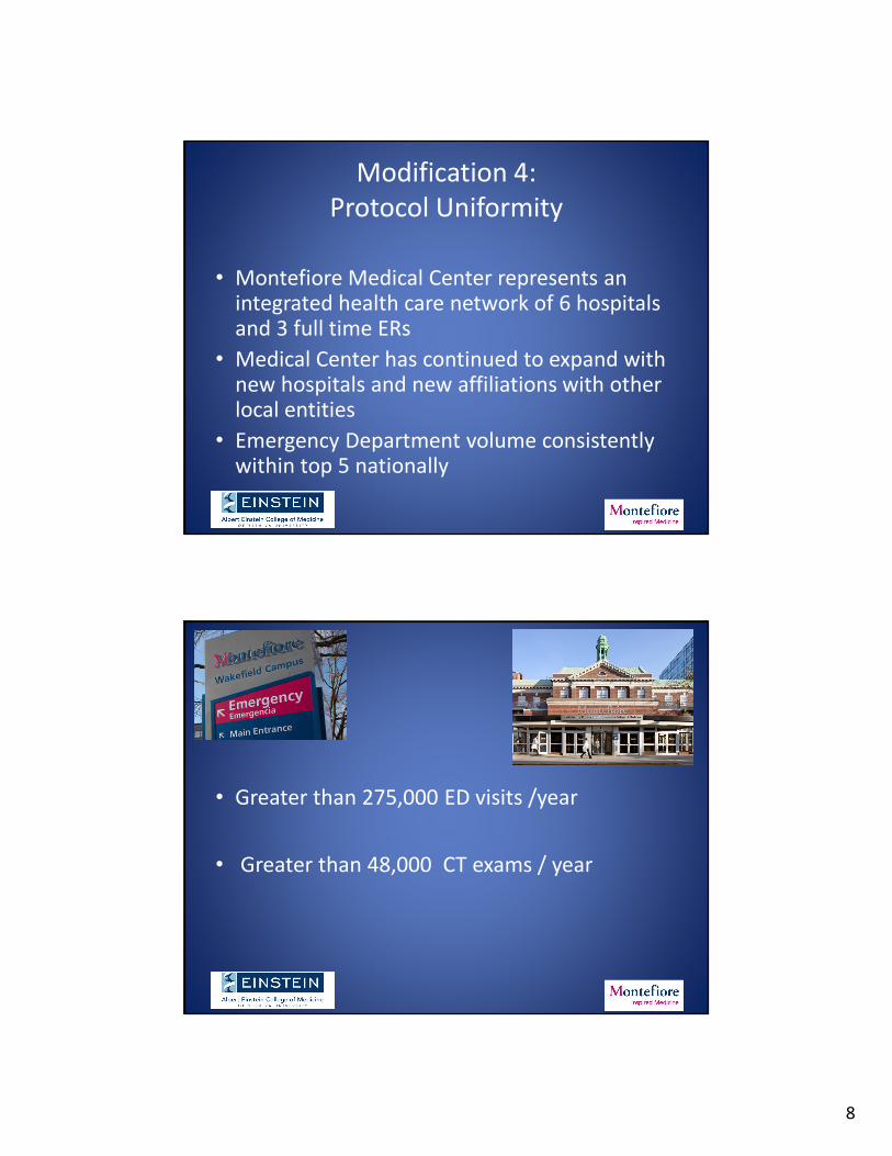
Modification 4:
Protocol Uniformity
• Montefiore Medical Center represents an integrated health care network of 6 hospitals and 3 full time ERs
• Medical Center has continued to expand with new hospitals and new affiliations with other local entities
• Emergency Department volume consistently within top 5 nationally
• Greater than 275,000 ED visits /year
• Greater than 48,000 CT exams / year

9
Modification 4:
Protocol Uniformity
• Recognition of heterogeneous imaging
protocols that existed across hospital
enterprise
• Unified protocol with standard study name
“CT Aortic Dissection”
Modification 5:
Referring Physician Collaboration
• Research and audit using an interdisciplinary
approach with active involvement of our
primary referral base, ED physicians
• Development and publication of clinical
algorithm based on retrospective data
• Collaborative initiative yielded a focused effort
on optimizing patient selection

10
Modification 6:
Prospective Data Accrual
• Order Entry for new imaging study allowed for
a priori development of unique interface to
require all relevant research data to be
entered prospectively
• Effective tool for prospective research reliant
on clinical records and for validation of
proposed clinical algorithm

11
Results:
• 6 months of data accrual (8/13-1/14)
– 192 cases of “CT Aortic Dissection” performed
• Comparison made to published institutional
data which predated this multifaceted
approach
Control Study Population
Cases (N=) 1465 192
Mean Effective Dose 43 + 20 mSv 13 + 6 mSv p = 0.0001
Incidence rate 2.7 % 4.3 % p = 0.14
Dose Recorded 61% 100% p < 0.05

12
Results:
Goal:
Successful
radiation
reduction
Elimination of
unnecessary
scans
Technical
Modifications
Inter-
Departmental
Collaboration
Attention to
Quality
Improvement
Processes
Goal:
Successful
radiation reduction
Elimination of unnecessary
scans
Technical Modifications
Inter-Departmental
Collaboration
Attention to Quality
Improvement Processes
• Optimized voltage
• Limitation of
multiphase acquisition
• Resulted in 70%
reduction in Effective
Dose
Technical Modifications

13
Elimination of unnecessary
scans
Goal:
Successful
radiation reduction
Elimination of unnecessary
scans
Technical Modifications
Inter-Departmental
Collaboration
Attention to Quality
Improvement Processes
• Trend to increased
incidence in the
imaged population
implies more
appropriate patient
selection
• Elimination of pre-
contrast abdominal
imaging
Attention to Quality
Improvement Processes
Goal:
Successful
radiation reduction
Elimination of unnecessary
scans
Technical Modifications
Inter-Departmental
Collaboration
Attention to Quality
Improvement Processes
• Standardized technique
across large enterprise
• Consistent archive of
radiation dose
• Uniform exam title
allows for easy and
focused audit

14
Goal:
Successful
radiation reduction
Elimination of unnecessary
scans
Technical Modifications
Inter-Departmental
Collaboration
Attention to Quality
Improvement Processes
• Development of clinical algorithm
• Assist in implementation of optimization techniques
• Builds rapport and respect for colleagues across multiple specialties
• Collaborative research opportunities
Inter-Departmental Collaboration
Discussion I:
• Complexity contributes to many challenges in
the Radiology community
• Solutions must address various components
that contribute to practice optimization

15
Discussion II:
• Often, the solutions to these complex challenges require alterations both in radiology practice and clinical interactions
• Involvement of clinical services facilitates effective problem solving, increases the likelihood of successful implementation and contributes to robust clinical research
Discussion III:
• The Radiology community serves a crucial role
in stewardship in Quality Assurance measures
and leading interdisciplinary problem solving

16
References
Bansal RC, Chandrasekaran K, Ayala K, et al. Frequency and explanation of false negative diagnosis of aortic dissection by aortography and transesophageal echocardiography. J Am Coll Cardiol1995;25(6):1393–401.
Klompas M. Does this patient have an acute thoracic aortic dissection? JAMA 2002;287(17):2262–72.
Lovy AJ, Bellin E, Levsky JM, Esses D, Haramati LB. Preliminary development of a clinical decision rule for acute aortic syndromes. American Journal of Emergency Medicine 2013:31:1546-1550
Meszaros I, Morocz J, Szlavi J, et al. Epidemiology and clinicopathology of aortic dissection. Chest 2000;117(5):1271–8.
Olsson C, Thelin S, Stahle E, et al. Thoracic aortic aneurysm and dissection: increasing prevalence and improved outcomes reported in a nationwide population-based study of more than 14,000 cases from 1987 to 2002. Circulation 2006;114(24):2611–8.
von Kodolitsch Y, Schwartz AG, Nienaber CA. Clinical prediction of acute aortic dissection. Arch Intern Med 2000;160(19):2977–82.
Thank You
Goal: Successful radiation reduction