multifunctional materials such as mcm-41/fe3o4 as · pdf filemultifunctional materials such as...
TRANSCRIPT
Rom J Morphol Embryol 2016, 57(2):483–489
ISSN (print) 1220–0522 ISSN (online) 2066–8279
OORRIIGGIINNAALL PPAAPPEERR
Multifunctional materials such as MCM-41/Fe3O4/folic acid as drug delivery system
SIMONA POPESCU, IOANA LAVINIA ARDELEAN, DRAGOŞ GUDOVAN, MARIUS RĂDULESCU, DENISA FICAI, ANTON FICAI, BOGDAN ŞTEFAN VASILE, ECATERINA ANDRONESCU
Faculty of Applied Chemistry and Material Science, Politehnica University of Bucharest, Romania
Abstract In this study, MCM-41 mesoporous silica nanoparticles (NPs) and MCM-41/Fe3O4 mesoporous silica NPs were prepared by sol–gel method using CTAB (cetyltrimethylammonium bromide) as template and TEOS (tetraethyl orthosilicate) as silica precursor in order to use these materials as drug delivery system (DDS) for different biologically active agents. The MCM-41 and MCM-41/Fe3O4 mesoporous silica NPs were characterized using specific physico-chemical methods [transmission electron microscopy (TEM), scanning electron microscopy (SEM), nitrogen adsorption and desorption studies – BET (Brunauer–Emmett–Teller) method, X-ray diffraction (XRD) and Fourier transform infrared (FTIR) spectroscopy], while the release studies were done by a high-performance liquid chromatography (HPLC)-modified method. The pH dependence of the delivery of folic acid from the mesoporous structures was analyzed and found that the release is pH sensitive. The lower delivery at strongly acid pH comparing with neutral/slightly alkaline pH could be beneficial because in stomach the folic acid can be destroyed.
Keywords: mesoporous silica, drug delivery, folic acid release, gastric protection.
Introduction
In the recent years, much efforts has been done in order to develop novel drug delivery systems hich exhibit advantages over conventional forms, such as greater efficacy, enhanced biocompatibility, and safety, controlled and prolonged release profile over certain period of time and predictable therapeutic response [1–3]. Nowadays, it is also known that some of these systems can also act as a protective system, especially for the orally administered active components, which, under the strong acid pH and the presence of specific gastric enzymes can be altered, losing their biological activity. Folic acid is a such biological active agent, which is altered at strong acidic pH. Also, folic acid was found to lose, partially or entirely its biological activity at increasing temperature, pressure or even at light [4, 5].
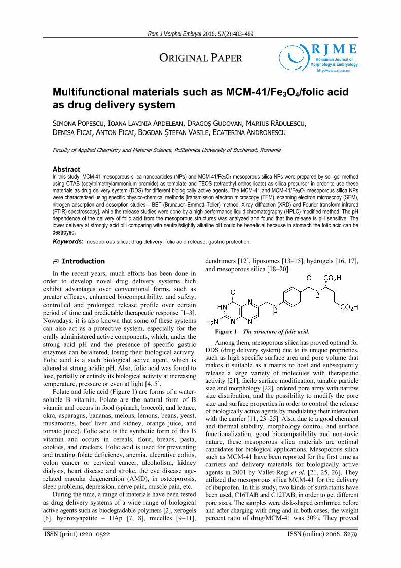
Folate and folic acid (Figure 1) are forms of a water-soluble B vitamin. Folate are the natural form of B vitamin and occurs in food (spinach, broccoli, and lettuce, okra, asparagus, bananas, melons, lemons, beans, yeast, mushrooms, beef liver and kidney, orange juice, and tomato juice). Folic acid is the synthetic form of this B vitamin and occurs in cereals, flour, breads, pasta, cookies, and crackers. Folic acid is used for preventing and treating folate deficiency, anemia, ulcerative colitis, colon cancer or cervical cancer, alcoholism, kidney dialysis, heart disease and stroke, the eye disease age-related macular degeneration (AMD), in osteoporosis, sleep problems, depression, nerve pain, muscle pain, etc.
During the time, a range of materials have been tested as drug delivery systems of a wide range of biological active agents such as biodegradable polymers [2], xerogels [6], hydroxyapatite – HAp [7, 8], micelles [9–11],
dendrimers [12], liposomes [13–15], hydrogels [16, 17], and mesoporous silica [18–20].
Figure 1 – The structure of folic acid.
Among them, mesoporous silica has proved optimal for DDS (drug delivery system) due to its unique proprieties, such as high specific surface area and pore volume that makes it suitable as a matrix to host and subsequently release a large variety of molecules with therapeutic activity [21], facile surface modification, tunable particle size and morphology [22], ordered pore array with narrow size distribution, and the possibility to modify the pore size and surface properties in order to control the release of biologically active agents by modulating their interaction with the carrier [11, 23–25]. Also, due to a good chemical and thermal stability, morphology control, and surface functionalization, good biocompatibility and non-toxic nature, these mesoporous silica materials are optimal candidates for biological applications. Mesoporous silica such as MCM-41 have been reported for the first time as carriers and delivery materials for biologically active agents in 2001 by Vallet-Regí et al. [21, 25, 26]. They utilized the mesoporous silica MCM-41 for the delivery of ibuprofen. In this study, two kinds of surfactants have been used, C16TAB and C12TAB, in order to get different pore sizes. The samples were disk-shaped confirmed before and after charging with drug and in both cases, the weight percent ratio of drug/MCM-41 was 30%. They proved
R J M ERomanian Journal of
Morphology & Embryologyhttp://www.rjme.ro/

Simona Popescu et al.
484
that the drug release plots show a different behavior depending on the method for charging the drug in the material but not on the type of the surfactant [21].
Rámila et al. investigated MCM-41 with amine groups as drug delivery systems for ibuprofen and better release kinetics were observed [27]. Aghaei et al. presented a hydroxyapatite composite with the mesoporous silica MCM-48 with potential applications as a drug delivery agent [1]. The obtained results showed a higher rate of ibuprofen release compared to pure MCM-48.
Vyskočilová et al. studied the modified MCM-41 with different groups (amino, chloro and oxo) as a drug delivery system for acetylsalicylic acid [28]. They shown that, on the MCM-41 using methyl, t-butyl ether as a solvent and MCM-41 without modification was loaded a height amount of acetylsalicylic acid after one hour (0.35 g per 1 g of the support) and on the MCM-41 modified by amino group after five hours (0.37 g per 1 g of the support).
In this study, MCM-41 mesoporous silica nanoparticles (NPs) and MCM-41/Fe3O4 mesoporous silica NPs were prepared by using sol–gel method using CTAB (cetyl-trimethylammonium bromide) as template and TEOS (tetraethyl orthosilicate) as silica precursor in order to use these materials as drug delivery system for folic acid. Also, the paper presents the effects of Fe3O4 on folic acid release.
Materials and Methods
Materials
Anhydrous iron (III) chloride – FeCl3 (Fluka), iron (II) chloride heptahydrate – FeCl2•7H2O (Fluka), sodium hydroxide – NaOH (Fluka), folic acid (Alfa Aesar), CTAB (ROTH), TEOS (Aldrich Chemistry), ammonia (S.C. Sial Trading SRL) and ethanol (Sigma Aldrich) were analytically grade and used without further purification. Distilled water was used throughout the experiments.
Methods
Preparation of MNPs (magnetic nanoparticles)
Synthesis of the MNPs was made by co-precipitation method from aqueous solutions, starting from equal volumes of FeCl2•7H2O (0.25 M) and FeCl3 (0.5 M) solutions as precursors [29]. Initially, sodium hydroxide was dissolved in distilled water (250 mL) under vigorous mechanical stirring, at room temperature, until a pH 13 was obtained. The MNPs precipitation was assured by the addition of the precursor solution (43 mL FeCl2, 43 mL FeCl3 and ~14 mL distilled water) into the sodium hydroxide solution, when the magnetic particles precipitate as pure Fe3O4. After maturation (24 hours), the MNPs were filtered and washed with distilled water until the pH 7 and negative reaction against AgNO3 0.1 M in order to verified the absence of chloride ions.
Preparation of MCM-41 mesoporous silica
0.5 g CTAB was dissolved in 96 mL distilled water and ultrasounded until the solution becomes clear. Subsequently, 34 mL of ethanol and 10 mL of an ammonia solution were added and stirring is continued until the solution becomes homogeneous. After mixing, 2 mL
TEOS were added and stirred for additional three hours at the same speed of rotation and a translucent precipitate is obtained. The precipitate thus obtained was filtered and washed with distilled water and ethanol. The last step of the synthesis consists in purification/washing with ethanol (20 mL) and water (three times with 20 mL) and drying for 12 hours at 1000C. The final product is annealed for nine hours at 5500C according to the following programme: 0–3000C at 80C/min., 300–5500C at 20C/min.
Preparation of MCM-41/Fe3O4
A suspension of 1 g Fe3O4, 0.5 g CTAB and 96 mL distilled water were shaken in ultrasonic bath for 30 minutes. After the homogenization, 34 mL of ethanol and 10 mL of 25% ammonia solution were added and stirred at a constant rate until the solution becomes homogeneous. Finally, 2 mL TEOS were added and stirred three hours at the same rate until a brown-black precipitate is obtained. The precipitate was filtered, washed with distilled water and ethanol and dried. After that, the filtrate was dried for 12 hours at 1000C. The final product is annealed for nine hours to 5500C, similar as described in the case of pure MCM-41.
Adsorption of folic acid on mesoporous silica
Adsorption of folic acid has been carried out by contacting with folic acid solution (0.05 g folic acid in 10 mL distilled water). In order to assure a good loading of folic acid inside the porous system, the loading was realized under vacuum, at room temperature. Prior to the addition of the folic acid solution, the MCM-41 was exposed 30 min. at 100 mbar. During this period, the air is partially removed from the pore system and when the solution is added, and the bottle is pressurized the solution is practically absorbed into the material. The as-obtained samples were dried at 1000C.
The amount of drug loaded was evaluated through high-performance liquid chromatography (HPLC) from the change in concentration in the pentane solution.
Characterization of mesoporous materials
The MCM-41 based mesoporous systems were characterized using specific physico-chemical methods: transmission electron microscopy (TEM), scanning electron microscopy (SEM), nitrogen adsorption and desorption studies – BET (Brunauer–Emmett–Teller), X-ray diffraction (XRD) and Fourier transform infrared (FTIR) spectroscopy.
XRD spectra were recorded on Panalytical X’Pert Pro MPD equipment, with Cu-Kα radiation.
FTIR analysis was performed using a Thermo IN50 MX FTIR microscope operated in reflection mode, was carried out to study their structural features.
The surface morphology of the samples was examined via a QUANTA INSPECT F electron microscope equipped with a field emission gun and an energy dispersive (EDS) detector, on samples covered with silver.
The transmission electron microscopy images were obtained on finely powdered samples using a Tecnai™ G2 F30 S-TWIN high-resolution transmission electron microscope (HRTEM) from FEI equipped with selected-area electron diffraction (SAED). The microscope was

Multifunctional materials such as MCM-41/Fe3O4/folic acid as drug delivery system
485
operated in transmission mode at 300 kV with TEM point resolution of 2 Å and line resolution of 1 Å.
BET analysis was performed on a Micrometrics Gemini V surface area and pore size analyzer. Magnetization curves were recorded using a Vibrating Sample Magne-tometer VSM-7304 Lake Shore (USA) operated up to 12 000 Oe.
Delivery studies
0.01 g mesoporous folic acid delivery systems (MCM-41/folic acid and MCM-41/Fe3O4/folic acid) were indivi-dually suspended in 50 mL simulated body fluid (SBF) or hydrochloric acid solution (pH~1.5), under vigorous mechanical stirring, at room temperature. At certain times (10 min., 30 min., 60 min., 2 h, 3 h and 24 h), 1 mL of each solution were taken and filtered through a 0.25 μm Agilent Econofilter syringe filter membrane and used for quantification. The HPLC analysis of the folic acid was done according to the conditions mentioned in Table 1.
Table 1 – Chromatographic parameters used for the Agilent 1260 Infinity LC and Agilent 1290 Infinity LC systems [30]
Parameter Agilent 1260 Infinity LC
Column oven thermostat 350C
Acquisition rate 20 Hz
Data acquisition 205, 214, 220, 232, 266, 268, 280 nm
Flow cell 60 mm path
Injection volume 5 μL (needle with wash, flush port active for 5 seconds)
Sample thermostat 50C
Mobile phase 25 mM K2HPO4, pH 7.0
Mobile phase B Acetonitrile
Gradient
At 0 min. → 1% B At 5 min. → 1% B At 15 min. → 30% B At 20 min. → 30% B At 20.1 min. → 1% B
Post run time 5 minutes
Flow rate 0.45 mL/min.
Results
X-ray diffraction (XRD)
Low angle XRD patterns of pure MCM-41, MCM-41/ folic acid, MCM-41/Fe3O4 and MCM-41/Fe3O4/folic acid (Figure 2) show a strong diffraction peak corresponding to (100) Bragg reflection and three other smaller peaks assigned to (110), (200), and (210) reflections characteristic for a long range ordered pore arrays [31]. The ordered mesoporous structure is basically preserved in both MCM-41 loaded samples as well as in the case of MCM-41/Fe3O4. Analyzing the four diffraction patterns, some interesting conclusions can be obtained. The synthesis of the meso-porous structure around the magnetite NPs can be done easily. The loading of the MCM-41 with folic acid, realized under vacuum, do not destroy the mesoporous structure of the silica but the dimension of the pores increase, especially for the pure MCM-41 sample, when the pore size increased from 3.575 to 3.648 nm. In the case of MCM-41/Fe3O4, the effect of loading of folic acid is practically marginal.
Figure 2 – XRD patterns of the mesoporous materials.
Infrared spectroscopy
In the IR spectra of the MCM-41, MCM-41/folic acid, MCM-41/Fe3O4 and MCM-41/Fe3O4/folic acid (Figure 3) the main characteristic bands of silica can be observed. The band from ~1245 and 1050 cm-1 can be assigned to the asymmetric stretching vibrations of Si–O–Si units; the band from 443 cm-1 can be assigned to the bending vibrations of Si–O–Si units of observed, the band from ~800 cm-1 can be assigned to the symmetric stretching vibrations of Si–O–Si units, while the band from ~474 cm-1 can be assigned to the O–Si–O symmetric stretching vibrations. The band from ~960–970 cm-1 is assigned to the silanolic groups of MCM-41 [32, 33].
Figure 3 – FTIR spectra of the mesoporous samples over 400–1300 cm-1.
The loading of the MCM-41 with folic acid induce important shifts (11.50 and 9.42 cm-1, respectively) of the most intense peaks of MCM-41, from 1046.84 and 434.19 cm-1 to 1058.34 and 443.61 cm-1, respectively. In the case of MCM-41/Fe3O4, these shifts are less important (7.79 and 1.31 cm-1), from 1053.98 and 439.94 cm-1 to 1061.66 and 441.25 cm-1, respectively. It is also evident that the presence of magnetite inside the forming MCM-41 network assist the synthesis of the silica network and lead to some shift, from 1046.84 to 1053.97 cm-1 (7.13 cm-1) and from 434.19 to 439.94 cm-1 (5.75 cm-1).
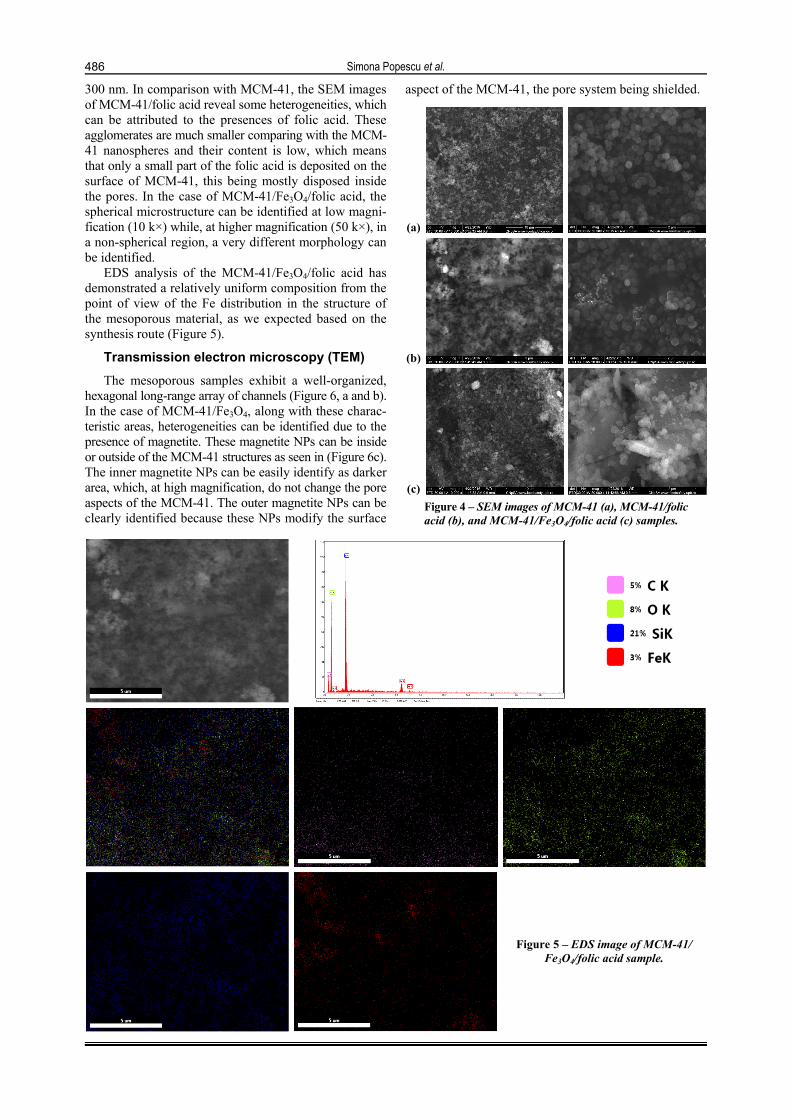
Scanning electron microscopy (SEM)
SEM images highlight some important differences between MCM-41, MCM-41/folic acid and MCM-41/ Fe3O4/folic acid (Figure 4). The MCM-41 sample consists in a very homogeneous nanometrical spheres of 250–
Simona Popescu et al.
486
300 nm. In comparison with MCM-41, the SEM images of MCM-41/folic acid reveal some heterogeneities, which can be attributed to the presences of folic acid. These agglomerates are much smaller comparing with the MCM-41 nanospheres and their content is low, which means that only a small part of the folic acid is deposited on the surface of MCM-41, this being mostly disposed inside the pores. In the case of MCM-41/Fe3O4/folic acid, the spherical microstructure can be identified at low magni-fication (10 k×) while, at higher magnification (50 k×), in a non-spherical region, a very different morphology can be identified.
EDS analysis of the MCM-41/Fe3O4/folic acid has demonstrated a relatively uniform composition from the point of view of the Fe distribution in the structure of the mesoporous material, as we expected based on the synthesis route (Figure 5).
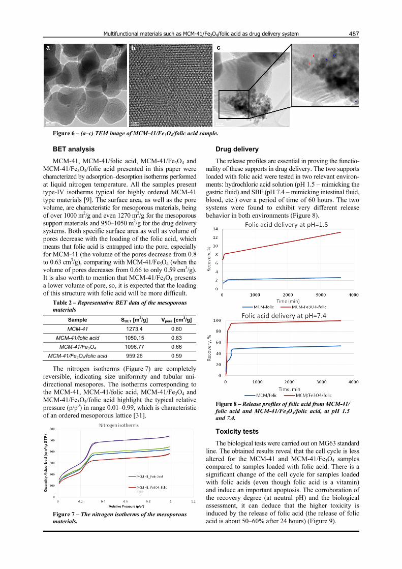
Transmission electron microscopy (TEM)
The mesoporous samples exhibit a well-organized, hexagonal long-range array of channels (Figure 6, a and b). In the case of MCM-41/Fe3O4, along with these charac-teristic areas, heterogeneities can be identified due to the presence of magnetite. These magnetite NPs can be inside or outside of the MCM-41 structures as seen in (Figure 6c). The inner magnetite NPs can be easily identify as darker area, which, at high magnification, do not change the pore aspects of the MCM-41. The outer magnetite NPs can be clearly identified because these NPs modify the surface
aspect of the MCM-41, the pore system being shielded.
(a)
(b)
(c) Figure 4 – SEM images of MCM-41 (a), MCM-41/folic acid (b), and MCM-41/Fe3O4/folic acid (c) samples.
Figure 5 – EDS image of MCM-41/ Fe3O4/folic acid sample.
Multifunctional materials such as MCM-41/Fe3O4/folic acid as drug delivery system
487
Figure 6 – (a–c) TEM image of MCM-41/Fe3O4/folic acid sample.
BET analysis
MCM-41, MCM-41/folic acid, MCM-41/Fe3O4 and MCM-41/Fe3O4/folic acid presented in this paper were characterized by adsorption–desorption isotherms performed at liquid nitrogen temperature. All the samples present type-IV isotherms typical for highly ordered MCM-41 type materials [9]. The surface area, as well as the pore volume, are characteristic for mesoporous materials, being of over 1000 m2/g and even 1270 m2/g for the mesoporous support materials and 950–1050 m2/g for the drug delivery systems. Both specific surface area as well as volume of pores decrease with the loading of the folic acid, which means that folic acid is entrapped into the pore, especially for MCM-41 (the volume of the pores decrease from 0.8 to 0.63 cm3/g), comparing with MCM-41/Fe3O4 (when the volume of pores decreases from 0.66 to only 0.59 cm3/g). It is also worth to mention that MCM-41/Fe3O4 presents a lower volume of pore, so, it is expected that the loading of this structure with folic acid will be more difficult.
Table 2 – Representative BET data of the mesoporous materials
Sample SBET [m2/g] Vpore [cm3/g]
MCM-41 1273.4 0.80
MCM-41/folic acid 1050.15 0.63
MCM-41/Fe3O4 1096.77 0.66
MCM-41/Fe3O4/folic acid 959.26 0.59
The nitrogen isotherms (Figure 7) are completely reversible, indicating size uniformity and tubular uni-directional mesopores. The isotherms corresponding to the MCM-41, MCM-41/folic acid, MCM-41/Fe3O4 and MCM-41/Fe3O4/folic acid highlight the typical relative pressure (p/p0) in range 0.01–0.99, which is characteristic of an ordered mesoporous lattice [31].
Figure 7 – The nitrogen isotherms of the mesoporous materials.
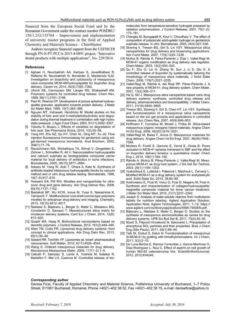
Drug delivery
The release profiles are essential in proving the functio-nality of these supports in drug delivery. The two supports loaded with folic acid were tested in two relevant environ-ments: hydrochloric acid solution (pH 1.5 – mimicking the gastric fluid) and SBF (pH 7.4 – mimicking intestinal fluid, blood, etc.) over a period of time of 60 hours. The two systems were found to exhibit very different release behavior in both environments (Figure 8).
Figure 8 – Release profiles of folic acid from MCM-41/ folic acid and MCM-41/Fe3O4/folic acid, at pH 1.5 and 7.4.
Toxicity tests
The biological tests were carried out on MG63 standard line. The obtained results reveal that the cell cycle is less altered for the MCM-41 and MCM-41/Fe3O4 samples compared to samples loaded with folic acid. There is a significant change of the cell cycle for samples loaded with folic acids (even though folic acid is a vitamin) and induce an important apoptosis. The corroboration of the recovery degree (at neutral pH) and the biological assessment, it can deduce that the higher toxicity is induced by the release of folic acid (the release of folic acid is about 50–60% after 24 hours) (Figure 9).

Simona Popescu et al.
488
Apoptosis can be identified for the MCM-41/Fe3O4 sample and is very important in the case of the samples
loaded with folic acid, when the “S” phase is also strongly increased (Figure 10).
MG63 – [34] MCM-41 MCM-41/Fe3O4 MCM-41/folic acid
Figure 9 – Optical microscopy images of cells that control cells and exposed to the material of interest.
MG63 – [34] MCM-41 MCM-41/Fe3O4 MCM-41/folic acid
Figure 10 – Cell cycle of MG63 cells control exposed to materials that interest. Discussion
MCM-41 as well as many other representatives of the mesoporous materials based on silica are extensively used as drug delivery systems for different biologically active principles [19, 21, 26, 28].
Usually, they are not pH-responsive, but, in certain cases, they can act as pH-responsive DDSs. In this particular case, the pH-responsivity is assured by the difference of the solubility of folic acid in acid and neutral/basic media. In that case, we tasted the two materials, loaded with folic acid in two relevant environments: hydrochloric acid solution (pH 1.5 – mimicking the gastric fluid) and SBF (pH 7.4 – mimicking intestinal fluid, blood, etc.) over a period of time of 60 hours. As expected based on the BET data (higher volume of the pores, which can host larger amount of folic acid), the release of the MCM-41/Fe3O4/ folic acid is much faster comparing with the MCM-41/ folic acid (4.6 times faster in acidic conditions and 1.8 times faster in neutral conditions).
From the controlled release profiles, the release in acidic environment is slower and with a sustained rate. This makes this type of delivery vector viable for delivery in acidic environment, typically in gastric acid. From the release curve, after an initial instability in the first minute, the concentration remains stable for longer period of time. In the case of SBF solution, at physiological pH, the release rate is very rapid (burst like). The effect of Fe3O4 NPs on the release behavior is that it tends to increase the desorption rate, in the case of acidic environment this being a benefit due to the very slow release in the case of the unmodified material. In the case of the SBF release, this is an unwanted effect, resulting in almost complete release of the folic acid loaded in the pores. A conclusion that can be drawn is that the unmodified material is better for release at physiological pH, while the modified material
is better for release at acidic pH (such as gastric environ-ment).
Conclusions
The work presents the synthesis of MCM-41 and MCM-41/Fe3O4, their loading with folic acid and the characterization of the support materials and drug delivery systems. A special attention of the work is related to the loading of the folic acid into the mesoporous systems and, based on the BET data, 21.25 and 10.6% of the total volume of the pores are filled due to the loading of folic acid in MCM-41 and MCM-41/Fe3O4, respectively. The delivery of folic acid depends on pH and the nature of the used support materials. Regardless the pH, the delivery is faster for MCM-41/Fe3O4/folic acid comparing with MCM-41/folic acid (4.6 times at pH 1.5 and 1.8 times at pH 7.4) because in the latter case, the folic acid is loaded mainly into the pores. The pH sensitivity is explained based on the limited solubility of folic acid in acidic conditions and the high solubility in neutral/basic conditions. The as-obtained systems can be potential candidates for oral administration, the folic acid being mostly released in the intestines and not in stomach and so, protecting folic acid against the undesired degradations induced by acidic pH and gastric enzymes. Further active components will be also tested from the point of view of absorption capacity inside the pore system, their release profile and especially their potential applications for biomedical applications.
Conflict of interests The authors declare that they have no conflict of
interests.
Acknowledgments This research was financially supported by Sectoral
Operational Programme Human Resources Development,
Multifunctional materials such as MCM-41/Fe3O4/folic acid as drug delivery system
489
financed from the European Social Fund and by the Romanian Government under the contract number POSDRU/ 156/1.2/G/135764 – Improvement and implementation of university master programs in the field of Applied Chemistry and Materials Science – ChimMaster.
Authors recognize financial support from the UEFISCDI through PN-II-PT-PCCA-2013-4-0891 project, “Innovative dental products with multiple applications”, No. 229/2014.
References [1] Aghaei H, Nourbakhsh AA, Karbasi S, JavadKalbasi R,
Rafienia M, Nourbakhsh N, Bonakdar S, Mackenzie KJD. Investigation on bioactivity and cytotoxicity of mesoporous nano-composite MCM-48/hydroxyapatite for ibuprofen drug delivery. Ceram Int, 2014, 40(5):7355–7362.
[2] Uhrich KE, Cannizzaro SM, Langer RS, Shakesheff KM. Polymeric systems for controlled drug release. Chem Rev, 1999, 99(11):3181–3198.
[3] Paul W, Sharma CP. Development of porous spherical hydroxy-apatite granules: application towards protein delivery. J Mater Sci Mater Med, 1999, 10(7):383–388.
[4] Nguyen MT, Indrawati, Hendrickx M. Model studies on the stability of folic acid and 5-methyltetrahydrofolic acid degra-dation during thermal treatment in combination with high hydro-static pressure. J Agric Food Chem, 2003, 51(11):3352–3357.
[5] Yakubu S, Muazu J. Effects of variables on degradation of folic acid. Der Pharmacia Sinica, 2010, 1(3):55–58.
[6] Yang HH, Zhu QZ, Qu HY, Chen XL, Ding MT, Xu JG. Flow injection fluorescence immunoassay for gentamicin using sol-gel-derived mesoporous biomaterial. Anal Biochem, 2002, 308(1):71–76.
[7] Rauschmann MA, Wichelhaus TA, Stirnal V, Dingeldein E, Zichner L, Schnettler R, Alt V. Nanocrystalline hydroxyapatite and calcium sulphate as biodegradable composite carrier material for local delivery of antibiotics in bone infections. Biomaterials, 2005, 26(15):2677–2684.
[8] Itokazu M, Yang W, Aoki T, Ohara A, Kato N. Synthesis of antibiotic-loaded interporous hydroxyapatite blocks by vacuum method and in vitro drug release testing. Biomaterials, 1998, 19(7–9):817–819.
[9] Husseini GA, Pitt WG. Micelles and nanoparticles for ultra-sonic drug and gene delivery. Adv Drug Deliver Rev, 2008, 60(10):1137–1152.
[10] Bastakoti BP, Wu KCW, Inoue M, Yusa S, Nakashima K, Yamauchi Y. Multifunctional core-shell-corona-type polymeric micelles for anticancer drug-delivery and imaging. Chemistry, 2013, 19(15):4812–4817.
[11] Nastase S, Bajenaru L, Berger D, Matei C, Moisescu MG, Constantin D, Savopol T. Mesostructured silica matrix for irinotecan delivery systems. Cent Eur J Chem, 2014, 12(8): 813–820.
[12] Quadir MA, Haag R. Biofunctional nanosystems based on dendritic polymers. J Control Release, 2012, 161(2):484–495.
[13] Allen TM, Cullis PR. Liposomal drug delivery systems: from concept to clinical applications. Adv Drug Deliv Rev, 2013, 65(1):36–48.
[14] Sawant RR, Torchilin VP. Liposomes as ‘smart’ pharmaceutical nanocarriers. Soft Matter, 2010, 6(17):4026–4044.
[15] Wang S. Ordered mesoporous materials for drug delivery. Microporous Mesoporous Mater, 2009, 117(1–2):1–9.
[16] Caliceti P, Salmaso S, Lante A, Yoshida M, Katakai R, Martellini F, Mei LH, Carenza M. Controlled release of bio-
molecules from temperature-sensitive hydrogels prepared by radiation polymerization. J Control Release, 2001, 75(1–2): 173–181.
[17] Changez M, Burugapalli K, Koul V, Choudhary V. The effect of composition of poly(acrylic acid)-gelatin hydrogel on gentamicin sulphate release: in vitro. Biomaterials, 2003, 24(4):527–536.
[18] Slowing II, Trewyn BG, Giri S, Lin VSY. Mesoporous silica nanoparticles for drug delivery and biosensing applications. Adv Funct Mater, 2007, 17(8):1225–1236.
[19] Muñoz B, Rámila A, Pérez-Pariente J, Díaz I, Vallet-Regí M. MCM-41 organic modification as drug delivery rate regulator. Chem Mater, 2003, 15(2):500–503.
[20] Qu F, Zhu G, Lin H, Zhang W, Sun J, Li S, Qiu S. A controlled release of ibuprofen by systematically tailoring the morphology of mesoporous silica materials. J Solid State Chem, 2006, 179(7):2027–2035.
[21] Vallet-Regí M, Rámila A, del Real RP, Pérez-Pariente J. A new property of MCM-41: drug delivery system. Chem Mater, 2001, 13(2):308–311.
[22] He Q, Shi J. Mesoporous silica nanoparticle based nano drug delivery systems: synthesis, controlled drug release and delivery, pharmacokinetics and biocompatibility. J Mater Chem, 2011, 21(16):5845–5855.
[23] Trewyn BG, Slowing II, Giri S, Chen HT, Lin VSY. Synthesis and functionalization of a mesoporous silica nanoparticle based on the sol–gel process and applications in controlled release. Acc Chem Res, 2007, 40(9):846–853.
[24] Hoffmann F, Cornelius M, Morell J, Fröba M. Silica-based mesoporous organic–inorganic hybrid materials. Angew Chem Int Ed Engl, 2006, 45(20):3216–3251.
[25] Vallet-Regí M, Balas F, Arcos D. Mesoporous materials for drug delivery. Angew Chem Int Ed Engl, 2007, 46(40):7548–7558.
[26] Mortera R, Fiorilli S, Garrone E, Verné E, Onida B. Pores occlusion in MCM-41 spheres immersed in SBF and the effect on ibuprofen delivery kinetics: a quantitative model. Chem Eng J, 2010, 156(1):184–192.
[27] Rámila A, Muñoz B, Pérez-Pariente J, Vallet-Regí M. Meso-porous MCM-41 as drug host system. J Sol Gel Sci Technol, 2003, 26(1):1199–1202.
[28] Vyskočilová E, Luštická I, Paterová I, Machová L, Červený L. Modified MCM-41 as a drug delivery system for acetylsalicylic acid. Solid State Sci, 2014, 38:85–89.
[29] Andronescu E, Ficai M, Voicu G, Ficai D, Maganu M, Ficai A. Synthesis and characterization of collagen/hydroxyapatite: magnetite composite material for bone cancer treatment. J Mater Sci Mater Med, 2010, 21(7):2237–2242.
[30] Joseph S. Analysis of water-soluble vitamins from multivitamin tablets for nutrition labeling. Agilent Application Solution, Application Note, Agilent Technologies, 2011, 1–12, https:// www.agilent.com/cs/library/applications/5990-7950EN.pdf.
[31] Băjenaru L, Năstase S, Matei C, Berger D. Studies on the synthesis of mesoporous aluminosilicates as carries for drug delivery systems. UPB Sci Bull Ser B, 2011, 73(4):45–50.
[32] Musić S, Filipović-Vinceković N, Sekovanić L. Precipitation of amorphous SiO2 particles and their properties. Braz J Chem Eng (São Paulo), 2011, 28(1):89–94.
[33] Taib NI, Endud S, Katun N. Functionalization of mesoporous Si-MCM-41 by grafting with trimethylchlorosilane. Int J Chem, 2011, 3(3):2–10.
[34] De Luna-Bertos E, Ramos-Torrecillas J, García-Martínez O, Díaz-Rodríguez L, Ruiz C. Effect of aspirin on cell growth of human MG-63 osteosarcoma line. ScientificWorldJournal, 2012, 2012:834246.
Corresponding author Denisa Ficai, Faculty of Applied Chemistry and Material Science, Politehnica University of Bucharest, 1–7 Polizu Street, 011061 Bucharest, Romania; Phone +4021–402 38 52, Fax +4021–402 38 15, e-mail: [email protected] Received: February 7, 2016 Accepted: July 30, 2016