multimodal imaging for radiotherapy and its benefit
TRANSCRIPT
ICRP, Oct. 2017
Multimodal Imaging for Radiotherapy
and its Benefit.
Vincent GREGOIRE, MD, PhD, hon. FRCR (UK), hon. FRCR (IE)
Radiation Oncology Dept. Head and Neck Oncology Program &
Center for Molecular Imaging, Radiotherapy and Oncology,
Université Catholique de Louvain, St-Luc University Hospital,
Brussels, Belgium
ICRP, Oct. 2017
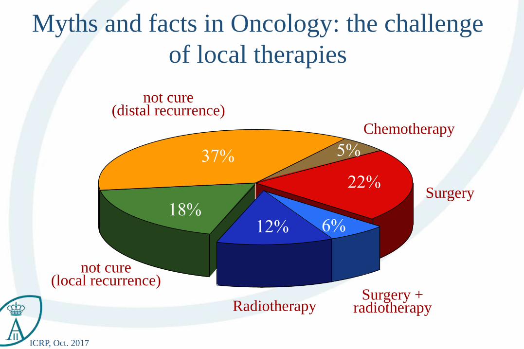
Myths and facts in Oncology: the challenge
of local therapies
Chemotherapy
Surgery
Surgery +radiotherapyRadiotherapy
not cure(local recurrence)
not cure(distal recurrence)
37%
18%
5%
22%
6%12%
ICRP, Oct. 2017
Table of Content
• Set the scene
• Multimodality imaging
• Future prospects
ICRP, Oct. 2017
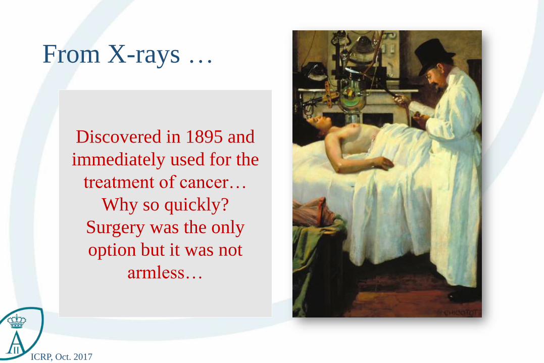
From X-rays …
Discovered in 1895 and
immediately used for the
treatment of cancer…
Why so quickly?
Surgery was the only
option but it was not
armless…
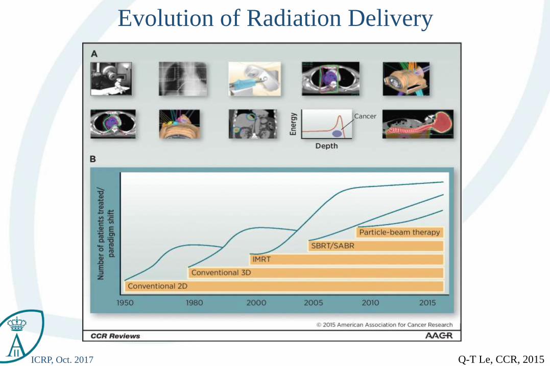
ICRP, Oct. 2017 Q-T Le, CCR, 2015
Evolution of Radiation Delivery
ICRP, Oct. 2017
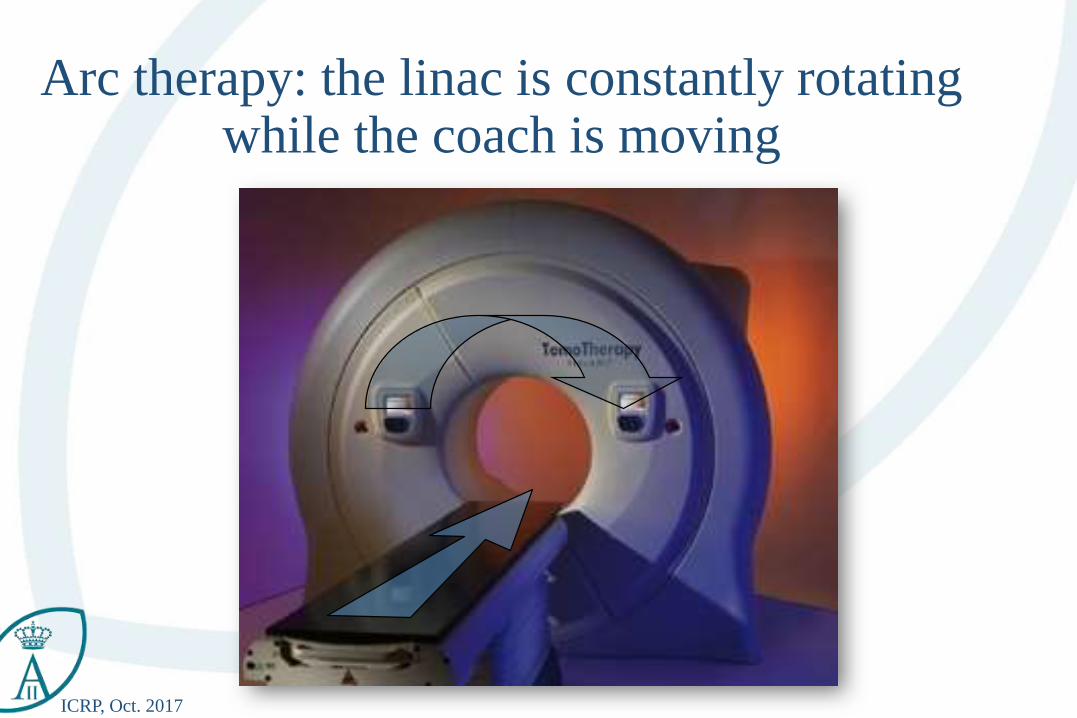
Arc therapy: the linac is constantly rotating while the coach is moving
ICRP, Oct. 2017
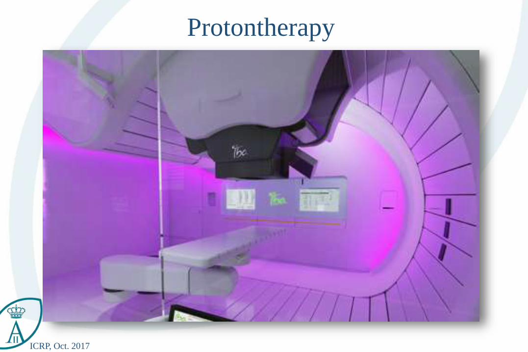
Protontherapy
ICRP, Oct. 2017
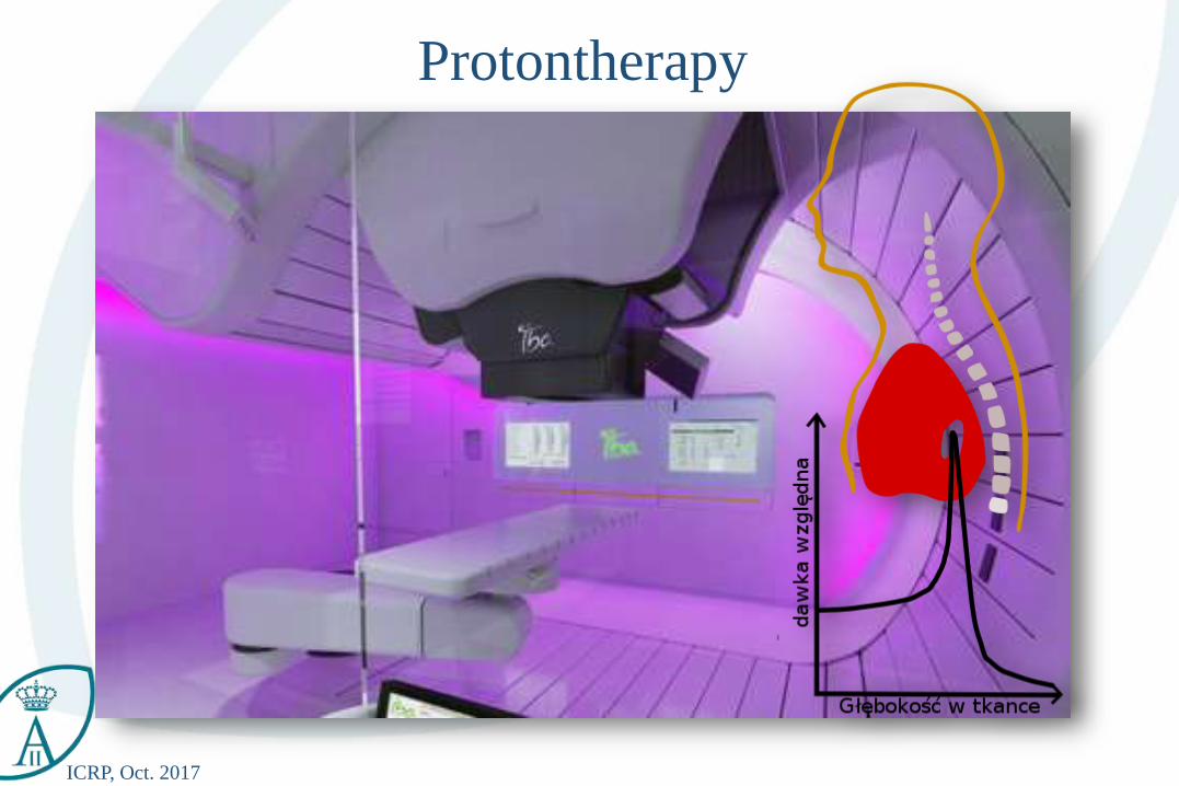
Protontherapy
ICRP, Oct. 2017
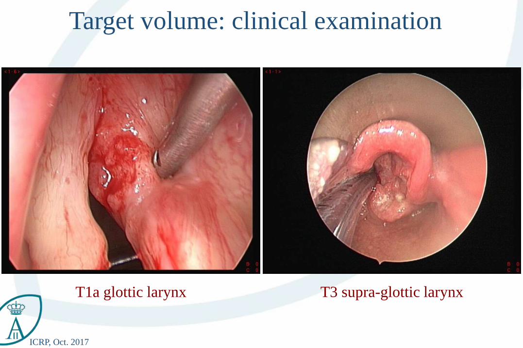
Target volume: clinical examination
T1a glottic larynx T3 supra-glottic larynx
ICRP, Oct. 2017
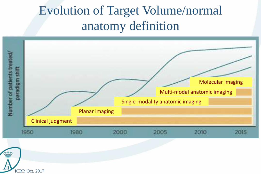
Evolution of Target Volume/normal
anatomy definition
Clinical judgment
Planar imaging
Multi-modal anatomic imaging
Molecular imaging
Single-modality anatomic imaging
ICRP, Oct. 2017
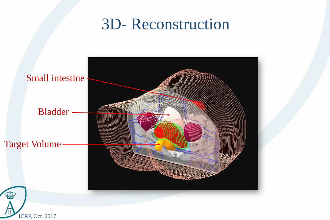
3D- Reconstruction
Small intestine
Bladder
Target Volume
ICRP, Oct. 2017
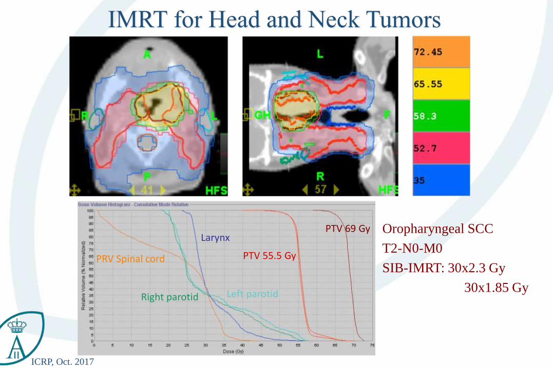
Oropharyngeal SCC
T2-N0-M0
SIB-IMRT: 30x2.3 Gy
30x1.85 Gy
PRV Spinal cord
Left parotidRight parotid
Larynx
PTV 55.5 Gy
PTV 69 Gy
IMRT for Head and Neck Tumors
ICRP, Oct. 2017
Table of Content
• Multimodality imaging
ICRP, Oct. 2017
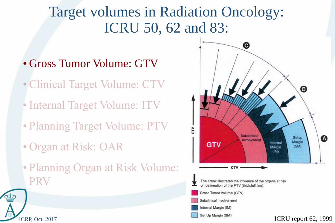
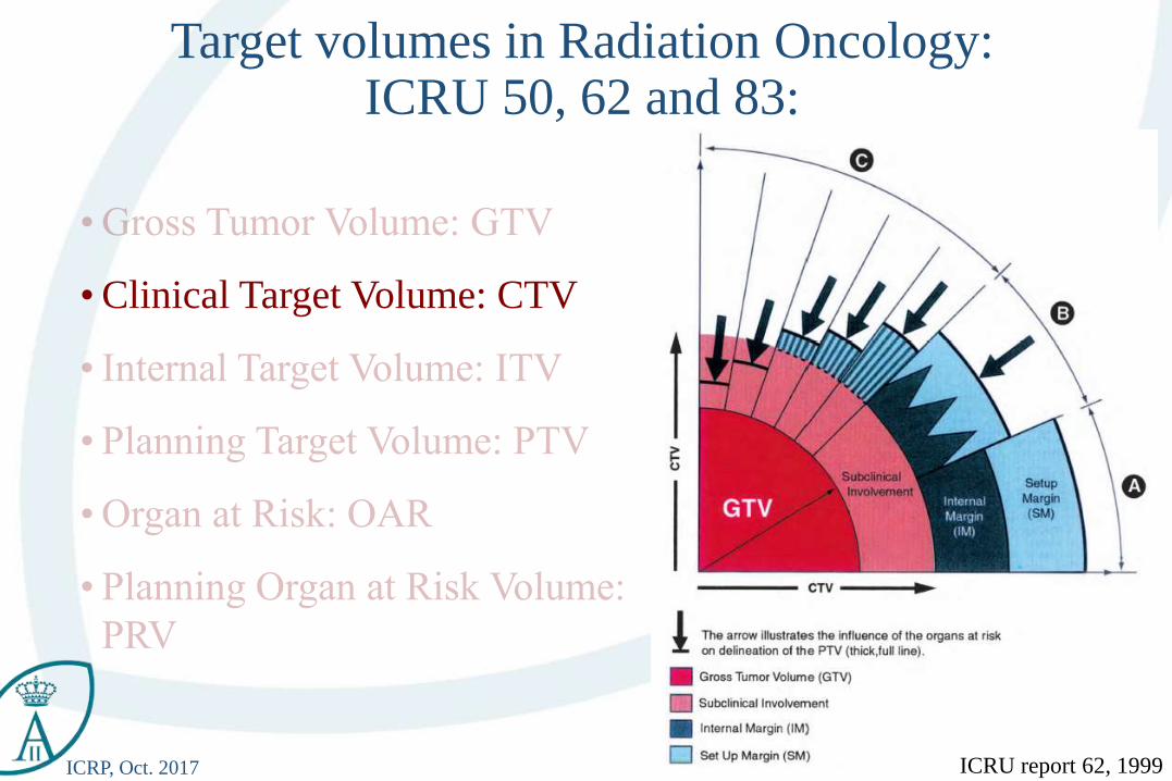
• Gross Tumor Volume: GTV
Target volumes in Radiation Oncology:ICRU 50, 62 and 83:
ICRU report 62, 1999
ICRP, Oct. 2017
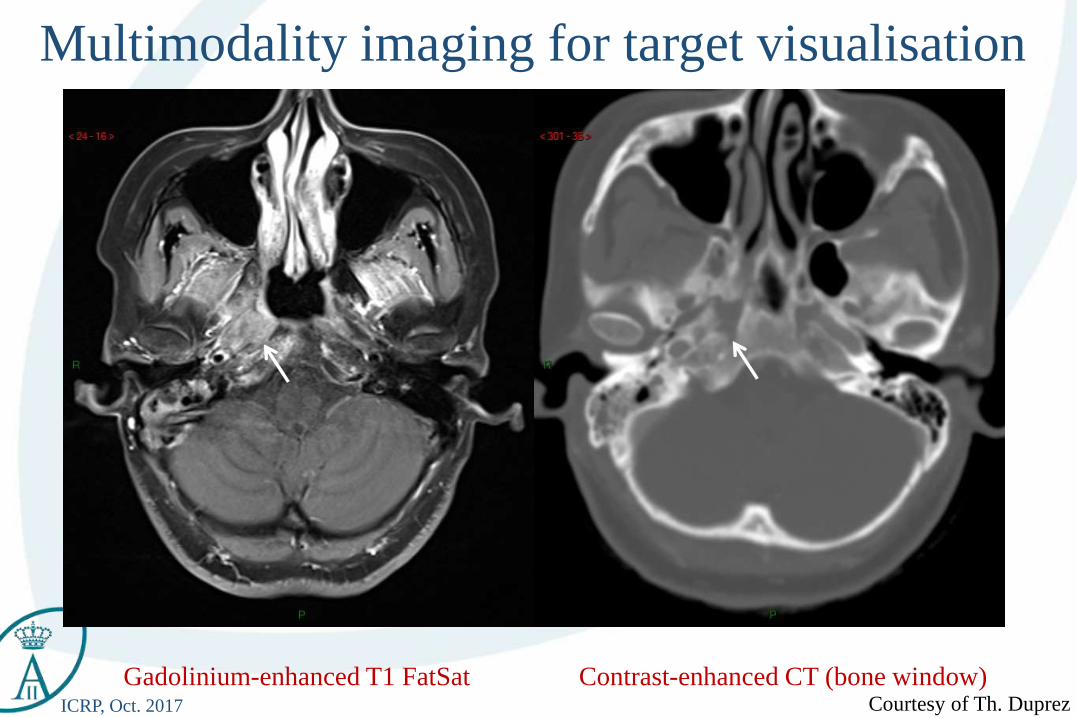
Multimodality imaging for target visualisation
Gadolinium-enhanced T1 FatSat Contrast-enhanced CT (bone window)Courtesy of Th. Duprez
ICRP, Oct. 2017
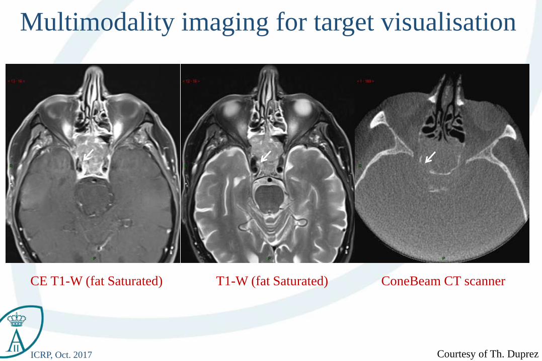
Multimodality imaging for target visualisation
Courtesy of Th. Duprez
CE T1-W (fat Saturated) T1-W (fat Saturated) ConeBeam CT scanner
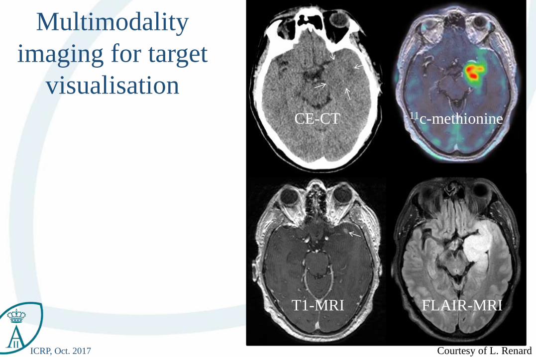
ICRP, Oct. 2017 Courtesy of L. Renard
11c-methionineCE-CT
T1-MRI FLAIR-MRI
Multimodality
imaging for target
visualisation
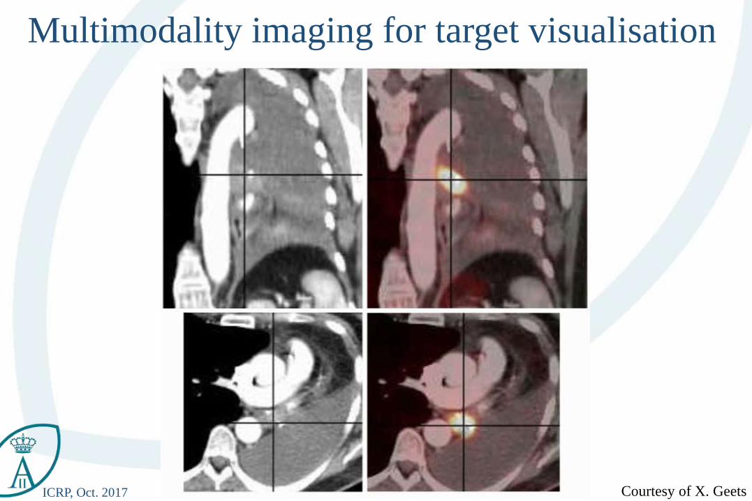
ICRP, Oct. 2017 Courtesy of X. Geets
Multimodality imaging for target visualisation
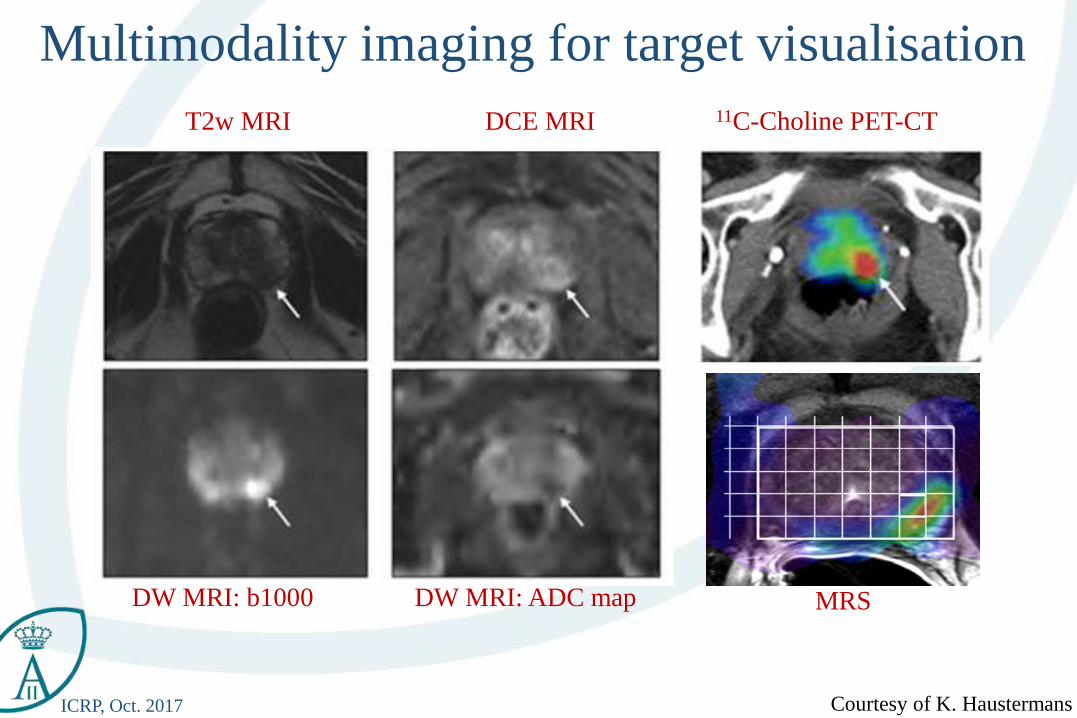
ICRP, Oct. 2017 Courtesy of K. Haustermans
T2w MRI DCE MRI
DW MRI: b1000 DW MRI: ADC map
11C-Choline PET-CT
MRS
Multimodality imaging for target visualisation
ICRP, Oct. 2017 Courtesy of K. Haustermans
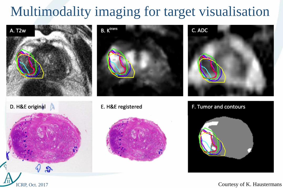
Multimodality imaging for target visualisation
ICRP, Oct. 2017
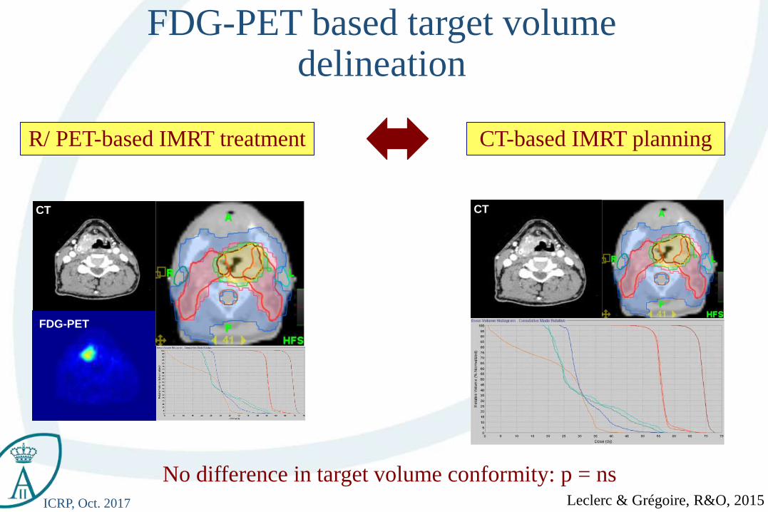
R/ PET-based IMRT treatment
MR T2
FDG-PET based target volume delineation
CT MR T2 FS
FDG-PET
CT
CT-based IMRT planning
No difference in target volume conformity: p = nsLeclerc & Grégoire, R&O, 2015
ICRP, Oct. 2017 S. Differding, RO, 2017
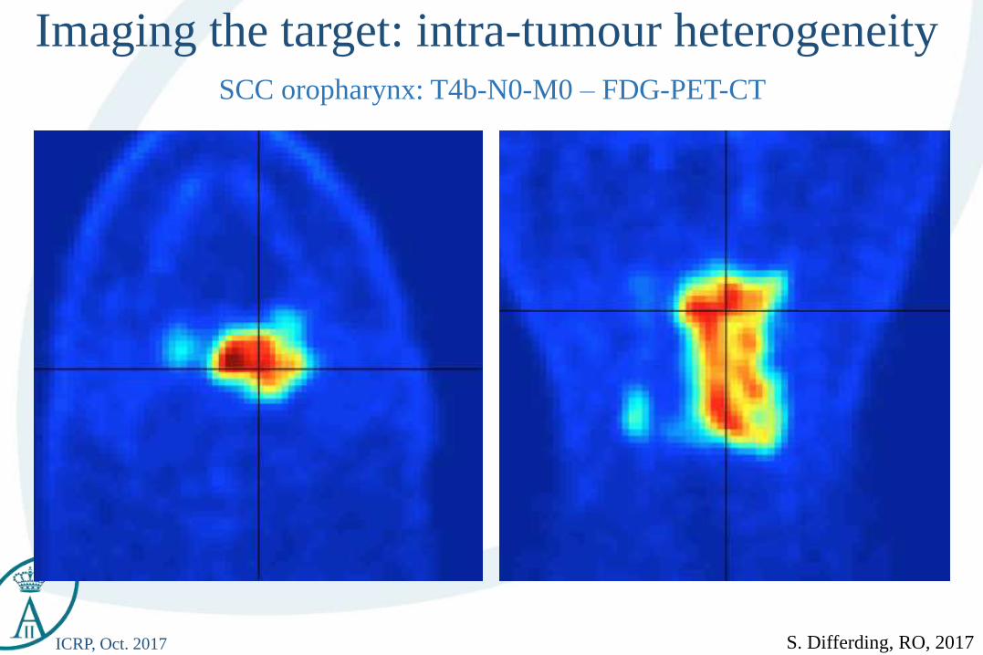
SCC oropharynx: T4b-N0-M0 – FDG-PET-CT
Imaging the target: intra-tumour heterogeneity
ICRP, Oct. 2017
• Clinical Target Volume: CTV
Target volumes in Radiation Oncology:ICRU 50, 62 and 83:
ICRU report 62, 1999
ICRP, Oct. 2017
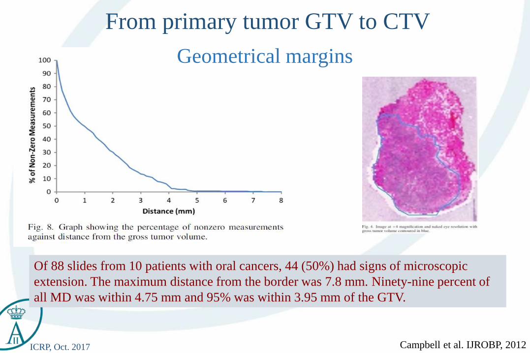
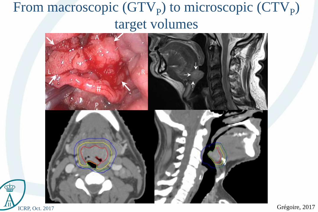
Of 88 slides from 10 patients with oral cancers, 44 (50%) had signs of microscopic
extension. The maximum distance from the border was 7.8 mm. Ninety-nine percent of
all MD was within 4.75 mm and 95% was within 3.95 mm of the GTV.
Campbell et al. IJROBP, 2012
From primary tumor GTV to CTV
Geometrical margins
ICRP, Oct. 2017
#
A
P
RL
From macroscopic (GTVP) to microscopic (CTVP)
target volumes
Grégoire, 2017
ICRP, Oct. 2017
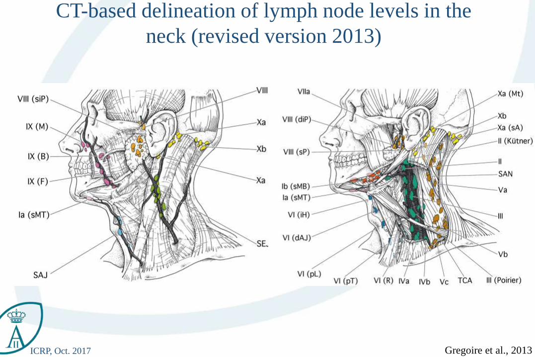
CT-based delineation of lymph node levels in the
neck (revised version 2013)
Gregoire et al., 2013
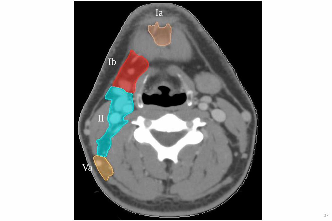
ICRP, Oct. 2017 27
II
Ib
Ia
Va
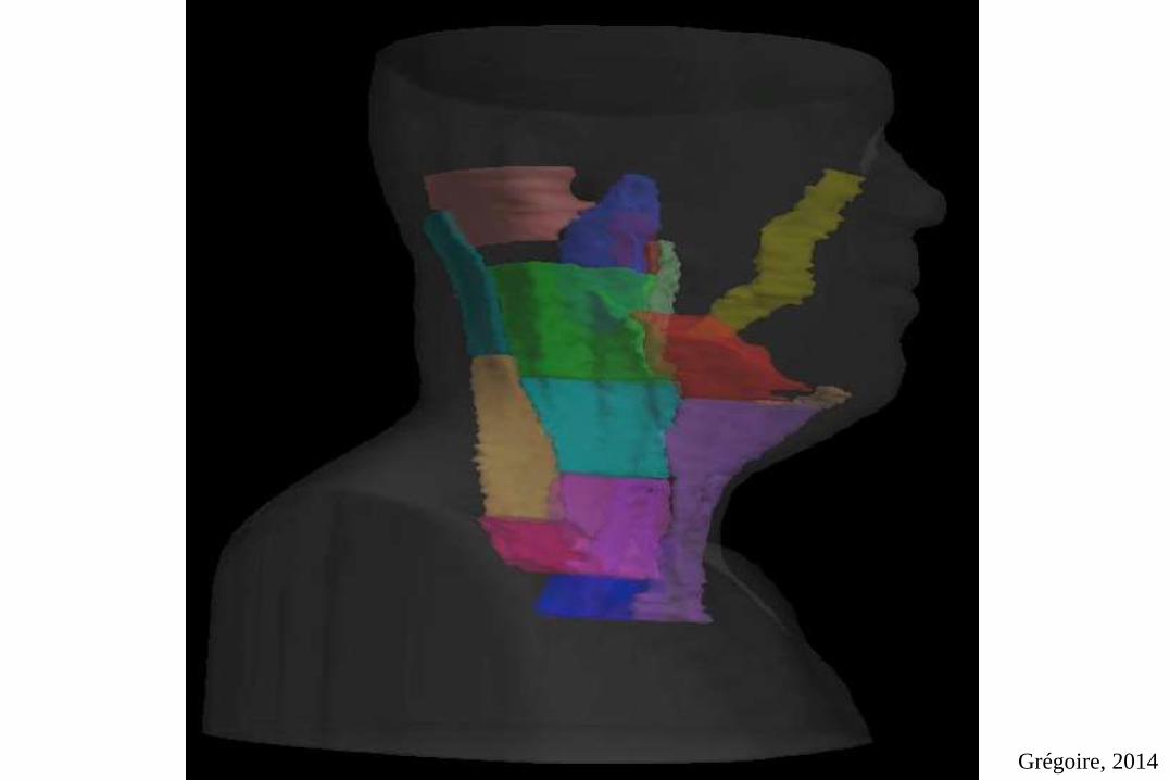
ICRP, Oct. 2017 Grégoire, 2014
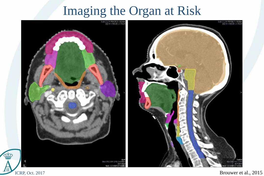
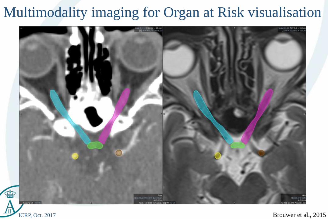
ICRP, Oct. 2017 Brouwer et al., 2015
Imaging the Organ at Risk
ICRP, Oct. 2017 Brouwer et al., 2015
Multimodality imaging for Organ at Risk visualisation

ICRP, Oct. 2017 DAHANCA.dk
Thirty years of progresses: the Danish example
Courtesy of Overgaard 2015
Standard 2013
Standard 1985
Loco
-reg
ion
al c
on
tro
l
1985
2013
2D
IMRT
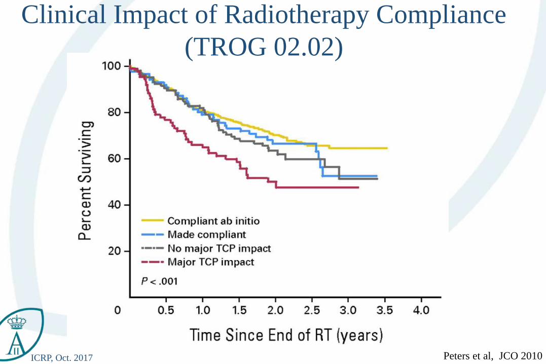
ICRP, Oct. 2017 Peters et al, JCO 2010
Clinical Impact of Radiotherapy Compliance
(TROG 02.02)
ICRP, Oct. 2017
Table of Content
• Future prospects
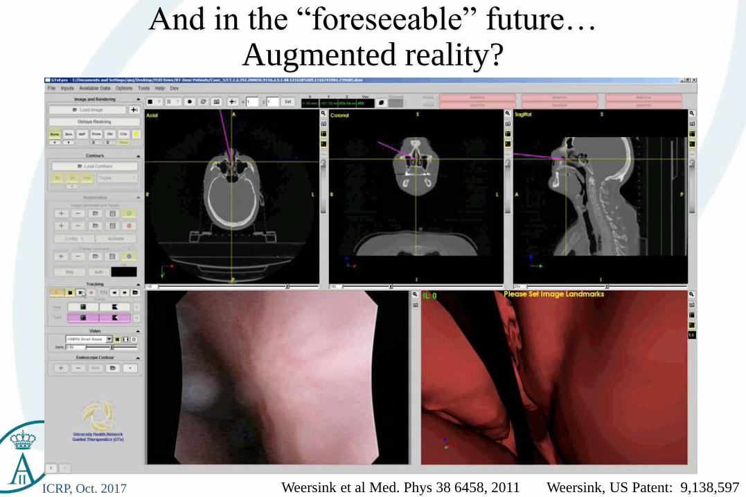
ICRP, Oct. 2017 Weersink et al Med. Phys 38 6458, 2011 Weersink, US Patent: 9,138,597
And in the “foreseeable” future…Augmented reality?
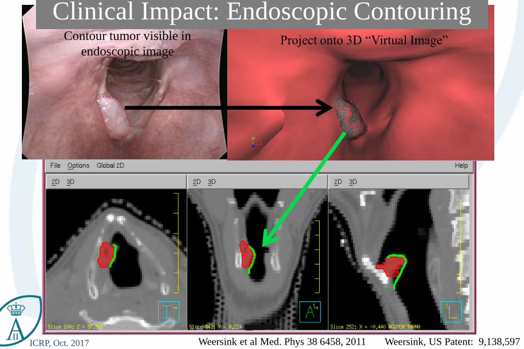
ICRP, Oct. 2017 Weersink et al Med. Phys 38 6458, 2011 Weersink, US Patent: 9,138,597
Contour tumor visible in
endoscopic imageProject onto 3D “Virtual Image”
Clinical Impact: Endoscopic Contouring
ICRP, Oct. 2017
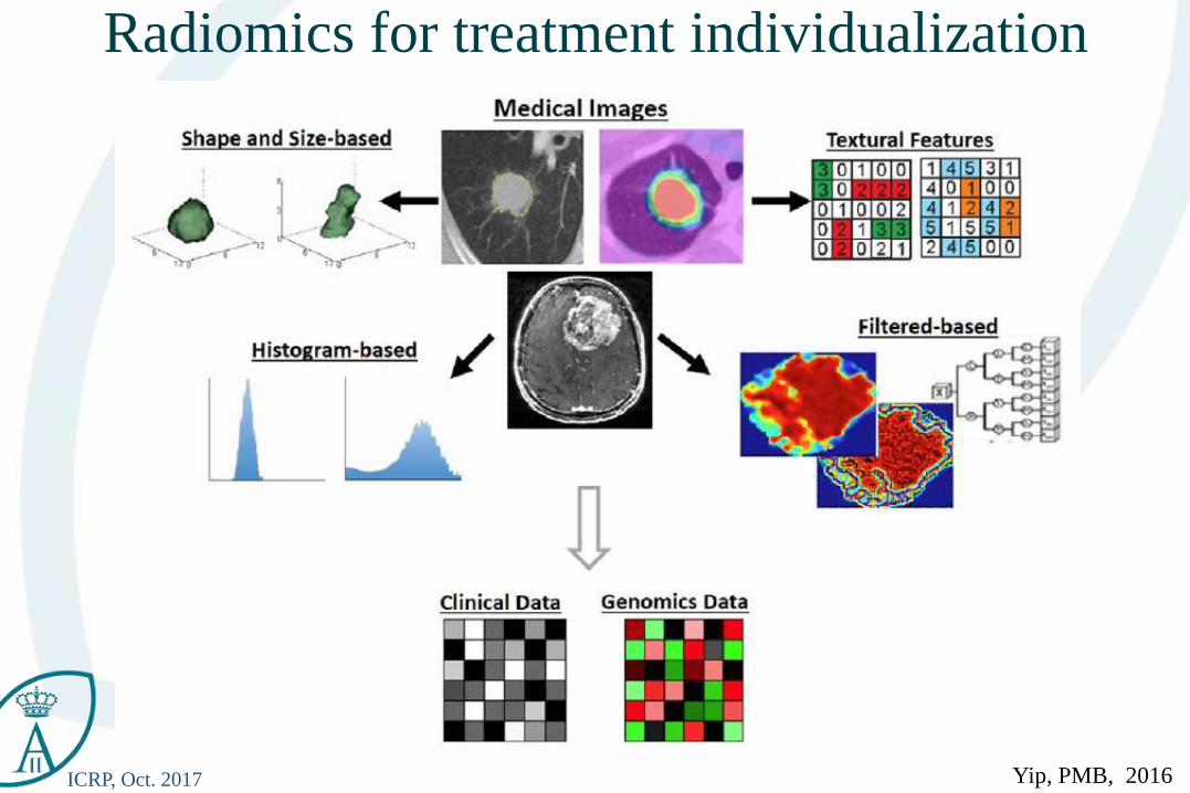
Radiomics for treatment individualization
Yip, PMB, 2016
ICRP, Oct. 2017
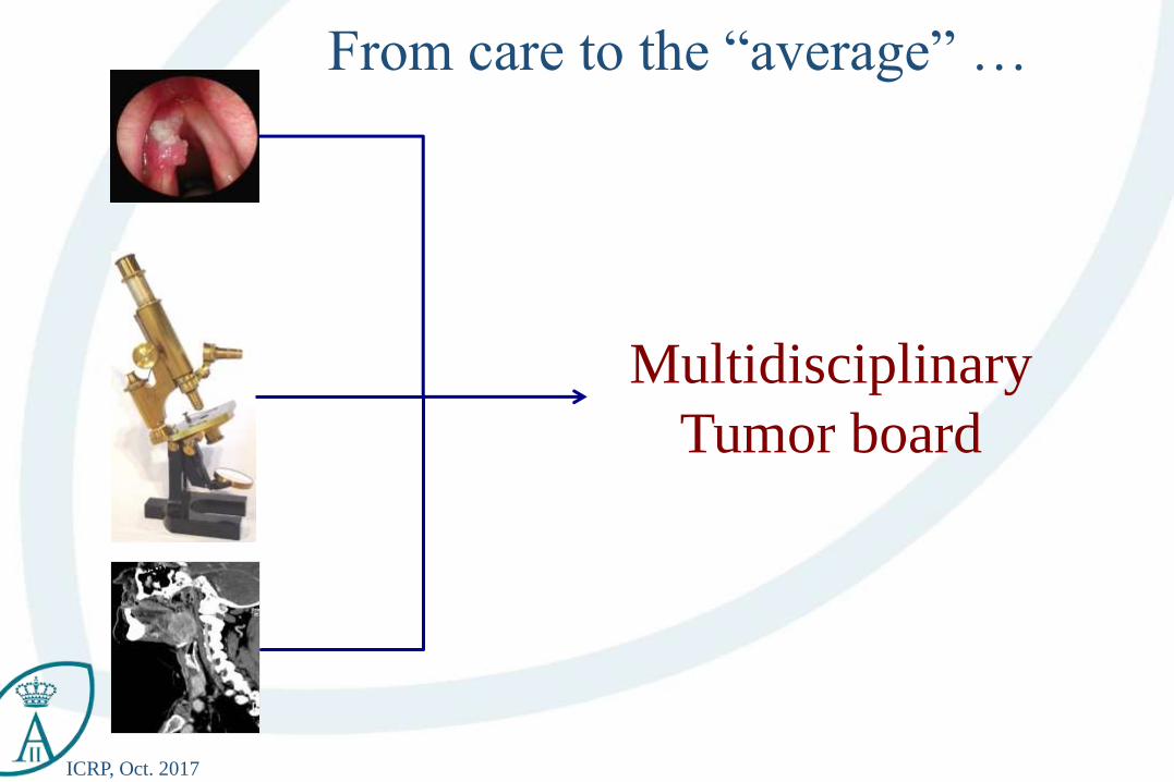
Multidisciplinary
Tumor board
From care to the “average” …
ICRP, Oct. 2017
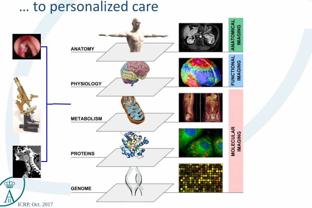
… to personalized care
ICRP, Oct. 2017
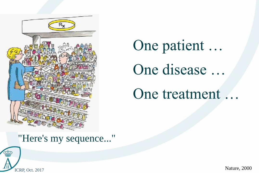
"Here's my sequence..."
Nature, 2000
One patient …
One disease …
One treatment …