multimodal longitudinal imaging of focal status...
TRANSCRIPT

276
Status epilepticus (SE), a condition characterized bypersistent seizures presents a difficult clinical problem. While itis clear that major motor seizures can lead to permanentpathological damage and altered physiological function, thechanges accompanying more focal discharges are less clear.1
Epilepsia partialis continua is a form of SE characterized bycontinuous, well-localized, clonic, focal motor myotonias.Epilepsia partialis continua is notoriously refractory to anti-
ABSTRACT: Background: Little is understood about the evolution of structural and functional brain changes during the course ofuncontrolled focal status epilepticus in humans. Methods: We serially evaluated and treated a nine-year-old girl with refractory focalstatus epilepticus. Long-term EEG monitoring, MRI, MRA, SPECT, intraoperative visualization of affected cortex, andneuropathological examination of a biopsy specimen were conducted over a three year time span. Imaging changes were correlated withsimultaneous treatment and EEG findings. Results: The EEG monitoring showed almost continuous spike discharges emanating initiallyfrom the right frontocentral area. These EEG abnormalities were intermittently suppressed by treatment with anesthetics. Over time,additional brain areas developed epileptiform EEG abnormalities. Serial MRI studies demonstrated an evolution of changes fromnormal, through increased regional T2 signal to generalized atrophy. An MRAdemonstrated dilatation of the middle cerebral artery stemon the right compared to the left with a broad distribution of flow-related enhancement. An 18FDG-PET scan showed a dramaticallyabnormal metabolic profile in the same right frontocentral areas, which modulated in response to treatment during the course of theillness. A right frontotemporal craniotomy revealed a markedly hyperemic cortical focus including vascular shunting. A sample ofresected cortex showed severe gliosis and neuronal death. Conclusions: The co-registration of structural and functional imaging and itscorrelation with operative and pathological findings in this case illustrates the relentless progression of regional and generalizedabnormalities in intractable focal status epilepticus that were only transiently modified by exhaustive therapeutic interventions.Increased flow through large vessels appeared to be shunted and did not translate into increased microvascular perfusion.
RÉSUMÉ: Imagerie longitudinale multimodale du status épilepticus focal. Introduction: La compréhension de l’évolution des changementsstructuraux et fonctionnels pendant le status épilepticus focal dans le cerveau humain est très limitée. Méthodes: Nous avons effectué une évaluationsériée chez une fillette de neuf ans que nous suivons pour un status épilepticus focal réfractaire au traitement. Le monitorage ÉEG à long terme, l’IRM,l’ARM, la TEMP, la visualisation peropératoire du cortex atteint et l’examen neuropathologique d’un spécimen anatomopathologique ont été réaliséssur une période de trois ans. Les changements à l’imagerie étaient corrélés au traitement et aux observations à l’ÉEG. Résultats: Le monitorage ÉEG amontré des décharges de pointes émanant initialement de la zone fronto-centrale droite. Ces anomalies électroencéphalographiques étaient suppriméesde façon intermittente par les anesthésiques. Avec le temps, des anomalies épileptiformes à l’ÉEG sont apparues dans d’autres régions du cerveau.L’IRM en série a montré une évolution des changements dans les zones initialement normales, soit d’un signal régional T2 augmenté à une atrophiegénéralisée. L’ARM a montré une dilatation du tronc de l’artère cérébrale moyenne du côté droit par rapport au gauche, avec une distribution large durehaussement relié au flot. Un PETscan au 18FDG a montré un profil métabolique très anormal dans cette zone fronto-centrale droite qui était modulépar le traitement pendant l’évolution de la maladie. À la craniotomie fronto-temporale droite, on a observé un foyer cortical très hyperhémique avecdérivation vasculaire. Une biopsie du cortex a montré une gliose sévère avec mort neuronale. Conclusions: L’imagerie structurelle et fonctionnelleutilisée conjointement, en corrélation avec les observations chirurgicales et anatomopathologiques, illustrent la progression implacable des anomaliesrégionales. Elles se sont éventuellement généralisées chez cette patiente présentant un status épilepticus réfractaire au traitement, et n’étaient modifiéesque temporairement par des interventions thérapeutiques exhaustives. L’accroissement du flot sanguin dans les gros vaisseaux semblait dévié etn’entraînait pas d’augmentation de la perfusion microvasculaire.
Can. J. Neurol. Sci. 2004; 31: 276-281
276 THE CANADIAN JOURNALOF NEUROLOGICALSCIENCES
Multimodal Longitudinal Imaging ofFocal Status EpilepticusColin P. Doherty, Andrew J. Cole, P. Ellen Grant, Alan Fischman, Elizabeth Dooling, Daniel B. Hoch, Tessa Hedley White, G. Rees Cosgrove
From the Epilepsy Service, Neurology Service, (CPD, AJC, DBH); Department ofNeuroradiology, (PEG); Nuclear Medicine Unit, Department of Radiology, (AF);Epilepsy Surgery Unit, Neurosurgical Service, (GRC); Pediatric Neurology,Department of Neurology, (ED); Department of Neuropathology, (THW)Massachusetts General Hospital and Harvard Medical School, Boston, USA.
RECEIVED APRIL 28, 2003. ACCEPTED INFINALFORM NOVEMBER 17, 2003.Reprint requests to: Andrew J. Cole, MGH Epilepsy Service, VBK-830,Massachusetts General Hospital, Fruit Street, Boston, Massachusetts 02114 USA
CASE REPORT

LE JOURNAL CANADIEN DES SCIENCES NEUROLOGIQUES
Volume 31, No. 2 – May 2004 277
epileptic medical therapy but may respond better to subpialtransection2 or resection. Epilepsia partialis continua is often asign of a serious progressive brain disease, perhaps mostcommonly Rasmussen’s chronic encephalitis1 but in some casesno clear etiology is established. We present a case of persistentand continuous focal motor seizures, which fulfilled the criteriafor epilepsia partialis continua. The evolution of multipleanatomic and functional imaging studies are discussed in light ofintra-operative and pathological findings.
CASE REPORT
A previously healthy nine-year-old left-handed girl presentedinitially to a local hospital with episodes of left facial twitching.Magnetic resonance imaging (MRI) was unremarkable, and she wastreated with carbamazepine. Six months later she woke with unremittingtwitching involving the left arm and face. She was admitted to a localhospital and required pentobarbital coma to control the events. Magneticresonance imaging on admission and one week later were normal. Shecontinued to have seizures after several attempts to wean frompentobarbital coma. On hospital day 14 she was transferred to this
hospital. On arrival she was unconscious but breathing spontaneously ona respirator. Clinically obvious focal seizure activity affecting the leftside was observed in episodes lasting 15 minutes every one to two hours(Figure 1). The EEG examination showed continuous sharp dischargesoccurring at 1 Hz arising from the right central region. An extensivebiochemical, metabolic and infectious work-up failed to reveal a cause.Cerebrospinal fluid examination on two occasions showed no cells, totalprotein of 11 and 16 mg/dl, appropriate glucose levels, minimallyelevated lactate (2.6 and 3.4 mmol/l, upper range of normal 2.2) and anormal electrophoresis pattern without oligoclonal bands. Bacterial,fungal and viral studies were negative. GluR3 antibodies were notdetected in serum or CSF. Magnetic resonance imaging of the brainshowed increased T2 signal in right hemisphere involving cortex,adjacent white matter and thalamus (Figure 2A). These changes wereconcordant with widespread metabolic abnormalities on 1 8f l u r o - 2 -deoxy-glucose positron emission tomography (18FDG-PET)(Figure 2B).A magnetic resonance angiogram (MRA) showed massive dilation of theright middle cerebral artery with increased flow related enhancement inthe middle cerebral artery territory (Figure 2C). Single photon emittedcomputed tomography (SPECT) scan after injection with 99Tc-HMPAO
Figure 3: Composite figure of imaging on day 28 of status epilepticus,one week after pentobarbital coma was initiated, two days beforesurgery. Axial FLAIR image showing improved signal change in rightfrontotemporal areas with only minimal subcortical hyperintensityremaining (2A). A 18FDG-PET from the same day scan showingimprovement in abnormal uptake of radio-labeled glucose in rightfrontotemporal regions, now highly localized to motor cortex.
Figure 1: 22 Lead EEG, day 13 of status epilepticus showinggeneralized background slowing and continuous 1 Hz sharp dischargesin the right central regions (C4) which were only occasionallyassociated with clinical seizure activity in the left arm and face.
Figure 2: Composite figure of imagingon day 14 of status epilepticus. Axialfluid attenuated inversion re c o v e ry(FLAIR) image showing incre a s e dsignal in cortical and white matterregions in right frontotemporal areas(2A). 1 8F D G - P E T scan showingi n c reased uptake of radio-labeledglucose in the same rightfrontotemporal region (2B). An MRangiogram showing dilatation of theright middle cerebral artery branchespresumably reflecting increased bloodflow to the active cortex (2C).
278
was normal. After a week in a pentobarbital coma, repeat MRIdemonstrated improvement of the T2 changes which were now almostresolved apart from mild subcortical hyperintensity (Figure 3A). Therewas a similar normalization of glucose uptake seen on an 18FDG-PETdone the same day (Figure 3B). However, with every attempted weanfrom the drug-induced coma the seizures returned.
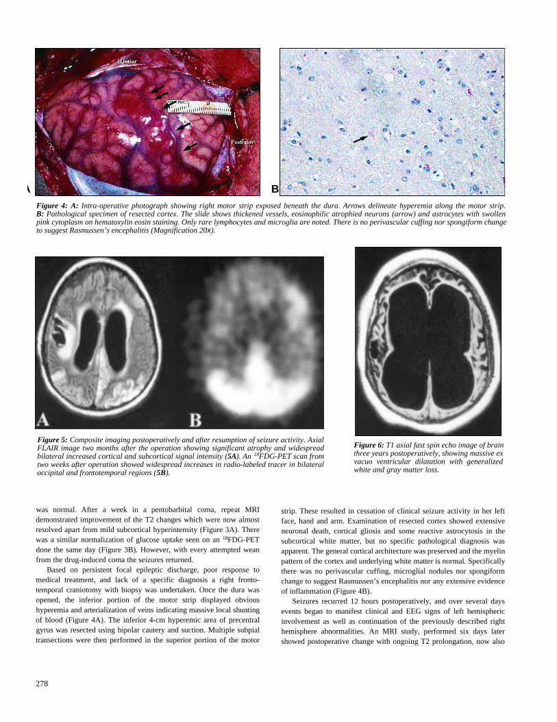
Based on persistent focal epileptic discharge, poor response tomedical treatment, and lack of a specific diagnosis a right fronto-temporal craniotomy with biopsy was undertaken. Once the dura wasopened, the inferior portion of the motor strip displayed obvioushyperemia and arterialization of veins indicating massive local shuntingof blood (Figure 4A). The inferior 4-cm hyperemic area of precentralgyrus was resected using bipolar cautery and suction. Multiple subpialtransections were then performed in the superior portion of the motor
strip. These resulted in cessation of clinical seizure activity in her leftface, hand and arm. Examination of resected cortex showed extensiveneuronal death, cortical gliosis and some reactive astrocytosis in thesubcortical white matter, but no specific pathological diagnosis wasapparent. The general cortical architecture was preserved and the myelinpattern of the cortex and underlying white matter is normal. Specificallythere was no perivascular cuffing, microglial nodules nor spongiformchange to suggest Rasmussen’s encephalitis nor any extensive evidenceof inflammation (Figure 4B).
Seizures recurred 12 hours postoperatively, and over several daysevents began to manifest clinical and EEG signs of left hemisphericinvolvement as well as continuation of the previously described righthemisphere abnormalities. An MRI study, performed six days latershowed postoperative change with ongoing T2 prolongation, now also
A B
Figure 6: T1 axial fast spin echo image of brainthree years postoperatively, showing massive exvacuo ventricular dilatation with generalizedwhite and gray matter loss.
Figure 4: A: Intra-operative photograph showing right motor strip exposed beneath the dura. Arrows delineate hyperemia along the motor strip. B: Pathological specimen of resected cortex. The slide shows thickened vessels, eosinophilic atrophied neurons (arrow) and astrocytes with swollenpink cytoplasm on hematoxylin eosin staining. Only rare lymphocytes and microglia are noted. There is no perivascular cuffing nor spongiform changeto suggest Rasmussen’s encephalitis (Magnification 20x).
Figure 5: Composite imaging postoperatively and after resumption of seizure activity. AxialFLAIR image two months after the operation showing significant atrophy and widespreadbilateral increased cortical and subcortical signal intensity (5A). An 18FDG-PET scan fromtwo weeks after operation showed widespread increases in radio-labeled tracer in bilateraloccipital and frontotemporal regions (5B).

LE JOURNAL CANADIEN DES SCIENCES NEUROLOGIQUES
Volume 31, No. 2 – May 2004 279
appearing to a lesser degree in the left hemisphere at a time whenongoing seizure activity appeared to involve the right side of the body.The patient was placed in a pentobarbital coma again. A follow-up studydone two months later showed the progressive nature of the process withgeneralized cerebral atrophy and multifocal areas of T2 prolongationconsistent with independent focal seizures from both hemispheres(Figure 5A). The new focus of activity on the left was confirmed by lefthemisphere abnormalities on concurrent 1 8F D G - P E T e x a m i n a t i o n(Figure 5B). Over the next six weeks several attempts to wean fromdrug-induced coma were made but the seizures invariably returned.Eventually on a combination of primidone, valproate and gabapentin,she was weaned off pentobarbital for the last time. She continued toremain refractory to medical treatment and was finally discharged tolong-term care in a persistent vegetative state with intermittentindependent seizure activity affecting both sides of her body, whichcontinues to this day. A final imaging study, done three years afterpresentation, demonstrates near complete loss of cortical volume withmassive compensatory dilatation of the ventricular system (Figure 6).
DISCUSSION
This case documents the evolution of imaging findings duringa catastrophic course of persistent and intractable focal SE. Wedemonstrate a clear correlation between the clinical,neurophysiological, intra-operative, and functional and anatomicimaging findings. Key findings include the correlation offunctional imaging abnormalities with disease activity andanatomic changes, the striking increase in local bloodflow, andthe lack of a primary pathological diagnosis. These findingssuggest that some cases of focal SE are idiopathic, in the originalsense of the word, “disease of the self”. While animal studies ofthe effects of prolonged status are well-described, the effects onthe human brain of such a catastrophic course are not well-understood.1,3
In the last 20 years, nuclear imaging studies such as SPECTand PET using both 18fluro-2-deoxy-glucose and 15oxygen havebeen used to noninvasively localize seizure foci through theanalysis of ictal bloodflow and interictal metabolism (seeSpencer for review4). Despite these studies, little is understoodabout the dynamics of flow in the microcirculation. Someevidence from animal studies suggests that there is uncoupling ofmetabolism and blood flow in focal seizures with therequirements of metabolism often outstripping the delivery ofoxygenated blood.5 On the other hand evidence from humanstudies suggests that in fact perfusion outstrips metabolicdemand.6 In 1933, Penfield7 described evidence of increasedlocal blood flow in the ictal zone during and after seizureactivity:
After the attack the cerebral arteries pulsate violently …Their color becomes a bright red and arteries which werenot seen to pulsate before the seizure may now begin to doso visibly. In fact this recovery may go so far that the veinsthemselves take on an arterial hue.7
This dramatic 70-year-old description is uncannily similar tothe gross operative findings in this case.
Advances in structural imaging have further supplementedfunctional localization in SE. Neuroimaging of the brain directlyafter prolonged epileptic seizures, either focal or generalized,may reveal focal cerebral abnormalities that are typically
transient. Cerebral hypodensities that enhance with contrast oncomputed tomography (CT) have been reported since 1980.8 Inmost cases the changes suggested the development of focalcerebral cytotoxic edema. By the early 1990s, reports of T2hyperintensity changes in focal areas associated with SE on MRIbegan appearing in the literature.8-18 A variety of metabolicevents which lead to edema have been invoked to explain thechanges including cytotoxic edema due to swelling of glial cells,increases in intracellular sodium concentration secondary toNa/K pump failure19 and vasogenic edema caused by excessiveglutamate release.12
There are several imaging features in this case that meritdiscussion. Firstly the initial imaging study, obtained six monthsprior to the onset of status was normal, as was an MRI obtaineda week after onset of SE. Widespread regional T2 changes firstappeared a full two weeks after SE began and were concordantwith both EEG and metabolic abnormalities on 18FDG-PET. Bycontrast, perfusion at the microvascular level as assessed by99TC-HMPAO-SPECT remained normal. We hypothesize thatthe increased blood flow visualized by MRA was due toincreased arterio-venous shunting as visualized intraoperatively.Magnetic resonance angiogram changes of this nature have beenreported in only one patient previously.12 The attenuation of theT2 and the metabolic abnormalities during pentobarbital comahas not been reported previously nor has concordance of theseT2 changes with visualized arterialization of surface veins andhyperemia in the same area at craniotomy. Unlike previousreports in which the T2 changes tended to resolve, favoring thecytotoxic edema hypothesis, the changes in this case persistedfor months after the onset of status presumably reflecting thedevelopment of dense gliosis and neuronal death asdemonstrated neuropathologically. To g e t h e r, our findingssuggest that cerebrovascular responses to SE in this patient,which appeared to be largely microvascular, were insufficient toadequately supply local neuronal demand, contributing to celldeath.
Perhaps the most dramatic structural finding in this case wasthe relentless progressive generalized brain atrophy over thecourse of the illness. The literature in this area includes detaileddescriptions of progressive focal atrophy on serial MRIs after SEin mesial structures.14,20 Similarly in more intractable diseasesuch as epilepsia partialis continua secondary to Rasmussen’sencephalitis, atrophy is extensive but appears limited to thea ffected hemisphere.2 1 The explanation for the widespreadatrophy in our patient is not clear. The role of three weeks ofintravenous dexamethasone in generalized brain atrophy in thiscase must be considered, but the degree of atrophy incombination with previous experience with steroid treatment inother disease states suggests that additional factors must havecontributed to the development of widespread atrophy. A tpresent we cannot differentiate between the roles of numerousbiochemical consequences of repeated seizures such as ionicfluxes, kinase activation, early and late gene expression, proteinexpression and modification, synaptic reorganization, the rolesof indirect consequences of seizures such as local hypoxia,hypotension, and acidosis (see Cole3 for review).
Catastrophic epilepsy arising in childhood may be due tostructural, infectious or metabolic disturbances. Structuralabnormalities include cortical dysgenesis, congenital

THE CANADIAN JOURNAL OF NEUROLOGICAL SCIENCES
280
demyelinating disease, and vascular insults such as venousocclusion, anoxic injury and major trauma. Infectious etiologiesinclude viral encephalitis, focal or multifocal cerebritis, andperhaps chronic encephalitis (Rasmussen’s disease), although nocausative agent has been identified, and recent studies havesuggested a potential autoimmune basis for the latter. Metabolicdisorders as a cause of catastrophic seizures are generallydescribed in terms of seizure types such as childhoodencephalopathy with myoclonus and myoclonic–astatic seizures(see Shields22 for discussion). Recently there have been anumber of case reports of rare metabolic disorders engenderingsevere intractable seizures, including Alpers syndrome,2 3 , 2 4
selenium deficiency, 2 5 peroxisomal disease,2 6 g l u t h a t h i o n esynthetase deficiency,27 glucose transporter deficiency syndromeand sulphite oxidase deficiency.28 Most of these were identifiedin association with infantile seizures soon after birth rather thanin later childhood. These protean conditions demonstrate thatwhile there is no single etiology for, nor common pathologicalabnormality in catastrophic epilepsy arising in childhood, all ofthese conditions are associated with a poor neurologicaloutcome. The presence of epilepsia partialis continua appears toconfer a particularly poor prognosis regardless of the cause.29
Large case series of catastrophic childhood epilepsy are rare.The natural history of Rasmussen’s encephalitis has beenrecently documented in a longitudinal study of 16 patientsdemonstrating typical neuroradiographic features and also asubgroup of slightly older patients who may have a slight betterprognosis.30 In a study of 15 children without Rasmussen’sencephalitis, although it was possible to classify the patients withrespect to seizure localization, either multi-focal or lateralized,the outcome in all cases was poor.24 Of the 15 cases, only eighthad a unifying diagnosis. Three had the Alpers syndrome, threewere thought to have cerebral dysgenesis, one had anoxicdamage, one had congenital cytomegalovirus, and seven patientshad no specific diagnosis. Other efforts at defining the syndromeof catastrophic epilepsy by seizure phenomenology have beenequally unrevealing. Coppola31 described an epileptic conditionin a number of patients characterized by intractable focalseizures and poor intellectual outcomes. Inclusion in the grouprequired an unidentified cause, but the coining of term“malignant developmental arrest” may have been the most usefulcontribution. In those that do have a common identifiable causesuch as acquired cortical damage, knowledge of the etiologyappears to have little impact on the course of the disease. In aretrospective review of catastrophic epilepsy in 42 patients witha history of encephalitis, only a small subset of those withseizures localized by EEG to one temporal lobe appeared to havea favourable prognosis.32
Recent guidelines for evaluating patients with catastrophicepilepsy have concentrated on imaging in an effort to identifylocalized abnormalities such as cerebral dysgenesis so that eitherfocal cortical resection or hemispherectomy may be added to thetreatment options. This approach is based on follow-up studies ofsmall numbers of surgical patients with catastrophic seizureswho have had sustained periods of seizure freedom and evendemonstrated a tendency to “catch-up” developmentally ifoperated on early enough.33
In our case the etiology remains obscure. The biochemicaland metabolic evaluation was negative. The neuropathological
examination of the biopsy specimen did not reveal a specificdiagnosis, although it was taken from the center of the mostconsistently involved cortical region and included surroundingtissue and subcortical white matter. Detailed examinationrevealed only evidence of cell death and gliosis, with noindication of cerebral dysgenesis, inflammatory changes, viralinclusions or tumor. While it is possible that subtle dysgenesiscould have been missed, especially after considerable neuronalloss had occurred, no evidence of a structural abnormality wasapparent on MRI prior to the development of seizure-relatedstructural changes. We suggest that SE in this patient was indeedidiopathic in the strict sense of the word, that is a consequence ofsome unique constellation of activity, connectivity, andstimulation in this individual’s brain that resulted indestabilization of the normal neuronal network architecture. Thisview, which is conjectural, is proposed as a hypothesis that mightbe tested using techniques of computational modeling.
This detailed case study of the catastrophic course ofepilepsia partialis continua in a child reveals several intriguingassociations. The employment of structural and functionalimaging illustrates the tight correlation of abnormal electrical,metabolic activity and the visualized cortical blood flow.Furthermore, the development of local and generalized structuraland metabolic changes were only transiently affected by a rangeof therapies demonstrating the relentless progressive nature ofthe damage caused by such constant activity.
ACKNOWLEDGEMENTS
We thank the patient and her family for their cooperation and themedical and surgical staff at Massachusetts General Hospital for theircare over the course of the patient’s illness.
REFERENCES
1. Wasterlain C, Fujikawa D, Penix L, Sankar R. Pathophysiologicalmechanisms of brain damage from status epilepticus. Epilepsia1993; 34: S37-S53.
2. Molyneux P, Barker R, Thom M, et al. Successful treatment ofintractable epilepsia partialis continua with multiple subpialtranssections. JNNP1998; 65: 137-138.
3. Cole A. Is epilepsy a progressive disease? The neurobiologicalconsequences of epilepsy. Epilepsia 2002; 41(Suppl 2): S13-S22.
4. Spencer S, Bautista E. Functional neuroimaging in localization ofthe ictal onset zone. In: Henry T, Duncan J (Eds.) FunctionalImaging in the Epilepsies, Philadelphia: Lippincott Williams andWilkins, 2000: 285-296.
5. Bruehl C, Hagemann G, Witte O. Uncoupling of blood flow andmetabolism in focal epilepsy. Epilepsia 1998; 39: 1235-1242.
6. Franck G, Sadzot B, Salmon E, et al. Regional cerebral bloodflowand metabolic rates in human focal epilepsy and statusepilepticus. Adv Neurol 1986; 44 : 935-948.
7. Penfield W. The evidence of a cerebral vascular mechanism inepilepsy. Ann Intern Med 1933; 7: 303-310.
8. Henry T, Drury I, Brunberg J, et al. Focal cerebral magneticresonance changes associated with partial status epilepticus.Epilespia 1994; 35: 35-41.
9. Callahan D, Noetzel M. Prolonged absence status epilepticusassociated with carbamazepine therapy, increased intracranialpressure and transient MRI abnormalities. Neurology 1992; 42:2198-2201.
10. Fazekas F, Kapeller P, Schmidt R, et al. Magnetic resonanceimaging and spectroscopy findings after focal status epilepticus.Epilepsia 1995; 36: 946-949.
11. Kramer R, Luders H, Lesser R, et al. Transient focal abnormalities

LE JOURNAL CANADIEN DES SCIENCES NEUROLOGIQUES
Volume 31, No. 2 – May 2004 281
of neuroimaging studies during focal status epilepticus. Epilepsia1987; 28: 528-532.
12. Lansberg M, O’Brien M, Norbash A, et al. MRI abnormalitiesassociated with partial status epilepticus. Neurology 1999;52:1021-1027.
13. Lazeyras F, Blanke O, Zimine I, et al. MRI, 1H-MRS, andfunctional MRI during and after prolonged non-convulsiveseizure activity. Neurology 1999; 55: 1677-1682.
14. Meierkord H, Wieshmann U, Niehaus L, Lehmann R. Structuralconsequences of status epilepticus demonstrated with serialmagnetic resonance imaging. Acta Neurologica Scadinavica1997; 96: 127-132.
15. Najm I, Wang Y, Shedid D, et al. MRS metabolic markers ofseizures and seizure-induced neuronal damage. Epilepsia 1998;39: 244-250.
16. Riela A, Sires B, Penry J. Transient magnetic resonance imagingabnormalities during partial status epilepticus. J Child Neurol1991; 6:143-145.
17. Wieshmann U, Woermann F, Lemieux L, et al. Development ofhippocampal atrophy: a serial magnetic resonance imaging studyin a patient who developed epilepsy after generalized statusepilepticus. Epilepsia 1997; 38: 1238-1241.
18. Yaffe K, Ferriero D, Barkovich J, Rowley H. Reversible MRIabnormalities following seizures. Neurology 1995; 45: 104-108.
19. Zhong J, Petroff O, Prichard J. Barbiturate-reversible reduction ofwater diffusion coefficient in flurothyl-induced status epilepticusin rats. Magn Reson Med 1995; 33: 253-256.
20. Nohria V, Lee N, Tien R, et al. Magnetic resonance imagingevidence of hippocampal sclerosis in progression: a case report.Epilepsia 1994; 35: 1332-1336.
21. Bien C, Urbach H, Deckert M, et al. Diagnosis and staging ofR a s m u s s e n ’s encephalitis by serial and histopathology.Neurology 2002; 58: 250-257.
22. Shields WD. Catastrophic epilepsy in childhood. Epilepsia 2000;41(Suppl 2):S2-S6.
23. Gauthier-Villars M, Landrieu P, Cromier-Daire V, et al. Respiratorychain deficiency in Alpers syndrome. Neuropediatrics 2001;32:150-152.
24. Ishii K, Oguni H, Hayashi K, et al. Clinical study of catastrophicinfantile epilepsy with focal seizures. Pediatr Neurol 2002; 27:369-377.
25. Ramaekers VT, Calomme M, Vanden Berghe D, Makropoulos W.Selenium deficiency triggering intractable seizures.Neuropediatrics 1994; 25:217-223.
26. Takahashi Y, Suzuki Y, Kumazakai K, et al. Epilepsy in peroxisomaldiseases. Epilepsia 1997; 38:182-188.
27. Ristoff E, Mayatepek E, Larsson A. Long-term clinical outcome inpatients with glutathione synthetase deficiency. J Pediatr 2001;139:79-84.
28. Slot HM, Overweg-Plandsoen W, Bakker HD, et al. Molybdenum-cofactor deficiency: an easily missed cause of neonatalconvulsions. Neuropediatrics 1993; 24:139-142.
29. Baram TZ, Mitchell WG, Snead OC. Prognostic significance ofacute epilepsia partialis continua. Pediatr Neurol 1991; 7(2): 144-146.
30. Bien CG, Widman G, Urbach H, et al. The natural history ofRasmussen’s encephalitis. Brain 2002; 125(8): 1751-1759.
31. Coppola G, Plouin P, Chiron C, Robain O, Dulac O. Migratingpartial seizures in infancy: a malignant disorder with develop-mental arrest. Epilepsia 1995; 36:1017-1024.
32. Trinka E, Dubeau F, Andermann F, et al. Clinical findings, imagingcharacteristics and outcome in catastrophic post-encephaliticepilepsy. Epileptic Disord 2000; 2(3):153-162.
33. Wyllie E. Surgery for catastrophic localization-related epilepsy ininfancy. Epilepsia 1996; 37(Suppl 1): S22-S25.