multimodal ultrasound tomography for breast imaging: a ...keywords: breast cancer, mammography,...
TRANSCRIPT
TECHNICAL NOTE Open Access
Multimodal ultrasound tomography forbreast imaging: a prospective study ofclinical feasibilityS. Forte* , S. Dellas, B. Stieltjes and B. Bongartz
Abstract
Background: To describe the clinical set-up and evaluate the feasibility of multimodal ultrasound tomography (MUT)for breast imaging.
Methods: Thirty-two consecutive patients referred for breast imaging and 24 healthy volunteers underwent MUT. Inthe 32 patients, the examination discomfort was compared to that of mammography (n = 31), handheld ultrasound(HUS) (n = 27) and magnetic resonance imaging (MRI) (n = 4) on a scale from 1 (lowest discomfort) to 10 (highestdiscomfort). MUT investigation time was recorded. Findings automatically detected by MUT were correlated withconventional imaging and biopsy results.
Results: Breast MUT was well tolerated by all 56 participants; 55 bilateral exams were uneventful. During one exam,the digitalisation card failed and the exam was successfully repeated within three days. Mean examination discomfortwas 1.6 (range = 1–5) for MUT, 1.5 (range = 1–5) for HUS, 5.3 (range = 3–7) for MRI, and 6.3 (range = 1–10) formammography. MUT examination time was 38 ± 6 min (mean ± standard deviation). In the patients referredfor breast imaging, MUT detected four lesions and indicated malignancy in three of these cases. These findings wereconfirmed by additional imaging and biopsy.
Conclusion: MUT is feasible in a clinical context considering examination time and patient acceptance. These interestinginitial diagnostic findings warrant further studies.
Keywords: Breast cancer, Mammography, Multimodal ultrasound tomography (MUT), Three-dimensional (3D),Transmission, Ultrasound
Key points
� This is the first report on the use of MUT in aclinical setting.
� MUT is feasible in a clinical setting, safe, and welltolerated.
� MUT showed a potential for automated lesiondetection and differentiation of breast lesions.
BackgroundBreast cancer is the most common malignancy and theleading cause of cancer-related mortality in female popu-lation [1, 2]. Since an effective primary prevention of
breast cancer is not attainable, the main focus of medicalcare is on secondary prevention, i.e. early detection.Screening programs based on x-ray mammography areestablished. However, mammography faces several chal-lenges. First, the density of glandular tissue may obscurelesions; second, it requires a non-negligible radiationdose [3]; and third, breast compression causes patientdiscomfort. Several studies have demonstrated that fordense breasts, handheld ultrasound (HUS) enables thedetection of additional and smaller lesions [2, 4–6], butthe time and skill necessary and lack of uniformity hasdiscouraged its widespread use in a screening context.Automated breast ultrasound systems have been pro-posed as alternative reducing operator dependence com-pared to HUS [2, 7], but the number of radiologists* Correspondence: [email protected]
University of Basel Hospital, Clinic for Radiology and Nuclear Medicine,Petersgraben 4, 4031 Basel, Switzerland
European RadiologyExperimental
© The Author(s). 2017 Open Access This article is distributed under the terms of the Creative Commons Attribution 4.0International License (http://creativecommons.org/licenses/by/4.0/), which permits unrestricted use, distribution, andreproduction in any medium, provided you give appropriate credit to the original author(s) and the source, provide a link tothe Creative Commons license, and indicate if changes were made.
Forte et al. European Radiology Experimental (2017) 1:27 DOI 10.1186/s41747-017-0029-y
trained for interpreting this modality is still limited anda comparable recall rate to HUS was reported [7–9].Thus, a method that can detect breast cancer in an early
stage is needed. It should be independent from the glan-dular density, radiation-free, investigator independent, andcost-effective. In 2011, a novel three-dimensional (3D) im-aging technology for detection of breast cancer, multi-modal ultrasound tomography (MUT), was introduced[10]. This method uses ultrasound in transmission modeto obtain tomographic images of the breast. Acousticattributes of each voxel like refractivity, frequency-dependent attenuation, and dispersion can be recordedand combined to yield an automated lesion detection anddifferentiation [11–14].However, previous studies reported findings in se-
lected, high-risk women and feasibility has not beenevaluated previously. In the present technical develop-ment, we describe the clinical setup and evaluate thefeasibility of MUT-based breast imaging.
MethodsThis prospective feasibility study was approved by ourInstitutional Review Board (EKBB196/12) and writteninformed consent was obtained from all participants.From January till March 2014, we included 24 healthyvolunteers (mean age 43 ± 11 years [mean ± standard de-viation {SD}], range 27–63 years). Furthermore, withinthe same time frame, we sought to include patients re-ferred for breast imaging in a consecutive fashion. Exclu-sion criteria for patients were: mammography or HUSimages of the breast older than one month; open wound;breast diameter > 20 cm; lactation; age < 18 years; andlack of informed consent. In three months, 856 patientswere referred for breast imaging; 32 (4%) were included(mean age 55 ± 9 years, range 39–76 years). The mainreason for exclusion was lack of informed consent. Ofthese 32 participants, four showed suspicious findingsand underwent ultrasound-guided core needle biopsy(14-gauge), one patient had additional magnetic resonanceimaging (MRI)-guided (8-gauge) vacuum-assisted biopsy.For MUT examination, we used the first available
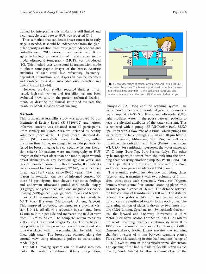
MUT Mark II system (Mastoscopia, Athens, Greece).This improved prototype, compared to a previous ver-sion [10, 15, 16] allows a reduction in scan time from15 min to 9 min per side and increased the field of viewfrom 16 cm to 20 cm. The complete system measures210 × 130 × 110 cm and weighs 350 kg. The participantwas positioned in the prone position and one breast at atime was placed within the scanning chamber which wasfilled with water. The breast was scanned along thecoronal view using ultrasound pulses in transmissionmode (Fig. 1).The MUT imaging system can be divided into two
parts: the water conditioner (Onda Corporation,
Sunnyvale, CA, USA) and the scanning system. Thewater conditioner continuously degasifies, de-ionises,heats (kept at 25–30 °C), filters, and ultraviolet (UV)-light irradiates water in the pause between patients tokeep the physical attributes of the water constant. Thisis achieved with a pump (SE-PSH000HM1000, SEKOSpa, Italy) with a flow rate of 2 l/min, which pumps thewater from the tank through a 5-μm and 10-μm filter intandem (Pentek, Milwaukee, WI, USA) as well as amixed-bed de-ionisation resin filter (Pentek, Sheboygan,WI, USA). For sanitisation purposes, the water passes anUV-C lamp (Puro-Tap, Puro-Parma, Italy). The othercycle transports the water from the tank into the scan-ning chamber using another pump (SE-PSH000HM1000,SEKO Spa, Italy) with a maximum flow rate of 2 l/minand once more passes an identical UV-C lamp.The scanning system includes two translating plates
(receiver and transmitter) with two columns of 4-mm-sized transducers each (Imasonic, Voray sur l’Ognon,France), which define four coronal scanning planes withan inter-plane distance of 16 mm. The distance betweenthe two columns of transducers is 100 mm. The distancebetween the plates is 240 mm and transmit–receivetransducers are positioned exactly facing each other. Thetranslating motion of plates is driven by two linear mo-tors (PS01 Linmot, Spreitenbach, Switzerland) that con-trol the forward and backward movement. A thirdmotor (Flex Drive Baldor, Fort Smith, AR, USA) rotatesthe whole scanning chamber continuously from 0 to180° at each scanning plane and a fourth motor (R88mOmron/Yaskawa, Kioto, Japan) elevates the scanningchamber in steps of 4 mm between scanning planes.This allows 3D scanning in four half-rotations (range of0–180°) over 64 mm in the vertical/coronal dimension.The opening of the bed is made of flexible Lexan (Sabic,Riyadh, Saudi Arabia) to allow scanning close to the
Fig. 1 Schematic image of patient positioning and setting for MUT.The patient lies prone. The breast is positioned through an openinginto the scanning chamber (1). The combined transducer andreceiver rotate and scan the breast (2). Courtesy of Mastoscopia
Forte et al. European Radiology Experimental (2017) 1:27 Page 2 of 6

chest wall. This renders the starting scanning positionpatient-dependent. To standardise this position, thescanning chamber is raised until it touches the Lexan.An automated control system assures the safety of thisinitialisation, followed by lowering the scanning chamberby 16 mm before starting the scan. A customised elec-tronic pulser device (Mastoscopia, Athens, Greece) gen-erates sequences of broadband ultrasound pulses(frequency range of 1–5 MHz). The first arrival pulse iscaptured by the receiver and digitised (Octopus GaGe,Lockport, NY, USA).As these signals are captured at multiple azimuthal-
angular positions for each 180° rotation for each coronalplane, an inverse Radon transform allowed the formationof a two-dimensional tomographic image for each acous-tic attribute extracted from the received pulse in relationto its transmitted counterpart [11, 12]. The extractedacoustic attributes are the refractivity index, frequency-dependent attenuation, and dispersion. The values ofthese attributes are normalised by their water-throughcounterparts in order to obtain a universal validity ofthese parameters. A specially designed algorithm com-bines the multimodal information for each pixel to yielda composite image that contains the diagnostically inter-pretable information. Based on pilot studies [15], thethus derived composite index determines the probabilityof malignancy within a tissue voxel.A putative diagnostic threshold of composite index
= 1.0 was set for malignant lesions while precancerousconditions corresponded to values in the range of0.6–1.0, benign lesions to values < 0.6, and normalbreast tissue < 0.1 [15]. For improved visualisation, thecomposite index value was transformed into a diagnosticindex (DI) where values from –1.0 to 0.0 represent water(dark blue), values of 0.1–1.5 represent fat (light blue),values of 1.5–3.0 represent normal glandular breast tissue(turquoise/yellow), values of 3.1–5.0 represent benignlesions (orange), and values > 5.0 represent malignancy(red). The different maps were displayed within 3 minafter the scan.Total exam time was measured from entering the door
until leaving the examination room.In the 32 patients, mammography, HUS, and breast
MRI were also performed, using standard protocols,meaning for mammography two views of each breast,cranio-caudal and mediolateral oblique views (SeleniaDimensions, Hologic, Bedford, MA, USA), for HUS alinear 15-4 MHz broadband linear array transducer(Aixplorer, SuperSonic Imagine, Aix-en-Provence,France) performed by a radiologist with documentationof at least each quadrant and retromamillar region, andMRI with T2-weighted fast spin echo and T1-weighteddynamic contrast-enhanced images (3 T Skyra, Siemens,Erlangen, Germany).
To these 32 patients, a questionnaire was adminis-tered. The exams were rated on a scale of 1–10, where 1represented no discomfort at all and 10 represented a re-fusal to repeat. All MUT scans were compared to theimaging available (Table 1). Score distributions of examdiscomfort were tested for normality using the Shapiro–Wilk W test and significant differences in discomfort be-tween different exam types were evaluated using a two-sided Student’s t-test. A significance level of 0.050 wasdeemed significant. Statistical evaluation was performedusing SPSS (Version 22.0). Furthermore, on the experi-ence of the 24 volunteers, all 32 patients were asked toevaluate MUT-specific discomfort on a scale of 0–2(none, slight and strong), which was pain at the costalarch and neck, but also the temperature of the water.
ResultsOf the 56 participants, 55 were investigated uneventfullywith no aborts. One participant had to be re-scanned asa digitisation card failed. Scores for patient discomfortwere normally distributed for MUT, HUS, and mam-mography (p = 0.956, 0.660, and 0.693, respectively).MRI could not be evaluated statistically since only fourparticipants underwent MRI. MUT was rated significantlybetter compared to mammography (p = 0.001) and slightlybut not significantly better compared to HUS (p = 0.205)(Table 1). The time required from entering the room todiscussing the results was 38 ± 6 min (mean ± SD). Con-sidering MUT-related patient acceptance, most of the 32patients referred no or just slight discomfort (Table 2).The reading took just a few seconds as the possible
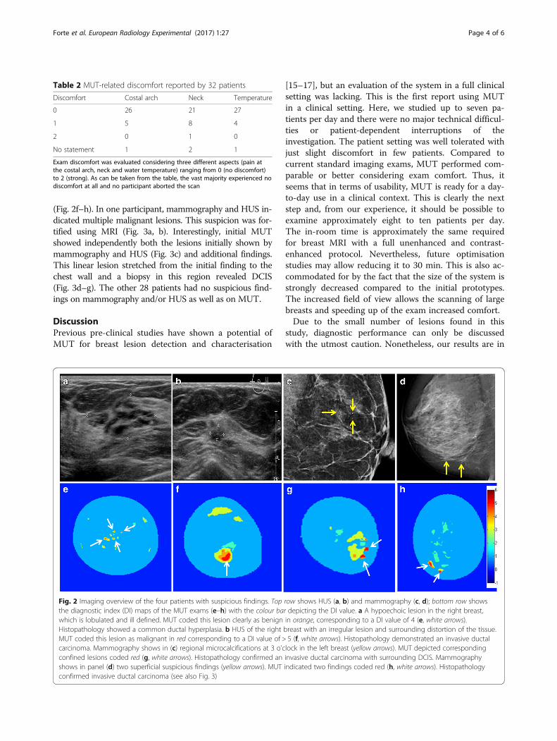
malignancy is marked red. Of the 32 patients, four had asuspicious finding at mammography and/or HUS(Fig. 2a–d) and underwent biopsy. One of the four suspi-cious findings was proven to be fibrosis with commonductal hyperplasia. The other three findings were proven tobe breast cancers: one ductal carcinoma in situ (DCIS) andtwo invasive ductal carcinomas. As shown in Fig. 2e–h,MUT allowed to detect all four suspicious finding. Further-more, the benign lesion was correctly coded orange (Fig. 2e)and the three malignancies were correctly coded red
Table 1 Examination discomfort reported by patients for thefour modalities
Modality Mammography Handheld US MRI MUT
Mean score 6.3 1.6 5.3 1.5
Score SD (range) 2.6 (1–10) 1.0 (1–5) 2.1 (3–7) 0.7 (1–5)
Patients 31 27 4 32
SD standard deviationDiscomfort level was scored ranging from 1 (no discomfort) to 10 (unwilling torepeat the exam) for mammography, HUS, MRI, and MUT. Indicated are themean score (second line) and the SD as well as the range of the score (thirdline). The number of available exams can be found in the last row (patients).Significant differences in discomfort level were found between MUT andmammography (see text)
Forte et al. European Radiology Experimental (2017) 1:27 Page 3 of 6
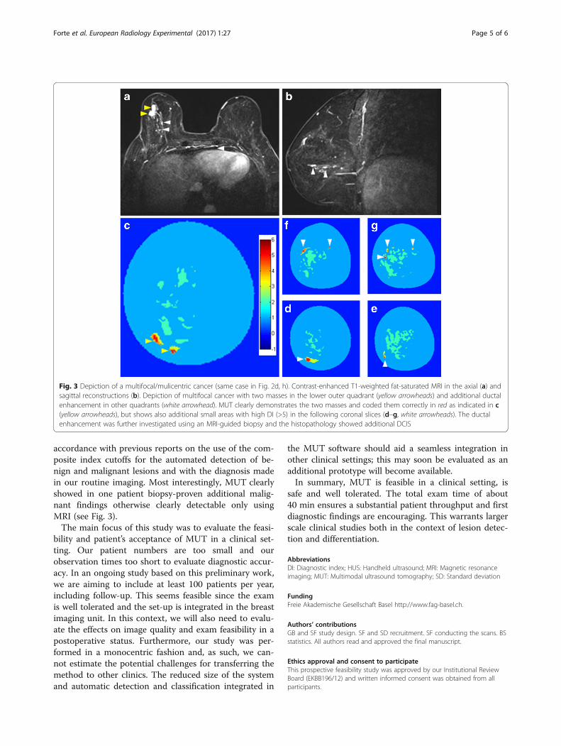
(Fig. 2f–h). In one participant, mammography and HUS in-dicated multiple malignant lesions. This suspicion was for-tified using MRI (Fig. 3a, b). Interestingly, initial MUTshowed independently both the lesions initially shown bymammography and HUS (Fig. 3c) and additional findings.This linear lesion stretched from the initial finding to thechest wall and a biopsy in this region revealed DCIS(Fig. 3d–g). The other 28 patients had no suspicious find-ings on mammography and/or HUS as well as on MUT.
DiscussionPrevious pre-clinical studies have shown a potential ofMUT for breast lesion detection and characterisation
[15–17], but an evaluation of the system in a full clinicalsetting was lacking. This is the first report using MUTin a clinical setting. Here, we studied up to seven pa-tients per day and there were no major technical difficul-ties or patient-dependent interruptions of theinvestigation. The patient setting was well tolerated withjust slight discomfort in few patients. Compared tocurrent standard imaging exams, MUT performed com-parable or better considering exam comfort. Thus, itseems that in terms of usability, MUT is ready for a day-to-day use in a clinical context. This is clearly the nextstep and, from our experience, it should be possible toexamine approximately eight to ten patients per day.The in-room time is approximately the same requiredfor breast MRI with a full unenhanced and contrast-enhanced protocol. Nevertheless, future optimisationstudies may allow reducing it to 30 min. This is also ac-commodated for by the fact that the size of the system isstrongly decreased compared to the initial prototypes.The increased field of view allows the scanning of largebreasts and speeding up of the exam increased comfort.Due to the small number of lesions found in this
study, diagnostic performance can only be discussedwith the utmost caution. Nonetheless, our results are in
Table 2 MUT-related discomfort reported by 32 patients
Discomfort Costal arch Neck Temperature
0 26 21 27
1 5 8 4
2 0 1 0
No statement 1 2 1
Exam discomfort was evaluated considering three different aspects (pain atthe costal arch, neck and water temperature) ranging from 0 (no discomfort)to 2 (strong). As can be taken from the table, the vast majority experienced nodiscomfort at all and no participant aborted the scan
Fig. 2 Imaging overview of the four patients with suspicious findings. Top row shows HUS (a, b) and mammography (c, d); bottom row showsthe diagnostic index (DI) maps of the MUT exams (e–h) with the colour bar depicting the DI value. a A hypoechoic lesion in the right breast,which is lobulated and ill defined. MUT coded this lesion clearly as benign in orange, corresponding to a DI value of 4 (e, white arrows).Histopathology showed a common ductal hyperplasia. b HUS of the right breast with an irregular lesion and surrounding distortion of the tissue.MUT coded this lesion as malignant in red corresponding to a DI value of > 5 (f, white arrows). Histopathology demonstrated an invasive ductalcarcinoma. Mammography shows in (c) regional microcalcifications at 3 o’clock in the left breast (yellow arrows). MUT depicted correspondingconfined lesions coded red (g, white arrows). Histopathology confirmed an invasive ductal carcinoma with surrounding DCIS. Mammographyshows in panel (d) two superficial suspicious findings (yellow arrows). MUT indicated two findings coded red (h, white arrows). Histopathologyconfirmed invasive ductal carcinoma (see also Fig. 3)
Forte et al. European Radiology Experimental (2017) 1:27 Page 4 of 6
accordance with previous reports on the use of the com-posite index cutoffs for the automated detection of be-nign and malignant lesions and with the diagnosis madein our routine imaging. Most interestingly, MUT clearlyshowed in one patient biopsy-proven additional malig-nant findings otherwise clearly detectable only usingMRI (see Fig. 3).The main focus of this study was to evaluate the feasi-
bility and patient’s acceptance of MUT in a clinical set-ting. Our patient numbers are too small and ourobservation times too short to evaluate diagnostic accur-acy. In an ongoing study based on this preliminary work,we are aiming to include at least 100 patients per year,including follow-up. This seems feasible since the examis well tolerated and the set-up is integrated in the breastimaging unit. In this context, we will also need to evalu-ate the effects on image quality and exam feasibility in apostoperative status. Furthermore, our study was per-formed in a monocentric fashion and, as such, we can-not estimate the potential challenges for transferring themethod to other clinics. The reduced size of the systemand automatic detection and classification integrated in
the MUT software should aid a seamless integration inother clinical settings; this may soon be evaluated as anadditional prototype will become available.In summary, MUT is feasible in a clinical setting, is
safe and well tolerated. The total exam time of about40 min ensures a substantial patient throughput and firstdiagnostic findings are encouraging. This warrants largerscale clinical studies both in the context of lesion detec-tion and differentiation.
AbbreviationsDI: Diagnostic index; HUS: Handheld ultrasound; MRI: Magnetic resonanceimaging; MUT: Multimodal ultrasound tomography; SD: Standard deviation
FundingFreie Akademische Gesellschaft Basel http://www.fag-basel.ch.
Authors’ contributionsGB and SF study design. SF and SD recruitment. SF conducting the scans. BSstatistics. All authors read and approved the final manuscript.
Ethics approval and consent to participateThis prospective feasibility study was approved by our Institutional ReviewBoard (EKBB196/12) and written informed consent was obtained from allparticipants.
Fig. 3 Depiction of a multifocal/mulicentric cancer (same case in Fig. 2d, h). Contrast-enhanced T1-weighted fat-saturated MRI in the axial (a) andsagittal reconstructions (b). Depiction of multifocal cancer with two masses in the lower outer quadrant (yellow arrowheads) and additional ductalenhancement in other quadrants (white arrowhead). MUT clearly demonstrates the two masses and coded them correctly in red as indicated in c(yellow arrowheads), but shows also additional small areas with high DI (>5) in the following coronal slices (d–g, white arrowheads). The ductalenhancement was further investigated using an MRI-guided biopsy and the histopathology showed additional DCIS
Forte et al. European Radiology Experimental (2017) 1:27 Page 5 of 6
Competing interestsThe authors declare that they have no competing interests.
Publisher’s NoteSpringer Nature remains neutral with regard to jurisdictional claims inpublished maps and institutional affiliations.
Received: 15 June 2017 Accepted: 3 November 2017
References1. Berry DA, Cronin KA, Plevritis SK et al (2005) Effect of screening and
adjuvant therapy on mortality from breast cancer. New Engl J Med 353:1784–1792
2. Berg WA, Blume JD, Cormack JB et al (2008) Combined screening withultrasound and mammography vs mammography alone in women atelevated risk of breast cancer. JAMA 299:2151–2163
3. Svahn TM, Houssami N, Sechopoulos I et al (2015) Review of radiation doseestimates in digital breast tomosynthesis relative to those in two-view full-field digital mammography. Breast 24:93–99
4. Berg WA (2004) Supplemental screening sonography in dense breasts.Radiol Clin North Am 42:845–851
5. Crystal P, Strano SD, Shcharynski S et al (2003) Using sonography to screenwomen with mammographically dense breasts. AJR Am J Roentgenol 181:177–182
6. Kolb TM, Lichy J, Newhouse JH (2002) Comparison of the performance ofscreening mammography, physical examination, and breast US andevaluation of factors that influence them: an analysis of 27,825 patientevaluations. Radiology 225:165–175
7. Chang JM, Moon WK, Cho N et al (2011) Radiologists' performance in thedetection of benign and malignant masses with 3D automated breastultrasound (ABUS). Eur J Radiol 78:99–103
8. Kelly KM, Dean J, Comulada WS et al (2010) Breast cancer detection usingautomated whole breast ultrasound and mammography in radiographicallydense breasts. Eur Radiol 20:734–742
9. Kelly KM, Dean J, Lee SJ et al (2010) Breast cancer detection: radiologists’performance using mammography with and without automated whole-breast ultrasound. Eur Radiol 20:2557–2564
10. Zografos E, Koulocheri D, Liakou P et al (2011). Detection of breast cancervia 3D multimodal ultrasound tomography. European Congress ofRadiology 2011; poster N. 5349. http://doi.org/10.1594/ecr2011/C-1135
11. Jeong JW, Kim T-S, Shin DC et al (2005) Soft tissue differentiation usingmultiband signatures of high resolution ultrasonic transmissiontomography. IEEE Trans Med Imaging 24:399–408
12. Jeong JW, Shin DC, Do S et al (2006) Segmentation methodology forautomated classification and differentiation of soft tissues in multibandimages of high-resolution ultrasonic transmission tomography. IEEE TransMed Imaging 25:1068–1078
13. Kim T-S, Do S-H, Marmarelis VZ (2003). Multiband tissue differentiation inultrasonic transmission tomography, SPIE International Symposium. http://doi.org/10.1117/12.479888
14. Marmarelis VZ, Kim T-S, Shehada REN (2003). High resolution ultrasonictransmission tomography. SPIE International Symposium. http://doi.org/10.1117/12.479887
15. Zografos G, Koulocheri D, Liakou P et al (2013) Novel technology ofmultimodal ultrasound tomography detects breast lesions. Eur Radiol 23:673–683
16. Zografos G, Liakou P, Koulocheri D et al (2015) Differentiation of BIRADS-4small breast lesions via multimodal ultrasound tomography. Eur Radiol 25:410–418
17. Jeong JW, Shin DC, Do SH et al (2008) Differentiation of cancerous lesionsin excised human breast specimens using multiband attenuation profilesfrom ultrasonic transmission tomography. J Ultrasound Med 27:435–451
Forte et al. European Radiology Experimental (2017) 1:27 Page 6 of 6