multiple copies of the psbq protein in a cyanobacterial ... · multiple copies of the psbq protein...
TRANSCRIPT
REGULAR PAPER
Multiple copies of the PsbQ protein in a cyanobacterialphotosystem II assembly intermediate complex
Haijun Liu1• Daniel A. Weisz1,2
• Himadri B. Pakrasi1
Received: 23 January 2015 / Accepted: 15 March 2015
� Springer Science+Business Media Dordrecht 2015
Abstract Photosystem II (PSII) undergoes frequent
damage owing to the demanding electron transfer chem-
istry it performs. To sustain photosynthetic activity, dam-
aged PSII undergoes a complex repair cycle consisting of
many transient intermediate complexes. By purifying PSII
from the cyanobacterium Synechocystis sp. PCC 6803 us-
ing a histidine-tag on the PsbQ protein, a lumenal extrinsic
subunit, a novel PSII assembly intermediate was isolated in
addition to the mature PSII complex. This new complex,
which we refer to as PSII-Q4, contained four copies of the
PsbQ protein per PSII monomer, instead of the expected
one copy. In addition, PSII-Q4 lacked two other lumenal
extrinsic proteins, PsbU and PsbV, which are present in the
mature PSII complex. We suggest that PSII-Q4 is a late
PSII assembly intermediate that is formed just before the
binding of PsbU and PsbV, and we incorporate these re-
sults into an updated model of PSII assembly.
Keywords Photosynthesis � Photosystem II assembly �Membrane protein � Thylakoid membrane
Introduction
Photosystem II (PSII) is a large membrane bound pigment-
protein complex that uses light to drive reduction of plas-
toquinone, while oxidizing water to molecular oxygen.
PSII is found in all oxygenic photosynthetic organisms,
including cyanobacteria, algae, and higher plants, and its
composition and function have been largely conserved
across species and evolutionary time (Vinyard et al. 2013).
Active PSII is generally found as a mixture of monomers
and dimers, with dimers being the major and more-active
species (Rogner et al. 1987; Watanabe et al. 2009; Nixon
et al. 2010; Nickelsen and Rengstl 2013). Based on the
available crystal structures of PSII and biochemical evi-
dence, each monomer contains 20 proteins, as well as nu-
merous protein-bound cofactors, including 35 Chlorophyll
a (Chl a) molecules, two pheophytins, two plastoquinones,
11 b-carotenes, two hemes and one non-heme iron, four
manganese ions, one calcium, and one chloride ion (Roose
et al. 2007a, b; Suga et al. 2015; Umena et al. 2011). The
D1 and D2 proteins primarily coordinate the major cofac-
tors involved in PSII photochemistry, including P680, the
chlorophyll pair whose light-driven oxidation provides the
driving force for water oxidation (Vinyard et al. 2013;
Rappaport and Diner 2008). CP43 and CP47 are the two
core chlorophyll-binding antenna proteins and funnel ex-
citation energy to P680 to initiate the reaction (Bricker and
Frankel 2002). Twelve low-molecular-weight membrane-
spanning proteins associate with PSII as well, each of
which appears to have a role in optimizing PSII structure
and activity under various environmental conditions, and/
or in assisting in PSII assembly and repair (Shi and
Schroder 2004; Shi et al. 2012). Water oxidation is ac-
complished via a Mn4CaO5 cluster, also called the water
oxidation complex (WOC), that is coordinated by D1 and
Haijun Liu and Daniel A. Weisz have contributed equally to this
study.
& Himadri B. Pakrasi
1 Department of Biology, CB1137, Washington University,
1 Brookings Drive, St. Louis, MO 63130, USA
2 Department of Chemistry, Washington University, St. Louis,
MO 63130, USA
123
Photosynth Res
DOI 10.1007/s11120-015-0123-z
CP43 near the lumenal side of the membrane (Vinyard
et al. 2013), and whose structure has been determined re-
cently to near-atomic resolution (Umena et al. 2011). Three
extrinsic, soluble proteins, PsbO, PsbU, and PsbV, bind to
the lumenal side of cyanobacterial and red algal PSII,
helping stabilize the WOC and allowing the cell to main-
tain high rates of oxygen evolution (Roose et al. 2007a, b;
Bricker et al. 2012). Green algae and higher plants also
contain PsbO, but lack PsbU and PsbV. They instead
contain PsbP, PsbQ, and PsbR, which appear to serve as
their functional replacements (Bricker et al. 2012).
Although it is not observed in the available PSII crystal
structures from either Thermosynechococcus elongatus BP-
1 or Thermosynechococcus vulcanus (Suga et al. 2015;
Umena et al. 2011; Zouni et al. 2001; Kamiya and Shen
2003; Ferreira et al. 2004; Loll et al. 2005; Guskov et al.
2009), experimental evidence indicates that active
cyanobacterial PSII contains a fourth extrinsic protein,
PsbQ (Roose et al. 2007a, b, Kashino et al. 2002; Thornton
et al. 2004). Recently, we used chemical cross-linking and
mass spectrometry to demonstrate that PsbQ binds to the
lumenal side of PSII, and interacts closely with both PsbO
and CP47 (Liu et al. 2014). This study also detected a close
interaction between copies of PsbQ present in two different
monomers. This result indicates that PsbQ is located near
the PSII dimer interface, and suggests that a PsbQ–PsbQ
interaction may help stabilize the PSII dimer.
Given its size and structural complexity, assembly of
PSII is a highly ordered and tightly regulated process
(Nickelsen and Rengstl 2013; Komenda et al. 2012). The
first major assembly intermediate formed is the reaction
center (RC) complex, consisting of the D1, D2, PsbE,
PsbF, and PsbI proteins (Nickelsen and Rengstl 2013;
Komenda et al. 2004). Binding of CP47 and several low-
molecular-mass proteins to RC leads to the formation of
the RC47 complex (Boehm et al. 2012). CP43 and several
other low-molecular-mass proteins then bind to RC47
(Nixon et al. 2010). After C-terminal processing of the D1
protein and dissociation of Psb27 from the lumenal side of
CP43, the manganese cluster is able to assemble at the
lumenal interface of D1 and CP43 (Roose and Pakrasi
2008; Grasse et al. 2011; Liu et al. 2011, 2013). The ex-
trinsic proteins are able to bind at this stage, stabilizing the
WOC (Komenda et al. 2012; Liu et al. 2011). Photoacti-
vation of the WOC produces a PSII complex capable of
oxygen evolution (Dasgupta et al. 2008; Becker et al.
2011), which dimerizes to form the primary active PSII
complex (Komenda et al. 2012; Nowaczyk et al. 2006).
Thus, at any given instant in the cell, a small subset of PSII
complexes are in various stages of assembly, in addition to
the major subset of fully assembled, active complexes.
The D1 protein (and, to a lesser extent, other PSII pro-
teins) incur frequent damage as a result of the high
oxidizing potentials produced at P680? and other cofac-
tors, and from the production of singlet oxygen that can
result from certain unproductive charge recombination
pathways during electron transfer (Aro and Allahverdiyeva
2012; Tyystjarvi 2013; Krieger-Liszkay et al. 2008; Adir
et al. 2003). Such damage leads to photoinactivation of
PSII, triggering a complex repair mechanism. After D1 is
damaged, it is believed that PSII is partially disassembled
(possibly to the RC47 stage), the damaged D1 is removed
and degraded, a newly synthesized copy of D1 is inserted,
and the full complex is reassembled (Nickelsen and
Rengstl 2013; Nowaczyk et al. 2006). Significant progress
has recently been made in understanding the PSII assembly
and repair cycles (Nickelsen and Rengstl 2013; Nowaczyk
et al. 2006). Nonetheless, the precise natures of many of
the intermediate complexes still remain elusive.
In the present study, we have identified a PSII complex
that contains four copies of PsbQ per PSII monomer. Based
on its subunit composition, we propose that it is a late PSII
assembly intermediate formed after the binding of PsbO,
but before the binding of PsbU and PsbV.
Experimental procedures
Cyanobacterial culture and PSII purification
The HT3 strain of Synechocystis 6803 was a generous gift
from Dr. Terry M. Bricker (Bricker et al. 1998). Generation
of the Q-His strain of Synechocystis 6803 was previously
reported (Roose et al. 2007a). Cyanobacterial strains were
grown in BG11 medium. Purification of histidine-tagged
PSII complexes was performed as described previously
(Kashino et al. 2002) with minor modifications as follows:
the first step of the two-step elution gradient was a linear
increase from 0 to 50 mM histidine for 10 min at 0.5 mL/
min, followed by a hold at 50 mM histidine for 5 min. The
histidine concentration in the elution buffer was then
switched to 200 mM histidine, and held at this concentra-
tion for an additional 23.5 min.
77 K fluorescence spectroscopy
Fluorescence emission spectra at 77 K were recorded on a
Fluoromax-2 fluorometer (JobinYvon, Longjumeau,
France) with excitation at 435 nm. PSII samples were di-
luted to 0.1-mg Chl a/mL in a buffer previously reported
(Bricker et al. 1998). Fluorescence emission curves in
Fig. 1b were normalized at 683 nm.
Photosynth Res
123
Oxygen evolution measurements
The steady-state rate of oxygen evolution by PSII was
measured on a Clark-type electrode at 5-lg Chl a/mL in
50-mM MES-NaOH (pH 6.5), 20-mM CaCl2, 0.5-M su-
crose, at 30 �C. Buffer contained 1-mM potassium ferri-
cyanide and 0.5-mM 2,6-dichloro-p-benzoquinone as
electron acceptors. Samples were incubated in the dark at
30 �C for 1 min before the onset of the measurement. Ir-
radiance of 8250 lmol photons m-2 s-1 was used during
the measurement.
Protein gel electrophoresis and immunodetection
SDS-PAGE was performed as described previously
(Kashino et al. 2001, 2002) unless otherwise indicated.
After electrophoresis, proteins were transferred onto PVDF
membranes (Millipore), and PSII proteins were probed
using specific antisera. Bands were visualized with
chemiluminescence reagents (West Pico; Pierce) on an
ImageQuant LAS-4000 imager (GE Healthcare). Pixel
densitometry analysis was performed using the Image-
Quant TL software. Blue-native gel electrophoresis was
performed as described previously (Schagger and von
Jagow 1991). Silver staining of protein gels following
SDS-PAGE was performed using metallic silver (Ag)
protein stain according to the manufacturer’s protocol
(Thermo Scientific, Rockford, IL, USA).
Results
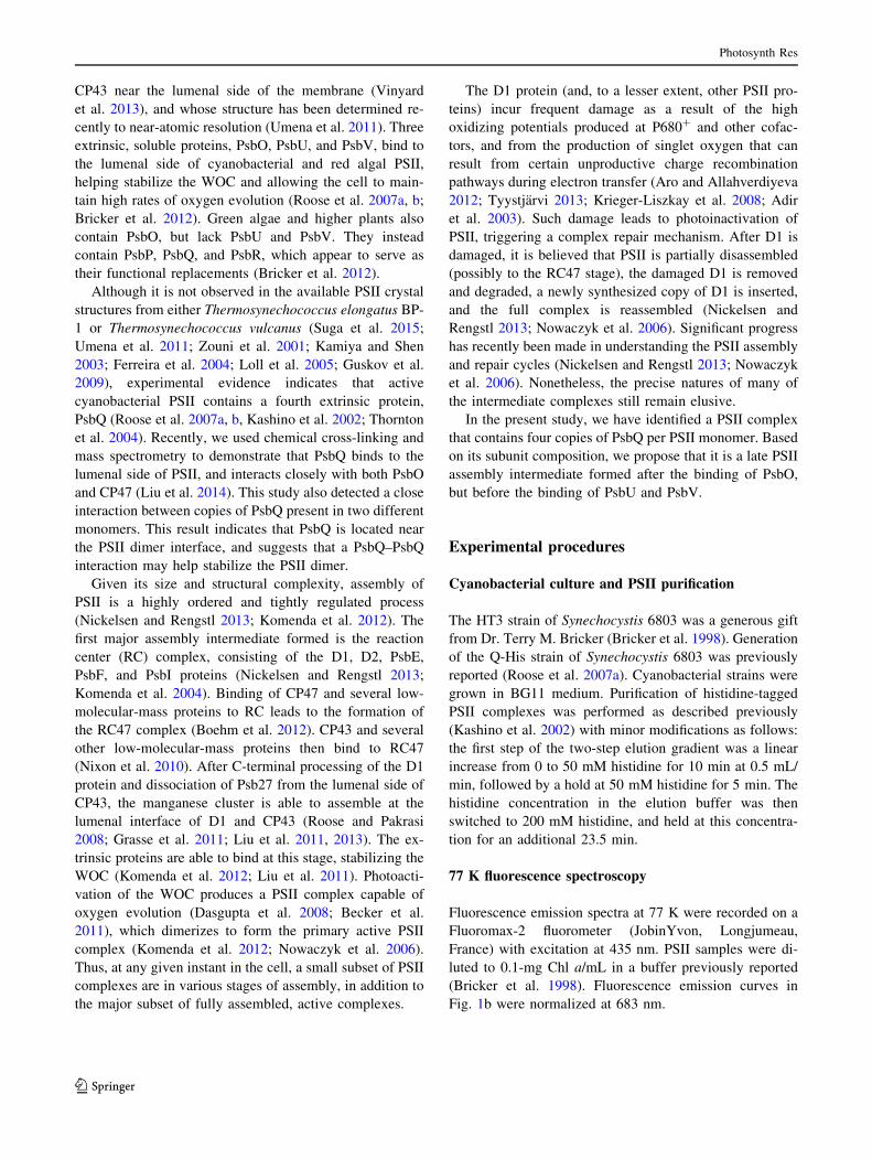
PSII from the Q-His strain was purified by nickel-affinity
chromatography using a two-step gradient of histidine-
containing buffer (Fig. 1a, red line). The two elution peaks
labeled in the chromatogram correspond to two distinct
protein complexes, referred to as complex 1 and complex 2
(later identified as the PSII dimer (PSII-D) and PSII-Q4,
respectively). They were collected separately and subjected
to biochemical characterization. Complexes 1 and 2 rep-
resent approximately 53 and 47 % of the total PSII yield,
respectively. In one of our previous studies in which PSII
from the Q-His strain was purified, elution was performed
with a single-linear gradient (Roose et al. 2007a), and the
different elution conditions used in this study likely ac-
count for the isolation of a second PSII complex.
To determine if both elution peaks correspond to PSII
complexes, 77 K fluorescence emission spectra were ob-
tained. Both samples displayed characteristic ‘‘F685’’ (at
683 nm) and ‘‘F695’’ (at 692 nm) fluorescence signatures
indicative of an assembled PSII reaction center (Fig. 1b)
(Satoh 1980; Liu et al. 2011). The relative intensities of the
two peaks were, however, slightly different, reflecting a slight
structural difference between the two PSII complexes. A
larger difference between the two complexes was observed by
measuring their oxygen evolution activity; complex 2
evolved oxygen at only 66 % of the saturated rate of complex
1 (841 and 1270 lmol O2�mgChl a-1�h-1, respectively).
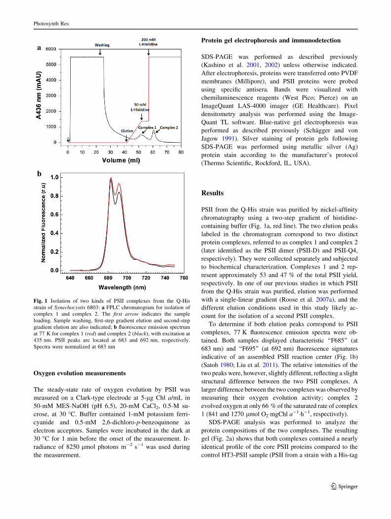
SDS-PAGE analysis was performed to analyze the
protein compositions of the two complexes. The resulting
gel (Fig. 2a) shows that both complexes contained a nearly
identical profile of the core PSII proteins compared to the
control HT3-PSII sample (PSII from a strain with a His-tag
Fig. 1 Isolation of two kinds of PSII complexes from the Q-His
strain of Synechocystis 6803: a FPLC chromatogram for isolation of
complex 1 and complex 2. The first arrow indicates the sample
loading. Sample washing, first-step gradient elution and second-step
gradient elution are also indicated; b fluorescence emission spectrum
at 77 K for complex 1 (red) and complex 2 (black), with excitation at
435 nm. PSII peaks are located at 683 and 692 nm, respectively.
Spectra were normalized at 683 nm
Photosynth Res
123
on the CP47 protein). However, Western blot analysis
showed that complex 2 lacked the PsbU and PsbV proteins
(Fig. 2b). In contrast, PsbO, the other lumenal extrinsic
protein observed in the PSII crystal structure, was present
in equal levels across all three complexes.
Surprisingly, the gel indicated that on a per-chlorophyll
basis, complex 2 contained significantly more PsbQ than
complex 1. Immunoblot analysis of these three complexes
using a PsbQ-specific antibody confirmed this result
(Fig. 2b). The Western blot indicates that complex 1 and
HT3-PSII contained roughly equal levels of PsbQ, imply-
ing that complex 2 contains more copies of PsbQ per PSII
monomer than both complex 1 and HT3-PSII.
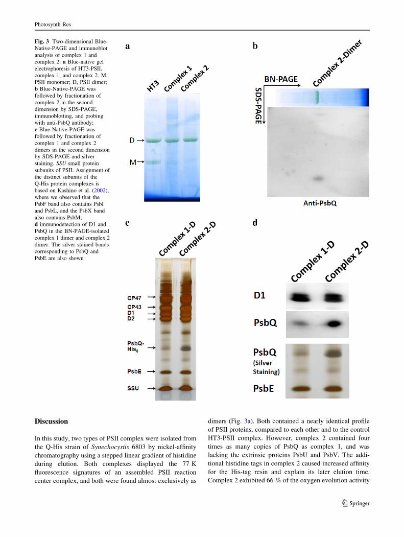
To determine if the additional copies of PsbQ in com-
plex 2 were a contamination from free copies of the protein
which are in fact unassociated with PSII, blue-native gel
electrophoresis followed by denaturing gel electrophoresis
and immunodetection was performed. The mild blue-native
gel conditions (Schagger and von Jagow 1991) keep PSII
complexes intact during electrophoresis, while unassoci-
ated proteins migrate separately due to their smaller size.
Subsequent denaturing gel electrophoresis of individual
excised native-gel bands allows characterization of the
proteins that are present in a particular PSII complex. The
native gel (Fig. 3a) showed that both complex 1 and
complex 2 were present almost exclusively as dimers,
consistent with our previous studies of Q-His-PSII (Liu
et al. 2014). The dimer band was excised and analyzed by
denaturing gel electrophoresis followed by silver staining
(Fig. 3c, d), as well as by immunodetection (Fig. 3b, d).
Both techniques confirmed the initial observation that
PsbQ was present at an elevated level in complex 2 com-
pared to complex 1. These results indicate that the addi-
tional copies of PsbQ found in complex 2 are indeed bound
to the PSII complex, and are not unassociated-protein
contaminants in the sample.
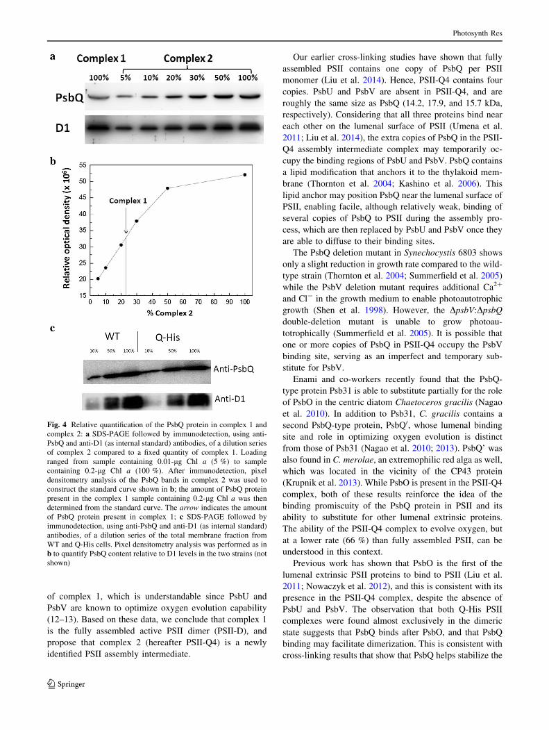
To quantify the increased level of PsbQ in complex 2,
PsbQ content in a dilution series of complex 2 was detected
by Western blot (Fig. 4a). Pixel densitometry analysis was
performed to obtain a calibration curve of band intensity
versus PsbQ content. By fitting PsbQ band intensity from
complex 1 (from the same blot) to the calibration curve
(Fig. 4b), we concluded that Complex 1 contains 23 % of
the PsbQ content of complex 2, on a per chlorophyll basis;
i.e. complex 2 contains four times as many copies of PsbQ
as complex 1.
It is conceivable that the Q-His strain could produce a
significantly higher quantity of PsbQ than the WT strain,
potentially leading to an artifactual Q-His-PSII complex
with higher PsbQ content. This possibility was ruled out by
comparing PsbQ levels in the total membrane fraction of
the Q-His and WT strains (Fig. 4c) by immunoblotting.
After normalizing to D1 levels in the two strains, we de-
termined that the PsbQ content of the Q-His strain was 1.1
(±0.3)-fold of that of the WT strain. This result indicates
that the Q-His strain does not synthesize dramatically
elevated levels of PsbQ compared to WT. Thus, the addi-
tional PsbQ content observed in complex 2 cannot be at-
tributed to a change in the expression level of PsbQ in the
Q-His strain.
Fig. 2 Polypeptide compositions of complex 1 and complex 2:
a SDS-PAGE protein profile with Coomassie Brilliant Blue staining.
Each lane contained sample with 2.4 lg of Chl a. HT3, a PSII
complex with a C-terminally polyhistidine-tagged version of CP47, is
used as a reference. Major PSII subunits are labeled. PsbQHis8 is
indicated on the right. Asterisk indicates a protein band containing
Psb28 and Sll1130 encoded protein. Assignment of the PSII subunits
is based on Kashino et al. (2002); b immunodetection of PSII
polypeptides in the isolated complexes after SDS-PAGE. Each lane
contained sample with 0.2 lg of Chl a. Specific antibodies against
CP47, CP43, D1, PsbO, PsbU, PsbV, and PsbQ were used for
immunodetection of the corresponding proteins
Photosynth Res
123
Discussion
In this study, two types of PSII complex were isolated from
the Q-His strain of Synechocystis 6803 by nickel-affinity
chromatography using a stepped linear gradient of histidine
during elution. Both complexes displayed the 77 K
fluorescence signatures of an assembled PSII reaction
center complex, and both were found almost exclusively as
dimers (Fig. 3a). Both contained a nearly identical profile
of PSII proteins, compared to each other and to the control
HT3-PSII complex. However, complex 2 contained four
times as many copies of PsbQ as complex 1, and was
lacking the extrinsic proteins PsbU and PsbV. The addi-
tional histidine tags in complex 2 caused increased affinity
for the His-tag resin and explain its later elution time.
Complex 2 exhibited 66 % of the oxygen evolution activity
Fig. 3 Two-dimensional Blue-
Native-PAGE and immunoblot
analysis of complex 1 and
complex 2: a Blue-native gel
electrophoresis of HT3-PSII,
complex 1, and complex 2. M,
PSII monomer; D, PSII dimer;
b Blue-Native-PAGE was
followed by fractionation of
complex 2 in the second
dimension by SDS-PAGE,
immunoblotting, and probing
with anti-PsbQ antibody;
c Blue-Native-PAGE was
followed by fractionation of
complex 1 and complex 2
dimers in the second dimension
by SDS-PAGE and silver
staining. SSU small protein
subunits of PSII. Assignment of
the distinct subunits of the
Q-His protein complexes is
based on Kashino et al. (2002),
where we observed that the
PsbF band also contains PsbI
and PsbL, and the PsbX band
also contains PsbM;
d immunodetection of D1 and
PsbQ in the BN-PAGE-isolated
complex 1 dimer and complex 2
dimer. The silver-stained bands
corresponding to PsbQ and
PsbE are also shown
Photosynth Res
123
of complex 1, which is understandable since PsbU and
PsbV are known to optimize oxygen evolution capability
(12–13). Based on these data, we conclude that complex 1
is the fully assembled active PSII dimer (PSII-D), and
propose that complex 2 (hereafter PSII-Q4) is a newly
identified PSII assembly intermediate.
Our earlier cross-linking studies have shown that fully
assembled PSII contains one copy of PsbQ per PSII
monomer (Liu et al. 2014). Hence, PSII-Q4 contains four
copies. PsbU and PsbV are absent in PSII-Q4, and are
roughly the same size as PsbQ (14.2, 17.9, and 15.7 kDa,
respectively). Considering that all three proteins bind near
each other on the lumenal surface of PSII (Umena et al.
2011; Liu et al. 2014), the extra copies of PsbQ in the PSII-
Q4 assembly intermediate complex may temporarily oc-
cupy the binding regions of PsbU and PsbV. PsbQ contains
a lipid modification that anchors it to the thylakoid mem-
brane (Thornton et al. 2004; Kashino et al. 2006). This
lipid anchor may position PsbQ near the lumenal surface of
PSII, enabling facile, although relatively weak, binding of
several copies of PsbQ to PSII during the assembly pro-
cess, which are then replaced by PsbU and PsbV once they
are able to diffuse to their binding sites.
The PsbQ deletion mutant in Synechocystis 6803 shows
only a slight reduction in growth rate compared to the wild-
type strain (Thornton et al. 2004; Summerfield et al. 2005)
while the PsbV deletion mutant requires additional Ca2?
and Cl- in the growth medium to enable photoautotrophic
growth (Shen et al. 1998). However, the DpsbV:DpsbQ
double-deletion mutant is unable to grow photoau-
totrophically (Summerfield et al. 2005). It is possible that
one or more copies of PsbQ in PSII-Q4 occupy the PsbV
binding site, serving as an imperfect and temporary sub-
stitute for PsbV.
Enami and co-workers recently found that the PsbQ-
type protein Psb31 is able to substitute partially for the role
of PsbO in the centric diatom Chaetoceros gracilis (Nagao
et al. 2010). In addition to Psb31, C. gracilis contains a
second PsbQ-type protein, PsbQ0, whose lumenal binding
site and role in optimizing oxygen evolution is distinct
from those of Psb31 (Nagao et al. 2010; 2013). PsbQ’ was
also found in C. merolae, an extremophilic red alga as well,
which was located in the vicinity of the CP43 protein
(Krupnik et al. 2013). While PsbO is present in the PSII-Q4
complex, both of these results reinforce the idea of the
binding promiscuity of the PsbQ protein in PSII and its
ability to substitute for other lumenal extrinsic proteins.
The ability of the PSII-Q4 complex to evolve oxygen, but
at a lower rate (66 %) than fully assembled PSII, can be
understood in this context.
Previous work has shown that PsbO is the first of the
lumenal extrinsic PSII proteins to bind to PSII (Liu et al.
2011; Nowaczyk et al. 2012), and this is consistent with its
presence in the PSII-Q4 complex, despite the absence of
PsbU and PsbV. The observation that both Q-His PSII
complexes were found almost exclusively in the dimeric
state suggests that PsbQ binds after PsbO, and that PsbQ
binding may facilitate dimerization. This is consistent with
cross-linking results that show that PsbQ helps stabilize the
Fig. 4 Relative quantification of the PsbQ protein in complex 1 and
complex 2: a SDS-PAGE followed by immunodetection, using anti-
PsbQ and anti-D1 (as internal standard) antibodies, of a dilution series
of complex 2 compared to a fixed quantity of complex 1. Loading
ranged from sample containing 0.01-lg Chl a (5 %) to sample
containing 0.2-lg Chl a (100 %). After immunodetection, pixel
densitometry analysis of the PsbQ bands in complex 2 was used to
construct the standard curve shown in b; the amount of PsbQ protein
present in the complex 1 sample containing 0.2-lg Chl a was then
determined from the standard curve. The arrow indicates the amount
of PsbQ protein present in complex 1; c SDS-PAGE followed by
immunodetection, using anti-PsbQ and anti-D1 (as internal standard)
antibodies, of a dilution series of the total membrane fraction from
WT and Q-His cells. Pixel densitometry analysis was performed as in
b to quantify PsbQ content relative to D1 levels in the two strains (not
shown)
Photosynth Res
123
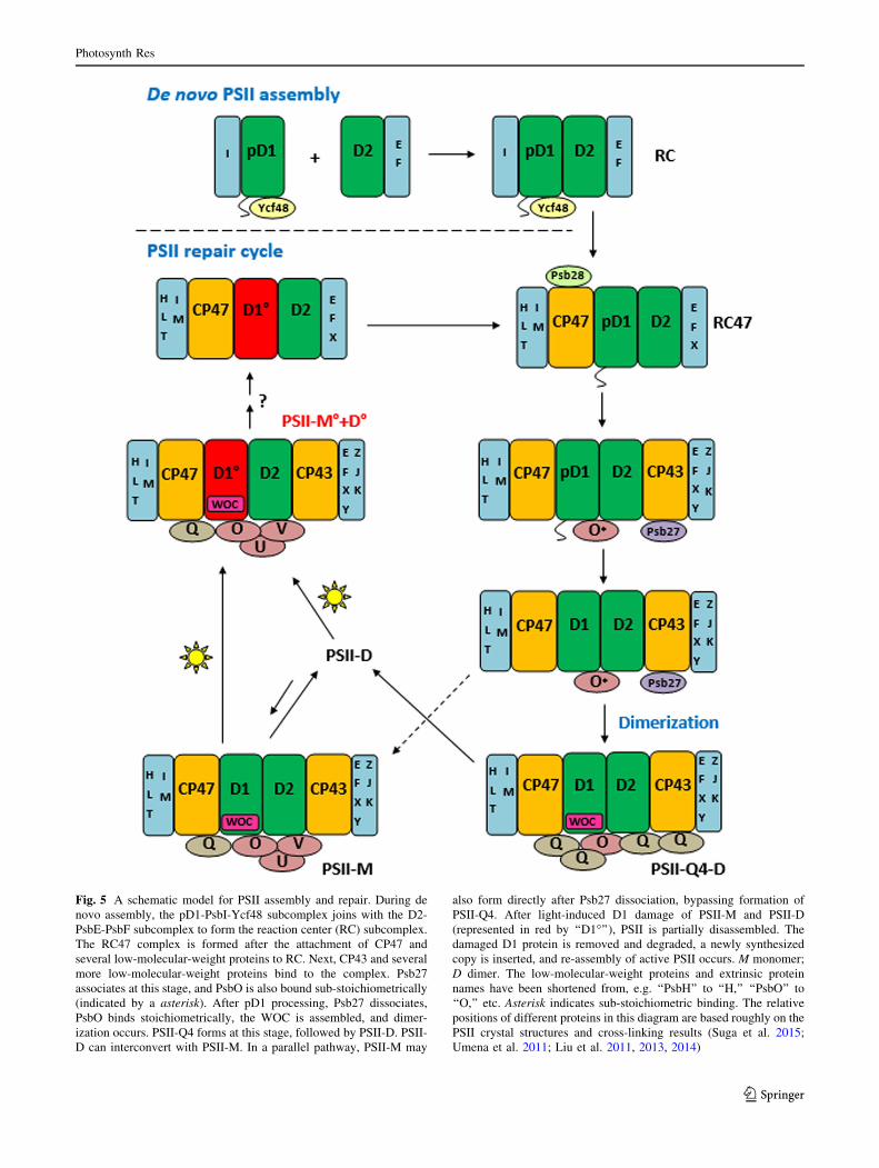
Fig. 5 A schematic model for PSII assembly and repair. During de
novo assembly, the pD1-PsbI-Ycf48 subcomplex joins with the D2-
PsbE-PsbF subcomplex to form the reaction center (RC) subcomplex.
The RC47 complex is formed after the attachment of CP47 and
several low-molecular-weight proteins to RC. Next, CP43 and several
more low-molecular-weight proteins bind to the complex. Psb27
associates at this stage, and PsbO is also bound sub-stoichiometrically
(indicated by a asterisk). After pD1 processing, Psb27 dissociates,
PsbO binds stoichiometrically, the WOC is assembled, and dimer-
ization occurs. PSII-Q4 forms at this stage, followed by PSII-D. PSII-
D can interconvert with PSII-M. In a parallel pathway, PSII-M may
also form directly after Psb27 dissociation, bypassing formation of
PSII-Q4. After light-induced D1 damage of PSII-M and PSII-D
(represented in red by ‘‘D1�’’), PSII is partially disassembled. The
damaged D1 protein is removed and degraded, a newly synthesized
copy is inserted, and re-assembly of active PSII occurs. M monomer;
D dimer. The low-molecular-weight proteins and extrinsic protein
names have been shortened from, e.g. ‘‘PsbH’’ to ‘‘H,’’ ‘‘PsbO’’ to
‘‘O,’’ etc. Asterisk indicates sub-stoichiometric binding. The relative
positions of different proteins in this diagram are based roughly on the
PSII crystal structures and cross-linking results (Suga et al. 2015;
Umena et al. 2011; Liu et al. 2011, 2013, 2014)
Photosynth Res
123
PSII dimer interface (Liu et al. 2014). PSII-Q4 thus appears
to be a late PSII assembly intermediate that is formed just
before the binding of PsbU and PsbV. Although we cannot
exclude the possibility that PSII-Q4 may also form during
the disassembly of PSII (after the dissociation of PsbU and
PsbV), the relatively high oxygen evolution rate of PSII-Q4
(66 % of fully assembled PSII) suggests that the majority
of PSII-Q4 complexes contain an undamaged D1 protein.
We have incorporated our results into an updated model
of PSII assembly (Fig. 5). We suggest that PSII dimer-
ization occurs after dissociation of Psb27 and formation of
the WOC. Four copies of PsbQ bind during or immediately
after this step, stabilizing this active dimer. Though we did
nit observe a monomeric PSII-Q4 intermediate, it is pos-
sible that such a complex forms transiently in between
Psb27 dissociation and PSII dimerization. The PSII-Q4
dimer is the first intermediate during PSII assembly that is
capable of oxygen evolution, albeit at around two-thirds the
rate of mature PSII. An active WOC increases PSII vul-
nerability to oxidative damage. As discussed above, due to
the presence of a lipid anchor of PsbQ, multiple copies of
PsbQ in PSII-Q4 serve as readily available substitutes for
PsbU and PsbV, helping to stabilize the WOC and protect
PSII as soon as it gains oxygen-evolving capability. The
additional copies of PsbQ must bind relatively weakly,
since PsbU and PsbV replace them on the lumenal surface
of PSII once they are able to diffuse to their binding sites,
forming the fully assembled, fully protected, PSII dimer
(PSII-D).Though we hypothesize that formation of PSII-Q4
stabilizes the active dimer until PsbU and PsbV bind, PSII-
M and PSII-D are still able to form in the absence of PsbQ
(Liu et al. 2014). It is therefore possible that a portion of
the complexes bypass PSII-Q4 formation during assembly,
as indicated by a dashed line in Fig. 5. Light-induced D1
damage triggers partial disassembly of PSII-M and PSII-D,
possibly to the RC47 stage (Nickelsen and Rengstl 2013)
(see Fig. 5).It is possible that the PSII-Q4 complex also
forms after the dissociation of PsbU and PsbV during PSII
disassembly. A new copy of D1 replaces the damaged copy
and re-assembly of active PSII occurs.
Changing environmental conditions can alter the rate of
PSII damage, making it difficult for the cell to maintain its
required rate of real-time energy production. The PSII-Q4
dimer may assist by serving as a pool of nearly assembled
PSII that can be rapidly converted to PSII-Din response
to shifts in the equilibrium concentration of PSII-D and
PSII-M.
In summary, we have isolated a PSII subcomplex (PSII-
Q4) with four copies of the PsbQ protein. Based on our
results, we conclude that this complex is a late PSII
assembly intermediate, formed after the binding of PsbO
and before the binding of PsbU and PsbV. This complex
helps to stabilize PSII immediately after it becomes capable
of water oxidation. Our results provide further evidence for
the binding promiscuity of PsbQ and its ability to substitute
for other lumenal extrinsic PSII proteins.
Acknowledgments We thank Dr. Terry Bricker for the kind gift of
the HT3 strain of Synechocystis 6803, and all members of the Pakrasi
lab for collegial discussions. This work was supported by funding
from the National Science Foundation (NSF-MCB0745611) to H.B.P.
References
Adir N, Zer H, Shochat S, Ohad I (2003) Photoinhibition-a historical
perspective. Photosynth Res 76:343–370
Aro E-M, Allahverdiyeva Y (2012) Photosynthetic responses of
plants to excess light: mechanisms and conditions for photoin-
hibition, excess energy dissipation, and repair. In: Eaton JJ,
Tripathy BC, Sharkey TD (eds) Photosynthesis: plastid biology,
energy conversion and carbon assimilation. Advances in photo-
synthesis and respiration. Springer, Dordrecht, pp 275–297
Becker K, Cormann KU, Nowaczyk MM (2011) Assembly of the
water-oxidizing complex in photosystem II. J Photochem Pho-
tobiol B 104:204–211
Boehm M, Yu J, Reisinger V, Beckova M, Eichacker LA, Schlodder
E, Komenda J, Nixon PJ (2012) Subunit composition of CP43-
less photosystem II complexes of Synechocystis sp. PCC 6803:
implications for the assembly and repair of photosystem II.
Philos Trans R Soc B 367:3444–3454
Bricker T, Frankel L (2002) The structure and function of CP47 and
CP43 in photosystem II. Photosynth Res 72:131–146
Bricker TM, Morvant J, Masri N, Sutton HM, Frankel LK (1998)
Isolation of a highly active photosystem II preparation from
Synechocystis 6803 using a histidine-tagged mutant of CP47.
Biochim Biophys Acta 1409:50–57
Bricker TM, Roose JL, Fagerlund RD, Frankel LK, Eaton-Rye JJ
(2012) The extrinsic proteins of photosystem II. Biochim
Biophys Acta 1817:121–142
Dasgupta J, Ananyev GM, Dismukes GC (2008) Photoassembly of
the water-oxidizing complex in photosystem II. Coord Chem
Rev 252:347–360
Ferreira KN, Iverson TM, Maghlaoui K, Barber J, Iwata S (2004)
Architecture of the photosynthetic oxygen-evolving center.
Science 303:1831–1838
Grasse N, Mamedov F, Becker K, Styrin S, Rogner M, Nowaczyk
MM (2011) Role of novel dimeric photosystem II (PSII)-Psb27
protein complex in PSII repair. J Biol Chem 286:29548–29555
Guskov A, Kern J, Gabdulkhakov A, Broser M, Zouni A, Saenger W
(2009) Cyanobacterial photosystem II at 2.9-A resolution and the
role of quinones, lipids, channels, and chloride. Nat Struct Mol
Biol 16:334–342
Kamiya N, Shen J-R (2003) Crystal structure of oxygen-evolving
photosystem II from Thermosynechococcus vulcanus at 3.7-A
resolution. Proc Natl Acad Sci USA 100:98–103
Kashino Y, Koike H, Satoh K (2001) An improved sodium dodecyl
sulfate-polyacrylamide gel electrophoresis system for the
analysis of membrane protein complexes. Electrophoresis
22:1004–1007
Kashino Y, Lauber WM, Carroll JA, Wang Q, Whitmarsh J, Satoh K,
Pakrasi HB (2002) Proteomic analysis of a highly active
photosystem II preparation from the cyanobacterium Syne-
chocystis sp. PCC 6803 reveals the presence of novel polypep-
tides. Biochemistry 41:8004–8012
Kashino Y, Inoue-Kashino N, Roose JL, Pakrasi HB (2006) Absence
of the PsbQ protein results in destabilization of the PsbV protein
Photosynth Res
123
and decreased oxygen evolution activity in cyanobacterial
photosystem II. J Biol Chem 281:20834–20841
Komenda J, Reisinger V, Muller BC, Dobakova M, Granvogl B,
Eichacker LA (2004) Accumulation of the D2 protein is a key
regulatory step for assembly of the photosystem II reaction
center complex in Synechocystis PCC 6803. J Biol Chem
279:48620–48629
Komenda J, Sobotka R, Nixon PJ (2012) Assembling and maintaining
the photosystem II complex in chloroplasts and cyanobacteria.
Curr Opin Plant Biol 15:245–251
Krieger-Liszkay A, Fufezan C, Trebst A (2008) Singlet oxygen
production in photosystem II and related protection mechanism.
Photosynth Res 98:551–564
Krupnik T, Kotabova E, van Bezouwen LS, Mazur R, Garstka M,
Nixon PJ, Barber J, Kana R, Boekema EJ, Kargul J (2013) A
reaction center-dependent photoprotection mechanism in a
highly robust Photosystem II from an extremophilic Red alga,
Cyanidioschyzon merolae. J Biol Chem 288:23529–23542
Liu H, Roose JL, Cameron JC, Pakrasi HB (2011) A genetically
tagged Psb27 protein allows purification of two consecutive
photosystem II (PSII) assembly intermediates in Synechocystis
6803, a cyanobacterium. J Biol Chem 286:24865–24871
Liu H, Chen J, Huang RY-C, Weisz D, Gross ML, Pakrasi HB (2013)
Mass spectrometry-based footprinting reveals structural dynam-
ics of loop E of the chlorophyll-binding protein CP43 during
photosystem II assembly in the cyanobacterium Synechocystis
6803. J Biol Chem 288:14212–14220
Liu H, Zhang H, Weisz DA, Vidavsky I, Gross ML, Pakrasi HB
(2014) MS-based cross-linking analysis reveals the location of
the PsbQ protein in cyanobacterial photosystem II. Proc Natl
Acad Sci USA 111:4638–4643
Loll B, Kern J, Saenger W, Zouni A, Biesiadka J (2005) Towards
complete cofactor arrangement in the 3.0 A resolution structure
of photosystem II. Nature 438:1040–1044
Nagao R, Moriguchi A, Tomo T, Niikura A, Nakajima S, Suzuki T,
Okumura A, Iwai M, Shen J-R, Ikeuchi M, Enami I (2010)
Binding and functional properties of five extrinsic proteins in
oxygen-evolving photosystem II from a marine centric diatom,
Chaetoceros gracilis. J Biol Chem 285:29191–29199
Nagao R, Suga M, Niikura A, Okumura A, Koua FHM, Suzuki T,
Tomo T, Enami I, Shen J-R (2013) Crystal structure of Psb31, a
novel extrinsic protein of photosystem II from a marine centric
diatom and implications for its binding and function. Biochem-
istry 52:6646–6652
Nickelsen J, Rengstl B (2013) Photosystem II assembly: from
cyanobacteria to plants. Annu Rev Plant Biol 64:609–635
Nixon PJ, Michoux F, Yu J, Boehm M, Komenda J (2010) Recent
advances in understanding the assembly and repair of photosys-
tem II. Ann Bot 106:1–16
Nowaczyk MM, Hebeler R, Schlodder E, Meyer HE, Warscheid B,
Rogner M (2006) Psb27, a cyanobacterial lipoprotein, is
involved in the repair cycle of photosystem II. Plant Cell
18:3121–3131
Nowaczyk MM, Krause K, Mieseler M, Sczibilanski A, Ikeuchi M,
Rogner M (2012) Deletion of psbJ leads to accumulation of
Psb27–Psb28 photosystem II complexes in Thermosynechococ-
cus elongatus. Biochim Biophys Acta 1817:1339–1345
Rappaport F, Diner BA (2008) Primary photochemistry and energetic
leading to the oxidation of the (Mn)4Ca cluster and to the
evolution of molecular oxygen in photosystem II. Coord Chem
Rev 252:259–272
Rogner M, Dekker JP, Boekema EJ, Witt HT (1987) Size, shape, and
mass of the oxygen-evolving photosystem II complex from the
thermophilic cyanobacterium Synechococcus sp. FEBS Lett
219:207–211
Roose JL, Pakrasi HB (2008) The Psb27 protein facilitates manganese
cluster assembly in photosystem II. J Biol Chem 283:4044–4050
Roose JL, Kashino Y, Pakrasi HB (2007a) The PsbQ protein defines
cyanobacterial photosystem II complexes with highest activity
and stability. Proc Natl Acad Sci USA 104:2548–2553
Roose JL, Wegener KM, Pakrasi HB (2007b) The extrinsic proteins
of photosystem II. Photosynth Res 92:369–387
Satoh K (1980) F-695 emission from the purified photosystem II
chlorophyll a-protein complex. FEBS Lett 110:53–56
Schagger H, von Jagow G (1991) Blue native electrophoresis for
isolation of membrane protein complexes in enzymatically
active form. Anal Biochem 199:223–231
Shen J-R, Qian M, Inoue Y, Burnap RL (1998) Functional charac-
terization of Synechocystis sp. PCC 6803 DpsbU and DpsbV
mutants reveals important roles of cytochrome c-550 in
cyanobacterial oxygen evolution. Biochemistry 37:1551–1558
Shi L-X, Schroder WP (2004) The low molecular mass subunits of the
photosynthetic supracomplex, photosystem II. Biochim Biophys
Acta 1608:75–96
Shi L-X, Hall M, Funk C, Schroder WP (2012) Photosystem II, a
growing complex: updates on newly discovered components and
low molecular mass proteins. Biochim Biophys Acta 1817:13–25
Suga M, Akita F, Hirata K, Ueno G, Murakami H, Nakajima Y,
Shimizu T, Yamashita K, Yamamoto M, Ago H, Shen JR (2015)
Native structure of photosystem II at 1.95 A resolution viewed
by femtosecond X-ray pulses. Nature 517:99–103
Summerfield TC, Shand JA, Bentley FK, Eaton-Rye JJ (2005) PsbQ
(Sll1638) in Synechocystis sp. PCC 6803 is required for
photosystem II activity in specific mutants and in nutrient-
limiting conditions. Biochemistry 44:805–815
Thornton LE, Ohkawa H, Roose JL, Kashino Y, Keren N, Pakrasi HB
(2004) Homologs of plant PsbP and PsbQ proteins are necessary
for regulation of photosystem II activity in the cyanobacterium
Synechocystis 6803. Plant Cell 16:2164–2175
Tyystjarvi E (2013) Photoinhibition of photosystem II. Int Rev Cell
Mol Biol 300:243–303
Umena Y, Kawakami K, Shen JR, Kamiya N (2011) Crystal structure
of oxygen-evolving photosystem II at a resolution of 1.9 A.
Nature 473:55–61
Vinyard DJ, Ananyev GM, Dismukes GC (2013) Photosystem II: the
reaction center of oxygenic photosynthesis. Annu Rev Biochem
82:577–606
Watanabe M, Iwai M, Narikawa R, Ikeuchi M (2009) Is the
photosystem II complex a monomer or a dimer? Plant Cell
Physiol 50:1674–1680
Zouni A, Witt HT, Kern J, Fromme P, Krauss N, Saenger W, Orth P
(2001) Crystal structure of photosystem II from Synechococcus
elongatus at 3.8 A resolution. Nature 409:739–743
Photosynth Res
123