multiple-file vs. single-file endodontics in dental ...(sf) endodontics (waveone-instruments,...
TRANSCRIPT
Submitted 13 July 2016Accepted 6 November 2016Published 7 December 2016
Corresponding authorAndreas Bartols,[email protected]
Academic editorRobert Druzinsky
Additional Information andDeclarations can be found onpage 19
DOI 10.7717/peerj.2765
Copyright2016 Bartols et al.
Distributed underCreative Commons CC-BY 4.0
OPEN ACCESS
Multiple-file vs. single-file endodontics indental practice: a study in routine careAndreas Bartols1,2, Gunter Laux3 and Winfried Walther1
1Dental Academy for Continuing Professional Development Karlsruhe, Karlsruhe, Germany2Clinic for Conservative Dentistry and Periodontology, Christian-Albrechts-University Kiel, Kiel, Germany3University Hospital Heidelberg, Department of General Practice and Health Services Research, University ofHeidelberg, Heidelberg, Baden-Württemberg, Germany
ABSTRACTBackground. Little is known about the differences of rotary multiple file endodon-tic therapy and single-file reciprocating endodontic treatment under routine careconditions in dental practice. This multicenter study was performed to compare theoutcome of multiple-file (MF) and single-file (SF) systems for primary root canaltreatment under conditions of general dental practice regarding reduction of pain witha visual analogue scale (VAS 100), improvement of oral-health-related quality of life(OHRQoL) with the german short version of the oral health impact profile (OHIP-G-14) and the speed of root canal preparation.Materials andMethods. Ten general dental practitioners (GDPs) participated in thestudy as practitioner-investigators (PI). In the first five-month period of the study, theGDPs treated patients with MF systems. After that, the GDPs treated the patients inthe second five-month period with a SF system (WaveOne). The GDPs documentedthe clinical findings at the beginning and on completion of treatment. The patientsdocumented their pain and OHRQoL before the beginning and before completion oftreatment.Results. A total of 599 patients were included in the evaluation. 280 patients were inthe MF group, 319 were in the SF WaveOne group. In terms of pain reduction andimprovement in OHIP-G-14, the improvement in both study groups (MF and SF) wasvery similar based on univariate analysis methods. Pain reduction was 34.4 (SD 33.7)VAS (MF) vs. 35.0 (SD 35.4) VAS (SF) (p= 0.840) and the improvement inOHIP-G-14score was 9.4 (SD 10.3) (MF) vs. 8.5 (SD 10.2) (SF) (p= 0.365). The treatment time perroot canal was 238.9 s (SD 206.2 s) (MF) vs. 146.8 sec. (SD 452.8 sec) (SF) (p= 0.003).Discussion. Regarding improvement of endodontic pain and OHRQoL measure withOHIP-G-14, there were no statistical significant differences between the SF und theMFsystems. WaveOne-prepared root canals significantly faster than MF systems.
Subjects DentistryKeywords Endodontics, Health services research, Patient outcomes, Dental public health,WaveOne, Single-file endodontics, Multiple-file systems, BioRaCe, Mity Roto Files, Clinical trial
INTRODUCTIONClinical endodontic research is mainly conducted by specialists or specialized universitycenters (Friedman, Furberg & DeMets, 2010; Ng et al., 2007). The predominant types ofsuch studies are retrospective observational studies, prospective cohort studies and a few
How to cite this article Bartols et al. (2016), Multiple-file vs. single-file endodontics in dental practice: a study in routine care. PeerJ4:e2765; DOI 10.7717/peerj.2765
randomized controlled trials (RCTs) (Ng et al., 2007). On account of the controlled studydesign, these studies have greater internal evidence and are classified as efficacy studies(Pfaff, Nellessen-Martens & Scriba, 2011). The effectiveness of endodontic interventionsunder everyday general dental care conditions has so far been hardly investigated (Nixdorfet al., 2012). Yet, patients treated in specialized centers can differ systematically frompatients treated in routine care (Hulley, 2013).
A commonality of many experimental endodontic studies is the low number of cases(Peters & Wesselink, 2002; Pettiette, Delano & Trope, 2001;Weiger, Rosendahl & Lost, 2000).Larger case numbers are described for retrospective observational studies and prospectivecohort studies, which, however, are often conducted without controls (Ng et al., 2007).Convincing results though can be obtained in studies if they include an adequate numberof cases (Hulley, 2013). Since only few experimental endodontic studies have been madeand many of them are lacking sufficient patient numbers, one could assume this to be anindication of considerable feasibility problems of such studies.
Reciprocating single-file (SF) systems are the latest stage of development of nickel-titanium (NiTi) instruments for the preparation of root canals (Bürklein, Benten &Schäfer, 2013; Yared, 2008). During the last years several systems as Reciproc (VDW,Munich, Germany), WaveOne (Dentsply, Konstanz, Germany), Genius files (Ultradent,South Jordan, UT, USA) or the Twisted Files Adaptive System (Kerr, Orange, CA, USA)with a combination of rotary and reciprocating movement were introduced into themarket. Our knowledge of the clinical effects of using different systems for root canalpreparation is limited (Schäfer, Schulz-Bongert & Tulus, 2004). The Swedish Council onHealth Technology Assessment stated in its Systematic Review ofMethods of Diagnosis andTreatment in Endodontics that the use of new tools facilitates the technical procedures ofroot canal treatment and that therefore investigations are needed regarding what influencethese techniques have on everyday general practice (Bergenholtz et al., 2012).
Typically, new instrument systems are investigated in in-vitro-studies with extractedteeth (Bürklein, Benten & Schäfer, 2013) or root canalmodels (Goldberg, Dahan & Machtou,2012). In such studies, the outcomes are mainly surrogate parameters, such as root canalstraightening, preparation faults, preparation time in a workbench situation etc. the clinicalsignificance of which can only be estimated to a limited extent (Hülsmann, 2013). Mostof the few clinical trials available investigated only one instrument system (Fleming etal., 2010; Su, Wang & Ye, 2011) and rarely allow a comparison with other instrumentsystems (Schäfer, Schulz-Bongert & Tulus, 2004). Recently some randomized controlledtrials were published, that investigated single and multiple file systems for endodontictreatment regarding pain reduction after treatment and improvement in quality of life(Kherlakian et al., 2016; Pasqualini et al., 2016; Relvas et al., 2016). It is unclear if thereexists an effectiveness-gap (Pfaff, Nellessen-Martens & Scriba, 2011) between the results ofthese controlled studies under the optimal treatment conditions of specialized treatmentproviders and the use of rotary multiple-file (MF) and SF systems in general dental practice.
Therefore, research is needed when new endodontic techniques are introduced intodental practice. The study we performed investigates the effects of these endodontictechniques on dental practice. For this purpose, it uses the methods of health services
Bartols et al. (2016), PeerJ, DOI 10.7717/peerj.2765 2/24
research which studies care processes under everyday conditions of dental practice(Pfaff et al., 2009). Short-term patient-relevant outcomes were in the center of the study.
The design we chose was a multicenter study in routine care. We started by evaluatingthe outcome of endodontic treatment using conventional MF instrument systems for rootcanal preparation. Then, the practitioner-investigators (PIs) were trained in single-file(SF) endodontics (WaveOne-Instruments, Dentsply Maillefer, Ballaigues, Switzerland).Subsequently we evaluated the outcome of endodontic treatments using WaveOne.
The following research hypotheses were investigated in our study:Primary outcome criterionDoes root canal preparation using SF root canal instruments lead to more or lessreduction of patients’ endodontic pain compared to using rotary MF instrumentsystems?Secondary outcome criterionDoes root canal preparation using SF root canal instruments lead to more or lessreduction of patients’ oral-health-related quality of life compared to using rotary MFinstrument systems?Tertiary outcome criterionDoes root canal preparation using a single-file system require less time compared tothe MF systems?
METHODSStudy designWe performed the present study as a multicenter clinical study. For the purpose of thisstudy we formed a network of ten general dental practitioners (GDPs). They acted asPIs. We conducted the study in two phases (Fig. 1). In the first 5-month phase the GDPsperformed the endodontic therapy with different rotary nickel-titanium (NiTi)MF systems(Table 1). Subsequently the GDPs were trained for the use of the WaveOne SF system(Maillefer, Ballaigues, Switzerland). In the second 5-month phase the PIs treated thepatients with SF WaveOne instruments exclusively. After each 5-month phase there was a3-month follow-up so that treatments started could be completed.
The authors of this study acted solely as investigators and did not treat patients.The study was conducted in conformity with the Declaration of Helsinki and the
ProfessionalCode for Physicians of theMedical Council of the State of Baden-Württemberg.The Ethics Committee of the Baden-Württemberg Medical Council reviewed the studyand approved it (AZ: F-2011-034-z).
ParticipantsPatient eligibility and recruitmentAll patients of the ten PIs who required endodontic therapy were consecutively assessedfor eligibility.
The following inclusion criteria were defined: patients had to be at least 18 years old andin need of initial orthograde root canal treatment.
Bartols et al. (2016), PeerJ, DOI 10.7717/peerj.2765 3/24
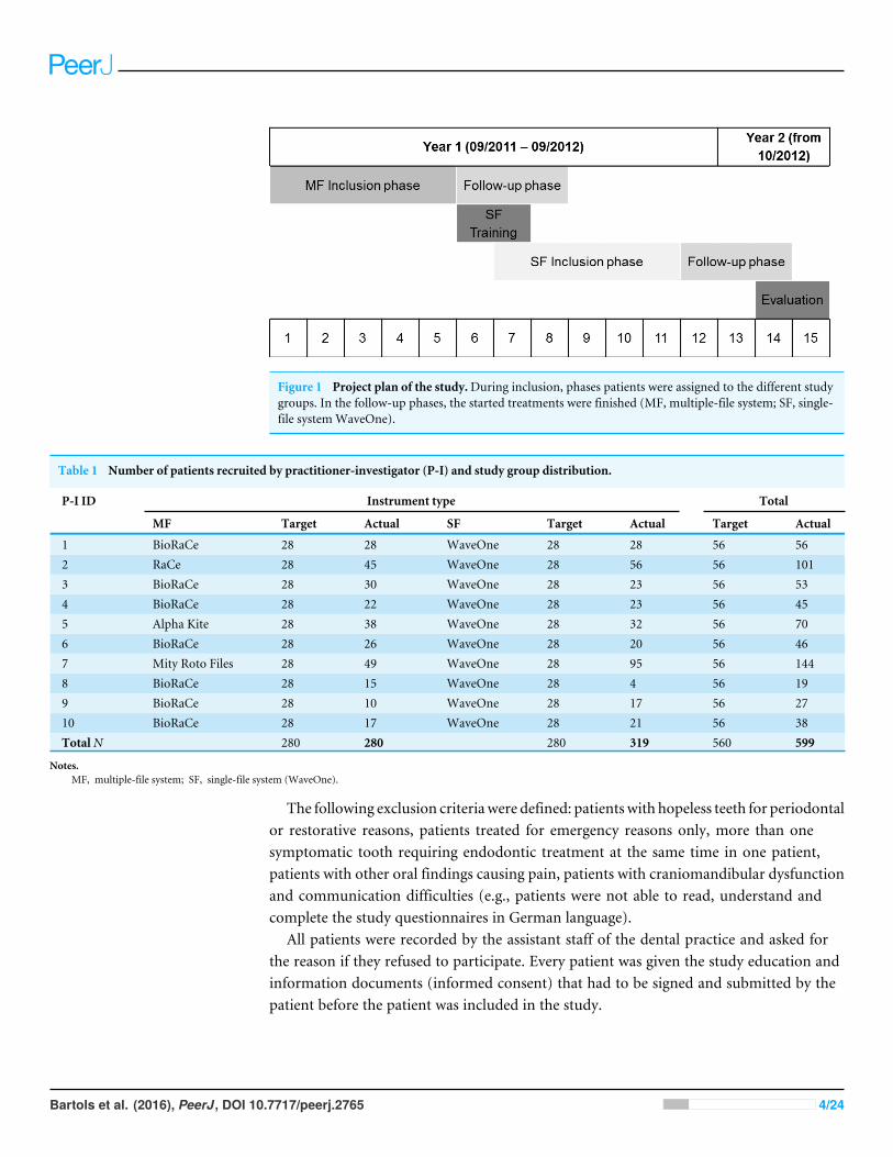
Figure 1 Project plan of the study.During inclusion, phases patients were assigned to the different studygroups. In the follow-up phases, the started treatments were finished (MF, multiple-file system; SF, single-file systemWaveOne).
Table 1 Number of patients recruited by practitioner-investigator (P-I) and study group distribution.
P-I ID Instrument type Total
MF Target Actual SF Target Actual Target Actual
1 BioRaCe 28 28 WaveOne 28 28 56 562 RaCe 28 45 WaveOne 28 56 56 1013 BioRaCe 28 30 WaveOne 28 23 56 534 BioRaCe 28 22 WaveOne 28 23 56 455 Alpha Kite 28 38 WaveOne 28 32 56 706 BioRaCe 28 26 WaveOne 28 20 56 467 Mity Roto Files 28 49 WaveOne 28 95 56 1448 BioRaCe 28 15 WaveOne 28 4 56 199 BioRaCe 28 10 WaveOne 28 17 56 2710 BioRaCe 28 17 WaveOne 28 21 56 38TotalN 280 280 280 319 560 599
Notes.MF, multiple-file system; SF, single-file system (WaveOne).
The following exclusion criteriawere defined: patientswith hopeless teeth for periodontalor restorative reasons, patients treated for emergency reasons only, more than onesymptomatic tooth requiring endodontic treatment at the same time in one patient,patients with other oral findings causing pain, patients with craniomandibular dysfunctionand communication difficulties (e.g., patients were not able to read, understand andcomplete the study questionnaires in German language).
All patients were recorded by the assistant staff of the dental practice and asked forthe reason if they refused to participate. Every patient was given the study education andinformation documents (informed consent) that had to be signed and submitted by thepatient before the patient was included in the study.
Bartols et al. (2016), PeerJ, DOI 10.7717/peerj.2765 4/24

Practitioner investigators (PIs)The ten dentists who participated in the study were general dental practitioners with at leasttwo years of professional experience in a general dental practice and without endodonticspecialization. All participating dentists worked under the conditions of the German‘‘Statutory Health Insurance.’’ The PIs were chosen as a convenient sample of dentists thatwanted to change their endodontic treatment to single file systems within the next 6–12months anyway. All practices were located in southwest Germany.
All PIs were familiar with root canal preparation using rotary NiTi instruments(Table 1) and used them routinely in their practice. All dentists followed the ‘‘GoodClinical Practice: Root Canal Treatment’’ Guideline of DGZMK (German Society ofDental, Oral and Craniomandibular Sciences) (Hülsmann & Schäfer, 2005) which containsessential key points of the Quality guidelines for endodontic treatment of the EuropeanSociety of Endodontology (2006).
Study initiation at the PIsBefore the study started, all participating PIs were visited by the principal investigator (AB)in their practice. The dentists were informed about the object and purpose of the study andits practical implementation. Each PI was given a copy of the study protocol and all otherstudy relevant files. The dentists were informed about the planned procedure with regardto patient recruitment, education/information and treatment.
InterventionsIn the first phase of the study, from 09/2011 to 02/2012, all endodontic treatments wereperformed with rotary NiTi MF systems (Fig. 1). All MF systems were used accordingto the manufacturer’s instructions. In 03/2012 the PIs were trained for the SF system.The training course explained the theoretical bases of the WaveOne System (Maillefer,Ballaigues, Switzerland) and provided hands-on training on extracted teeth. After this one-day training course every participating dentist was able to prepare root canals by the newmethod in a reliable way. The training was followed by a two-week implementation phasein all participating dental offices. During that time, the PIs should learn to treat patientswith the new instruments and gain experience. In case of difficulties, this procedure offeredthe chance of clarifying problems. In the second phase of the trial, from 04/2012 to 08/2012,all endodontic treatments were performed with the SF system. All other variables of thepractice setting and the treatment procedures remained unchanged.
Before treatment, the affected tooth was anesthetized by local anesthesia. After localanesthesia the endodontic access cavity was prepared. All teeth were isolated with arubberdam. Root canals were probed with K-steel files of ISO sizes 06, 08, 10 and 15, inorder to create a glidepath up to ISO 15 throughout all phases of the study. The workinglength was determined electrometrically and/or by X-ray. The dentists prepared the rootcanals according to the details provided by the manufacturers of the different rotarypreparation systems. In the second study phase, the root canals were prepared with theWaveOne instruments according to the manufacturer’s instructions. If the dentists neededan apical preparation size that is not included in theWaveOne System, the last ISO size was
Bartols et al. (2016), PeerJ, DOI 10.7717/peerj.2765 5/24

followed up by a single hand instrument of the desired size. During rotary or reciprocatingpreparation the root canals were rinsed with 1–3%NaOCl between every rotary instrumentor in case of the SF system between every 3–4 picks with the WaveOne file. After completepreparation of the root canals they were irrigated with a final irrigation of NaOCl 1–3%and a calcium hydroxide dressing or the root canal filling was placed. After that the toothwas sealed provisionally bacteria-proof with a temporary bacteria tight seal. In the lastappointment the root canal filling was placed or in case of single-visit endodontics adefinitive coronal filling was applied.
OutcomesThe primary outcome of reduction of endodontic pain and the secondary outcomeof improvement of oral-health-related quality of life was measured with a patientquestionnaire. The questionnaire assessed the pain by the Visual Analog Scale (VAS100) (Turk, 2011) and the oral-health-related quality of life with the items of the shortversion of the oral health impact profile (OHIP-G-14) (John, Micheelis & Biffar, 2004) wichis the German translation of OHIP-14 (Slade, 1997). The patients were asked about thebiggest complaints (consisting of the VAS 100 and OHIP-G14) without pain medication inthe week before treatment and in the week before completion of treatment. This was twoweeks after initial treatment and in connection with either the placement of the root canalfilling or the definitive coronal filling of the tooth. The questionnaires were filled in by thepatients before treatment started or while local anesthesia was taking effect. Any patientquestions were answered by the dental team.
The time needed for root canal preparation was measured by the dental assistantstaff. The measurement started when the first rotating or reciprocating instrument wasplaced in the root canal and ended when the last instrument was removed. The rootcanal recapitulations during preparation and the irrigations were included in the timemeasurement. Probing and glidepath preparation before using the rotary instruments werenot included in the time measurement. Nor were the final irrigation of the root canalsand the placement of a dressing included. When a tooth had several root canals, the totalpreparation time of all canals was measured and divided by the number of root canals inorder to determine the preparation time per canal.
Further questionnaires and data collectionIn the PI questionnaires the clinical findings (dental chart, sensitivity test, percussiontest, apical pressure point, periodontal probing depth, radiographic presence of apicalperiodontitis, number of prepared root canals, presence of fistulae), the time needed forroot canal preparation, instrument fractures and procedural events were documented. Inaddition, a consecutive patient log was introduced to record, if possible, the patient’s reasonfor rejecting participation. The patient forms included the patient’s informed consent toparticipate in the trial, a questionnaire asking for demographic and basic medical data,and the above described pain questionnaire which consisted of the VAS and the Items ofOHIP-G-14.
All patients that qualified for participation in the study were informed about the studyby the PI personally. All PI forms were filled in by the assistant dental staff. The patient
Bartols et al. (2016), PeerJ, DOI 10.7717/peerj.2765 6/24

questionnaires were filled in by the patients themselves. All patient questionnaires werepseudonymized and collected in a sealed box. The PI forms were pseudonymized in thesame way to be able to match the patient data and the PI data in the subsequent evaluation.
The pain questionnaires were completed by the patients immediately before treatmentstarted. The demographic data could be provided at any time, but were requested oncompletion of the treatment at the latest. The pain questionnaires were completed by thepatients again 14 days after treatment. The PIs’ treatment was taken down on record. Thetime required for root canal preparation was measured by the dental assistant staff.
The questionnaires were handed over to the principal investigator (AB) at the end ofthe first and at the end of the second trial phase for evaluation.
Safety measuresBefore treatment started, each patient participating in the study was informed aboutthe endodontic risks in the same way as it is usually done before endodontic therapy.The patient was informed in particular about events, such as instrument fractures andother complications that may occur during root canal preparation and could lead to theextraction of the tooth affected. The patient was also informed, that root canal treatment isthe last attempt to save a tooth. The information was provided by the PI and an additionaleducation and information questionnaire. If in the course of the trial the complication ofan instrument fracture occurred, the patient would be informed about it. This informationwas provided by an information questionnaire for instrument fractures.
Sample size calculation and statisticsFor sample size calculation we had to consider the sample design which was characterizedby a 2-stage structure (dental practice, patient). This cluster sample made special demandson both sample size planning and the analysis of the results (Donner & Klar, 2000).
To calculate the case number, the following parameters were defined: Significance level:0.05, Power: 80% and Number of clusters (dental practices): 10.
Moreover, based on the analysis of similar studies (Pak, 2012), the most realisticassumptions possible were made about the clinically relevant difference of the VAS 100(Visual Analog Scale) and the ICC (Intra Cluster Correlation Coefficient) which is ameasure for the homogeneity in relation to a target variable of interest within the cluster:1VAS= 20 (20%) and ICC = 0.04.
For case number calculation, a validated software tool was used which determinedthe number of trial units (patients) per cluster (practice) on the basis of the parametersspecified above (Campbell et al., 2004).
The resulting number of patients was 28 per dental practice and every trial phase. Thisnumber appeared realistic in terms of feasibility. In view of the basic statistical data ondental care in Germany (KZBV, 2011), a conservative estimate showed that one GDPperforms about 60 root canal treatments per year. This means that 10 participating GDPsshould be able to recruit the required case number in each of the trial phases.
The results were calculated with the SPSS (Version 21, Win x64) statistical system andSAS (Version 9.2, Win x64). With the PROC MIXED procedure (Singer, 1998) SAS offers
Bartols et al. (2016), PeerJ, DOI 10.7717/peerj.2765 7/24
options for explicitly considering potential cluster effects (here: several data collection unitsper dental office) in the overall regression model.
Assessment of potential covariatesBesides collecting the data for the primary outcome we assessed other dentist- and patient-related as well as treatment-specific covariates. This was done with a questionnaire fordemographic information which also documented the patients’ basic medical data. Inaddition, the PIs recorded the dental chart and treatment-specific findings (tooth sensitivitybefore treatment, apical translucency, percussion test, apical pressure point and fistula).
Study termination criteriaIt was planned to terminate the study when two weeks after root canal preparation by thenew single-file method the patient’s pain was 40% above the expected level. The studywould also have been terminated if single-file endodontics would have caused markedlymore instrument fractures than expected. If during the study more than three instrumentfractures had already occurred in the first 20 single-file treatment cases, the study wouldhave been terminated.
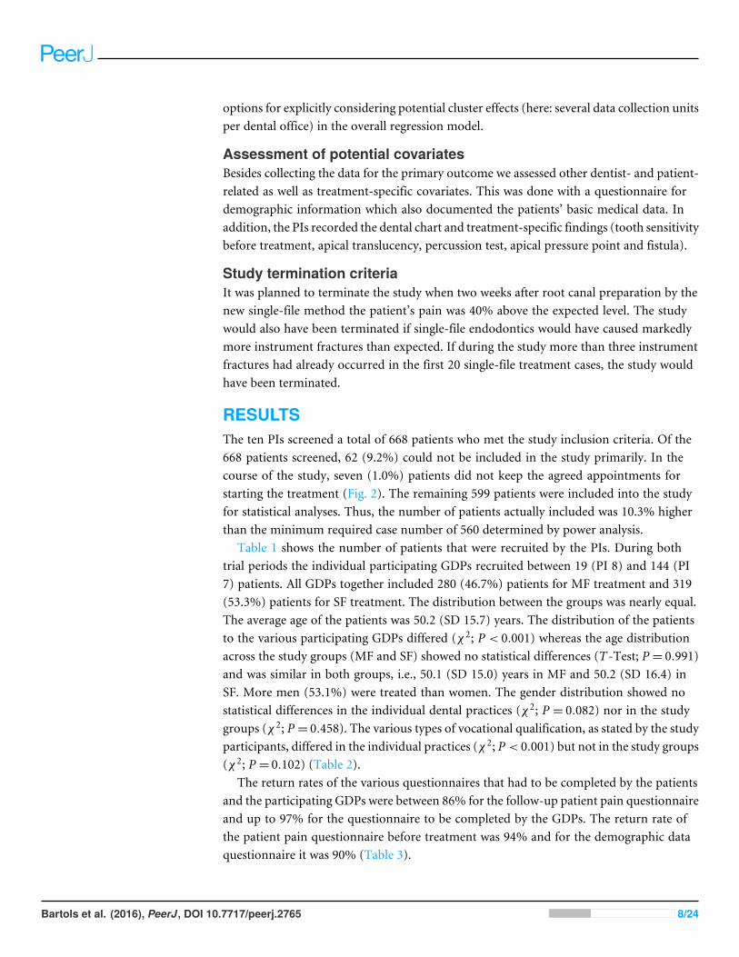
RESULTSThe ten PIs screened a total of 668 patients who met the study inclusion criteria. Of the668 patients screened, 62 (9.2%) could not be included in the study primarily. In thecourse of the study, seven (1.0%) patients did not keep the agreed appointments forstarting the treatment (Fig. 2). The remaining 599 patients were included into the studyfor statistical analyses. Thus, the number of patients actually included was 10.3% higherthan the minimum required case number of 560 determined by power analysis.
Table 1 shows the number of patients that were recruited by the PIs. During bothtrial periods the individual participating GDPs recruited between 19 (PI 8) and 144 (PI7) patients. All GDPs together included 280 (46.7%) patients for MF treatment and 319(53.3%) patients for SF treatment. The distribution between the groups was nearly equal.The average age of the patients was 50.2 (SD 15.7) years. The distribution of the patientsto the various participating GDPs differed (χ2; P < 0.001) whereas the age distributionacross the study groups (MF and SF) showed no statistical differences (T -Test; P = 0.991)and was similar in both groups, i.e., 50.1 (SD 15.0) years in MF and 50.2 (SD 16.4) inSF. More men (53.1%) were treated than women. The gender distribution showed nostatistical differences in the individual dental practices (χ2; P = 0.082) nor in the studygroups (χ2; P = 0.458). The various types of vocational qualification, as stated by the studyparticipants, differed in the individual practices (χ2; P < 0.001) but not in the study groups(χ2; P = 0.102) (Table 2).
The return rates of the various questionnaires that had to be completed by the patientsand the participating GDPs were between 86% for the follow-up patient pain questionnaireand up to 97% for the questionnaire to be completed by the GDPs. The return rate ofthe patient pain questionnaire before treatment was 94% and for the demographic dataquestionnaire it was 90% (Table 3).
Bartols et al. (2016), PeerJ, DOI 10.7717/peerj.2765 8/24
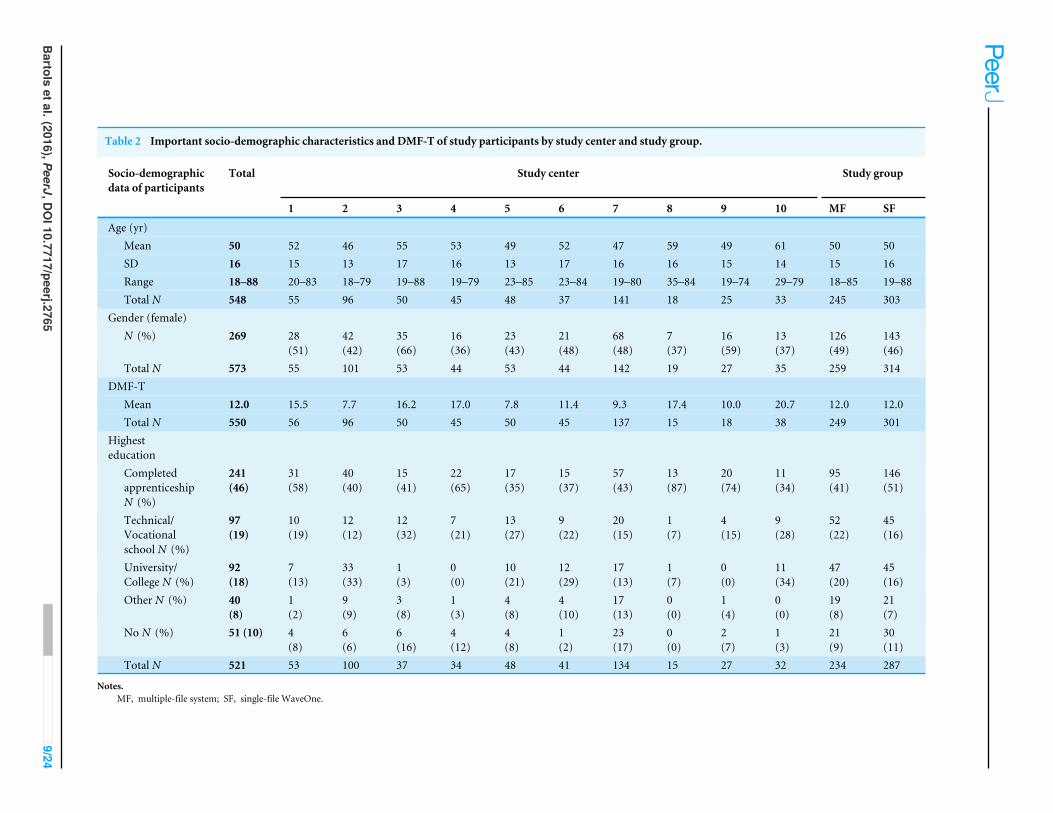
Table 2 Important socio-demographic characteristics and DMF-T of study participants by study center and study group.
Socio-demographicdata of participants
Total Study center Study group
1 2 3 4 5 6 7 8 9 10 MF SF
Age (yr)Mean 50 52 46 55 53 49 52 47 59 49 61 50 50SD 16 15 13 17 16 13 17 16 16 15 14 15 16Range 18–88 20–83 18–79 19–88 19–79 23–85 23–84 19–80 35–84 19–74 29–79 18–85 19–88Total N 548 55 96 50 45 48 37 141 18 25 33 245 303
Gender (female)N (%) 269 28
(51)42(42)
35(66)
16(36)
23(43)
21(48)
68(48)
7(37)
16(59)
13(37)
126(49)
143(46)
Total N 573 55 101 53 44 53 44 142 19 27 35 259 314DMF-T
Mean 12.0 15.5 7.7 16.2 17.0 7.8 11.4 9.3 17.4 10.0 20.7 12.0 12.0Total N 550 56 96 50 45 50 45 137 15 18 38 249 301
Highesteducation
CompletedapprenticeshipN (%)
241(46)
31(58)
40(40)
15(41)
22(65)
17(35)
15(37)
57(43)
13(87)
20(74)
11(34)
95(41)
146(51)
Technical/Vocationalschool N (%)
97(19)
10(19)
12(12)
12(32)
7(21)
13(27)
9(22)
20(15)
1(7)
4(15)
9(28)
52(22)
45(16)
University/College N (%)
92(18)
7(13)
33(33)
1(3)
0(0)
10(21)
12(29)
17(13)
1(7)
0(0)
11(34)
47(20)
45(16)
Other N (%) 40(8)
1(2)
9(9)
3(8)
1(3)
4(8)
4(10)
17(13)
0(0)
1(4)
0(0)
19(8)
21(7)
No N (%) 51 (10) 4(8)
6(6)
6(16)
4(12)
4(8)
1(2)
23(17)
0(0)
2(7)
1(3)
21(9)
30(11)
Total N 521 53 100 37 34 48 41 134 15 27 32 234 287
Notes.MF, multiple-file system; SF, single-file WaveOne.
Bartols
etal.(2016),PeerJ,DO
I10.7717/peerj.27659/24

Figure 2 Flow diagram.
Table 3 Questionnaire return rates for the enrolled participants.
Description Timing N(received)
N(expected)
%
Patient survey demographic data Before root filling 538 599 90Pain and OHIP-14 survey before treatment 1st appointment 565 599 94Dentist survey for treatment parameters All appointments 582 599 97Patient 2 weeks follow-up survey 2 weeks after RCT 518 599 86
Bartols et al. (2016), PeerJ, DOI 10.7717/peerj.2765 10/24

In the course of the study slightly more maxillary (53.7%) teeth were treated (Table 4).The distribution of the different types of teeth showed no significant differences betweenthe two study groups (χ2; P = 0.255).
Evaluation of the primary outcome criterionFor the evaluation of the primary outcome, i.e., post-operative reduction of patients’endodontic pain and improvement of oral-health-related quality of life, we measured painreduction via VAS 100 and the OHIP-G14 score. Both values were measured before rootcanal treatment and 14 days after treatment. Then we compared the different study groups(MF and SF).
The mean pain score before root canal treatment for MF was 42.3 (SD 32.6) VAS andfor SF 43.9 (SD 32.0) VAS and decreased to 10.0 (SD 18.6) VAS (MF) and 9.3 (SD 19.2)VAS (SF).
The mean OHIP-G 14 score before root canal treatment for MF was 12.5 (SD 10.6) andfor SF 13.0 (SD 10.8) and decreased to 3.6 (SD 5.1) (MF) and 4.6 (SD 6.5) (SF).
For pain reduction andOHRQoL, univariate analysis showed a very similar improvementin both study groups (MF and SF):(a) Pain reduction 34.4 (SD 33.7) VAS (MF) vs. 35.0 (SD 35.4) VAS (SF) (p= 0.8).(b) Improvement of oral-health-related quality of life according to OHIP-14 score: 9.4
(SD 10.3) (MF) vs. 8.5 (SD 10.2) (SF) (p= 0.4).The differences between the study groups were not significant.Multivariate analysis of variance (MANOVA) taking the additional factor ‘‘single vs.
multiple-visit treatment’’ into account did not reveal any significant influence of the factors‘‘study group (MF or SF)’’, ‘‘single vs. multiple-visit’’ or an interaction of the two regardingpain reduction. For improvement of OHIP-14 score there was an overall significantinfluence (p= 0.03) with ‘‘single-visit’’ treatments having a significantly (p= 0.01) lowerimprovement than ‘‘multiple-visit’’. But further analyses showed that OHIP-14 scores for‘‘single-visit’’ treatments (10.7 (SD 9.9)) were already significantly (p= 0.06) lower beforetreatment than for ‘‘multiple-visit’’ treatments (13.6 (SD 10.9)) and dropped to almost thesame levels before completion of treatment (4.1 (SD 5.9) vs. 4.2 (SD 6.0)).
Evaluation of the secondary outcome criterionFor the speed of root canal preparation, univariate analysis showed a significant differencebetween the study groups MF and SF:(c) Duration of treatment per root canal (in sec): 239 (MF) vs. 147 (SF) (P = 0.003).For (c) a multivariate analysis was made taking into consideration the dentist- andpatient-related as well as treatment-specific covariates:
• Gender of dentist• Gender of patient• Age of patient• Comorbidities of the patient (none, hypertension, DM(I or II), asthma)• DMFT Index• Tooth
Bartols et al. (2016), PeerJ, DOI 10.7717/peerj.2765 11/24

Table 4 Descriptive data of teeth treated by location, study center (PI) and study group.
Tooth type Study center Study group
1 2 3 4 5 6 7 8 9 10 Multiple file Single file Total
Mean (SD) N Mean (SD) N Mean (SD) N Mean (SD) N Mean (SD) N Mean (SD) N Mean (SD) N Mean (SD) N Mean (SD) N Mean (SD) N Mean (SD) N Mean (SD) N Mean (SD) N
Maxillaryanteriors
No. of treatedroot canals
1.0 0.0 7 1.0 0.0 13 1.0 0.0 11 1.0 0.0 5 1.0 0.0 2 1.0 0.0 7 1.1 0.3 23 1.0 0.0 2 1.0 0.0 2 1.0 0.0 8 3.1 0.5 58 3.0 0.5 80 3.1 0.5 138
Time fortreatment pertooth (s)
225.4 145.6 7 139.7 83.1 13 114.2 53.1 11 132.8 77.7 5 169.0 58.0 2 163.6 193.8 7 157.7 200.3 22 50.0 14.1 2 93.0 52.3 2 1500.0 2342.3 8 836.8 747.5 57 375.5 751.8 78 570.3 781.4 135
Time fortreatment perroot canal (s)
225.4 145.6 7 139.7 83.1 13 114.2 53.1 11 132.8 77.7 5 169.0 58.0 2 163.6 193.8 7 155.0 201.8 22 50.0 14.1 2 93.0 52.3 2 1500.0 2342.3 8 273.1 246.1 57 120.3 204.8 77 185.3 235.0 134
Maxillarypremolars
No. of treatedroot canals
1.6 0.5 11 1.4 0.5 17 1.7 0.5 7 2.0 0.0 9 2.0 0.0 5 1.7 0.5 6 1.8 0.4 20 2.0 0.0 2 1.6 0.5 8 1.3 0.5 7 1.7 0.5 53 1.6 0.5 39 1.7 0.5 92
Time fortreatment pertooth (s)
276.6 115.0 11 188.1 104.8 17 274.7 116.9 7 150.6 42.6 10 322.2 45.8 5 165.5 84.4 6 299.2 271.9 19 87.5 17.7 2 249.1 150.0 8 950.0 435.0 6 303.0 200.2 53 257.0 326.1 38 283.8 259.6 91
Time fortreatment perroot canal (s)
180.0 75.0 11 141.6 69.4 17 171.1 105.5 7 74.3 22.4 9 161.1 22.9 5 97.6 31.2 6 162.1 130.8 19 43.8 8.8 2 148.2 70.0 8 710.0 210.1 6 192.5 154.3 52 164.4 197.8 38 180.7 173.5 90
Maxillarymolars
No. of treatedroot canals
3.2 0.4 13 2.9 0.4 26 3.1 0.7 11 3.0 0.7 9 3.3 0.5 14 3.1 0.4 8 3.1 0.3 37 2.3 0.6 3 3.2 0.8 6 3.0 0.4 11 1.0 0.0 37 1.1 0.3 43 1.0 0.2 80
Time fortreatment pertooth (s)
908.6 1616.5 12 310.7 143.9 26 354.3 162.0 11 207.6 54.4 9 484.9 132.4 14 179.4 182.3 9 503.7 690.3 35 255.0 336.6 2 433.7 205.9 6 2100.0 523.8 11 274.9 229.6 37 294.8 1115.9 42 285.5 824.0 79
Time fortreatment perroot canal (s)
252.3 397.1 12 109.3 53.2 26 113.0 42.1 11 70.3 17.0 9 151.0 47.5 14 63.7 63.7 8 164.9 226.7 35 86.4 110.2 2 133.7 46.9 6 700.9 141.5 11 274.9 229.6 37 293.4 1116.2 42 284.7 824.2 79
Mandibularanteriors
No. of treatedroot canals
1.0 0.0 1 1.0 0.0 3 1.0 0.0 7 1.0 0.0 3 1.0 0.0 2 1.0 0.0 2 1.1 0.4 14 1.0 0.0 3 1.0 0.0 2 1.0 0.0 2 3.0 0.6 61 3.0 0.6 78 3.0 0.6 139
Time fortreatment pertooth (s)
114.0 0.0 1 122.7 76.2 3 113.7 38.4 7 55.0 35.5 3 229.5 113.8 2 50.0 31.1 2 271.1 268.9 14 45.0 5.0 3 172.0 1.4 2 1320.0 1018.2 2 669.7 662.4 62 278.0 324.5 78 451.5 537.6 140
Time fortreatment perroot canal (s)
114.0 0.0 1 122.7 76.2 3 113.7 38.4 7 55.0 35.5 3 229.5 113.8 2 50.0 31.1 2 249.3 259.6 14 45.0 5.0 3 172.0 1.4 2 1320.0 1018.2 2 219.6 205.0 60 93.6 107.3 77 148.8 169.1 137
Mandibularpremolars
No. of treatedroot canals
1.0 0.0 5 1.0 0.0 15 1.0 0.0 6 1.0 0.0 7 1.0 0.0 3 1.0 0.0 13 1.1 0.3 28 1.0 0.0 2 1.0 0.0 6 1.0 0.0 2 1.0 0.2 35 1.0 0.2 52 1.0 0.2 87
Time fortreatment pertooth (s)
162.2 101.9 5 104.9 74.0 15 162.8 65.2 6 99.3 55.8 7 327.0 96.0 3 122.5 128.2 13 196.8 294.5 28 75.0 21.2 2 204.2 149.4 6 810.0 466.7 2 260.9 250.0 35 115.5 173.9 52 174.0 218.6 87
Time fortreatment perroot canal (s)
162.2 101.9 5 104.9 74.0 15 162.8 65.2 6 99.3 55.8 7 327.0 96.0 3 122.5 128.2 13 162.7 188.8 27 75.0 21.2 2 204.2 149.4 6 810.0 466.7 2 237.7 156.6 34 114.2 174.4 52 163.0 177.4 86
Mandibularmolars
No. of treatedroot canals
3.0 0.0 19 2.8 0.5 25 2.7 0.7 10 2.8 0.8 11 3.2 0.5 28 2.9 0.9 9 3.3 0.5 20 2.8 0.4 6 2.7 0.6 3 3.3 0.5 8 1.1 0.3 16 1.0 0.2 23 1.1 0.2 39
Time fortreatment pertooth (s)
378.8 195.7 19 274.6 166.3 27 247.9 92.2 10 198.4 48.5 11 426.3 169.0 28 295.4 193.0 9 592.9 822.7 19 153.2 63.0 6 277.0 23.5 3 2040.0 521.1 8 276.1 239.8 16 195.6 421.0 23 228.6 356.2 39
Time fortreatment perroot canal (s)
126.3 65.2 19 98.6 57.3 25 96.6 42.4 10 71.8 13.1 11 135.3 53.1 28 93.7 53.8 9 181.9 263.5 19 53.9 22.2 5 108.6 33.4 3 635.6 168.4 8 259.2 229.2 16 194.1 421.5 23 220.8 353.1 39
Totals No. of treatedroot canals
2.3 1.0 56 2.0 1.0 99 1.9 1.0 52 2.1 1.0 44 2.8 0.9 54 1.8 1.0 45 2.0 1.0 142 1.9 0.9 18 1.9 1.0 27 2.1 1.1 38 2.1 1.0 260 2.1 1.0 315 2.1 1.0 575
Time fortreatment pertooth (s)
429.9 788.1 55 222.2 148.5 101 217.8 134.9 52 157.3 68.9 45 409.5 160.7 54 176.2 164.8 46 345.7 524.3 137 117.0 114.5 17 266.0 170.6 27 1658.9 1218.4 37 496.2 555.8 260 268.9 607.3 311 372.4 594.8 571
Time fortreatment perroot canal (s)
180.2 200.7 55 115.9 66.0 99 123.8 61.7 52 82.2 41.3 44 157.1 69.2 54 106.1 109.7 45 173.5 211.9 136 57.2 34.8 16 150.7 86.3 27 900.4 1114.8 37 238.9 206.2 256 147.1 453.5 309 188.7 365.5 565
Bartols
etal.(2016),PeerJ,DO
I10.7717/peerj.276512/24
• Tooth sensitivity before treatment (positive/negative)• Apical translucency (yes/no)• Percussion test (positive/negative)• Apical pressure point (yes/no)• Fistula (yes/no).For the WO SF system a significantly shorter duration resulted in comparison to the MF
systems (121 s; SD 37.40; p= 0.01). The adjusted reduction in required preparation timewas 32.8% with the SF-System.
The root canal preparation with the WaveOne System produced the same resultsregarding reduction of patients’ endodontic related pain and oral health related quality oflife, but the preparation speed per root canal was faster.
DISCUSSIONOur study showed that root canal treatment with MFs as well as with the SF WaveOneSystem reduced the patients’ endodontic related pain and improved oral health relatedquality of life without statistically significant differences under conditions of general dentalpractice. The root canal preparation with the SF system was faster.
Measuring patients’ endodontic pain and oral-health-related qualityof lifePain intensity can be measured with various methods, e.g., the Visual Analog Scale (VAS),the Numerical Rating Scale (NRS) or the Verbal (Categorical) Rating Scale (VRS). The VRSis an easy-to-apply measuring method, but forces the study subjects to select a wordingwhich may not represent an adequate description of the pain they feel. Moreover, theVRS depends on a clear and unequivocal understanding of the language (Turk, 2011). TheNRS and VAS are uncomplicated measuring methods for the pain felt and both showgood evidence of construct validation (Turk, 2011). In the present study, the VAS wasselected because it offers a large number of scores. This makes the VAS more sensitive tochanges in the pain intensity felt than other scales offering fewer reply categories (Turk,2011). In addition, the VAS is widely used in the endodontic literature (King et al., 2012;Martin-Gonzalez et al., 2012; Pak, 2012; Udoye & Jafarzadeh, 2011). The limitation to onescale is both feasible and adequate (Attar et al., 2008).
Pain assessment alone gives no information about the patients’ oral-health-relatedquality of life which represents a patient-relevant outcome (Pfaff et al., 2009). The onlyvalidated German-language measuring instrument for the oral-health-related quality oflife is the OHIP (John et al., 2003). To limit the questionnaire to a practicable length, theOHIP-G-14 was used in our study (John, Micheelis & Biffar, 2004). In 2011, when the studywas planned, there existed two endodontic studies which chose the OHIP-14 questionnairefor endodontic issues (Dugas et al., 2002; Gatten et al., 2011).
Description of the resultsIn the present study, we compared two clinical short-term outcome parameters of twobasic methods for root canal preparation.
Bartols et al. (2016), PeerJ, DOI 10.7717/peerj.2765 13/24

We defined the most important patient-relevant outcomes as reduction of pain byendodontic therapy and the improvement of oral-health-related quality of life. Whenwe were planning this study in 2011, shortly after the introduction of reciprocating SFendodontic instruments (WaveOne and Reciproc), there was no information about theclinical performance of these instruments. The new system should have at least a similaror an enhanced clinical outcome compared to the conventional (MF) systems. This is animportant condition when a new technology is introduced. Moreover, we expected greaterspeed and thus greater efficiency of the SF system as a relevant result. As far as the authorsknow, no studies had been made that compared the clinical outcome of MF and SF systemsat the time of the study planning in 2011.
The reduction of pain and the improvement of oral health related quality of life as aresult of endodontic treatment were not different in the two experimental groups (MF andSF) in the second week after treatment. Both methods were equally effective in reducingendodontic pain. The mean pain intensity of about 43.2 (SD 32.2) VAS before root canaltreatment and about 9.5 (SD 19.0) VAS after treatment agrees with the results obtainedby other researchers who investigated pain reduction after endodontic therapy (Ehrmann,Messer & Adams, 2003; Pak & White, 2011). A review found that pain levels beforeendodontic therapy of 54 VAS and a standard deviation of 24 VAS are a common averagein endodontic studies and decrease to less than 10 VAS on average within 7 days (Ehrmann,Messer & Adams, 2003; Pak & White, 2011). Also Ehrmann, Messer & Adams (2003) foundvery similar values to those in our study (44.4 (SD 26.9) VAS). The mean pain measured4 days after endodontic therapy decreased to 7.5 (SD 15.5) VAS. The mean pain reductionof 36.9 (SD 29.0) VAS was very similar to the values in our study. The improvement of theOHIP-14 with mean scores of 12.8 (SD 10.6) before therapy found in our study are verycomparable to another study where a mean OHIP-14 score of 15.4 (SD 10.5) was foundbefore endodontic treatment (Liu, McGrath & Cheung, 2014). Also other studies reportthat endodontic treatment leads to an improvement of the oral-health-related quality of life(Dugas et al., 2002; Hamasha & Hatiwsh, 2013). This finding was confirmed by our study.
Recently a couple of randomized controlled trials (RCT) have been published, thatevaluated the pain reduction and/or the improvement in quality of life of single filesystems (Kherlakian et al., 2016; Pasqualini et al., 2016; Relvas et al., 2016). In the firstRCT (Kherlakian et al., 2016) two SF reciprocating systems (Reciproc (VDW, Munich,Germany) and WaveOne (Dentsply)) and one MF system (ProTaper Next (Dentsply))were compared. Only asymptomatic vital teeth were treated with the different systems.Therefore patients did not have pain before treatment. Also after treatment, pain rateson a categorized VAS 100 score were also very low and showed no significant differencesbetween the systems. The second RCT (Relvas et al., 2016) compared one reciprocating SFsystem (Reciproc (VDW, Munich, Germany)) with a MF system (ProTaper (Dentsply)).Only asymptomatic teeth with apical periodontitis were included in the trial. Thereforepatients were pain-free before treatment. Pain measurement was not performed with theVAS. Therefore, results can be hardly compared with our study. The different instrumentsystems showed no statistically significant differences in postoperative pain scores after24 h and 72 h. The third RCT (Pasqualini et al., 2016) investigated the ProTaper MF and
Bartols et al. (2016), PeerJ, DOI 10.7717/peerj.2765 14/24

the WaveOne SF system. Compared were primary root canal treatments of every clinicalcondition (symptomatic, asymptomatic, vital and non-vital cases). Mean pain on VAS was35.2 for SF before treatment and 24.6 for MF decreasing to very low rates of 1.3 (SF) and0.9 (MF) after seven days. This is different to our study, where the mean pain scores werehigher before treatment, but more equal. Also, our mean pain scores in the second weekafter treatment were higher than the score found in the above mentioned study. If thiscan be interpreted as an effectiveness gap regarding the success of these instruments ingeneral dental practice compared to specialized care providers remains unclear, because ourinitial VAS scores were higher and therefore perhaps naturally need a longer time periodto drop to low scores. Recently our research group published a smaller, but very similarstudy comparing hand instrumentation with Reciproc for root canal preparation underconditions of general dental practice (Bartols et al., 2016). The mean pain score before rootcanal treatment with hand instruments was 43.6 (SD 30.7) VAS and with Reciproc it was41.2 (SD 27.7) VAS, which is perfectly comparable with the initial values of the presentstudy where the initial VAS scores were 42.3 (SD 32.6) VAS for MF and 43.9 (SD 32.0) VASfor SF. Within the same period of time the scores decreased in all four groups to valuesin the range between 9.3 and 11.5 (with SDs of 16.5–19.2) VAS therefore only showedminimal differences. Also, regarding OHRQoL, the OHIP-G-14 scores of all four groupsshow only minimal differences before treatment and before completion of therapy (handinstruments 9.2 (SD 9.6) decreasing to 3.4 (SD 5.4), Reciproc 10.4 (SD 9.6) decreasingto 3.5 (SD 6.1), MF 12.5 (SD 10.6) decreasing to 3.6 (SD 5.1) and SF WaveOne 13.0 (SD10.8) decreasing to 4.6 (SD 6.5)). Therefore, it can be concluded that all four techniquesinvestigated show the same clinical outcome regarding pain reduction and improvementin OHRQoL under routine care conditions.
As the main outcome parameter was the reduction of endodontic pain after treatment,the different root canal preparation techniques and their influence on postoperative painhas to be considered. Recently it was demonstrated that different root canal preparationtechniques lead to the expression of different levels of inflammatory neuropeptides inthe periapical periodontal ligament linked with the possible emergence of symptomaticapical periodontitis (Caviedes-Bucheli et al., 2013). It is believed that this is connected tothe different amounts of extruded debris beyond the apical foramen (Caviedes-Bucheliet al., 2016). Since nearly all root canal instrumentation techniques including handinstrumentation as well as engine driven instruments lead to apical extrusion of debris(Al-Omari & Dummer, 1995; Bürklein & Schäfer, 2012; Capar et al., 2014; De-Deus et al.,2010) in most cases there will be an inflammatory response to a certain extent. In an in-vitrostudy reciprocating instruments extruded more debris than rotary instruments (Bürklein& Schäfer, 2012) with Reciproc producing most debris while another in-vitro study foundReciproc to produce significantly less extruded debris compared to rotary techniques(Kocak et al., 2013). The only clinical studies measuring the expression of inflammatoryneuropeptides in the periodontal ligament found that the instrument design of enginedriven root canal instruments has a greater impact on expression of neuropeptides than theinstrumentation technique (Caviedes-Bucheli et al., 2016). Because of this contradictorydata situation and the limited knowledge, if the amount of expressed neuropeptides
Bartols et al. (2016), PeerJ, DOI 10.7717/peerj.2765 15/24

can be directly correlated to the perceived pain it remains unclear if there is an impacton the postoperative pain levels of patients after root canal treatment. In our study thepreoperative levels of pain, their improvement and the postoperative VAS pain levels werevery similar and very much comparable to our previously published study (Bartols et al.,2016). Therefore in the heterogeneous situation of clinical cases, the impact of the rootcanal preparation systems used seems to be limited regarding postoperative pain relief.
A significant differencewas found in the speed of root canal preparation. The preparationtime required when using WO instruments was on average 92 s shorter than the timerequired with MF systems. This time is probably saved because the WO system doesnot require any instrument changes. As changing instruments cannot be avoided withMF systems, the time needed for it was included in the time measurement. An in-vitrostudy reported that root canal preparation with WaveOne instruments in contrast toMF systems is about 100 s faster (Bürklein et al., 2012). This time benefit per canal wasalso observed in our study. In both study designs the instrumentation time includedinstrument changes, cleaning of instruments and irrigation of the root canal. Thereforeresults are comparable. Thus, there is nearly no effectiveness gap of the method. Thiswas not necessarily to be expected as unlike root canal preparation in the laboratory thepreparation in the patient’s mouth is more complicated due to patient-related factors,such as mouth opening, restlessness of the patient etc. The time saved in canal preparationcan be beneficially reinvested in additional root canal disinfection (Van der Sluis, Wu &Wesselink, 2009).
In general, the endodontic literature proves that pain that existed before endodontictherapy will be reduced by root canal therapy (Ehrmann, Messer & Adams, 2003; Genet,Wesselink & Thoden van Velzen, 1986; Pak & White, 2011). Comparative studies onendodontic pain have so far mainly compared different types of pain medication (Attaret al., 2008; Ryan et al., 2008), different types of root canal dressings (Ehrmann, Messer &Adams, 2003; Torabinejad et al., 1994) and differences between single-visit vs. multiple-visittreatment (Prashanth et al., 2011; Su, Wang & Ye, 2011). For single- versus multiple-visittreatment, studies found no differences for one week postoperative pain levels (Figini et al.,2008; Prashanth et al., 2011). This suits our results, because we also did not find differencesin our analyses regarding single- versus multiple-visit treatments regarding pain reduction.For OHRQoL there was a difference in improvement of OHIP-14 scores between single-and multiple-visit treatments. But as the initial OHIP-14 scores were significantly lower inthe single-visit group than the initial scores in the multiple-visit group, we conclude thatthe PIs primarily treated ‘‘safe’’ cases with low initial OHIP-14 scores as single-visit.
Clinical trials comparing pain after root canal preparation with different instrumentsystems are rare (Gambarini et al., 2013; Kherlakian et al., 2016; Pasqualini et al., 2016;Relvas et al., 2016) and have mostly low case numbers (N = 30–70 per experimentalgroup). The authors do not know of any large-scale clinical comparative studies with highcase numbers reliably reflecting the dental practice reality. Research in practice networksoffers an environment which allows to generate case numbers high enough for clinicaltrials (Nixdorf et al., 2012). In this way, new research opportunities are created that canalso be applied to other issues of endodontics or other fields of dentistry.
Bartols et al. (2016), PeerJ, DOI 10.7717/peerj.2765 16/24
All three outcome parameters reflect patient relevant short term success criteria, thatare not necessarily connected to the long term success of the treatments performed. Tothe knowledge of the authors, until now there is only one study that investigated also thelong term success of technological change from stainless steel instrumentation to rotaryinstrumentation in a general dental practitioner situation in the Swedish Public DentalService (Koch et al., 2015). While tooth survival was higher in teeth treated post-educationwith rotary instruments there was no improvement in periapical health. Only surrogateparameters like the technical quality of the treatments improved. Studies investigating thelong term outcome after technological change to reciprocating technique in endodonticsare not known to the authors. Therefore, further research regarding long term results ofreciprocating techniques in general dental practice is needed.
Study design and feasibilityGenerally this study was planned as a health services research study. Therefore, it was neverintended to compare two treatment groups in a classical clinical trial setting. We wantedto investigate the effects of technological change in endodontic treatment in general dentalpractices and chose therefore a study design in a timeline sequence and not a study designwith parallel treatments groups. We accompanied the technological change in endodontictreatment methods in everyday dental practice to uncover possible ‘‘shortcomings’’ oreffectiveness gaps by structured observation, which were basically not found for short-termoutcomes as pain reduction and improvement of OHRQoL.
In the present study, the number of recruited patients agreed with the initial casenumber planning. The planning therefore seemed to be based on realistic assumptions.The return rates of the collected study data and questionnaires were high and the patientswere adequately followed up. Judged by these requirements, research can be conducted indental practices in an adequate way (Kohout et al., 2015).
Ten GDPs agreed to participate in the study as PIs. This exactly equaled the numberunderlying the power analysis. The recruitment of the minimum number of required GDPsposes the risk that the case numbers aimed at cannot be reached. As studies of this type arerare in endodontics, there are no broadly-based typical figures available on the experienceregarding the recruitment of PIs. There is only one study pursuing a similar approach byobserving the results obtained in dental practices (Nixdorf et al., 2012). That study wasdesigned as an observational study to measure pain and burden connected with initialorthograde root canal treatment. A total of 62 GDPs participated in the study, whereas 48had been aimed for in case number planning. This corresponds to an over-recruitmentrate of 29% (Nixdorf et al., 2012).
Contrary to that study (Nixdorf et al., 2012), the present study takes an approach tocompare different treatment methods, which makes considerably higher demands on theparticipating GDPs. The GDPs had to undergo training to learn how to prepare the rootcanals with the SF WaveOne instruments and, at the same time, they had to care for twotherapeutic groups and to recruit themselves the patients for each. The GDPs did notget any financial support. As an incentive, they were offered a payment of ¤5 for everyevaluable/analyzable case which, however, most colleagues did not take. The training for
Bartols et al. (2016), PeerJ, DOI 10.7717/peerj.2765 17/24

the use of WaveOne instruments was provided free of charge to the GDPs. In addition,in the second study phase Dentsply Maillefer (Ballaigues, Switzerland) made available therequiredWaveOne files free of charge and loaned the GDPs the Wave-One motors. In viewof the fact that the literature describes serious resentments of German physicians againstpractice-based clinical trials (Hummers-Pradier et al., 2012; Hummers-Pradier et al., 2008),it is a special success to recruit 10 GDPs. Moreover a recently published similar study ofour research group showed, that 3 of 9 PIs could not cope with the organizational demandsof a study like this and could not contribute any cases for evaluation (Bartols et al., 2016).
The participating GDPs documented treatments that were required anyway. Thepractice routine had to be changed for the documentation requirements of the study, butthe organizational work with the study participants was mainly delegated to the assistantdental staff. This certainly is one reason for the good feasibility of the study.Moreover, therewere no special demands on the patients, so that their willingness to participate in the studywas very high. The GDPs screened 668 patients and actually enrolled 599 in the study so thaton average every GDP screened about 1.1 patients to include one in the study. Comparedwith the study of Nixdorf et al. (2012) who screened 1.5 patients for each subject includedin the study, this is a high rate of inclusion and shows the patients’ great willingness toparticipate in a clinical trial of the extent described here. Altogether 599 participants wererecruited, whereas 560 would have been needed. This is an over-recruitment of not quite7%, so that, on average, the case number aimed for was reached. However, the individualcase numbers differed very much (Table 1).
The two-phase study design split into separate periods increased the GDPs’ willingnessto participate in the clinical study because it limited the organizational effort for thestudy. Although this means that the present study was not based on randomization,generally considered the optimum study design (Friedman, Furberg & DeMets, 2010;Hulley, 2013), the clear time split of the study groups prevented randomization errors andselection bias at the level of the participating dental practices in the sense of manipulatingthe patient randomization to each of the study groups and was also used in anotherstudy investigating endodontic technological change in general dental practice (Kochet al., 2015). The consecutive sample used in the present study also counteracted thevolunteer bias (volunteerism) (Hulley, 2013). Additionally the broad inclusion criteria forthe participating patients made recruitment feasible for the PIs and reflects in this way theconditions of everyday general dental practice.
CONCLUSIONConcerning the reduction of endodontic pain and improvement of oral-health-relatedquality of life, the WaveOne SF system shows no statistical difference to MF systems underthe conditions of general dental practice. The speed of preparation of root canals appearsto be higher with the WaveOne SF instruments.
Bartols et al. (2016), PeerJ, DOI 10.7717/peerj.2765 18/24
ACKNOWLEDGEMENTSThe authors would like to thank all the participating dental practices and all patients fortheir cooperation. We are deeply grateful for the opportunity to conduct this study.
ADDITIONAL INFORMATION AND DECLARATIONS
FundingAndreas Bartols was funded within the young scientists’ program of the German network‘‘Health Services Research Baden-Württemberg’’ of the Ministry of Science, Research andArts, in collaboration with the Ministry of Employment and Social Order, Family, Womenand Senior Citizens, Baden-Württemberg, Germany. Dentsply Maillefer made WaveOnemotors available on loan and WaveOne files free of charge for the purpose of this study.The funders had no role in study design, data collection and analysis, decision to publish,or preparation of the manuscript.
Grant DisclosuresThe following grant information was disclosed by the authors:‘‘Health Services Research Baden-Württemberg’’ of the Ministry of Science, Research andArts.Social Order, Family, Women and Senior Citizens, Baden-Württemberg, Germany.Dentsply Maillefer.
Competing InterestsThe authors declare there are no competing interests.
Author Contributions• Andreas Bartols conceived and designed the experiments, performed the experiments,analyzed the data, contributed reagents/materials/analysis tools, wrote the paper,prepared figures and/or tables, reviewed drafts of the paper.• Gunter Laux and Winfried Walther conceived and designed the experiments, analyzedthe data, contributed reagents/materials/analysis tools, wrote the paper, prepared figuresand/or tables, reviewed drafts of the paper.
Human EthicsThe following information was supplied relating to ethical approvals (i.e., approving bodyand any reference numbers):
The study was conducted in conformity with the Declaration of Helsinki and theProfessionalCode for Physicians of theMedical Council of the State of Baden-Württemberg.The Ethics Committee of the Baden-Württemberg Medical Council reviewed the studyand approved it (AZ: F-2011-034-z).
Data AvailabilityThe following information was supplied regarding data availability:
The raw data has been supplied as a Data S1.
Bartols et al. (2016), PeerJ, DOI 10.7717/peerj.2765 19/24
Supplemental InformationSupplemental information for this article can be found online at http://dx.doi.org/10.7717/peerj.2765#supplemental-information.
REFERENCESAl-Omari MA, Dummer PM. 1995. Canal blockage and debris extrusion with eight
preparation techniques. Journal of Endodontics 21:154–158DOI 10.1016/S0099-2399(06)80443-7.
Attar S, BowlesWR, BaisdenMK, Hodges JS, McClanahan SB. 2008. Evaluation ofpretreatment analgesia and endodontic treatment for postoperative endodontic pain.Journal of Endodontics 34:652–655 DOI 10.1016/j.joen.2008.02.017.
Bartols A, Reutter CA, Robra B-P,WaltherW. 2016. Reciproc vs. hand instrumentationin dental practice: a study in routine care. PeerJ 4:e2182 DOI 10.7717/peerj.2182.
Bergenholtz G, Axelsson S, Davidson T, Frisk F, HakebergM, Helgesson G, HåkansonK, Kedebring T, Kvist T, Lindblom J, Mejàre I, Norlund A, Petersson A, PortenierI, Sandberg H, Tranæus S. 2012.Methods of diagnosis and treatment in endodon-tics. Swedish Council on Health Technology Assessment, Stockholm. Available athttp://www.sbu.se/ en/publications/ sbu-assesses/methods-of-diagnosis-and-treatment-in-endodontics/.
Bürklein S, Benten S, Schäfer E. 2013. Shaping ability of different single-file systemsin severely curved root canals of extracted teeth. International Endodontic Journal46:590–597 DOI 10.1111/iej.12037.
Bürklein S, Hinschitza K, Dammaschke T, Schäfer E. 2012. Shaping ability and cleaningeffectiveness of two single-file systems in severely curved root canals of extractedteeth: reciproc and WaveOne versus Mtwo and ProTaper. International EndodonticJournal 45:449–461 DOI 10.1111/j.1365-2591.2011.01996.x.
Bürklein S, Schäfer E. 2012. Apically extruded debris with reciprocating single-file andfull-sequence rotary instrumentation systems. Journal of Endodontics 38:850–852DOI 10.1016/j.joen.2012.02.017.
Campbell MK, Thomson S, Ramsay CR, MacLennan GS, Grimshaw JM. 2004. Samplesize calculator for cluster randomized trials. Computers in Biology and Medicine34:113–125 DOI 10.1016/S0010-4825(03)00039-8.
Capar ID, Arslan H, AkcayM, Ertas H. 2014. An in vitro comparison of apically extrudeddebris and instrumentation times with ProTaper Universal, ProTaper next, twistedfile adaptive, and HyFlex instruments. Journal of Endodontics 40:1638–1641DOI 10.1016/j.joen.2014.04.004.
Caviedes-Bucheli J, Castellanos F, Vasquez N, Ulate E, Munoz HR. 2016. The influenceof two reciprocating single-file and two rotary-file systems on the apical extrusionof debris and its biological relationship with symptomatic apical periodontitis. Asystematic review and meta-analysis. International Endodontic Journal 49:255–270DOI 10.1111/iej.12452.
Bartols et al. (2016), PeerJ, DOI 10.7717/peerj.2765 20/24

Caviedes-Bucheli J, Moreno JO, Carreno CP, Delgado R, Garcia DJ, Solano J, DiazE, Munoz HR. 2013. The effect of single-file reciprocating systems on SubstanceP and Calcitonin gene-related peptide expression in human periodontal ligament.International Endodontic Journal 46:419–426 DOI 10.1111/iej.12005.
De-Deus G, BrandaoMC, Barino B, Di Giorgi K, Fidel RA, Luna AS. 2010. Assessmentof apically extruded debris produced by the single-file ProTaper F2 techniqueunder reciprocating movement. Oral Surgery, Oral Medicine, Oral Pathology, OralRadiology, and Endodontology 110:390–394 DOI 10.1016/j.tripleo.2010.04.020.
Donner A, Klar N. 2000.Design and analysis of cluster randomization trials in healthresearch. London: Arnold Co., Oxford University Press.
Dugas NN, Lawrence HP, Teplitsky P, Friedman S. 2002. Quality of life and satisfactionoutcomes of endodontic treatment. Journal of Endodontics 28:819–827DOI 10.1097/00004770-200212000-00007.
Ehrmann EH,Messer HH, Adams GG. 2003. The relationship of intracanal medicamentsto postoperative pain in endodontics. International Endodontic Journal 36:868–875DOI 10.1111/j.1365-2591.2003.00735.x.
European Society of Endodontology. 2006. Quality guidelines for endodontic treatment:consensus report of the European Society of Endodontology. International Endodon-tic Journal 39:921–930 DOI 10.1111/j.1365-2591.2006.01180.x.
Figini L, Lodi G, Gorni F, Gagliani M. 2008. Single versus multiple visits for endodontictreatment of permanent teeth: a Cochrane systematic review. Journal of Endodontics34:1041–1047 DOI 10.1016/j.joen.2008.06.009.
Fleming CH, Litaker MS, Alley LW, Eleazer PD. 2010. Comparison of classic endodontictechniques versus contemporary techniques on endodontic treatment success.Journal of Endodontics 36:414–418 DOI 10.1016/j.joen.2009.11.013.
Friedman LM, Furberg CD, DeMets DL. 2010. Fundamentals of clinical trials. 4thedition. New York: Springer.
Gambarini G, Testarelli L, De LucaM,Milana V, Plotino G, Grande NM, Rubini AG,Al Sudani D, Sannino G. 2013. The influence of three different instrumentationtechniques on the incidence of postoperative pain after endodontic treatment. Annalidi Stomatologia 4:152–155.
Gatten DL, Riedy CA, Hong SK, Johnson JD, Cohenca N. 2011. Quality of life ofendodontically treated versus implant treated patients: a University-based qualitativeresearch study. Journal of Endodontics 37:903–909 DOI 10.1016/j.joen.2011.03.026.
Genet JM,Wesselink PR, Thoden van Velzen SK. 1986. The incidence of preoperativeand postoperative pain in endodontic therapy. International Endodontic Journal19:221–229 DOI 10.1111/j.1365-2591.1986.tb00482.x.
GoldbergM, Dahan S, Machtou P. 2012. Centering ability and influence of expe-rience when using waveone single-file technique in simulated canals. Int J Dent2012:206321 DOI 10.1155/2012/206321.
Hamasha AA, Hatiwsh A. 2013. Quality of life and satisfaction of patients after nonsur-gical primary root canal treatment provided by undergraduate students, graduate
Bartols et al. (2016), PeerJ, DOI 10.7717/peerj.2765 21/24

students and endodontic specialists. International Endodontic Journal 46:1131–1139DOI 10.1111/iej.12106.
Hulley SB. 2013.Designing clinical research. Philadelphia: Wolters Kluwer/LippincottWilliams & Wilkins.
HülsmannM. 2013. Research that matters—canal preparation, retreatment and workinglength studies. International Endodontic Journal 46:293–295 DOI 10.1111/iej.12063.
HülsmannM, Schäfer E. 2005. ‘‘Good clinical practice’’: Die Wurzelkanalbehandlung.Stellungnahme der DGZ/DGZMK. Deutsche Zahnärztliche Zeitschrift 60:418–419.
Hummers-Pradier E, Bleidorn J, Schmiemann G, Joos S, Becker A, Altiner A, ChenotJ-F, Scherer M, Network’ tGCTiGGP. 2012. General practice-based clinical trials inGermany—a problem analysis. Trials 13:205 DOI 10.1186/1745-6215-13-205.
Hummers-Pradier E, Scheidt-Nave C, Martin H, Heinemann S, KochenMM, HimmelW. 2008. Simply no time? Barriers to GPs’ participation in primary health careresearch. Family Practice 25:105–112 DOI 10.1093/fampra/cmn015.
JohnMT, LeResche L, Koepsell TD, Hujoel P, Miglioretti DL, Micheelis W. 2003.Oral health-related quality of life in Germany. European Journal of Oral Sciences111:483–491 DOI 10.1111/j.0909-8836.2003.00079.x.
JohnMT, Micheelis W, Biffar R. 2004. Reference values in oral health-related qualityof life for the abbreviated version of the Oral Health Impact Profile. SchweizerMonatsschrift fur Zahnmedizin 114:784–791.
Kherlakian D, Cunha RS, Ehrhardt IC, ZuoloML, Kishen A, Da silveira Bueno CE.2016. Comparison of the incidence of postoperative pain after using 2 reciprocatingsystems and a continuous rotary system: a prospective randomized clinical trial.Journal of Endodontics 42:171–176 DOI 10.1016/j.joen.2015.10.011.
King JW, Bair E, Duggan D, MaixnerW, Khan AA. 2012. The relationship betweenresting arterial blood pressure and acute postoperative pain in endodontic patients.Journal of Orofacial Pain 26:321–327.
Kocak S, KocakMM, Saglam BC, Turker SA, Sagsen B, Er O. 2013. Apical extrusion ofdebris using self-adjusting file, reciprocating single-file, and 2 rotary instrumentationsystems. Journal of Endodontics 39:1278–1280 DOI 10.1016/j.joen.2013.06.013.
KochM,Wolf E, Tegelberg A, Petersson K. 2015. Effect of education intervention onthe quality and long-term outcomes of root canal treatment in general practice.International Endodontic Journal 48:680–689 DOI 10.1111/iej.12367.
Kohout GD, He J, Primus CM, Opperman LA,Woodmansey KF. 2015. Comparison ofQuick-Set and mineral trioxide aggregate root-end fillings for the regeneration ofapical tissues in dogs. Journal of Endodontics 41:248–252DOI 10.1016/j.joen.2014.10.005.
KZBV. 2011. Jahrbuch statistische Basisdaten zur vertragszahnärztlichen Versorgung;einschließlich GOZ-Analyse. Köln: Kassenzahnärztliche Bundesvereinigung.
Liu P, McGrath C, Cheung G. 2014.What are the key endodontic factors associatedwith oral health-related quality of life? International Endodontic Journal 47:238–245DOI 10.1111/iej.12139.
Bartols et al. (2016), PeerJ, DOI 10.7717/peerj.2765 22/24

Martin-Gonzalez J, Echevarria-Perez M, Sanchez-Dominguez B, Tarilonte-DelgadoML, Castellanos-Cosano L, Lopez-Frias FJ, Segura-Egea JJ. 2012. Influence of rootcanal instrumentation and obturation techniques on intra-operative pain duringendodontic therapy.Medicina Oral Patologia Oral y Cirugia Bucal 17:e912–e918DOI 10.4317/medoral.18234.
Ng YL, Mann V, Rahbaran S, Lewsey J, Gulabivala K. 2007. Outcome of primary rootcanal treatment: systematic review of the literature—part 1. Effects of study char-acteristics on probability of success. International Endodontic Journal 40:921–939DOI 10.1111/j.1365-2591.2007.01322.x.
Nixdorf DR, Law AS, Look JO, Rindal DB, Durand EU, KangW, Agee BS, FellowsJL, Gordan VV, Gilbert GH. 2012. Large-scale clinical endodontic research in thenational dental practice-based research network: study overview and methods.Journal of Endodontics 38:1470–1478 DOI 10.1016/j.joen.2012.08.002.
Pak JG. 2012. Patient-based endodontic outcomes, systematic reviews of pathology andpain prevalences. Master’s thesis, UCLA.
Pak JG,White SN. 2011. Pain prevalence and severity before, during, and after root canaltreatment: a systematic review. Journal of Endodontics 37:429–438DOI 10.1016/j.joen.2010.12.016.
Pasqualini D, Corbella S, Alovisi M, Taschieri S, Del FabbroM,Migliaretti G,Carpegna GC, Scotti N, Berutti E. 2016. Postoperative quality of life followingsingle-visit root canal treatment performed by rotary or reciprocating instrumen-tation: a randomized clinical trial. International Endodontic Journal 49:1030–1039DOI 10.1111/iej.12563.
Peters LB,Wesselink PR. 2002. Periapical healing of endodontically treated teeth in oneand two visits obturated in the presence or absence of detectable microorganisms.International Endodontic Journal 35:660–667 DOI 10.1046/j.1365-2591.2002.00541.x.
Pettiette MT, Delano EO, TropeM. 2001. Evaluation of success rate of endodontic treat-ment performed by students with stainless-steel K-files and nickel-titanium handfiles. Journal of Endodontics 27:124–127 DOI 10.1097/00004770-200102000-00017.
Pfaff H, Glaeske G, Neugebauer EA, SchrappeM. 2009.Memorandum III: ‘‘Meth-ods for Health Services Research’’ (Part 1). Gesundheitswesen 71:505–510DOI 10.1055/s-0029-1234066.
Pfaff H, Nellessen-Martens G, Scriba PC. 2011. Lehrbuch Versorgungsforschung System-atik - Methodik - Anwendung; mit 19 Tabellen ; [input, throughput, output, outcome].Stuttgart: Schattauer.
PrashanthMB, Tavane PN, Abraham S, Chacko L. 2011. Comparative evaluation ofpain, tenderness and swelling followed by radiographic evaluation of periapicalchanges at various intervals of time following single and multiple visit endodontictherapy: an in vivo study. The Journal of Contemporary Dental Practice 12:187–191.
Relvas JB, Bastos MM,Marques AA, Garrido AD, Sponchiado Jr EC. 2016. Assess-ment of postoperative pain after reciprocating or rotary NiTi instrumentationof root canals: a randomized, controlled clinical trial. Clinical Oral Investigations20:1987–1993 DOI 10.1007/s00784-015-1692-0.
Bartols et al. (2016), PeerJ, DOI 10.7717/peerj.2765 23/24

Ryan JL, Jureidini B, Hodges JS, BaisdenM, Swift JQ, BowlesWR. 2008. Genderdifferences in analgesia for endodontic pain. Journal of Endodontics 34:552–556DOI 10.1016/j.joen.2008.01.021.
Schäfer E, Schulz-Bongert U, Tulus G. 2004. Comparison of hand stainless steel andnickel titanium rotary instrumentation: a clinical study. Journal of Endodontics30:432–435 DOI 10.1097/00004770-200406000-00014.
Singer JD. 1998. Using SAS PROCMIXED to fit multilevel models, hierarchical models,and individual growth models. Journal of Educational and Behavioral Statistics23:323–355 DOI 10.2307/1165280.
Slade GD. 1997. Derivation and validation of a short-form oral health impact profile.Community Dentistry and Oral Epidemiology 25:284–290DOI 10.1111/j.1600-0528.1997.tb00941.x.
Su Y,Wang C, Ye L. 2011.Healing rate and post-obturation pain of single- versusmultiple-visit endodontic treatment for infected root canals: a systematic review.Journal of Endodontics 37:125–132 DOI 10.1016/j.joen.2010.09.005.
TorabinejadM, Dorn SO, Eleazer PD, FranksonM, Jouhari B, Mullin RK, Soluti A.1994. Effectiveness of various medications on postoperative pain following rootcanal obturation. Journal of Endodontics 20:427–431DOI 10.1016/S0099-2399(06)80031-2.
Turk DC. 2011.Handbook of pain assessment. New York: Guilford Press.Udoye CI, Jafarzadeh H. 2011. Pain during root canal treatment: an investigation of
patient modifying factors. Journal of Contemporary Dental Practice 12:301–304.Van der Sluis L, WuMK,Wesselink P. 2009. Comparison of 2 flushing methods used
during passive ultrasonic irrigation of the root canal. Quintessence International40:875–879.
Weiger R, Rosendahl R, Lost C. 2000. Influence of calcium hydroxide intracanaldressings on the prognosis of teeth with endodontically induced periapical lesions.International Endodontic Journal 33:219–226 DOI 10.1046/j.1365-2591.1999.00298.x.
Yared G. 2008. Canal preparation using only one Ni-Ti rotary instrument: preliminaryobservations. International Endodontic Journal 41:339–344DOI 10.1111/j.1365-2591.2007.01351.x.
Bartols et al. (2016), PeerJ, DOI 10.7717/peerj.2765 24/24