multiple sclerosis: conventional and advanced mri techniques · tetraparesis-most often, asymmetric...
TRANSCRIPT
1Multiple Sclerosis | www.smgebooks.comCopyright Li Y.This book chapter is open access distributed under the Creative Commons Attribution 4.0 Inter-national License, which allows users to download, copy and build upon published articles even for commercial purposes, as long as the author and publisher are properly credited.
Gr upSMMultiple Sclerosis: Conventional and Advanced
MRI Techniques
ABSTRACTMultiple Sclerosis (MS) is an autoimmune disease characterized by recurrent episodes of
central nervous system (CNS) demyelination leading to variable clinical outcomes. The latest revision of McDonald’s criteria allow for an earlier diagnosis of MS from a single baseline brain MRI providing improved sensitivity and specificity in the definite diagnosis of MS after the first clinically isolated syndrome (CIS). Conventional MRI plays a key role in diagnosing and in monitoring the disease course; however, it lacks sensitivity to gray matter lesions and diffuse damage throughtout the white matter. Advanced MRI techniques offer new biomarkers more closely linked to the pathological features of the disease, which are likely to improve the understanding of MS pathophysiology. However, the adoption of advanced techniques into clinical research and practice need to be refined and validated due to several limitations.
In this review, we have discussed the clinical characteristics and conventional MRI findings of MS, including the more recently emerged cutting edge techniques that capture structural and functional changes of the disease.
Keywords: Multiple Sclerosis, Autoimmune disease, Demyelination, MRI, CIS
Shambhu Kumar Sah1, Sanjeet Kumar Shah1, Silin Du1, Ping Yin1, Xiaoqing Shi1 and Yongmei Li1*1Department of Radiology, The First Affiliated Hospital of Chongqing Medical University, No. 1 Youyi Road, Yuzhong District, Chongqing 400016, China
*Corresponding author: Yongmei Li, Department of Radiology, The First Affiliated Hospital of Chongqing Medical University, No. 1 Youyi Road, Yuzhong District, Chongqing 400016, China, Tel: +86-23-68899931; Fax: +86-23-68811487; Email: [email protected]
Published Date: January 30, 2016
2Multiple Sclerosis | www.smgebooks.comCopyright Li Y.This book chapter is open access distributed under the Creative Commons Attribution 4.0 Inter-national License, which allows users to download, copy and build upon published articles even for commercial purposes, as long as the author and publisher are properly credited.
INTRODUCTIONMultiple Sclerosis (MS) is a chronic immune-mediated degenerative disease of the central
nervous system (CNS) characterized by localized areas of inflammation, demyelination, axonal loss and glial scar formation (sclerosis) in the brain and spinal cord [1,2]. The disease usually begins in young adults with onset at the age of 20-50, although it may affect children before 16 years of age and elderly after 60 years of age, and is 3 times more common in women than in men [3,4]. The exact etiology and pathogenesis of the disease still remains unknown; however, it is believed to occur as a result of genetic, environmental and immunological factors [5]. The estimated number of people affected globally is 2.5 million, which are unevenly distributed throughout the globe [3]. The prevalence of MS rises with increasing distance from the equator [6].
CLINICAL PRESENTATIONThe clinical course of MS is highly variable, including four main clinical types [7]:
• Relapsing-remitting MS (RRMS): On-off periods of symptoms with full or partial recovery in between flares. It accounts for approximately 85% of all MS cases.
• Primary progressive MS (PPMS): The illness begins as a progressive disease with occasional plateaus and temporary minor improvements. It accounts for approximately 15% of MS cases.
• Secondary progressive MS (SPMS): Progression of symptoms in RRMS with deterioration of baseline functioning. It is estimated that approximately 50% of RRMS convert to SPMS after 10 years and 90% after 25 years [8].
• Progressive relapsing MS (PRMS): It progresses from onset as does PPMS, but shows clear acute relapses, with or without full recovery.
The additional MS subtypes include clinically isolated syndrome (CIS) and radiologically isolated syndrome (RIS). CIS refers to a first clinical episode of inflammation or demyelination of nerve tissue suggestive of MS [9], but does not fulfill the criteria for MS. RIS refers to the incidental finding of typical MS lesions on magnetic resonance imaging (MRI) in the absence of clinical manifestations [10].
MS patients show diverse neurological symptoms and signs that originate in different parts of the CNS. The optic chiasm, brainstem, cerebellum, and spinal cord are commonly involved. Visual symptoms include diplopia, blurred vision, diminution or loss of visual acuity on one or both sides, and visual field defects [6]. The sudden onset of optic neuritis without any other CNS signs or symptoms is often interpreted as the first symptom of MS. Trigeminal lemniscus and corticobulbar tract involvement show impairment of pain sensation in the face and weakness of the facial muscles of the lower part of one side of the face. Limb weakness is the most common sign of corticospinal tract involvement, almost always present in advanced cases as monoparesis, hemiparesis, or tetraparesis-most often, asymmetric paraparesis. The cerebellum and its connections with the
3Multiple Sclerosis | www.smgebooks.comCopyright Li Y.This book chapter is open access distributed under the Creative Commons Attribution 4.0 Inter-national License, which allows users to download, copy and build upon published articles even for commercial purposes, as long as the author and publisher are properly credited.
brainstem are usually involved, thereby casing dysarthria, gait ataxia, tremor, and incoordination of the trunk or limbs. Paresthesias and sensory impairment are common in spinothalamic tract involvement. Frequently, patients feel tingling or numbness in the limbs, trunk, or face. The Lhermitte sign is a sensation of electricity down the back or extremities after passive or active flexion of the neck [6]. Other clinical manifestations may include paroxysmal dysarthria and itching, fatigue, urinary symptoms, sexual dysfunction, psychiatric disorder, cognitive, judgment, and memory disorders.
DIAGNOSISNo pathognomonic test for MS exists, but MRI, CSF examination (oligoclonal IgG bands),
and evoked potential studies are of greatest diagnostic value [11]. MRI plays a pivotal role in diagnosing and in monitoring disease progression and treatment efficacy in MS.
Conventional MRI lesions can be described as follows:
• T1: Lesions are typically hypointense, also called T1 black holes.
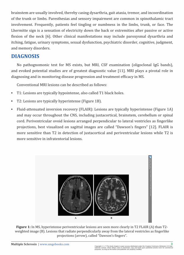
• T2: Lesions are typically hyperintense (Figure 1B).
• Fluid-attenuated inversion recovery (FLAIR): Lesions are typically hyperintense (Figure 1A) and may occur throughout the CNS, including juxtacortical, brainstem, cerebellum or spinal cord. Periventricular ovoid lesions arranged perpendicular to lateral ventricles as fingerlike projections, best visualized on sagittal images are called “Dawson’s fingers” [12]. FLAIR is more sensitive than T2 in detection of juxtacortical and periventricular lesions while T2 is more sensitive in infratentorial lesions.
Figure 1: In MS, hyperintense periventricular lesions are seen more clearly in T2 FLAIR (A) than T2-weighted image (B). Lesions that radiate perpendicularly away from the lateral ventricles as fingerlike
projections (arrow), called “Dawson’s fingers”.
4Multiple Sclerosis | www.smgebooks.comCopyright Li Y.This book chapter is open access distributed under the Creative Commons Attribution 4.0 Inter-national License, which allows users to download, copy and build upon published articles even for commercial purposes, as long as the author and publisher are properly credited.
• T1 C+: Gadolinium is useful in defining areas undergoing active inflammation. Active lesions often show incomplete peripheral enhancement called open ring sign. It is thought that the enhancing part of the ring represents advancing front of demyelination and indicates the white matter side of the lesion; whereas the open part of the ring usually point towards the grey matter.
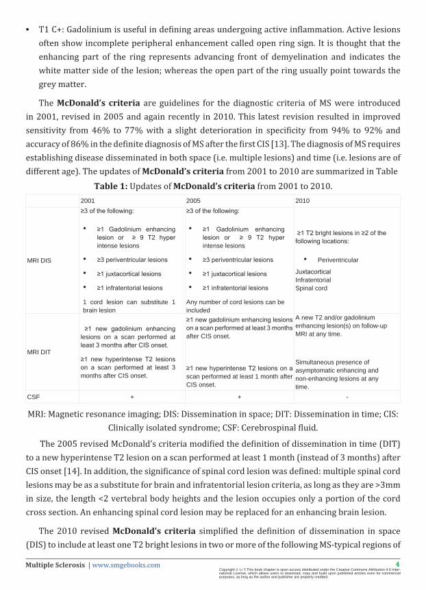
The McDonald’s criteria are guidelines for the diagnostic criteria of MS were introduced in 2001, revised in 2005 and again recently in 2010. This latest revision resulted in improved sensitivity from 46% to 77% with a slight deterioration in specificity from 94% to 92% and accuracy of 86% in the definite diagnosis of MS after the first CIS [13]. The diagnosis of MS requires establishing disease disseminated in both space (i.e. multiple lesions) and time (i.e. lesions are of different age). The updates of McDonald’s criteria from 2001 to 2010 are summarized in Table
Table 1: Updates of McDonald’s criteria from 2001 to 2010.2001 2005 2010
MRI DIS
≥3 of the following:
• ≥1 Gadolinium enhancing lesion or ≥ 9 T2 hyper intense lesions
• ≥3 periventricular lesions
• ≥1 juxtacortical lesions
• ≥1 infratentorial lesions
1 cord lesion can substitute 1 brain lesion
≥3 of the following:
• ≥1 Gadolinium enhancing lesion or ≥ 9 T2 hyper intense lesions
• ≥3 periventricular lesions
• ≥1 juxtacortical lesions
• ≥1 infratentorial lesions
Any number of cord lesions can be included
≥1 T2 bright lesions in ≥2 of the following locations:
• Periventricular
Juxtacortical Infratentorial Spinal cord
MRI DIT
≥1 new gadolinium enhancing lesions on a scan performed at least 3 months after CIS onset.
≥1 new hyperintense T2 lesions on a scan performed at least 3 months after CIS onset.
≥1 new gadolinium enhancing lesions on a scan performed at least 3 months after CIS onset.
≥1 new hyperintense T2 lesions on a scan performed at least 1 month after CIS onset.
A new T2 and/or gadolinium enhancing lesion(s) on follow-up MRI at any time.
Simultaneous presence of asymptomatic enhancing and non-enhancing lesions at any time.
CSF + + -
MRI: Magnetic resonance imaging; DIS: Dissemination in space; DIT: Dissemination in time; CIS: Clinically isolated syndrome; CSF: Cerebrospinal fluid.
The 2005 revised McDonald’s criteria modified the definition of dissemination in time (DIT) to a new hyperintense T2 lesion on a scan performed at least 1 month (instead of 3 months) after CIS onset [14]. In addition, the significance of spinal cord lesion was defined: multiple spinal cord lesions may be as a substitute for brain and infratentorial lesion criteria, as long as they are >3mm in size, the length <2 vertebral body heights and the lesion occupies only a portion of the cord cross section. An enhancing spinal cord lesion may be replaced for an enhancing brain lesion.
The 2010 revised McDonald’s criteria simplified the definition of dissemination in space (DIS) to include at least one T2 bright lesions in two or more of the following MS-typical regions of
5Multiple Sclerosis | www.smgebooks.comCopyright Li Y.This book chapter is open access distributed under the Creative Commons Attribution 4.0 Inter-national License, which allows users to download, copy and build upon published articles even for commercial purposes, as long as the author and publisher are properly credited.
the CNS (periventricular, juxtacortical, infratentorial, or spinal cord) [15]. Gadolinium enhancing lesion was no more required for DIS. The criteria for DIT were modified to include a new T2 and/or gadolinium enhancing lesion(s) on follow-up MRI at any time, or asymptomatic enhancing and non-enhancing lesions simultaneously present at any time [16]. These modifications implicate that DIT can be identified on a single baseline MRI scan resulting in early diagnosis of MS. CSF examination for oligoclonal IgG bands was no longer needed for the diagnosis of RRMS. Modifications of the McDonald’s criteria in each successive revision have resulted in earlier and easier diagnosis of MS.
Clinical Variants
There are several clinical variants of MS with specific clinical and radiological findings. They include:
Benign MS
It accounts for approximately 20% of all MS patients. The patients remain fully functional in all neurological systems for 15 years after the disease onset. Serial brain MRI shows few new or enlarging lesions with lower incidence of contrast enhancement [17].
Malignant (Fulminant) MS or Marburg disease
It has a rapid progressive course with severe relapses, leading to significant disability in various neurologic systems or death within weeks to months after the disease onset. MRI shows numerous smaller lesions that may coalesce to form large white matter plaques, disseminated throughout the brain. The lesions may show defined rings and incomplete ring enhancement [17].
Schilder’s disease (SD)
It is a rare progressive demyelinating disease that usually affects children and young adults. The clinical manifestations include aphasia, dementia, personality changes, balance instability, seizures, headache, and visual disturbances. MRI shows large ring-enhancing lesions in each brain hemisphere [18].
Baló’s concentric sclerosis (BCS)
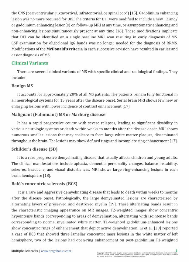
It is a rare and aggressive demyelinating disease that leads to death within weeks to months after the disease onset. Pathologically, the large demyelinated lesions are characterized by alternating layers of preserved and destroyed myelin [19]. These alternating bands result in the characteristic imaging appearance on MR images. T2-weighted images show concentric hypointense bands corresponding to areas of demyelination, alternating with isointense bands corresponding to normal myelinated white matter. T1-weighted gadolinium-enhanced lesions show concentric rings of enhancement that depict active demyelination. Li et al. [20] reported a case of BCS that showed three lamellar concentric mass lesions in the white matter of left hemisphere, two of the lesions had open-ring enhancement on post-gadolinium T1-weighted
6Multiple Sclerosis | www.smgebooks.comCopyright Li Y.This book chapter is open access distributed under the Creative Commons Attribution 4.0 Inter-national License, which allows users to download, copy and build upon published articles even for commercial purposes, as long as the author and publisher are properly credited.
imaging. Three small MS lesions were also visualized, two in the right periventricular white matter and the other in the left lateral ventricular triangle-area (Figure 2A-D). A follow-up MRI at 27 months showed resolution of the ring lesions and small plaques in the previous regions (Figure 2E, F), and the three MS lesions were stable. There was no other new lesion in the brain.
Figure 2: Images of a 37-year-old man (A) Axial T1-weighted image reveals three hypo-/isointense concentric rings in the supratentorial left cerebral white matter area, two of which were typical concentric images, (B) Axial T2-weighted image displays whirlpool hyperintense
concentric rings, two small plaque multiple sclerosis-like lesions were detected in the right frontal white matter, (C) Axial T1-weighted image after administration of gadolinium-DTPA shows an open-ring enhancement of the lesion, (D) The next slice of the 1c image shows the
patchy enhancement at the edge of the concentric lesions, (E) and (F) Axial T2-weighted images shows the concentric lesions markedly disappeared after 27 months, and transformed into
multiple sclerosis-like small plaques in the previous concentric lesions, the other three multiple sclerosis-like lesions were stable. There was no other new lesion in the brain.
7Multiple Sclerosis | www.smgebooks.comCopyright Li Y.This book chapter is open access distributed under the Creative Commons Attribution 4.0 Inter-national License, which allows users to download, copy and build upon published articles even for commercial purposes, as long as the author and publisher are properly credited.
Tumefactive MS
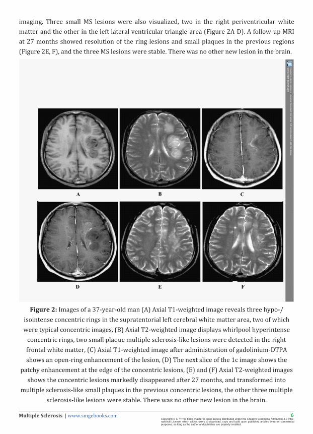
It presents as single or multiple focal lesions that may mimic a brain tumor clinically and radiologically. Lesions on T1-weighted imaging are hypointense, T2-weighted imaging are hyperintense, and T1-weighted gadolinium-enhanced are open ring-enhancement [21,22]. Tumefactive demyelination is distinguished from a brain tumor by the presence of multiple lesions and decrease in lesion size or detection of new lesions on serial brain MRI. A case of biopsy-proven tumefactive demyelinating lesion (TDL) shows an open-ring sign in the right front-parietal lobe subcortical white matter on post-gadolinium coronal T1 Flair MR imaging (Figure 3A) and on sagittal T1 Flair MR image with contrast enhancement shows marked enhancement of dilated veins running through the outside and inside of the lesion (Figure 3B).
Figure 3: Images of a 36-year-old man with biopsy-proven tumefactive demyelinating lesion (TDL). (A) Coronal T1 Flair MR image with contrast enhancement shows an open-ring sign in the
right front-parietal lobe subcortical white matter. (B) Sagittal T1 Flair MR image with contrast enhancement shows marked enhancement of dilated veins running through the outside and
inside of the lesion.
Advanced MRI Techniques
Proton magnetic resonance spectroscopy (1H-MRS)
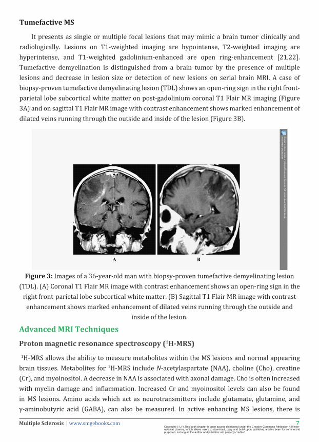
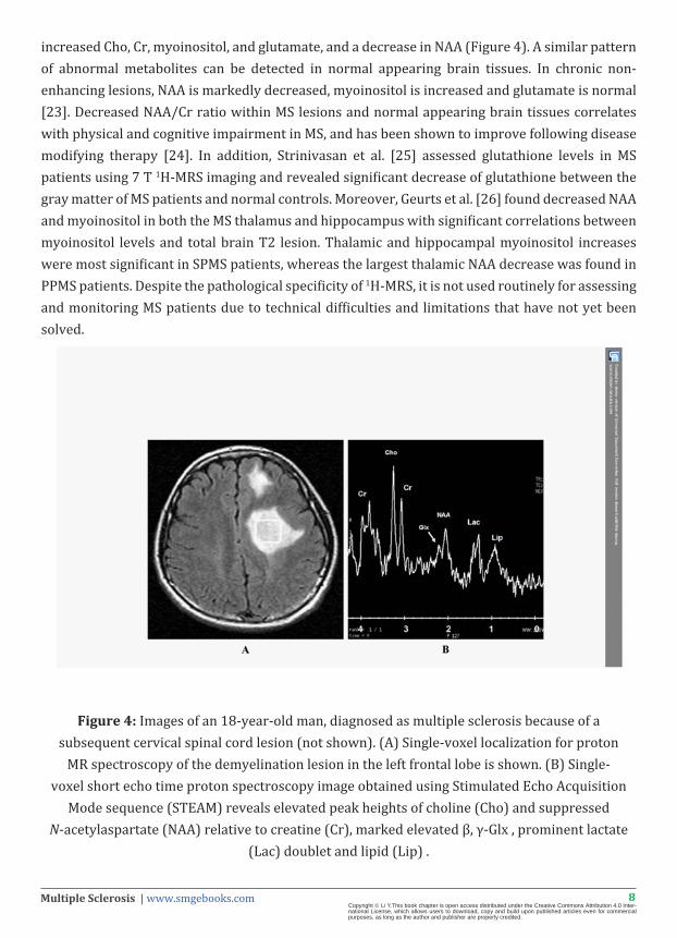
1H-MRS allows the ability to measure metabolites within the MS lesions and normal appearing brain tissues. Metabolites for 1H-MRS include N-acetylaspartate (NAA), choline (Cho), creatine (Cr), and myoinositol. A decrease in NAA is associated with axonal damage. Cho is often increased with myelin damage and inflammation. Increased Cr and myoinositol levels can also be found in MS lesions. Amino acids which act as neurotransmitters include glutamate, glutamine, and γ-aminobutyric acid (GABA), can also be measured. In active enhancing MS lesions, there is
8Multiple Sclerosis | www.smgebooks.comCopyright Li Y.This book chapter is open access distributed under the Creative Commons Attribution 4.0 Inter-national License, which allows users to download, copy and build upon published articles even for commercial purposes, as long as the author and publisher are properly credited.
increased Cho, Cr, myoinositol, and glutamate, and a decrease in NAA (Figure 4). A similar pattern of abnormal metabolites can be detected in normal appearing brain tissues. In chronic non-enhancing lesions, NAA is markedly decreased, myoinositol is increased and glutamate is normal [23]. Decreased NAA/Cr ratio within MS lesions and normal appearing brain tissues correlates with physical and cognitive impairment in MS, and has been shown to improve following disease modifying therapy [24]. In addition, Strinivasan et al. [25] assessed glutathione levels in MS patients using 7 T 1H-MRS imaging and revealed significant decrease of glutathione between the gray matter of MS patients and normal controls. Moreover, Geurts et al. [26] found decreased NAA and myoinositol in both the MS thalamus and hippocampus with significant correlations between myoinositol levels and total brain T2 lesion. Thalamic and hippocampal myoinositol increases were most significant in SPMS patients, whereas the largest thalamic NAA decrease was found in PPMS patients. Despite the pathological specificity of 1H-MRS, it is not used routinely for assessing and monitoring MS patients due to technical difficulties and limitations that have not yet been solved.
Figure 4: Images of an 18-year-old man, diagnosed as multiple sclerosis because of a subsequent cervical spinal cord lesion (not shown). (A) Single-voxel localization for proton
MR spectroscopy of the demyelination lesion in the left frontal lobe is shown. (B) Single-voxel short echo time proton spectroscopy image obtained using Stimulated Echo Acquisition
Mode sequence (STEAM) reveals elevated peak heights of choline (Cho) and suppressed N-acetylaspartate (NAA) relative to creatine (Cr), marked elevated β, γ-Glx , prominent lactate
(Lac) doublet and lipid (Lip) .
9Multiple Sclerosis | www.smgebooks.comCopyright Li Y.This book chapter is open access distributed under the Creative Commons Attribution 4.0 Inter-national License, which allows users to download, copy and build upon published articles even for commercial purposes, as long as the author and publisher are properly credited.
Magnetisation transfer imaging (MTI)
MTI is based on the interactions between protons in free fluids and protons bound to macromolecules. Magnetisation transfer is quantified using the magnetisation transfer ratio (MTR), where a reduced MTR is an indicator of damage to myelin and axonal membranes [27]. MTR correlates strongly with T2 lesion and brain atrophy. In addition, reduced MTR has been documented in the normal-appearing gray/white matter and are more pronounced with progressive forms of MS [28,29]. Filippi et al. [30] reported reduced MTR in the GM and normal-appearing gray matter of patients with different MS phenotypes, even from the earliest clinical stages of the disease. Due to lack of standardization in acquisition protocols, it is not used in routine clinical setting at the present time.
Diffusion weighted imaging (DWI) and diffusion tensor imaging (DTI)
Diffusion imaging is based on diffusion, or the random movement of molecules through a fluid. The diffusivity in brain tissue is lower than that of free water, called the apparent diffusion coefficient (ADC). Restricted diffusion causes increase in ADC values. Active plaques of MS may demonstrate restricted diffusion on DWI/ADC. Analogous to MTI, DTI is sensitive to tissue injury and is abnormal in MS lesions and normal appearing brain tissues [31]. Poonawalla et al. [32] reported that the fractional anisotropy (FA) and mean diffusivity (MD) values were significantly higher in cortical lesions as compared to healthy controls. DTI abnormalities have confirmed that damage to the brain in MS patients is not limited to focal and macroscopic lesions. This damage is also present in the normal-appearing gray matter, even at early stages of the disease. Abnormal gray matter diffusivity correlates with disease progression and cognitive impairment [33].
Perfusion MRI
Perfusion MRI uses either bolus tracking of exogenous tracers or endogenous arterial water (arterial spin labeling) to analyze brain tissue perfusion. Active MS lesions are characterized by increased perfusion, whereas normal-appearing brain tissues have relatively decreased perfusion [34]. Holland et al. [35] reported that reduced perfusion in the white matter correlated with chronic plaques and lesions were highly perfused in RRMS than SPMS. By perfusion MRI, we may be able to characterize the stage of the lesion and this could have a direct effect on disease management.
Susceptibility-weighted imaging (SWI)
SWI is a relatively new MRI sequence, which uses the paramagnetic susceptibility effect of iron to demonstrate susceptibility differences between tissues [36]. Veins associated with MS plaques and iron laden tissues can be visualized by SWI. The imaging of venous blood with SWI is a blood-oxygenation-level dependent (BOLD) technique which is why it was originally called BOLD venographic imaging. Haacke et al. [37] demonstrated that by combining conventional imaging and SWI, nearly 50% more lesions can be visualized. SWI is unable to provide enough specificity to distinguish MS from small vessel disease involving white matter lesions.

10Multiple Sclerosis | www.smgebooks.comCopyright Li Y.This book chapter is open access distributed under the Creative Commons Attribution 4.0 Inter-national License, which allows users to download, copy and build upon published articles even for commercial purposes, as long as the author and publisher are properly credited.
Functional MRI (fMRI)
fMRI is a technique used to obtain functional information by visualization of cortical activity. It detects subtle alteration in blood flow in response to stimuli or actions. An advantage of fMRI over other imaging techniques is that it provides evidence for plasticity in MS. It utilizes the different magnetic properties of oxygenated and deoxygenated blood to identify areas of increased or decreased cerebral blood flow, called BOLD imaging. Rocca et al. [38] demonstrated that the combination of measures of functional connectivity with measures of structural damage within specific white-matter fibre bundles is likely to improve our understanding of the relation between structural and functional abnormalities in RRMS patients. An abnormal pattern of brain activation has been shown to be correlated with fatigue [39].
Higher field imaging
High-field (3 T) and ultra-high field (>7 T) MRI scanners are more sensitive for detecting T2 and gadolinium-enhancing lesions than 1.5 T MRI scanner [40,41]. Higher filed imaging can improve the early diagnosis of MS [42]. Specific pulse sequences, such as double inversion recovery (DIR) imaging can further improve the ability to detect cortical MS lesions in vivo but are technically challenging to execute [43]. Calabrese et al. [44] reported that DIR imaging at higher field captures larger number of cortical lesions.
Differential Diagnosis
Multiple Sclerosis (MS) must be differentiated from the following clinical entities as the therapeutic interventions differ considerably:
Non-specific white matter T2 hyperintensities
Age, hypertension, diabetes, smoking, and migraine are all risk factors for non-specific T2 hyperintensities in the cerebral white matter. Leukoaraiosis or small vessel ischemic disease usually shows white matter T2 hyperintensities in elderly people. Smaller size and peripheral location of the lesions help to differentiate from MS plaques.
Acute disseminated encephalomyelitis (ADEM)
ADEM is an acute inflammatory and demyelinating disease of the central nervous system (CNS), usually preceded by viral or bacterial infections or vaccinations [45]. It occurs more commonly in children than adults, is typically monophasic, and presents with encephalopathy [46]. It predominantly involves the subcortical white matter, thalami and basal ganglia. The spinal cord is usually affected with large, tumefactive lesions [47]. Unlike lesions in MS, ADEM lesions are larger, more edematous, and more symmetric; usually enhance simultaneously or nearly simultaneously, underscoring their synchronous course [48]. It is estimated that 21% of ADEM convert to MS after a mean follow-up period of 2.36 years, and 27% after 5.64 years [49].
11Multiple Sclerosis | www.smgebooks.comCopyright Li Y.This book chapter is open access distributed under the Creative Commons Attribution 4.0 Inter-national License, which allows users to download, copy and build upon published articles even for commercial purposes, as long as the author and publisher are properly credited.
Neuromyelitis optica (NMO)
NMO, also known as Devic’s disease, is a severe demyelinating disease primarily affecting optic nerves and spinal cord. It usually presents with bilateral optic neuritis and myelitis. Spinal cord MR imaging is characterized by longitudinally extensive myelitis and cord lesions involving more than three vertebral segments [50]. Brain MR imaging usually shows bilateral swollen optic nerve with T2 hypersignal and contrast enhancement. Unlike MS, white matter lesions are absent or few, and non-specific [51]. If T2 lesions are present in the brain of NMO patients, they look atypical for MS and tend to border midline CSF spaces, where the expression of the aquaporin-4 (AQP4) water channel is high [52].
Neoplastic disease
Primary CNS tumors or systemic metastases may cause T2 hyperinensity on MRI, usually show gadolinium enhancement. Lymphoma can create multifocal lesions and should be included in the differential diagnosis of MS. Lymphoma and demyelination could be differentiated with a response to corticosteroid treatment, lymphoma shows recurrent enhancement on follow-up imaging, whereas demyelination shows persistent improvement on follow-up imaging [53]. Advanced MRI techniques, such as perfusion MRI can be helpful for the differentiation as both lymphoma and neoplastic disease present higher perfusion that that of the demyelinating lesions [54].
Rheumatologic diseases
Rheumatologic diseases such as systemic lupus erythematosus (SLE), Wegener granulomatosis, polyarteritis nodosa, etc may sometimes mimic the MRI lesions of MS. They can present with subcortical T2 hyperintensities, however the T2 lesion burden is far lower than in MS [55]. They are less common than MS and can be best differentiated clinically.
Sarcoidosis
The diagnosis of sarcoidosis is difficult due to non-specific clinical presentations and imaging findings, it may sometimes mimic MS. The characteristic MRI findings of meningeal thickening with gadolinium enhancement, hypothalamus involvement, and persistent enhancement on follow-up imaging despite treatment with corticosteroids differentiate sarcoidosis from MS [56].
CNS infectious diseases
Toxoplasmosis and lyme disease may show imaging findings that are similar to MS, however exposure histories and serologic results differ from MS [57]. Progressive multifocal leukoencephalopathy (PML) usually presents as subcortical, serpentine, T2 hyperintensities expanding over time and do not enhance on postcontrast T1-weighted images [53].
12Multiple Sclerosis | www.smgebooks.comCopyright Li Y.This book chapter is open access distributed under the Creative Commons Attribution 4.0 Inter-national License, which allows users to download, copy and build upon published articles even for commercial purposes, as long as the author and publisher are properly credited.
Cerebral autosomal dominant arteriopathy with subcortical infarcts and leukoencephalopathy (CADASIL)
CADASIL usually presents as confluent subcortical or periventricular areas of T2 hyperintensities and do not enhance after gadolinium administration. It mainly affects anterior temporal lobes and has a family history of neurologic disability [58].
CONCLUSIONSMultiple sclerosis (MS) is a chronic immune-mediated degenerative disease of the central
nervous system and is a major cause of acquired disability in young adults. The latest revision of McDonald’s criteria resulted in improved sensitivity and specificity in the definite diagnosis of MS after the first CIS. Conventional MRI plays an important role in diagnosing and in monitoring the disease course; however, it lacks sensitivity to gray matter lesions and diffuse damage throughtout the white matter. Advanced MRI techniques provide higher specificity and sensitivity to both lesions and normal-appearing gray/white matter contributing in better understanding of MS pathophysiology. The adoption of cutting edge techniques into clinical research and practice need to be refined and validated due to several limitations.
ACKNOWLEDGMENTSThis study was supported by National Key Clinical Specialties Construction Program of China
(No. 2013-544).
REFERENCES1. BererK,Krishnamoorthy G. Microbial view of central nervous system autoimmunity. FEBS Lett. 2014; 588: 4207-4213.
2. Frohman EM, Racke MK, Raine CS. Multiple sclerosis-the plaque and its pathogenesis. N Engl Med.2006; 354:942-955.
3. Milo R, Miller A. Revised diagnostic criteria of multiple sclerosis. Autoimmun Rev. 2014; 13: 518-524.
4. Krupp LB, Banwell B, Tenembaum S. International Pediatric MS Study Group. Consensus definitions proposed for pediatric multiple sclerosis and related disorders. Neurology. 2007; 68: S7-12.
5. Milo R, Kahana E. Multiple sclerosis: geoepidemiology, genetics and the environment.Autoimmun Rev. 2010; 9: A387-A394.
6. Compston A, Coles A. Multiple sclerosis. Lancet. 2008; 372: 1502-1517.
7. Lublin FD, Reingold SC. Defining the clinical course ofmultiple sclerosis: results of an international survey. National Multiple Sclerosis Society (USA) Advisory Committee on Clinical Trials of New Agents in Multiple Sclerosis. Neurology. 1996; 46: 907-911.
8. Weinshenker BG, Bass B, Rice GP, Noseworthy J, Carriere W, Baskerville J, et al. The natural history of multiple sclerosis: a geographically based study. I. Clinical course and disability. Brain. 1989; 112: 133-146.
9. Miller D, Barkhof F, Montalban X, Thompson A, Filippi M. Clinically isolated syndromes suggestive of multiple sclerosis, part I: natural history, pathogenesis, diagnosis, and prognosis. Lancet Neurol. 2005; 4: 281-288.
10. Okuda DT, Mowry EM, Beheshtian A, Waubant E, Baranzini SE, Goodin DS, et al. IncidentalMRI anomalies suggestive of multiple sclerosis: the radiologically isolated syndrome. Neurology. 2009; 72: 800-805.
11. Tsang BK, Macdonell R. Multiple sclerosis- diagnosis, management and prognosis. AustFam Physician. 2011; 40: 948-955.
12. Dawson JD. The histology of disseminated sclerosis. Trans Roy SocEdinb. 1916; 50: 517.
13. Swanton JK, Fernando K, Dalton CM, Miszkiel KA, Thompson AJ, Plant GT, et al. Modification of MRI criteria for multiple sclerosis in patients with clinically isolated syndromes. J NeurolNeurosurg Psychiatry.2006; 77: 830-833.
14. Polman CH, Reingold SC, Edan C, Filippy M, Hartung HP, Kappos L, et al. Diagnostic criteria for multiple sclerosis: 2005 revisions to the “McDonald Criteria”. Ann Neurol. 2005; 56: 840-846.
13Multiple Sclerosis | www.smgebooks.comCopyright Li Y.This book chapter is open access distributed under the Creative Commons Attribution 4.0 Inter-national License, which allows users to download, copy and build upon published articles even for commercial purposes, as long as the author and publisher are properly credited.
15. Polman CH, Reingold SC, Banwell B, Clanet M, Cohen JA, Filippi M, et al. Diagnostic criteria for multiple sclerosis: 2010 revisions to the McDonald criteria. Ann Neurol. 2010; 69: 292-302.
16. Montalban X, Tintoré M, Swanton J, Barkhof F, Fazekas F, Filippi M, et al. MRI criteria for MS in patients with clinically isolated syndromes. Neurology.2010; 74: 427-434.
17. Cañellas AR, Gols AR, Izquierdo JR, Subirana MT, Gairin XM.Idiopathic inflammatory-demyelinating diseases of the central nervous system. Neuroradiology. 2007; 49: 393-409.
18. Sastre-Garriga J, Rovira A, Río J, Tintoré M, Grivé E, et al. Clinically definite multiple sclerosis after radiological Schilder-like onset. J Neurol. 2003; 250: 871-873.
19. Gharagozloo AM, Poe LB, Collins GH.Antemortem diagnosis of Baló concentric sclerosis: correlative MR imaging and pathologic features. Radiology. 1994; 191: 817-819.
20. Li Y, Xie P, Fan X, Tang HBalò’s concentric sclerosis presenting with benign clinical course and multiple sclerosis-like lesions onmagnetic resonance images. Neurol India. 2009; 57: 66-68.
21. Jeong IH, Kim SH, Hyun JW, Joung A, Cho HJ, Kim HJ.Tumefactive demyelinating lesions as a first clinical event: Clinical, imaging, and follow-up observations. J Neurol Sci. 2015; 358: 118-124.
22. Mabray MC, Cohen BA, Villanueva-Meyer JE, Valles FE, Barajas RF, Rubenstein JL, et al. Performance of Apparent Diffusion Coefficient Values and Conventional MRI Features in Differentiating Tumefactive Demyelinating Lesions From Primary Brain Neoplasms. AJR Am J Roentgenol.2015; 205: 1075-1085.
23. Srinivasan R, Sailasuta N, Hurd R, Nelson S, Pelletier D. Evidence of elevated glutamate in multiple sclerosis using magnetic resonance spectroscopy at 3 T. Brain. 2005; 128: 1016-1025.
24. Khan O, Shen Y, Caon C, Bao F, Ching W, Reznar M, et al. Axonal metabolic recovery and potential neuroprotective effect of glatiramer acetate in relapsing-remitting multiple sclerosis. MultScler. 2005; 11:646-651.
25. Srinivasan R, Ratiney H, Hammond-Rosenbluth KE, Pelletier D, Nelson SJ. MR spectroscopic imaging of glutathione in the white and gray matter at 7 T with an application to multiple sclerosis. MagnReson Imaging. 2010; 28: 163-170.
26. Geurts JJ, Reuling IE, Vrenken H, Uitdehaag BM, Polman CH, Castelijns JA, et al. MR spectroscopic evidence for thalamic and hippocampal, but not cortical, damage in multiple sclerosis. MagnReson Med. 2006; 55: 478-483.
27. Wolff SD, Balaban RS. Magnetization transfer imaging: practical aspects and clinical applications. Radiology. 1994; 192: 593-599.
28. Filippi M, Iannucci G, Tortorella C, Minicucci L, Horsfield MA,Colombo B, et al. Comparison of MS clinical phenotypes using conventional and magnetization transfer MRI. Neurology. 1999; 52: 588-594.
29. Filippi M, Rocca MA. Magnetization transfer magnetic resonance imaging of the brain, spinal cord, and optic nerve. Neurotherapeutics. 2007; 4: 401-413.
30. Filippi M, Agosta F. Magnetization transfer MRI in multiple sclerosis. J Neuroimaging. 2007; 17: 22-26.
31. Henry RG, Oh J, Nelson SJ, Pelletier D. Directional tensor imaging in relapsing-remitting multiple sclerosis: a possible in vivo signature of walleriandegeneration. J MagnResonImaging. 2003; 18: 420-426.
32. Poonawalla AH, Hasan KM, Gupta RK, Ahn CW, Nelson F, Wolinsky JS, et al. Diffusion-tensor MR imaging of cortical lesions in multiple sclerosis: initial findings. Radiology. 2008; 246: 880-886.
33. Yu HJ, Christodoulou C, Bhise V, Greenblatt D, Patel Y, Serafin D, et al. Multiple white matter tract abnormalities underlie cognitive impairment in RRMS. Neuroimage. 2012; 59: 3713-3722.
34. Bakshi R, Thompson AJ, Rocca MA, Pelletier D, Dousset V, Barkhof F, et al.MRI in multiple sclerosis: current status and future prospects. Lancet Neurol. 2008; 7: 615-625.
35. Holland CM, Charil A, Csapo I, Liptak Z, Ichise M, et al. The relationship between normal cerebral perfusion patterns and white matter lesion distribution in 1,249patients with multiple sclerosis. J Neuroimaging. 2012; 22: 129-136.
36. Haacke EM,Xu Y, Cheng YC, Reichenbach JR. Susceptibility weighted imaging (SWI). MagnReson Med. 2004; 52: 612-618.
37. Haacke EM, Makki M, Ge Y, Maheshwari M, Sehgal V, Hu J, et al. Characterizing iron deposition in multiple sclerosis lesions using susceptibility weighted imaging. J MagnResonImaging. 2009; 29: 537-544.
38. Rocca MA, Pagani E, Absinta M, Valsasina P, Falini A, et al. Altered functional andstructural connectivities in patients with MS: a 3-T study. Neurology. 2007; 69: 2136-2145.
39. Rocca MA, Filippi M. Functional MRI in multiple sclerosis. J Neuroimaging. 2007; 1: 36S-41S.
40. Sicotte NL, Voskuhl RR, Bouvier S, Klutch R, Cohen MS,Mazziotta JC, et al. Comparison of multiple sclerosis lesions at 1.5 and 3.0 tesla. Invest Radiol. 2003; 38: 423-427.
14Multiple Sclerosis | www.smgebooks.comCopyright Li Y.This book chapter is open access distributed under the Creative Commons Attribution 4.0 Inter-national License, which allows users to download, copy and build upon published articles even for commercial purposes, as long as the author and publisher are properly credited.
41. Kollia K, Maderwald S, Putzki N, Schlamann M, Theysohn JM, Kraff O, et al. First clinical study on ultra-high-field MR imaging in patients with multiple sclerosis: comparison of 1.5T and 7T. AJNR Am J Neuroradiol. 2009; 30: 699-702.
42. Wattjes MP, Harzheim M, Kuhl CK, Gieseke J, Schmidt S, Klotz L, et al. Does high-field MR imaging have an influence on the classification of patients with clinically isolated syndromes according to current diagnostic MR imaging criteria for multiple sclerosis? AJNR Am J Neuroradiol. 2006; 27: 1794-1798.
43. Wattjes MP, Lutterbey GG, Gieseke J, Träber F, Klotz L, Schmidt S, et al. Double inversion recovery brain imaging at 3T: diagnostic value in the detection of multiple sclerosis lesions. AJNR Am J Neuroradiol. 2007; 28: 54-59.
44. Calabrese M, Battaglini M, Giorgio A, Atzori M, Bernardi V, Mattisi I, et al. Imaging distribution and frequency of cortical lesions in patients with multiple sclerosis. Neurology. 2010; 75: 1234-1240.
45. Menge T, Hemmer B, Nessler S, Wiendl H, Neuhaus O, Hartung HP, et al. Acute disseminated encephalomyelitis: an update. Arch Neurol. 2005; 62: 1673-1680.
46. Tenembaum S, Chitnis T, Ness J, Hahn JS. International Pediatric MS Study Group. Acute disseminated encephalomyelitis. Neurology; 2007;68: S23-S36.
47. Dale RC, Branson JA. Acute disseminated encephalomyelitis or multiple sclerosis: can the initial presentation help in establishing acorrect diagnosis? Arch Dis Child. 2005; 90: 636-639.
48. Bennetto L, Scolding N. Inflammatory/post-infectious encephalomyelitis. J NeurolNeurosurg Psychiatry. 2004; 75 Suppl 1: i22-28.
49. Hartung HP, Grossman RI .ADEM: distinct disease or part of the MS spectrum? Neurology. 2001; 56: 1257-1260.
50. Polman CH, Reingold SC, Banwell B, Clanet M, Cohen JA, Filippi M, et al. Diagnostic criteria for multiple sclerosis: 2010 revisions to the McDonald Criteria. Ann Neurol. 2011; 69: 292-302.
51. Ghezzi A, Bergamaschi R, Martinelli V, Trojano M, Tola MR, Merelli E, et al. Clinical characteristics, course and prognosis of relapsing Devic’sneuromyelitisoptica. J Neurol. 2004; 251: 47-52.
52. Pittock SJ, Weinshenker BG, Lucchinetti CF, Wingerchuk DM, Corboy JR, Lennon VA. Neuromyelitisoptica brain lesions localized at sites of high aquaporin 4 expression. Arch Neurol. 2006; 63: 964-968.
53. Bermel RA, Fox RJ. MRI in multiple sclerosis. Continuum. 2010; 16: 37-57.
54. Al-Okaili RN, Krejza J, Wang S, Woo JH, Melhem ER. Advanced MR imaging techniques in the diagnosis of intraaxial brain tumors in adults. Radiographs. 2006; 26 Suppl 1:S173-189.
55. Rovaris M, Viti B, Ciboddo G, Gerevini S, Capra R, Iannucci G, et al. Brain involvement in systemic immune mediated diseases: magnetic resonance and magnetisation transfer imaging study. J NeurolNeurosurg Psychiatry. 2000; 68: 170-177.
56. Joseph FG, Scolding NJ. Sarcoidosis of the nervous system. Pract Neurol. 2007; 7: 234-244.
57. Karussis D, Weiner HL, Abramsky O. Multiple sclerosis vs Lyme disease: a case presentation to a discussant and a review of the literature. MultScler. 1999; 5: 395-402.
58. Ringelstein EB, Nabavi DG. Cerebral small vessel diseases: cerebral microangiopathies. CurrOpin Neurol. 2005; 18: 179-188.