multivariate analysis of dengue in a …repository-tnmgrmu.ac.in/1429/1/200400713malini.pdfdengue...
TRANSCRIPT

MULTIVARIATE ANALYSIS OF DENGUE IN A TERTIARY CARE HOSPTIAL
Dissertation submitted in
Partial Fulfillment of the regulations required for the award of
M.D DEGREE
In
Microbiology – Branch IV
THE TAMILNADU DR.M.G.R MEDICAL UNIVERSITY,
CHENNAI – 600 032.
April 2013

DECLARATION
I Dr P.Malini solemnly declare that the dissertation entitled “Multivariate
Analysis of Dengue in a Tertiary Care Hospital” was done by me at Coimbatore
Medical College, Coimbatore during the period September 2011 to August 2012
under the supervision and guidance of Dr.K. RAJENDRAN BSc, M.D., Professor
and HOD Department of Microbiology Coimbatore Medical College Coimbatore
– 14.
This dissertation is submitted to the TamilNadu Dr.M.G.R Medical
University, Chennai towards the partial fulfillment of the requirement for the
award of M.D Degree (Branch – IV) in Microbiology to be held in April 2013.
I have not submitted this dissertation on any previous occasion to any
university for the award of any degree.
Dr.P.Malini
Place:
Date:
CERTIFICATE
This is to certify that the dissertation entitled “MULTIVARIATE
ANALYSIS OF DENGUE IN A TERTIARY CARE HOSPTIAL” is a
bonafide work done by Dr.P.Malini, post graduate student in the Department of
Microbiology, under the supervision of Dr.K.RAJENDRAN B.Sc, M.D.,
Professor & Head Department of Microbiology, Coimbatore Medical College,
Coimbatore, in fulfillment of the regulation of the Tamil Nadu Dr.M.G.R Medical
University, towards the award of M.D Degree (Branch – IV) in Microbiology.
DR.R.VIMALA M.D., DR.K.RAJENDRAN B.Sc,M.D.,
Dean Professor & Head
Coimbatore Medical College Department of Microbiology
Coimbatore – 14 Coimbatore Medical College
Coimbatore - 14


ACKNOWLEDGEMENT
I am grateful to the Dean,Dr.R.Vimala M.D., Coimbatore Medical College
and Hospital, for permitting me to carry out this study.
I thank Dr.Lalitha M.D., Vice Principal, Coimbatore Medical College for
her encouragement in completing this study.
I wish to place on the records my deep sense of gratitude and sincere thanks
to Professor & HOD Dr. K. Rajendran BSc, M.D., Department of Microbiology
for his precious guidance and valuable suggestions given throughout the study.
I express my sincere thanks to our Associate Professors
Dr. V.Sadhiqua M.D, D.G.O., Dr V.Dhanasekharan M.D, D.C.H,
Dr.N.Mythili M.D., Department of Microbiology, Coimbatore Medical College.
I would like to place my thanks to our Assistant Professors Dr.N.Bharathi
Santhose M.D., Dr S.Deepa M.D., Dr.B.Padmini M.D., Dr.R.Radhika M.D.,
for their constant help, guidance and encouragement given to me throughout this
study.
I express my immense thanks to Prof. Dr. Veerakesari, M.D., Professor
and Head of the Department, Department of Medicine and Dr. Neelakandan
M.D., Professor and Head of the Department of Paediatrics for their kind
cooperation in selection of cases, collection of specimens and datas.
I am thankful to my colleagues Dr.Lokeshwari, Dr.Aruna,
Dr.Ashwini,Dr.Bhuvaneshwari,Dr.kokilapriya,Dr.Deepa,Dr.Swaathy,
Dr.Vijayashree ,Dr.Sathyapriya, Dr.M.Preethi, Dr.Shanthi.

At last but not the least, I extend my thanks to all staff members, Department
of Microbiology for giving full cooperation and timely help in carrying out this
study.
I would like to thank the Institutional Ethical Committee for approving my
study.
I thank my family members for their immense help and support throughout
this study. Finally I thank the Almighty for his blessings in every moment in my
life.


CONTENTS S.No. Page No. 1. INTRODUCTION 1 2. AIM AND OBJECTIVES 4 3. REVIEW OF LITERATURE 5 4. MATERIALS AND METHODS 34 5. RESULTS 47 6. DISCUSSION 64 7. SUMMARY 78 8. CONCLUSION 82 9. BIBLIOGRAPHY APPENDICES
(i) APPENDIX 1 (ii) APPENDIX 2 (iii) LIST OF TABLES (iv) LIST OF CHARTS (v) LIST OF COLOUR PLATES (vi) LIST OF ABBREVIATIONS (vii) PROFORMA (viii) PROTOCOL (ix) WORKSHEET (x) CONSENT FORM (xi) MASTER SHEET

1
INTRODUCTION
Dengue virus is increasingly recognized as one of the world’s
major emerging infectious tropical diseases1.According to WHO, Dengue
fever or Dengue haemorrhagic fever is considered as the second most
important tropical disease next to malaria2 .Dengue in recent years has
become a major international health problem. Annually there are 100
million new dengue viral infections reported worldwide with 5 lakh cases
of Dengue haemorrhagic fever(DHF) and Dengue shock syndrome(DSS).
There are around 30000 deaths every year which is mostly among
children2,3,4. DHF/DSS is known as one of the leading causes of mortality
and morbidity among school going children in Tropical and Subtropical
countries5.
In India, dengue fever has been known for the past 2 centuries. In
India the virus was first isolated in 1945.All the four serotypes are
endemic in India6,7. In South India all 4 serotypes of dengue were isolated
in Vellore between 1956 &1960. Later on it started to be reported from
other parts of India.
Dengue virus is a single stranded RNA virus with four serotypes.
They are DEN1, DEN2, DEN3 &DEN4. Infection with one serotype of
Dengue does not confer cross protection against the other serotype. On
subsequent infection it may lead to serious forms of disease like Dengue

2
haemorrhagic fever and Dengue shock syndrome through immuno–
pathological enhancement8,7.
Clinically Dengue infection causes a wide spectrum of illness
ranging from undifferentiated dengue fever, Dengue haemorrhagic fever
and Dengue shock syndrome which can finally lead to death7.
Commonly used diagnostic methods for Dengue are viral isolation,
RT-PCR and serological methods. Viral isolation is a time consuming
and fastidious process that requires specialized laboratory equipments and
experienced personnel. RT-PCR even though significantly reduces the
processing time and detects the virus in early stage these methods remain
expensive and technically difficult particularly in laboratory settings of
the developing world9.
Serological diagnosis of Dengue has many advantages like more
flexibility, wide availability of reagents, low cost and requirement of less
equipments7. One of the definite methods to diagnose early Dengue
infection is to detect specific antigen which directly correlate with
underlying viremia and pathogenesis of infection10,9.
There is no specific treatment or vaccine for Dengue infection.
Hence in view of its life threatening complications and increased
mortality rate, it is imperative to have rapid and sensitive laboratory
methods for early recognition of disease. This helps to identify cases,

3
initiate treatment at the earliest and to reduce the complications
associated with it. Apart from serological parameters, haematological
profile and biochemical tests are also helpful as diagnostic markers in the
detection of Dengue infection.
In view of the increased occurrence of Dengue and its complications, the
study is undertaken to look for seropositivity of Dengue among patients
attending tertiary care hospital with categorisation and comparatively
evaluate the performance of ELISA and rapid card in the detection of
dengue antigen and antibody.
4
AIM AND OBJECTIVES
Aim: Multivariate analysis of Dengue in a tertiary care hospital.
Objectives of the study:
1. To study the significance of seropositivity of Dengue in a tertiary
care hospital.
2. To differentiate serologically between primary and secondary
dengue.
3. To detect Dengue NS1 antigen, Dengue IgM and IgG antibody by
rapid card method and ELISA technique.
4. To compare and evaluate the efficacy of rapid card with
immunocapture ELISA in the diagnosis of dengue infection.
5. To correlate Dengue clinically along with haematological and
biochemical markers.

5
REVIEW OF LITERATURE
History:
• First description of dengue was given by Benjamin Rush in 178011.
• Mosquito borne transmission of infection by Aedes aegypti was
demonstrated in 1903 by Graham, by Bancroft in 1906 and by
Cleland et al in 191812.
• Viral etiology was demonstrated in 1906 by Bancroft.
• Dengue viral serotypes were discovered in 19441.
• Dengue haemorrhagic fever was described after worldwar –II.
• First epidemic of DHF was reported from patients with
haemorrhagic disease during an epidemic in Manila in 1956.
• Sabin and his colleagues showed that virus strains from 3
geographical areas like Hawai, New guinea and India were
antigenically similar12 .
• The strain isolated from Hawai was called as DENV-1.
• Strain isolated from NewGuinea was called as DENV-2.
• DENV-3 & DENV-4 were isolated later on during epidemics in
Manila.

6
Epidemiology:
The first reported major epidemic was in 1779-80 in Asia, Africa
and North America which indicated that these viruses and their mosquito
vector had world wide distribution in tropics for more than 200 years7.
Earliest record of Dengue was seen in Chinese encyclopedia of disease
symptoms and remedies first published during the JIN dynasty and again
in 992 in northern sung dynasty. The disease was called as “water
poison” by the Chinese and was thought to be connected with flying
insects associated with water12.
Global pandemic of Dengue began in South East Asia .It was first
reported from Batavia in Jakarta of Indonesia12 and intensified after world
war II. Outbreaks of dengue fever epidemics were documented
sporadically with a long gap of 10-30 years. In Southeast Asia, epidemic
dengue haemorrhagic fever was first reported in 1950s. In 1975, it
became a leading cause of hospitalization and death among children in
many countries, with a case fatality rate of 1-5%13,14. After a long interval
of 10-40 years second pandemic occurred in Asia in 1980 including
China and Taiwan. Dengue viruses were reintroduced in the pacific
regions after 25 years in the early 1970s 12.

7
Recent epidemic in Srilanka and India was associated with multiple
serotypes but DEN-3 serotype was predominant and different from the
previous occurrence. In other countries of Asia where DHF is endemic,
the epidemics have become progressively larger in last 15 years.
Epidemics reported around the world:
• Chartertower,Australia-1897
• Beirut- 1910
• Taiwan -1916
• Greece- 1928
• Taiwan-193112
Currently dengue fever causes more illness and death than any other
Arboviral disease of humans.The incidence has increased over the last 50
years with 2.5 billion people now living in dengue endemic areas12. 100
million cases are reported every year worldwide with 5 lakh cases of
dengue haemorrhagic fever and dengue shock syndrome. Around 30,000
deaths /year are reported which is mostly among children of 1to 14 years
of age13.
India : The encounter of Dengue in India is interesting and intriguing.
The clinical entity of dengue fever has been known for the past two
centuries. Initially it was restricted to east coast of India which later
affected most parts of India. It has been endemic in several parts with

8
interspersed Epidemics. The first major Epidemic in India was reported
from Madras in 1780. The dengue virus was first isolated in Calcutta in
1944.Isolation of serotypes 1&4 was in 1964 followed by serotype 3 in
1968. In 1998 Dengue virus subtype 3 of Serotype 3 arose from India and
spread to the world15 .Since then serotypes were isolated from Vellore,
Pondicherry, Chennai, Mangalore, Kolkata, Assam, Lucknow, Delhi &
Haryana. Major outbreak of dengue in India was during October 2002 to
December 2003. Dengue seems to be an emerging disease in TamilNadu
according to Moorthy et al 200916 and in other parts of India where
Dengue fever & Dengue haemorrhagic fever have occurred.
Epidemics Reported in India:
Calcutta-1963, Vishakapatnam-1964, Vellore -1968, Ajmeer-1969,
Kanpur-1969, Delhi- 1970, Rajasthan- 1985, Delhi- 1996, Delhi -2003,
2006, 201016,14
Dengue on rise:
Increased incidence and emergence of epidemic DHF is due to
1. Ecological changes
2. Massive human movement during World War II
3. Economic development
4. Unplanned and uncontrolled population growth
5. Uncontrolled urbanization.

9
6. Air travel by humans.
7. Lack of effective mosquito control in dengue endemic areas17,18,12,19
8. Sub standard housing and inadequate water supply, sewage and waste
management systems20.
9. Presence of all four serotypes with secondary infection of a different
serotype in the Host21,22,17,12.
Dengue virus:
It is an enveloped positive sense ssRNA virus of 35- 40 nm
diameter belonging to the Genus Flavivirus and family Flaviviridae.
Genus has about 70 distinct viruses. Out of 70, 13 viruses are causing
disease in humans and 3 are found to cause increased mortality.
Genomic structure:
The genomic RNA is approx. 11 kb in length. It is composed of 3
structural protein genes that encodes nucleocapsid or core protein , a
membrane associated (m) protein ,envelope (e) protein and 7 non-
structural proteins namely NS1,NS2a,NS2b,NS3,NS4a,NS4b,NS5. NS1
protein is secreted from virus infected cells19. NS1protein is involved in
virion morphogenesis, NS2A and NS3 Protein has virus specific protease.
NS3 has helicase activity. The nucleocapsid core is encapsulated by a
lipid bilayer membrane.It has 180 copies of membrane proteins and 180

Dengue Virus
10
copies of envelope proteins which is anchored to it. DENV envelope is
found to be the dominant antigen23.
NS1 is 55KDa membrane bound glycoprotein whose function has not
been fully determined. NS1 protein is responsible for inducing the soluble
complement fixing antigen which is detected in virus infected cells .It is
said to contribute to the pathogenesis of Dengue 24,1. In children elevated
NS1 plasma concentration early in illness are associated with more severe
disease reflecting high viral burden7.
Serotypes of Dengue:
There are 4 serotypes of dengue DEN-1, DEN-2, DEN-3&DEN-4
depending on antigenic and genetic characterization, 19&16 .DEN 1and 3
serotypes share antigenic determinants. Close relation between DEN1 and
4 serotypes has been proved by cDNA hybridization probe. DEN 2
serotype alone shows low sequence homology with all other serotypes.
Dengue epidemic results from introduction of new serotype in
areas where already one serotype exists. Asymptomatic to symptomatic
infection ratio in dengue viral infection varies with virus strain, age and
immune status of the population.
Route of transmission:
Dengue fever is transmitted by the bite of the principal vector
mosquitoes called Aedes aegypti followed by other secondary vectors
11
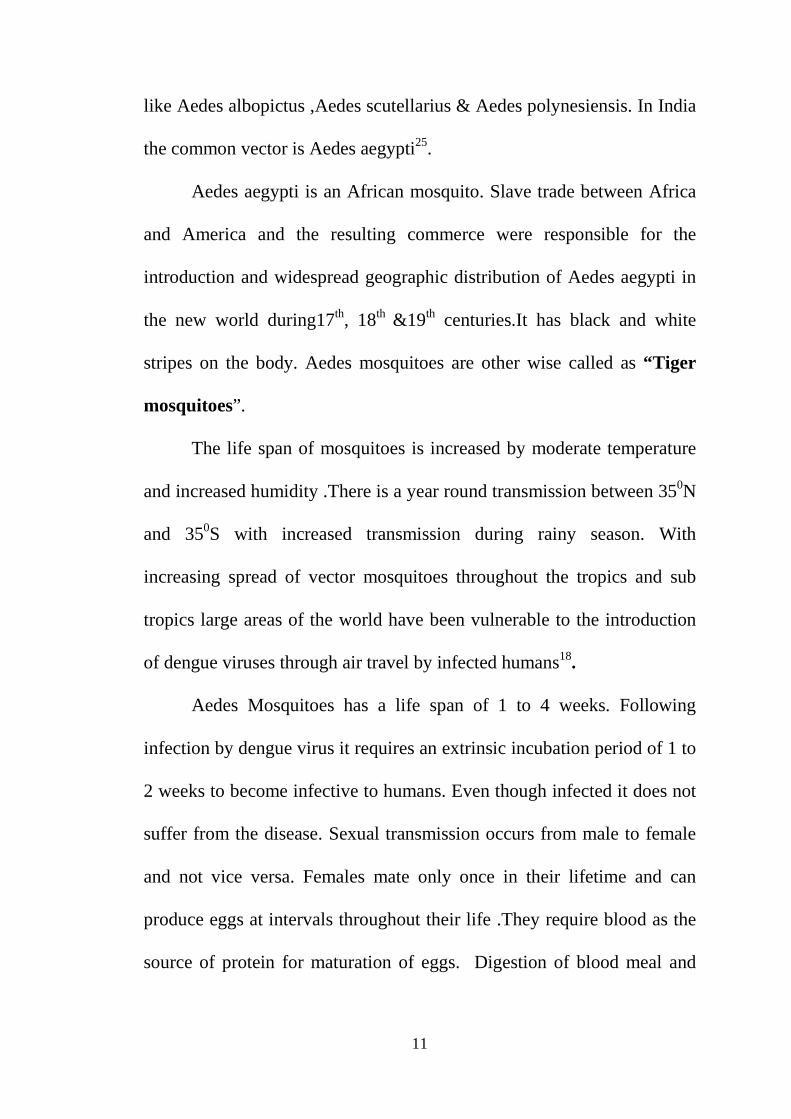
like Aedes albopictus ,Aedes scutellarius & Aedes polynesiensis. In India
the common vector is Aedes aegypti25.
Aedes aegypti is an African mosquito. Slave trade between Africa
and America and the resulting commerce were responsible for the
introduction and widespread geographic distribution of Aedes aegypti in
the new world during17th, 18th &19th centuries.It has black and white
stripes on the body. Aedes mosquitoes are other wise called as “Tiger
mosquitoes”.
The life span of mosquitoes is increased by moderate temperature
and increased humidity .There is a year round transmission between 350N
and 350S with increased transmission during rainy season. With
increasing spread of vector mosquitoes throughout the tropics and sub
tropics large areas of the world have been vulnerable to the introduction
of dengue viruses through air travel by infected humans18.
Aedes Mosquitoes has a life span of 1 to 4 weeks. Following
infection by dengue virus it requires an extrinsic incubation period of 1 to
2 weeks to become infective to humans. Even though infected it does not
suffer from the disease. Sexual transmission occurs from male to female
and not vice versa. Females mate only once in their lifetime and can
produce eggs at intervals throughout their life .They require blood as the
source of protein for maturation of eggs. Digestion of blood meal and
Vector – Female Aedes Aegypti
12
simultaneous development of eggs takes 2-3 days in the tropics but longer
in temperate climates. Female mosquitoes lays 30 -300 eggs at a time.
Transovarian transmission7 in mosquitoes leads to propagation of virus
to their progeny and it acts as reservoir for virus maintenance during Inter
epidemic periods. Eggs are laid above the water surface and hatch only in
water which can happen even in flower vases, water storage jars and
rainwater collected in small cups, tyres etc26, 19.If left dry they remain
viable for many weeks.
Adult mosquitoes are indoor daytime biting mosquitoes. Females
are said to be nervous feeders.Disruption of the feeding process of
mosquitoes takes place even at the slightest movement of the biting
person. Aedes will often feed on several persons for a single meal. During
this single blood meal, if the mosquito is infective it may transmit dengue
virus to multiple persons in a short period of time even if they only probe
without taking blood. Because of this behavior it can affect all the family
members which makes it an effective Epidemic vector12,19 .Mosquitoes
are normally attracted by body odours,CO2 and heat emitted from
animals/human beings18.
Adult mosquitoes normally shelter indoor and bite at an interval 1-
2 hrs particularly during early morning and late afternoon. The

13
commonest biting area preferred are over the ankles and exposed parts
of neck.
Other modes of transmission:
Needle stick injuries1.
Through blood transfusion in endemic areas during the viremic
period27,28.
Vertical transmission of dengue virus to neonates whose mothers had an
onset of primary /secondary dengue fever upto 5 weeks before delivery
has resulted in acute neonatal dengue manifesting as fever, apnoea,
mottling, hepatomegaly and decreased platelet count1.
Endemic transmission: In endemic countries 1/20 houses may contain
infected mosquitoes1.
Viral replication :
DENGUE DISEASE
“A TICKING TIME BOMB OF HUGE EPIDEMIC POTENTIAL ”
Following vector bite ,incoming genomic RNA serves directly as
messenger RNA .It has a large open reading frame which is translated
completely from its 5’ end to produce a large precursor polyprotein
which is again cleaved to produce individual proteins. About ¼th length
of genomic RNA from 5’ end encodes 3 structural proteins and 3’ end
encodes for non-structural proteins. Premembrane precursor protein is

14
cleaved and matures to form ‘M’ (membrane). NS3&NS5 proteins form
RNA dependent RNA polymerase complex. NS3 is responsible for co-
translational cleavage of nascent polyprotein that yields the NS protein
whereas cellular signal peptidases affect other primary cleavage.
Replication occurs in the perinuclear foci leading to synthesis of
complementary minus strand which is the template for synthesis of more
positive strands during infection. Virion assembly takes place mainly on
the endoplasmic reticular membrane in vertebrates and in plasma
membrane in invertebrates. Fully formed cell is released usually by cell
lysis.
Pathogenesis and Immunological reaction
After the bite of an infected mosquito, the virus replicates in local
lymph nodes and within 2 to 3 days disseminates through the blood to
tissues. Interstitial dendritic cells located in the epithelia are believed to
constitute the first line of innate host immune response against invasion
of dengue virus. Since phagocytic cells are serving as first line of defense,
most of the infections are asymptomatic. Virus circulates in blood for 5

15
days in infected monocytes and macrophages and to lesser degree in B
cells and T cells. It also replicates in the skin, spleen lymphoid cells and
macrophages1. Macrophage inflammatory protein-1α has a pathogenic
role in dengue virus infection8.
Infection of monocyte and macrophage is central to the
pathogenesis of Dengue fever and origin of Dengue haemorrhagic fever
or shock syndrome16. First infection with any of the dengue serotypes
results in self limiting febrile illness. Recovery from first infection is
usually accompanied by generation of immunological responses. Hence
secondary infection with a heterotypic virus will result in severe illness.
Epitopes present on Envelope protein are capable of inducing
homologous as well as heterologous antibodies.
The plasma leakage is induced by C3a,C5a and several mediators
leading to the increased vascular permeability during the acute febrile
stage .The plasma leakage is prominent during “critical period ” which
lasts from 24 hrs before and 24 hours after the day of defervescence of
fever1. The evidence of plasma leakage includes hemoconcentration,
hypoproteinemia/hypoalbuminemia, pleural effusion, ascites, threatened
shock or profound shock7.
Vascular permeability in Dengue Haemorrhagic fever is associated
with increased immune activation as manifested by increased levels of

16
plasma soluble TNF receptor (sTNFR/75) IL-8, IFN-γ, local endothelial
proliferation of IL-8, RANTES with apoptotic endothelial cell death.
Immune complex formed by antigen and antibody leads to complement
activation1.
The cause of bleeding diathesis in Dengue is complex. It is caused
by vasculopathy, thrombocytopenia, platelet dysfunction and
coagulopathy29.Haemorrhagic diathesis is due to a combination of
cytokine activation and vascular injury. Viral antibodies binding to
platelets or cross reacting with plasminogen and other clotting factors,
decreased platelet function and survival leads to mild consumptive
coagulopathy.10
The pathogenesis of severe disease is not well understood and
multiple factors may be implicated. During secondary infection with
another serotype, the heterologous antibodies increases the viral uptake
not neutralized and replication takes place in Fc receptor bearing cells
This phenomenon is called as Antibody dependent enhancement
(ADE)30,31,29,12.This forms the basis of severe dengue infection and in
infants with primary infection. ADE- results in high viral load and
increased inflammatory response which accounts for capillary leak
syndrome6. From this it is clear that the dengue vaccine must induce
protection simultaneously to all serotypes32.
17
Dengue haemorrhagic fever is an infrequent but potentially lethal
form of illness usually due to secondary infection by heterologous
serotype. The pathophysiology of DHF is multifactorial involving viral
nature, host genetics, host immunity and previous exposure to the virus29.
Higher plasma viremia early in the course of infection and consequent T-
cell activation predispose to DHF8.
In patients with DHF, production of various proinflammatory
cytokines/chemokines and complement is increased. Genes encoding for
IL-10, IL-8, IL-1β, IL-32/NK4, IFN-γ, TNF-α, MIP-1β, RANTES,
CXCL7, CXLI, properdin, and factor D component of complement were
more strongly expressed in Peripheral blood mononuclear cells of
patients with DHF than DF5.
DHF in primary or secondary dengue is due to the occurrence of
abnormal immune response involving production of cytokines or
activation of T-lymphocytes and disturbance of the hemostatic
system29.According to Scott B Halstead33 DHF is not significantly
associated with secondary dengue infection as it can result from a virulent
dengue virus strain causing primary infection itself.
The mediators like C3a,C5a,TNF-α, interleukin (IL)-2, IL-6,IL-
10, IFN–α and histamine are elevated in DHF than Dengue fever.This
18
indicates enhanced activation of cross reactive T cell which leads to
increased viral uptake and replication in macrophages and
monocytes.This is called as “original antigenic sin”29.
Profound T cell activation and death of T cells, during acute dengue
infection may suppress or delay viral elimination. This further leads to the
higher viral loads and increased immunopathology found in patients with
DHF25.
Illness after infection with 2 serotypes occurs infrequently and illness
after 3 infection virtually never. Repeated episodes of DHF have been
recognized rarely, presumably because of immune factors that promote
immunopathologic response are outweighed by immune response that
clear infection.
Pathologic feature of Dengue in DHF/DSS-
Midzonal and hepatocellular necrosis, minimal inflammatory
response, councilman bodies, microvesicular fatty changes,hypercellular
and hyperplasia of mononuclear phagocytic cells in lymphoid tissue.
Atypical lymphocytosis in peripheral blood, widespread infection of
mononuclear phagocytes and endothelial cells are seen 12.The degree of
liver dysfunction in dengue infection varies from mild injury with
elevation of aminotransferases alone to severe injury with jaundice and
hepatic failure. The severity of dengue infection varies depending upon

19
type of infection and is more in complicated dengue. AST and ALT
levels are increased in DHF1
Malaise and flu like symptoms in Dengue are due to cytokine
response. Myalgia is due to pathological changes in muscle due to
moderate perivascular mononuclear infiltrate with lipid
accumulation. Severe Musculoskeletal pain indicates viral infection of
bone marrow including mobile macrophages and dendritic cells
(CD11b/CD18 [Mac-1] positive) and relatively non motile adventitial
reticular cells (nerve growth factor receptor positive).
Local suppression of erythrocytes, myelocytic and thrombocytopenic
precursors within 4 – 5 days which is reflected in peripheral cytopenia
resembling a picture of malignancy. Histopathologic examination of skin
show minor degree of lymphocytic dermal vasculitis and variably viral
antigen.
Thrombocytopenia and hemoconcentration are constant findings in
DHF.A drop in platelet count below 1lakh is seen from 3rd day to 8th day
of illness. Hemoconcentration with rise in hematocrit >20% is definitive
evidence of plasma leakage.WBC count varies ranging from leucopenia
to leucocytosis which is more common towards the end of febrile phase
of illness.

20
Turk cells are transformed lymphocytes.Presence of more than 20 %
of turk cells in buffy coat smear is a frequent finding of dengue
hemorrhagic fever 34.
Immunological Response to Dengue infection:
The acquired immune reponse following a dengue infection
consists of the production of IgM and IgG antibodies primarily directed
against the virus envelope proteins .The immune response differs for
primary and secondary infection.
In primary dengue IgM is the first immunoglobulin isotype to
appear. IgG is detectable in low titre at the end of first week and slowly
increases. In contrast, in secondary dengue antibody titres rise extremely
rapidly due to the presence of antibody to the previous infection and these
antibodies react broadly with other flaviviruses. High levels of IgG are
detectable even in acute phase and rise dramatically over the proceeding
2 weeks. IgM levels are significantly lower in secondary infections.
PAHO says that 80% of all dengue cases have IgM antibody by day 5 of
illness and 93-99% cases are detectable by 6-10 days and remain
detectable upto 90 days. The presence of antibodies in dengue is
protective to the infecting serotype but short lived for heterologous
serotype. The level of neutralizing antibodies corresponds with protection
against dengue virus.

21
Clinical features:
Incubation period of Dengue is 2 to 7 days and is characterized by
high fever, headache, retro orbital pain, lumbosacral pain, conjunctival
congestion and facial flushing. Biphasic fever pattern- fever upto 3 rd day
of onset which subsides and rises again from 5 th to 7 th day. Because of
regular fluctuations in temperature it is also called as ‘saddle back’ fever.
Fever is usually associated with severe generalized myalgia and
arthralgia which gives the name as ‘Breakbone fever’. Maculopapular
rash appears on the trunk on 1st /2nd day (non irritating rash) then spreads
centripetally to the face and limbs but spares palms and soles.
Generalised lymphadenopathy, cutaneous hyperaesthesia, altered taste
sensation can occur.
Unusual manifestations are neurological, hepatic, renal and isolated
organ involvement.
According to WHO Guidelines Dengue fever, Dengue haemorrhagic
fever and Dengue shock syndrome are defined as follows:
Dengue fever: fever of 2-7 days associated with symptoms like
Arthralgia, myalgia, vomiting, abdominal pain, rash, retro orbital pain,
conjunctival congestion, haemorrhagic manifestation,or leucopenia.
Dengue haemorrhagic fever: DHF is defined by the presence of above
symptoms plus thrombocytopenia, evidence of increased vascular

22
permeability (i.e.hemoconcentration, pleural or abdominal effusion,
hypoalbuminemia or hypoproteinemia). DHF has 4 grades.
Dengue shock syndrome: Defined by the presence of symptoms of DHF
with reduced perfusion towards the defervescence and early signs of
shock manifested as narrowing of pulse pressure or hypotension for age.
The symptoms of DSS are cold clammy extremities, flushed face,
diaphoresis, restlessness, irritability and mild epigastric pain12.
Induction of shock in Dengue is influenced by the following:
1. Presence of enhancing and non- reacting antibodies.
2. DHF/DSS is common upto 12 years of age and drops later.
3. Females are affected more than males.
4. Race- Caucasians are affected more.
5. Nutritional status- Malnutrition is said to be protective as they have
deficient immune response to infection.
6. Sequence of infection- Serotype 1 followed by Serotype 2 infection is
more dangerous.
7. Infecting serotype- Type 2 is apparently more dangerous than other
serotypes.
Period of shock is about 1-2 days18 so prompt supportive management
and good care with close monitoring during this period would prevent
complications and save the life of the patient .

23
� Apart from the above causes,WHO also defines the high risk group as
infants and elderly, obesity, Pregnancy , peptic ulcer disease, women
who have menstrual / abnormal vaginal bleeding.Other risks factors
like G-6PD deficiency ,thalessemia and hemoglobinopathies ,chronic
disease like DM, asthma, SHT are also said to be associated.35
Laboratory Diagnosis:
Efficient and accurate diagnosis of dengue is of primary importance in
detection of cases and early treatment. Laboratory diagnostic methods of
detection of dengue are
Serological methods
Viral isolation
Detection of viral nucleic acid.
In complete blood count platelet count and the total WBC count would
be reduced depending upon the type of fever. As the WBC count
becomes low, platelet counts also becomes very low but during the
recovery stage WBC count rises rapidly than Platelet count .Platelet count
is usually reduced more during the DHF and DSS.
Neutropenia with atypical lymphocytosis resembling malignancy could
be seen in peripheral smear. Packed cell volume should be estimated as it
rises in Dengue haemorrhagic fever rapidly.

24
In liver function test –Enzymes like Aspartate aminotransferase and
Alanine aminotransferase are elevated depending upon the type of injury.
Serological methods:
Rapid diagnostic tests:
Various commercial kits are available for dengue detection using
the envelope glycoproteins of dengue for IgM and IgG antibody detection
and NS1 antibody for antigen detection.
Lateral flow immunochromatographic test allows detection of both
antigen and antibody simultaneously with single serum sample,therefore
differentiation of primary and secondary dengue can be made with single
sample in contrast to multiple samples required for HAI. It produces
results within 15- 20 minutes and it has the advantage of ease of
performance also. But the sensitivity and specificity of these tests are not
known and are yet to be evaluated .According to WHO , these RDTs are
not be used for clinical management because of false negativity that can
lead to missing of diagnosis and fatal complications36.
Enzyme immunosorbent assay (ELISA):
Antigen detection:
Enzyme Immunoassays for antigen detection have four steps. An
antigen specific antibody is attached to a solid phase surface. Patient
serum that may contain the antigen is added next. An enzyme labeled

25
antibody specific to the antigen (conjugate) is added. Finally substrate is
added which changes colour in the presence of the enzyme. The amount
of colour that develops is proportional to the amount of antigen present in
the patient specimen.
NS1 protein can be detected by ELISA as soon as first day of fever
and can be found upto 9 days in serum even when PCR is negative and
where PCR is not available. It might serve as early prognostic marker for
severe dengue infections37.
Antibody detection:
Immunocapture ELISA : It is designed to detect a specific type of
antibody such as immunoglobulin M or immunoglobulin G.Antibody
specific for IgM or IgG is attached to solid phase surface.The Patient
specimen potentially containing IgM or IgG is added. Specific antigen is
added. Finally chromogenic substrate is added which in the presence of
the enzyme changes colour.The amount of colour that develops is
proportional to the amount of antigen specific IgM or IgG in the patient
serum.
Capture ELISA is found to be highly sensitive and is routinely used
in diagnosis of dengue infections. They are simple to perform and large
number of samples can be tested at the same time.
26
A.IgM capture : It is the widely used ELISA for detection of
antibodies.It shows good sensitivity and specificity only when used 5 or
more days after onset of fever. A positive result indicates recent infection
but cannot identify the serotype.There are different formats of capture
ELISA available like capture ultramicro ELISA, dot ELISA, Aubiodot
IgM capture ELISA and dipstick. Samples like saliva, serum on filter
paper could also be used. Different commercial kits are available with
varying sensitivity and specificity38.
B.IgG capture: This capture ELISA is commonly used for
classifying cases based on type of infection like primary and secondary
infection. When used along with IgM the ratio is calculated and according
to standard criteria it is classified as primary or secondary. In primary
dengue IgG is detected after 2 weeks and persists for life. In secondary
dengue IgG antibody to the previous infection rises immediately within 1-
2 days before the appearance of IgM. It is followed by IgM to the present
infection and IgG after 1week25.
Other serological methods available for the detection of dengue
antibodies:
Neutralisation tests: These tests are technically demanding and
time consuming and are performed only in reference laboratories25
Complement fixation test: One of the classic methods for demonstrating

27
the presence of antibody in a patient’s serum has been the complement
fixation test. As the procedure is cumbersome, requires highly trained
personnel and the reagents are thermolabile it is not used for routine
diagnosis39.
Heamagglutination inhibition test: This test can be used for suspected
DHF/DSS but results are delayed in time when compared to Capture
ELISA .Hence it cannot be used for routine diagnostic purposes40.
Indirect immunofluorescence test41
Dot Blot ELISA 42
Viral isolation:
Mosquito Cell lines, mammalian cell lines and adult mosquito
inoculation.
In acute phase of infection isolation and identification of the virus is the
only way to diagnose early dengue infection. Serum collected from
patient is applied to mosquito cell lines .After amplification of virus in
cell line the serotype is identified using monoclonal antibodies. This
technique is sensitive only if there is high viremia in the early stages.
common cell lines used are C6/36(Aedesaegypti),
AP61(pseudoscutellarius) The CPE on cell lines appears between 1-4
days post inoculation –cells become round and swollen leading to
multinucleated giant cells and syncytia formation.

28
In Mammalian cell culture (LLC-MK2) –CPE occurs after 14 days.
Least sensitive methods are Intra thoracic inoculation of mosquitoes.
Eventhough viral culture once considered as ”Goldstandard”, because of
its lower sensitivity, long time required and applicable only for acute
samples this procedure has been replaced by molecular diagnosis43.
Animal inoculation: Intracerebral inoculation of serum into suckling
mice of 1-2 days old done using tuberculin syringe into the lateral
hemisphere. After inoculation the mice is observed for sickness daily for
any sign of abnormal behavior and if positive it is euthanised, brain
tissue is harvested and subjected to relevant tests for confirmation. As this
is a tedious procedure and time consuming, it cannot be applied for
routine use43.
Detection of Nucleic acid: RT –PCR provides a rapid and simple method
for detection of RNA viruses in serum samples and tissue specimens.
Viral genome is amplified using oligonucleotide primers and the
amplified product is detected using reverse transcriptase enzyme.
Recently conventional RT-PCR has been replaced by Real time PCR
which can be automated. Sensitivity ,Specificity and rapid detection of
minute quantities of nucleic acid in patient’s serum makes RealTime -
PCR more sensitive with less contamination and easy identification of
circulating serotype by analyzing the unique sizes of amplicon. Other

29
methods available for nucleic acid detection are NASBA, Hybridization
probes44.
Differential diagnosis of Dengue fever: Scrub typhus, Leptospirosis,
Malaria, Chikungunya, Influenza, Measles, Rubella45.These diseases
could be ruled out by clinical examination and appropriate laboratory
tests
Dengue vaccine: More researches have been carried out to develop a
dengue vaccine that is safe and immunogenic against all 4 serotypres.
Attempts to develop an effective vaccine have been hampered by the lack
of understanding of the pathogenesis of disease and absence of suitable
experimental model. Dengue vaccine must provide solid and long
lasting protection against all dengue virus serotypes. Live attenuated
tetravalent candidate vaccine are in the late stage of development which
is produced by serial passage of wild type virus in primary dog kidney
cells or other cell types46. Sanofi Pasteur has reported successful results
of phase II trials of tetravalent recombinant live attenuated vaccine47.
Non- recombinant candidate vaccine using structural& non-structural
purified proteins and synthetic peptides 13
Recombinant subunit (Escherichia coli, baculovirus, yeast) 13
Recombinant subunit vaccine was produced by coating the β domain of
dengue serotypes 1-4 to the binding protein of E.coli .Its efficacy was

30
evaluated in mice as single or tetravalent vaccine.Neutralising antibody
titres to each individual serotype was significantly greater than cross
reactive neutralising titre compared to tetravalent vaccine 48.
Recombinant vector13
Infectious cDNA clone technology has also been exploited for
development of dengue vaccine .A chimeric Yellow fever dengue type -
2 vaccine prepared by using recombinant cDNA of a Yellow fever
vaccine strain as a backbone to which pre membrane and envelope gene
of DEN-2 were inserted. It is in Phase II-III trial12.
DNA vaccine is another novel and promising immunization approach.In
order to improve the immunogenicity of DEN-2 candidate vaccine
cytoplasmic region of envelope gene was replaced by lysosome
associated membrane protein.13.
Treatment: No specific drugs are available for the treatment of Dengue
Proper maintenance of fluid balance is the cornerstone of management in
Dengue.
Supportive and symptomatic treatment
- I.V. Fluids ,Bed rest
- Parcetamol for control of fever
- Aspirin and NSAIDS must be avoided.
31
- Platelet and HCT should be monitored daily beginning on the third
day of fever.
- Blood and platelet transfusion if required –depending upon the
occurrence of bleeding in GI tract but does not correlate with
platelet count.
- Early recognition of leakage phase with prompt resuscitation and
Close monitoring with oxygen, pulse, blood pressure and urine
output would help to reduce complications and improves the
treatment outcome.
Prevention: Neither vaccine nor specific drugs are available .Hence
prevention by vector control of Aedes aegypti plays a major role in
Dengue endemic countries.
Vector control:
Mosquitoes that rest indoors are easy to control. Female Aedes aegypti
are indoor daytime biters. It is difficult to avoid species biting in
daytime than those species that bite at night.
Removal or filling of breeding habitats in man made & natural
containers, burning of organic waste, screen fitting of mosquito proof
lids to drinking water storage containers, installing piped drinking
water supply.
Safe and effective larvicides to breeding sites.

32
Personal protective equipments for the daytime biting mosquitoes are
protective clothing like wearing long sleeved clothes,repellents &
house screening ,indoor space spraying ,mosquito coils, mats, bed nets
& air conditioning.
Applying insect repellents to both skin and clothing using permethrin is
found to be effective. The most effective repellents contain DEET.
Biological control
Antilarval measures:
1. Eliminating or changing the breeding place to make it unsuitable
for development of larvae.
2. Making the breeding place inaccessible to adult mosquitoes.
3. Releasing fish/predators that feed on larvae.
Gambusia affinis /Poecilin reticulate endotoxin producing
bacteria (Bacillus thuringiensis serotype H-14, Bacillus sphaeria)
and copepod crustaceans are currently used .These are suitable
for large containers that are not cleaned regularly as frequent
cleaning leads to depletion of nutrients available for the larvae.
4. Applying larvicides wherever necessary.
Antiadult measures:
1. Epidemic situations- outdoor space spraying with insecticides.
2. Insecticidal spray are usually applied to parts of town where

33
Abundant breeding sites are available supporting large
Population of Aedes.
3. Pyrethrin Knockdown sprays /Organo phosphate sprays can be
delivered in microdroplets49,26.

34
MATERIALS AND METHODS
Study design: This is a Prospective study
Study period: One Year from September 2011 to August 2012.Approval
from Ethical committee obtained for this study.
Inclusion criteria : Patients suffering from fever of 1-12 days duration
with any of these symptoms like myalgia, arthralgia, headache, rash,
anorexia, nausea, and vomiting, abdominal pain.
Exclusion criteria: Patients suffering from other nonspecific fevers
without clinical features suggestive of dengue fever, Urinary tract
infections, pneumonia and lung abscess etc.
Statistical analysis: Statistical analysis of results of this study was done
using SPSS version 17, p value obtained from chi-square test.
Sample collection: About 5 ml of venous blood was drawn from 350
suspected patients of dengue fever after taking informed consent, under
aseptic precautions in sterile containers .These samples were centrifuged
at 1500 rpm for 10 minutes & and serum separated .If the sample was not
tested immediately it was stored at -200 c.
35
Methodology
The serum samples collected were subjected to Dengue duo
NS1Ag +Ab combo cassette, NS1 capture ELISA, IgM Capture and IgG
Capture ELISA in the Department of Microbiology, at Coimbatore
medical college hospital.
Immunochromatography card test (ICT): Dengue duo combo card: The rapid test is an in-vitro immunochromatographic one step assay to
detect both dengue virus Nonstructural (NS1) antigen and differential
IgG/IgM antibodies to dengue virus in serum, plasma or whole blood.
The left side of Dengue duocard is the NS1 Ag rapid test for qualitative
determination of antigen. It has a circular well for addition of sample. The
test device contains a membrane strip precoated with anti-dengue NS1 Ag
capture on test band region.
The right side of the card is a solid phase immunochromatographic assay
for rapid, qualitative and differential detection of IgG and IgM antibodies
to dengue virus in serum, plasma and whole blood .The card has a square
well for sample addition, a circular well for buffer and a lateral flow

36
membrane with colloidal gold conjugate containing recombinant dengue
antigen and a control.
Principle of the test:
The dengue NS1 antigen card can identify dengue virus NS1
antigen in serum, plasma or whole blood specimens with a high degree of
sensitivity and specificity. Dengue IgG/IgM rapid test is designed to
simultaneously detect and differentiate IgG, IgM antibodies to dengue
virus.This test can also detect all 4 serotypes by using a mixture of
recombinant dengue envelope proteins.
When a specimen is added to the sample well anti-dengue IgG
&IgM antibodies in the specimen will react with recombinant dengue
envelope proteins –colloidal gold conjugates and forms a complex of
antibody-antigen. As this complex migrates along the length of test
device by capillary action, it will be captured by the relevant anti-human
IgG and /or anti –human IgM immobilized in 2 test lines across the test
device and generate a coloured line.
Procedure:
1. The card was placed on an even surface after being unwrapped
from the cover. Id no & date noted down on the card

37
2. With the dropper provided 10 µl of serum was added to the sample
well followed by 4 drops of buffer on the right side of the card.
3. With another dropper provided 100µl of sample was added to the
antigen well on the left side of the card.
4. Results were read after 15-20 minutes.
Results:
Antibody
1. Appearance of bands in all the lines on the antibody card
2. Appearance of control & “M” line alone
3. Appearance of control &”G” line alone.
4. Appearance of control line alone
Antigen: Appearance of bands in control area alone
Appearance of bands in control and test regions.
Interpretation :
1. Antibody :
a. Presence of control line alone- test is negative for antibodies.
b. Presence of all the lines control, M and G – both IgM &IgG
Positive -Indicates either late primary or early secondary infection
c. Presence of control and M line alone- primary dengue
d. Presence of control and G line alone- secondary or past dengue
38
e. Absences of control line – invalid and test to be repeated.
2. Antigen :
a. Presence of bands in both test &control area – positive for NS1
antigen.
b. Presence of band in control area alone- negative for antigen
c. No bands in the result window, it is invalid– test has to be
repeated.
Dengue EARLY ELISA
Principle of the test: NS1 antigen present in the serum binds to the
anti-NS1 coated on the microwell strips .The unbound antigen if present
is removed by the washing step.HRP conjugated monoclonal anti-NS1
antibody is added. A colourless substrate TMB is added which produces
blue colour .On addition of stop solution phosphoric acid it changes to
yellow colour.
Procedure:
1. 100µl of diluted sample & controls were added into the respective
wells
2. The plate was Covered &incubated for 1 hour at 370 c
3. At the end of 1 hour Plate was washed 6 times with diluted wash
buffer
39
4. 100µl of Horseradish peroxidase(HRP) conjugate was added into the
well.
5. The plate was Covered & incubated at 370 c for 1 hour.
6. Washed 6 times with diluted wash buffer at the end of incubation.
7. 100µl of Tetramethylbenzidine(TMB) was added into each well
8. Incubated for 10 mts at Roomtemperature(RT) & observed for blue
colour
9. 100µl of stop solution was added & the change of colour noted.
10. Absorbance value of each well measured at 450 nm with reference
filter 600-650nm spectrophotometrically.
Calculation:
Average absorbance of calibrator triplicate multiplied by the
calibration factor
Cut off value(COV) = absorbance of calibrator X cal. Factor
Cal .factor=0.57
Index value= sample OD/ cutoff value
Panbio units= Index value x 10

40
Test validity:
Negative absorbance < 0.250
COV >1.5 X Negative absorbance.
Positive control/ cutoff ratio= 1.1-7.0
Interpretation:
Index Panbiounits Result
<0.9 < 9 Negative
0.9-1.1 9-11 Equivocal
>1.1 > 11 Positive
Dengue IgM capture ELISA:
Principle of the test:
Serum antibodies of the IgM class, when present, combine with
anti-human IgM antibodies coated on the polystyrene surface of the
microwell test strips (assay plate). The diluted recombinant antigen is
mixed with equal volume of horseradish peroxidase (HRP) conjugated
monoclonal antibody (MAb) allowing the formation of antigen-MAb
complexes. Addition of complexed antigen-MAb complexes following
washing will bind to the serum dengue-specific IgM antibodies. After
incubation a colourless substrate tetramethylbenzidine/hydrogen peroxide

41
(TMB Chromogen) is added. The substrate is hydrolysed by HRP if
present, and the chromogen turns blue. After stopping the reaction with
acid, the TMB turns yellow. Colour development is indicative of the
presence of anti-dengue IgG antibodies in the test sample.
Procedure:
All the kit reagents were brought to room temperature before start of the
test.
ELISA Procedure:
1. Dispensed 100µl of diluted patient serum, positive and negative
control, calibrator in triplicate added to their respective wells in the
microtiter plate.
2. The microtiter plate was covered and incubated at 370 c for 1 hour
3. Antigen was diluted in 1/250 with antigen diluent i.e 10µl of antigen in
2.5ml of antigen diluent.
4. Required amount of diluted antigen is mixed with equal volume of
Monoclonal antibody tracer and incubated at room temperature until
required.
5. The plate was washed 6 times with diluted wash buffer at the end of
incubation.
42
6. Added 100µl of antigen antibody mixture to all the wells
7. The plate was covered and incubated at 370 c for 1 hour.
8. At the end of incubation, plate was washed 6 times with diluted wash
buffer
9. Added 100µl of TMB substrate to the wells and incubated in the dark
for 10 minutes.
10. 100µl of stop solution added to all the wells.
11. Absorbance value of each well measured spectophotometrically at
450nm wavelength with reference filter of 600-650nm.
Calculation:
• Average absorbance of the triplicate of the calibrator multiplied by
the calibration factor. This is the cutoff-value.
• An index value is calculated by dividing the sample absorbance by
the cut-off value
Index value= sample absorbance/ cut-off value
Panbio units= Indexvalue x 10

43
Test validity:
Cutoff value>1.5x neg.absorbance
Calibrator mean>negative absorbance
Positive control/cutoff value=1.1-8
Negative control<0.40
Calibration factor=0.99
Interpretation of results:
INDEX PANBIOUNITS RESULT
0.9 < 9 Negative
0.9-1.1 9-11 Equivocal
>1.1 >11 Positive
Dengue IgG capture ELISA
Principle of the test: Serum antibodies of the IgG class, when present,
combine with anti-human IgG antibodies coated on the polystyrene
surface of the microwell test strips (assay plate). An equal volume of
horseradish peroxidase (HRP) conjugated monoclonal antibody (MAb) is
added to the diluted antigen, allowing the formation of antigen-MAb
complexes. Residual serum is removed from the assay plate by washing,
and complexed antigen-MAb complexes added then bind to the serum
dengue-specific IgG antibodies. After incubation a colourless substrate

44
added to the washed wells and was followed by
tetramethylbenzidine/hydrogen peroxide (TMB Chromogen) addition.
The substrate is hydrolysed by HRP if present, and the chromogen turns
blue. After stopping the reaction with acid, the TMB turns yellow, Colour
development is indicative of the presence of anti-dengue IgG antibodies
in the test sample.
Preprocedure preparation:
All the kit reagents were brought to room temperature before the
commencement of the procedure.
Procedure:
Antigen diluted 1/250 using antigen diluent i.e. 10µl antigen mixed with
2.5ml of antigen diluent .About 0.5ml of diluted antigen is required per
strip.
Equal volume of diluted antigen mixed with monoclonal antibody tracer
solution. This is incubated at room temperature for 1 hour.
1. Dispensed 100 µl of diluted sample, positive control, negative
control and calibrators in triplicate to the respective wells
2. The plate was covered and Incubated at 37 0c for 1 hour.
3. Plate was washed 6 times with diluted wash buffer at the end of
incubation
45
4. Added 100 µl of Ag –MAb tracer solution to all the wells at the
end of incubation
5. Plate was covered and Incubated at 370c for I hour.
6. Washed 6 times with wash buffer at the end of incubation.
7. Added 100 µl of TMB was to all the wells.
8. Incubated at dark for 10 minutes.
9. Added 100µl of stop solution to all the wells
10. Absorbance value of each well measured at 450 nm wavelength
with 600-650 nm reference filter spectophotometrically.
Calculation:
1. The average absorbance of the triplicate of the calibrator is
multiplied by the calibration factor. This is the cut-off value.
2. Index value is calculated by dividing the sample absorbance by the
cut- off value.
Test validity:
Cutoff value= 1.5x neg.absorbance
Calibrator mean >negative absorbance
Positive control/Cutoff value= 1.1-6.0
46
Negative control<.350
Calibration factor=1
Index value= Sample absorbance / cutoff value
Panbio units= index value x 10
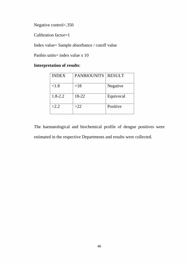
Interpretation of results:
INDEX PANBIOUNITS RESULT
<1.8 <18 Negative
1.8-2.2 18-22 Equivocal
>2.2 >22 Positive
The haematological and biochemical profile of dengue positives were
estimated in the respective Departments and results were collected.

Dengue Duo Combo Card Kit
NS1 Positive
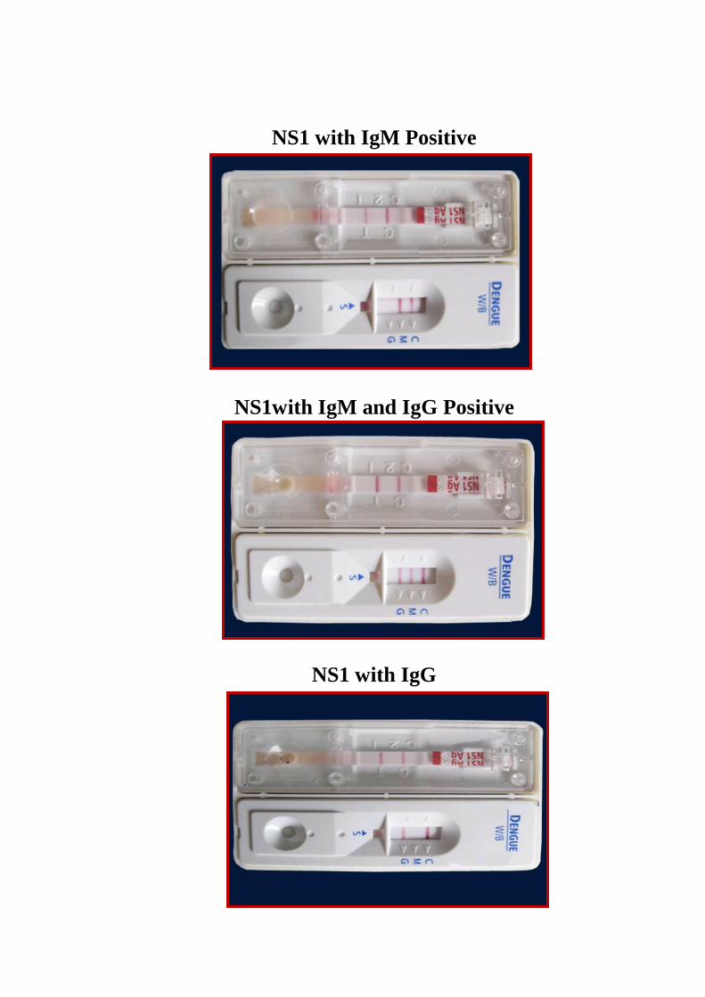
NS1 with IgM Positive
NS1with IgM and IgG Positive
NS1 with IgG

NS1 Negative with IgM and IgG Positive
IgM antibody positive
IgG Antibody Positive

NS1 Antigen ELISA Kit
IgM ELISA Kit

IgG ELISA Kit
ELISA Reader

Antigen Antibody Mixture of IgM & IgG
MicrotitrePlate with Samples

Microtitre Plate with Samples after substrate Addition
Samples after addition of Stop Solution
47
RESULTS
During the study period of one year Blood was collected from 350
clinically suspected dengue cases .The samples were subjected to Dengue
serology for both NS1 antigen and IgM,IgG antibody detection by ELISA
and ICT.The 107 seropositive samples by ELISA were further analysed for
demographic ,clinical and laboratory data including haematological and
biochemical profile. The Statistical analysis of results of this study was done
using SPSS version 17.p value obtained from pearson chi-square test.
In this study the seropositivity of dengue was 30 %( Ref Table 1).
The age of the seropositives ranged from 0-72 years and the
commonest age group affected in this study was 0-10years 45 (42%)
followed by 11-20 years 27 (25.2%), 21-30years 16 (14.9%), 31-40years 12
(11.2%) .The incidence was less in the age group of 41-50years and 51-60
years each constituting about 2 (1.8%) and >60years 3 (2.8%). (Ref Table2)
The male: female ratio was 1.6:1 in this study with males 67 (62.3%) and
females 40(37.7%) (Ref Table 2).
The incidence of dengue cases in this study month wise were
September 2 (16.6%), October 0 (0%) November 5 (21.7%), December 12

48
(25.5%), January 13 (31.7%), February 2 (13.3%), March 1 (4.3%), April 4
(23.5%), May 13 (26.5%), June 41 (50.6%) and July13 (52%) and August 1
(12.5%). (Ref Table 3).
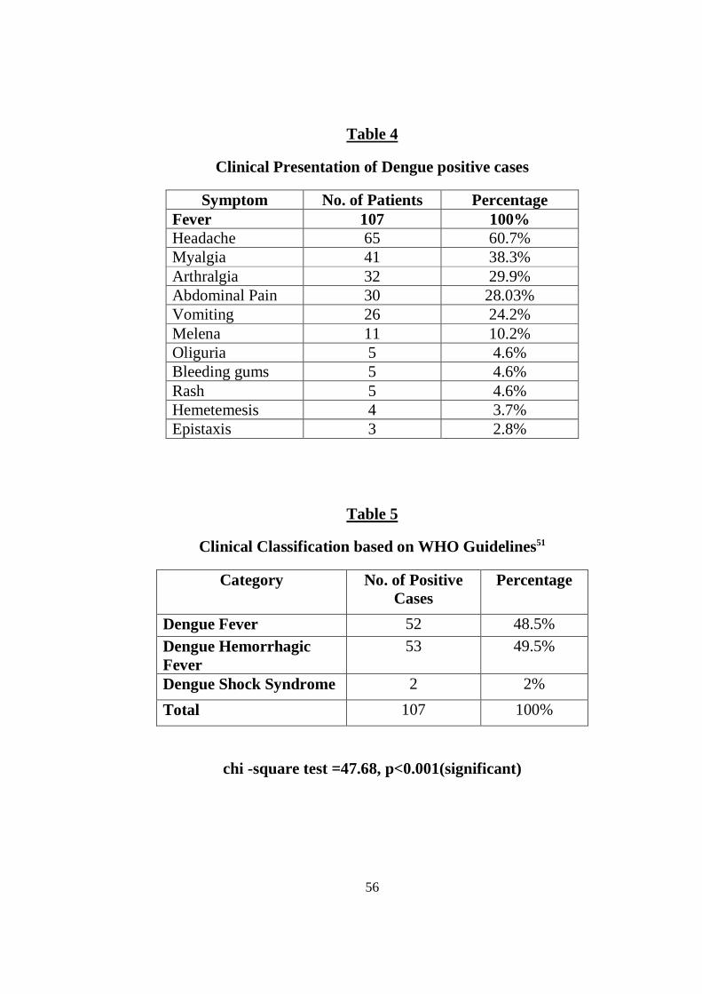
The common clinical symptom was fever 107 (100%), followed by
headache 65(60.7%),Myalgia 41(38.3%),Arthralgia 32 (29.9%),Abdominal
pain 30 (28.03%),vomiting 26 (24.2%) ,Oliguria and Rash was observed in 5
cases(4.6%).( Ref Table 4)
Melena 11(10.2%) was the commonest bleeding manifestation
followed by bleeding gums 5 (4.6%),Hemetemesis 4 (3.7%) and Epistaxis 3
(2.8%) (Ref Table 4)
The positive cases were classified as Dengue fever 52(48.5%),
Dengue haemorrhagic fever 53(49.5%) and Dengue shock syndrome 2(2%)
according to WHO guidelines. ( Ref Table 5).
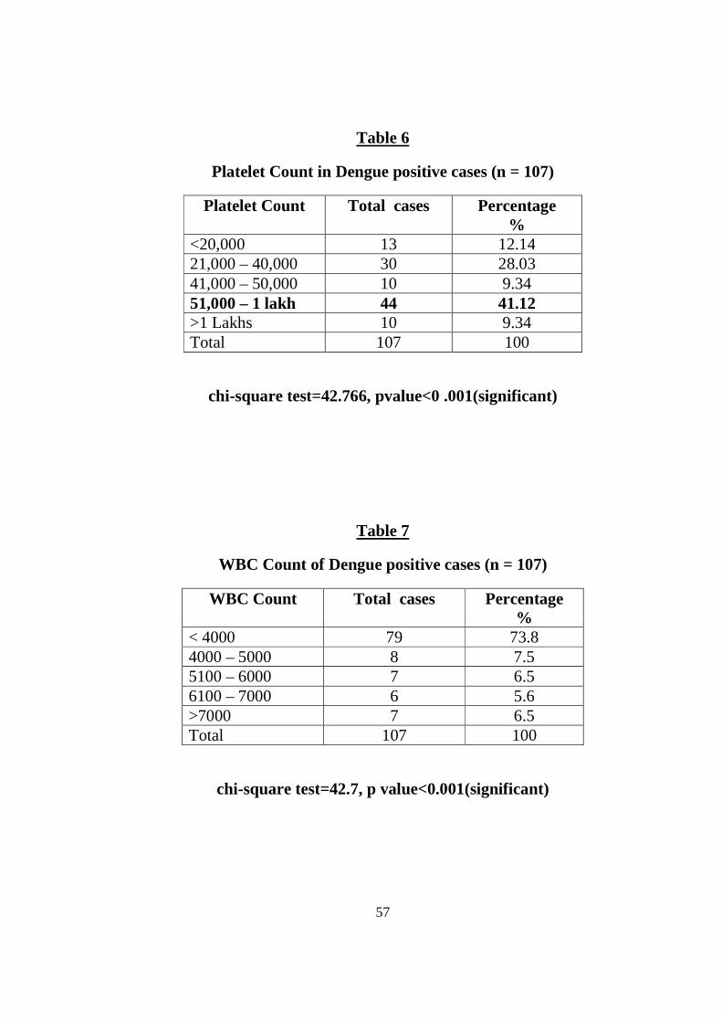
In this study thrombocytopenia was associated with majority of
Dengue positive cases .The common range was 51,000-1lakh44 (41.1%)
followed by 21000-40,000 30(28.03%),<20,000 13(12.14%),41,000-50,000
10(9.34%)and>1lakh 10(9.34%).Amongthrombocytopenia<1lakh 43(82.6%)

49
were associated with denguefever,52 (98%) with Dengue haemorrhagic
fever and 2 (100%)with Dengue shock syndrome. (Ref Table 6 & 9).
In this study leucopenia (count <4000) was observed in 79(73.8%)
dengue positive cases .Out of 52 Dengue fever cases 37 had
leucopenia(71.1%), out of 53 DHF 40 had leucopenia(75.4%) and out of 2
DSS both had leucopenia(100%). Eight cases (7.5%)had WBC count of
4000-5000, 7 were in 5100-6000(6.5%) range, 6 were in 6100-7000range
and 7 cases had count >7000(6.5%). (Ref Table 7&9).
Elevated Haematocrit > 45% was observed in 33(30.8%) cases.
Among them 5(9.6%) were associated with Dengue fever and 26(49.05%)
with Dengue haemorrhagic fever and 2(100%) with Dengue shock
syndrome.HCT level in other cases were 35-44 in 30(28.03%) cases, 25-34
in 34(31.7%) and <25in 10 case (9.3%) . (Ref Table 8&9)
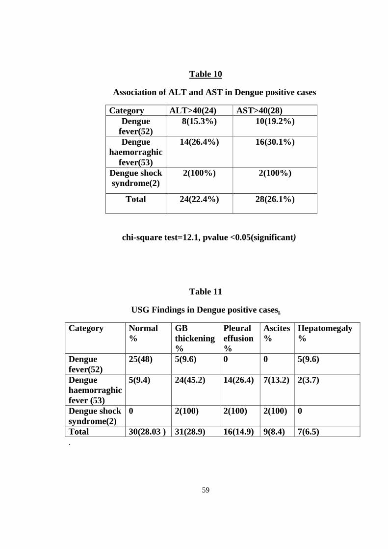
Liver parameters like ALT and AST were raised in 24(22.4%) and
28(26.1%) cases respectively. Among ALT elevated cases 8 (15.3%) were
associated with Dengue fever, 14(26.4%) were associated with Dengue
haemorrhagic fever and 2(100%) were associated with Dengue shock
syndrome. Among AST elevated cases10 (19.2%) were associated with

50
Dengue fever, 16(30.1%) were associated with Dengue haemorrhagic fever
and 2(100%) were associated with Dengue shock syndrome. (Ref Table 10)
In Ultrasonogram (USG) Gallbladder thickening 31(28.9%) was observed in
majority of Dengue positive cases followed by pleural effusion 16 (14.9%),
ascites 9 (8.4%) and hepatomegaly 7(6.5%). Among GB thickening 5 (9.6%)
were associated with Dengue fever, 24 (45.2%) with Dengue haemorrhagic
fever, 2(100%) with Dengue shock syndrome .Pleural effusion of 14(26.4%)
were associated with Dengue haemorrhagic fever and 2(100%) were
associated with Dengue shock syndrome and nil case in Dengue fever.
Ascites of 7(13.2%) were associated with Dengue haemorrhagic fever,
2(100%) with Dengue shock syndrome and nil case in Dengue fever.
Hepatomegaly was associated with dengue fever 5 (9.6%) and Dengue
haemorrhagic fever 2(3.7%) and nil association with Dengue shock
syndrome.USG was normal in 30(28.03%) cases which was 25 (48%) in
Dengue fever and 5(9.4%) in Dengue haemorrhagic fever.USG was not
available for 17 cases of Dengue fever and 1case in Dengue haemorrhagic
fever.(Ref Table 11)
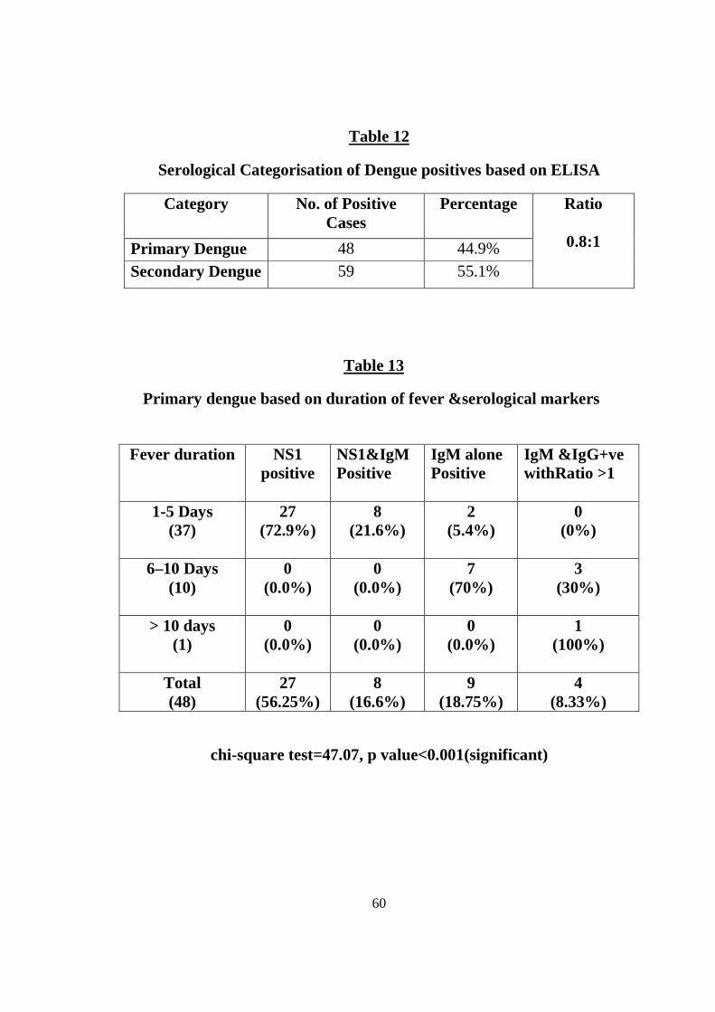
Seropositives were categorised as Primary 48(44.9%) and secondary
dengue 59(55.1%)with ratio0.8:1 based on ELISA .The serological markers

51
included for primary dengue were NS1 alone positive ,NS1 with IgM
positive, IgM alone positive and IgM&IgG positive with ratio >150. The
serological markers and their combination included for secondary dengue
were IgG positive with thrombocytopenia, NS1 & IgGpositive,NS1 ,IgM &
IgG positive and IgM & IgG positive with ratio <150 .(Ref Table 12)
Based on duration of fever ,primary dengue cases with above
mentioned markers were split into 1-5 days(37),6-10 days(10) and >10
days(1).
In primary Dengue(PD) NS1 alone showed positivity of 27(56.25%),
NS1 & IgM showed positivity of 8(16.6%) , IgM alone showed positivity
of 9(18.75%), IgM & IgG +ve with ratio >1 showed positivity of
4(8.33%).
In 1-5 days period NS1alone showed positivity of 27(72.9%) followed
by NS1 &IgM positivity of 8(21.6%) and IgM, alone positive of 2(5.4%). In
6-10 days duration IgM alone showed positivity of 70% followed by IgM
&IgG positive with ratio >1positivity of 3(30%). NS1 and NS1 with IgM
showed nil positivity. In >10 days IgM & IgG positive with ratio >1 alone
had positivity of 100% ,others were nil during this period. With

52
p value<0.001 it was found to be statistically significant. (Ref Table 13)
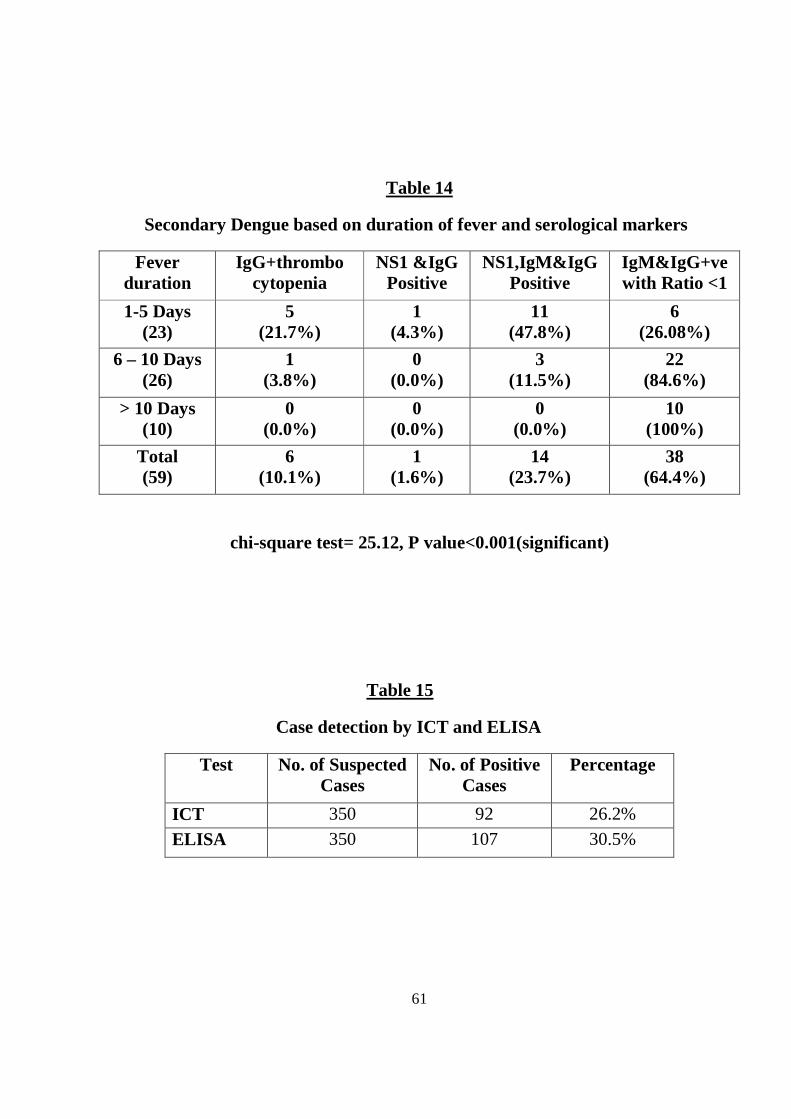
Similarly in secondary dengue(SD) IgG positive with thrombocytopenia
showed positivity of 6(10.1%) , NS1 & IgG of 1(1.6%) , NS1,IgM &IgG
of 14(23.7%), IgM &IgG positive with ratio <1 of 38(64.4%) .
In 1-5 days of secondary dengue NS1,IgM&IgG 11(47.8%) showed
higher positivity followed by IgM & IgG positive with ratio <1 6(26.08%),
IgG positive with thrombocytopenia were 5(21.7%),NS1&IgG positive
1(4.3%).
In 6-10 days IgM &IgG positive with ratio <1 showed positivity of
22(84.6%) followed by NS1,IgM,IgG positivity of 3(11.5%)and IgG
positive with thrombocytopenia1(3.8%). In > 10 days duration IgM &IgG
positive with ratio<1 showed higher positivity of 10(100%).Others showed
nil positivity. With p value<0.001 it was found to be statistically
significant.(Ref Table 14)
Case detection by ICT and ELISA showed 26.2% positivity for ICT
and 30.5% positivity for ELISA. (Ref Table 15)

53
Detection of serological markers like NS1, IgM and IgG by ICT and ELISA
showed positivity of 36(10.2%), 62(17.7%), 54(15.4%) by ICT and
50(14.2%), 73(20.8%), 65(18.2%) respectively by ELISA (Ref Table 15a).
Using ELISA as Reference assay, Sensitivity, Specificity, NPV and
PPV of the ICT were calculated for NS1 which showed 68%, 99.3%, 94.4%,
and 85.7% respectively. With p value of <0.001 it was found to be
statistically significant.(Ref Table 16)
Similarly for IgM the ICT showed sensitivity, specificity, PPV, NPV
of 80.8%, 98.9%, 95.1%, and 95.1% respectively. With p value<0.05 it was
found to be statistically significant.(Ref Table 17).
Sensitivity, Specificity, PPV, and NPV of IgG in ICT was 81.3%,
98.9%, 94.1%, 96.32% respectively. With p value<0.001it was found to be
statistically significant. (Ref Table 18)

54
Tables
.
Table 1
Seropositivity of Dengue
Total No. of Suspected fever
Cases
Total No. of positive cases
Percentage
350 107 30.5%
Table 2
Age and Sex Wise Distribution of Dengue positive cases
S.No. Age Group
Male Female Total
1. 0-10 Yrs 26(24.2%) 19(17.7%) 45(42%) 2. 11-20 Yrs 18(16.8%) 09(8.4%) 27(25.2%) 3. 21-30 Yrs 10(9.3%) 06(5.6%) 16(14.9%) 4. 31-40 Yrs 10(9.3%) 02(2.0%) 12(11.2%) 5. 41-50 Yrs 01(0.9%) 01(1%) 02(1.8%) 6. 51-60 Yrs - 02 (2.0%) 02(1.8%) 7. >60 Yrs 02(1.8%) 01(1.0%) 03(2.8%)
Total 67(62.3%) 40(37.7%) 107(100%) Male: Female ratio 1.6:1

55
Table 3
Seasonal Distribution of Dengue positive Cases
Month
2011-2012
Total no of cases
350
No of Positives
107
Percentage
%
September
12 2 16.6
October
9 0 0
November
23 5 21.7
December
47 12 25.5
January
41 13 31.7
February
15 2 13.3
March
23 1 4.3
April
17 4 23.5
May
49 13 26.5
June
81 41 50.6
July
25 13 52
August
8 1 12.5
chi square test =30.7, p value<.001(significant)
56
Table 4
Clinical Presentation of Dengue positive cases
Symptom No. of Patients Percentage Fever 107 100% Headache 65 60.7% Myalgia 41 38.3% Arthralgia 32 29.9% Abdominal Pain 30 28.03% Vomiting 26 24.2% Melena 11 10.2% Oliguria 5 4.6% Bleeding gums 5 4.6% Rash 5 4.6% Hemetemesis 4 3.7% Epistaxis 3 2.8%
Table 5
Clinical Classification based on WHO Guidelines51
Category No. of Positive Cases
Percentage
Dengue Fever 52 48.5%
Dengue Hemorrhagic Fever
53 49.5%
Dengue Shock Syndrome 2 2%
Total 107 100%
chi -square test =47.68, p<0.001(significant)
57
Table 6
Platelet Count in Dengue positive cases (n = 107)
Platelet Count
Total cases Percentage %
<20,000 13 12.14 21,000 – 40,000 30 28.03 41,000 – 50,000 10 9.34 51,000 – 1 lakh 44 41.12 >1 Lakhs 10 9.34 Total 107 100
chi-square test=42.766, pvalue<0 .001(significant)
Table 7
WBC Count of Dengue positive cases (n = 107)
WBC Count
Total cases Percentage %
< 4000 79 73.8 4000 – 5000 8 7.5 5100 – 6000 7 6.5 6100 – 7000 6 5.6 >7000 7 6.5 Total 107 100
chi-square test=42.7, p value<0.001(significant)

58
Table 8
Level of HCT in Dengue positive cases (n = 107)
HCT
Total cases Percentage %
>45 33 30.8 35-44 30 28.03 25-34 34 31.7 <25 10 9.3 Total 107 100
chi square test= 42.7, p value <0.001 (significant)
Table 9
Association of platelet, Haematocrit and WBC with Dengue positives
Category
Platelet count < 1 lakh(97)
Haematocrit >45(33)
WBC count <4000(79)
Dengue fever(52) 43(82.6%) 5(9.6%) 37(71.1%)
Dengue hamorrhagic fever(53)
52(98.1%) 26(49.05%) 40(75.4%)
Dengue shock syndrome(2)
2(100%) 2(100%) 2(100%)
Total 97 33 79
59
Table 10
Association of ALT and AST in Dengue positive cases
Category ALT>40(24) AST>40(28) Dengue
fever(52) 8(15.3%) 10(19.2%)
Dengue haemorraghic
fever(53)
14(26.4%) 16(30.1%)
Dengue shock syndrome(2)
2(100%) 2(100%)
Total 24(22.4%) 28(26.1%)
chi-square test=12.1, pvalue <0.05(significant)
Table 11
USG Findings in Dengue positive cases.
Category Normal %
GB thickening%
Pleural effusion %
Ascites %
Hepatomegaly %
Dengue fever(52)
25(48) 5(9.6) 0 0 5(9.6)
Dengue haemorraghic fever (53)
5(9.4) 24(45.2) 14(26.4) 7(13.2) 2(3.7)
Dengue shock syndrome(2)
0 2(100) 2(100) 2(100) 0
Total 30(28.03 ) 31(28.9) 16(14.9) 9(8.4) 7(6.5) .
60
Table 12
Serological Categorisation of Dengue positives based on ELISA
Category No. of Positive Cases
Percentage Ratio
0.8:1
Primary Dengue 48 44.9%
Secondary Dengue 59 55.1%
Table 13
Primary dengue based on duration of fever &serological markers
Fever duration NS1 positive
NS1&IgM Positive
IgM alone Positive
IgM &IgG+ve withRatio >1
1-5 Days
(37) 27
(72.9%) 8
(21.6%) 2
(5.4%)
0 (0%)
6–10 Days (10)
0 (0.0%)
0 (0.0%)
7 (70%)
3 (30%)
> 10 days (1)
0 (0.0%)
0 (0.0%)
0 (0.0%)
1 (100%)
Total (48)
27 (56.25%)
8 (16.6%)
9 (18.75%)
4 (8.33%)
chi-square test=47.07, p value<0.001(significant)
61
Table 14
Secondary Dengue based on duration of fever and serological markers
Fever duration
IgG+thrombo cytopenia
NS1 &IgG Positive
NS1,IgM&IgG Positive
IgM&IgG+ve with Ratio <1
1-5 Days (23)
5 (21.7%)
1 (4.3%)
11 (47.8%)
6 (26.08%)
6 – 10 Days (26)
1 (3.8%)
0 (0.0%)
3 (11.5%)
22 (84.6%)
> 10 Days (10)
0 (0.0%)
0 (0.0%)
0 (0.0%)
10 (100%)
Total (59)
6 (10.1%)
1 (1.6%)
14 (23.7%)
38 (64.4%)
chi-square test= 25.12, P value<0.001(significant)
Table 15
Case detection by ICT and ELISA
Test No. of Suspected Cases
No. of Positive Cases
Percentage
ICT 350 92 26.2%
ELISA 350 107 30.5%

62
Table 15(a)
Detection of NS1, IgM, IgG by ICT and ELISA
Test No of suspected
cases
ICT ELISA
NS1 350 36(10.2%) 50(14.2%) IgM 350 62(17.7%) 73(20.8%) IgG 350 54(15.4%) 65(18.2%)
Table 16
Cross tabulation of ICT and ELISA for NS1
ICT ELISA Total
Positive Negative Positive 34 2 36
Negative 16 298 314
Total 50 300 350
Sensitivity 68%, Specificity 99.3%, PPV 94.4%, NPV 85.7%
chi-square test =46.36, p value<0.001(significant)

63
Table 17
Cross tabulation of ICT and ELISA for IgM
ICT ELISA Total
Positive Negative Positive 59 3 62
Negative 14 274 288
Total 73 277 350
Sensitivity 80.8%, Specificity98.9%, PPV95.1%, NPV 95.1%
chi-square test =5.1, p value<0.05(significant)
Table 18
Cross tabulation of ICT and ELISA for IgG
ICT ELISA Total
Positive Negative Positive 51 3 54
Negative 14 282 296
Total 65 285 350
Sensitivity 81.3%, Specificity 98.9%, PPV 94.1%, NPV 96.32%
chi-square test=41.3, p value< 0.001(significant)
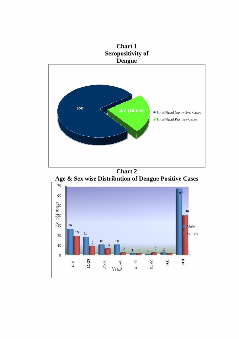
Chart 1 Seropositivity of
Dengue
Chart 2
Age & Sex wise Distribution of Dengue Positive Cases
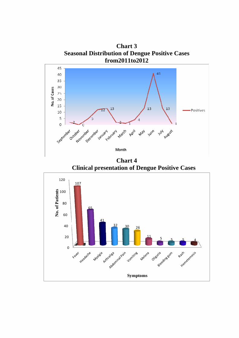
Chart 3 Seasonal Distribution of Dengue Positive Cases
from2011to2012
Chart 4
Clinical presentation of Dengue Positive Cases
0
20
40
60
80
100
120107
65
41
32 30 26
115 5 5 4
No
. of P
atie
nts
Symptoms
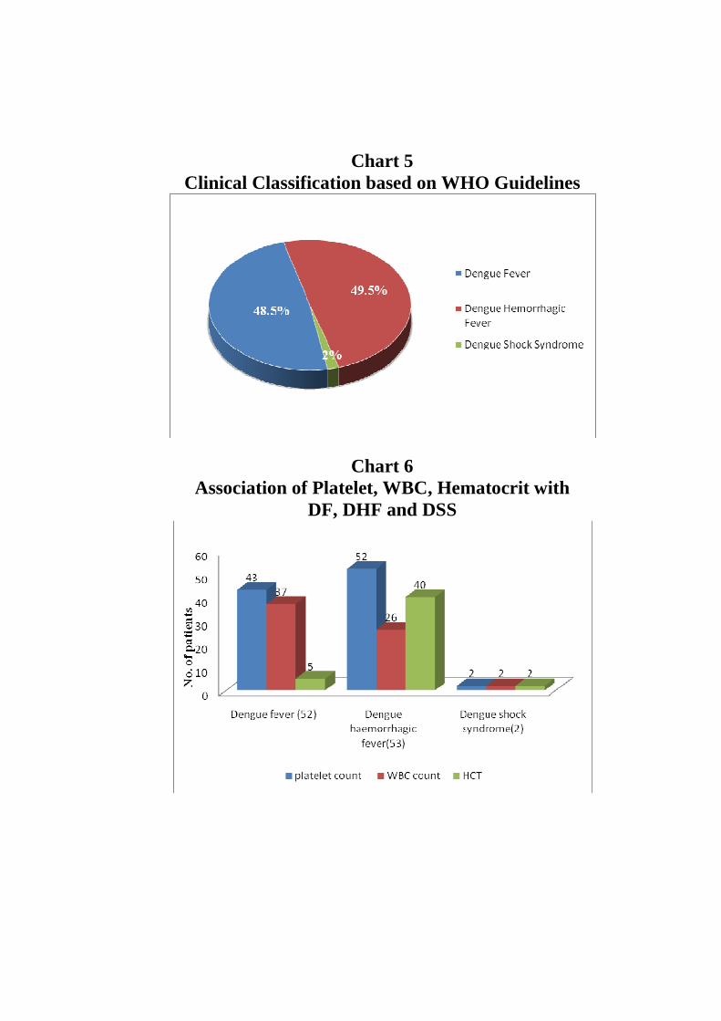
Chart 5
Clinical Classification based on WHO Guidelines
Chart 6 Association of Platelet, WBC, Hematocrit with
DF, DHF and DSS

Chart 7
Serological Categorisation of Dengue Positive Cases
Chart 8 Primary dengue based on duration of fever and serological
markers

Chart 9 Secondary Dengue based on duration of fever and serological
markers.
Table 10 Detection of NS1, IgM & IgG by ICT and ELISA
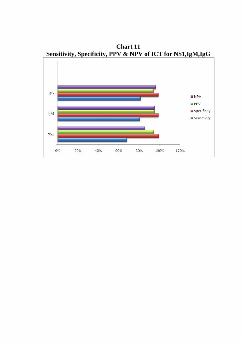
Chart 11 Sensitivity, Specificity, PPV & NPV of ICT for NS1,IgM,IgG

64
DISCUSSION
In order to provide timely information for the management of patients
with acute dengue and effectively control dengue outbreaks, it is important
to establish an accurate confirmation of acute dengue infection during the
first few days of clinical symptoms.
For a long time, detection of dengue specific IgM/IgG has been the main
stay of diagnosis of Dengue infection. The Dengue specific IgM antibodies
begin to appear only around fifth day of primary infection. The New
parameter called NS1 now available can be detected from the first day of
illness. Dengue NS1 is detected in the blood circulation as early as viral
RNA detected in PCR.
In this study the seropositivity of dengue infection was 30% .In a study by
Halstead et al 52 1969, Bangkok 29% of febrile illness was associated with
dengue virus infection similar to this study.
In a study by Chakravarti A et al 53 2011, New Delhi conducted on 145
samples noted seropositivity of 60.7% which is higher to this study.
In this study 62.3% of the affected populations were males and 37.7% were
females. The following studies were found to coincide with this study Aisha

65
sajid, Asim akram, Mubashir Ahmed54 2012, Syed Irfan ahmed et al55
2010 Fasalabad and Kurukumbi et al 21 2001.
In contrast to this study females were affected more than males in a study by
Murray smith and Skelly C 56 Queensland. But Susan shepherd et al 57 in
a study conducted at Pennisylvania showed that dengue infection can occur
irrespective of sex.
In this study Male: female ratio was 1.6:1 and this is due to males being
engaged in outdoor activities and due to their dressing culture when
compared to females which increases the exposure to mosquito bites. The
studies by Eng Eong Ooi58 2001 Singapore, Shrivastava et al59 2011 and
Rajoo Singh Chhina et al 60 2008 Punjab had similar M: F ratio.
In this study the commonest age group affected was 0-10 years (42.05%) in
both sexes. Increased incidence of infection in 0-10 years in this study is due
to increased mosquito bite exposure in the school premises during the day
time. The study by Shah GS et al61 Kathmandu revealed that the
commonest age group affected in their study was from 8months to 14 years
and Malavige et al 45 2004 Srilanka study showed age range from 1 month
to 12 years which is similar to this study.
66
In a study by Anita chakravarti et al 62 conducted at New Delhi the age
group commonly affected was 26-30 years which is in contrast to this study.
In this study incidence of dengue cases were more during the months of
November& December 2011 and January ,May , June, July of 2012 .
Increase in the incidence of Dengue cases occur usually during rainfall63.
Tamilnadu receives rainfall from October to November followed by post
monsoon period of December & January. The cause for the incidence of
cases during the above months like May, June and July in this study is that
the eggs can survive even in stored waters in the absence of rainfall.
According to NVBCDP25 2007 every year there is an upsurge in the cases of
Dengue /DHF from July to November. Ekta gupta et al 64 2006 in their 3
year study had shown an increase in incidence during Post monsoon period
(September – November).
A study by Louise Kelly –hope Mandeleine C.Thomson65 2008 in their
Study revealed that a climatic change has a role in the transmission of
Dengue. Umesh Isalkar66 , TNN 2009 said that whether changes cause
atypical viral fever.
In this study the commonest symptom was fever100% followed by
headache 60.7%, Myalgia 38.3%, arthralgia 29.9% ,abdominal pain 28.3%

67
,vomiting 24.2% ,oliguria &,rash 4.6% . Saadiah sulaiman et al 671999 in
their study had fever of 100% followed by headache (80.9%) arthralgia
(74%), myalgia (70%), rash (4.6%) and vomiting (62.7%) similar to this
study. Jonathen et al68 2010 in their study had fever (72%) as most
common symptom followed by epistaxis, abdominal pain and rashes.
Rahman et al69 2002 in their study had headache (90%) as predominant
symptom followed by myalgia / arthralgia (85%) and vomiting (64%).
In this study Dengue positive cases were classified according to WHO
guidelines. It showed 48.5% Dengue fever, 49.5% Dengue haemorrhagic
fever and 2% Dengue shock syndrome. In this study DHF is found to be
high because of increased incidence of infection in children. Because of the
increased surface area of the blood vessel per unit volume of skeletal muscle
and the immature blood vessel leading to increased vascular permeability,
DHF is common in children. Even though Dengue haemorrhagic fever was
high in this study and is said to be a complication of secondary dengue, DSS
was comparatively less because of the eminent care and management given
to the diagnosed cases in the respective wards.
In a study by S Kalyanarooj et al70 1997 Thailand, conducted on 60
seropositives showed 53% (32) Dengue fever, 47% (28) Dengue

68
haemorrhagic fever and Dengue shock syndrome. According to David
W.Vaughn and Sharone Green71 fatality rate is decreased in DHF /DSS
cases those who are hospitalised and treated promptly.
In this study thrombocytopenia was observed in 90% of the positive cases
the common range was between 51,000-1 lakh followed by 20,000- 40,000.
Thrombocytopenia is due to decreased platelet production in Dengue fever
and increased peripheral destruction in DHF. RD Kulkarni et al 62011
Karnataka, in their study had thrombocytopenia associated with majority of
dengue positive cases than dengue negative cases similar to this study. In a
study by Chandrakanta et al72 2008 revealed 60% of thrombocytopenia <
1lakh.
Karande et al73 2005 revealed that arthralgia and thrombocytopenia if
present in a child increases the chance of the child to be Dengue positive.
But a study by Col Banerjee et al74 revealed that thrombocytopenia may not
always be a feature of Dengue.
In this study leucopenia was observed in 79/107(73.8%) of dengue positive
cases. Out of 79, 37 were associated with dengue fever, 40 were associated
with Dengue haemorrhagic fever, 2 were associated with Dengue shock
syndrome.The association of leucopenia with Dengue fever,DHF and DSS is

69
71.1%,75.4% and 100% respectively(Table 9). Neutrophil adhesion to
dengue infected endothelial cell is one of the causes for leucopenia.
Kalyanarooj et al 70 1997 has stated that leucopenia in Dengue is due to
bone marrow suppression.In a study by Somachai insiripong MD75 2010
revealed that leucopenia in DHF is multifactorial .
In this study elevated Heamatocrit (>45%) was observed in 33 patients, 5
were associated with dengue fever, 26 were associated with Dengue
haemorraghic fever and 2 were associated with Dengue shock syndrome.
The association of elevated HCT with DF, DHF and DSS are 9.6%, 49.05%
and 100% respectively. Rise in haematocrit is due to increased vascular
permeability and plasma leakage in DHF /DSS. Malavige et al 762007 in
their study have considered hemoconcentration when the packed cell volume
was >45 in children.
In this study liver enzymes like AST (Aspartate aminotransferase)) and ALT
(Alanine aminotransferase) were elevated in 28 and 24 patients respectively.
Among ALT and AST rise, >40units majority were associated with DHF
and DSS. Rajoo Singh Chhina et al 60, Punjab in their study had
significant rise of AST (97%) and ALT (93%) in DHF/DSS as compared to

70
DF. They also concluded that raised AST can be an early indicator of
dengue infection.
In secondary dengue causing liver damage AST will be elevated more than
ALT which is due to the release of AST from damaged monocytes of dengue
infection and due to release of AST from skeletal muscles due to myalgia.
In this study, the Ultrasound findings showed more of Gall bladder
thickening followed by pleural effusion, ascites and hepatomegaly . Gall
bladder thickening is due to increased vascular permeability where the fluid
tends to collect in between the layers increasing the thickness of the Gall
bladder which occurs in Dengue haemorrhagic fever and Dengue shock
syndrome. Similarly the fluid collects in pleural and peritoneal cavity.
In the study conducted by Bala subramanian et al 772006 ultrasonography
was found to be superior to radiography in detecting pleural effusion. In the
study by Kumar et al78 2010 Karnataka, pleural effusion was noted in 4.29
% (20) of 466 patients which is less than this study. In this study by
Zulkarnain 79 2004 revealed 29.8% of Gall bladder edema which is higher
than this study.

71
In this study the incidence of Primary dengue was 48(44.8%) and Secondary
Dengue was 59(55.1%) in the proportion of 0.8:1. In a study by Jonathan G
et al68 2010 secondary dengue (81%) was more than primary dengue which
is similar to this study. In a study by Neeraja M et al22, Hyderabad showed
72.5% of secondary dengue conducted on 211patients which is similar to
this study.
In 48 samples of primary dengue NS1 showed higher positivity (56.25%)
followed by IgM alone positivity of 18.75% , NS1 &IgM positivity of
16.6% , and IgM & IgG positive with ratio >1of 8.33%.During the primary
infection ,NS1 gets secreted out from virus infected cells which indicates
underlying viremia and pathogenesis of infection. As there are low antibody
titres in primary dengue, the NS1 protein can be estimated more in serum
during the acute period. So NS1 detection can be used as a stand alone test
for Primary dengue in cases when IgM is not detectable and where PCR is
not available.
During the acute period of 1-5 days in primary dengue , NS1 detection
(72.9%) had higher positivity followed by NS1 & IgM(21.6%) and IgM
alone positive of 2(5.4%). As IgM antibodies tends to appear after 4-5 days
of infection and as many patients in this study presented within 5 days, the

72
sensitivity of NS1 &IgM ,IgM alone positive in primary dengue were less
when compared to NS1 positivity alone. Hence when IgM ELISA is
negative in the acute period, NS1 ELISA should be done for diagnosis which
significantly improves the detection rate without the requirement of paired
serum to demonstrate the rise in titre of antibodies. In a study by
Chakravarti A, Kumar A, Malik S 53 2011New Delhi conducted on 145
acute serum samples NS1 ELISA showed a positivity rate of 65.9% and
MAC-ELISA showed 60.2% positivity in the acute period. Veasna duong et
al9 study also showed NS1 positivity to be higher in first 3 days.
Minipritam singh et al 80 and Chua KB et al 812011 studies revealed that
NS1 antigen capture ELISA was more sensitive than RT-PCR and viral
isolation.
During the early convalescent period of 6-10 days in primary dengue, IgM
showed higher positivity (70%) than other serological markers. This is due
to the appearance of IgM antibodies after 4th day of illness and is the first
antibody to appear following Dengue infection. IgM antibody gradually
increases towards end of first week and it persists for 3 months thereafter.
The virus may be at undetectable levels in this period as they tend to be in
complex with IgM antibody. Hence NS1 positivity was less during this

73
period. IgM and IgG combination also showed less positivity (30%) during
this period as anti dengue IgG is detectable only at low titer during this
period as it appears still later. In a study by S Datta and Wattal 82 IgM
detection by MAC ELISA showed 93.6% positivity in early convalescent
period.
In more than 10 days duration, IgM &IgG positive with ratio > 1 showed
higher positivity (100%) as IgG antibodies would have appeared during this
period which increases further in the proceeding weeks and persists lifelong
thereafter.
In this study, in secondary dengue, IgM and IgG positive with ratio <1
showed higher positivity (64.4%) than other parameters followed by NS1,
IgM and IgG combination (23.7%). As the antibody titres rise extremely
rapidly, IgM and IgG are detected more in secondary dengue.
During the acute period of 1-5 days in secondary dengue NS1, IgM and IgG
combination showed higher positivity11(47.8%) than IgM & IgG
combination 6 (26.08%),NS1& IgG 1(4.3%) and IgG alone positive with
thrombocytopenia 5(21.7%).The NS1 antigen has reduced sensitivity in
secondary dengue because it is sequestered in immune complexes and that
target epitopes are not accessible to the plate bound mAb in NS1 ELISA.

74
High levels of IgG are detectable even in acute phase of secondary dengue
due to previous infection and rise dramatically over the proceeding 2 weeks
whereas IgM levels tend to be significantly lowered in secondary infection.
In the early convalescent period of 6-10 days in secondary dengue IgM
&IgG positive with ratio<1 22 (84.6%) showed higher positivity as IgM to
second infection appears during this period. NS1, IgM &IgG showed
positivity of 3(11.5%), IgG alone positive with thrombocytopenia 1(3.8%).
As IgG is present in both current and past infections, presence of IgG alone
does not indicate active infection. Hence it should be combined with platelet
count. Only if the platelet count is lowered it indicates current infection. If it
is normal, it shows past infection.
In more than 10 days duration of secondary dengue IgM & IgG positive with
ratio <1showed higher positivity (100%) which is due to the presence of
IgM &IgG antibodies to the second infection. Susana vazquez et al83
2007showed that IgM and IgG capture ELISA kits has high sensitivity and
specificity and offers good alternative for dengue diagnosis.

75
ICTs for the detection of NS1, IgM, IgG antibodies have been developed by
a number of commercial companies and have found wide applications
because of their ease of use and rapidity of results.
The case detection by ICT in this study was 10.2% for NS1, 17.7% for IgM,
15.4% for IgG, whereas in ELISA it was 14.2%, 20.8% and 18.2%
respectively. This clearly shows that case detection was more in ELISA. The
ICT is found to be less sensitive to ELISA and gives false negative results in
some cases.Cross reaction with other flaviviruses gives rise to false positives
in some cases. In a study by RD Kulkarni et al6 2011 showed NS1
positivity 29%, IgM positivity 50% and IgG positivity 2.8% in ICT.
Masao et al in their study 84 2011 in Japan had said that NS1 rapid test is
suitable for lab diagnosis due to its reliability and rapidity as it require only
20 minutes for detection. But in view of its low sensitivity it is not suitable
for screening purposes.
In a study by Subhash C Arya 85 2011the ICT was highly sensitive (88%)
and specific (98%).
As diagnostic assays are usually evaluated in terms of Sensitivity and
specificity that was calculated by 2x2 cross- tabulation, using a reference

76
standard (the test most widely used) is compared to determine diagnostic
accuracy.
Using ELISA as reference standard, the accuracy indices of the ICT were
calculated.
NS1 ICT had a sensitivity of 68%,specificity 99.3%, PPV 94.4%, NPV
85.7%.Using chi-square test p value of <0.001 was obtained which was
found to be statistically significant. In a study by Subhash C Arya et al85
2011 ICT showed 80-90% detection rate during first 7 days. In a Study by
Vu Ty Han et al10, Thailand NS1 LFRT was found to be 100% specific and
72% sensitive in primary than secondary dengue.
In this study the sensitivity and specificity of IgM by ICT was
80.8%&98.9%,PPV-95.1% &NPV-95.1%.Using chi-square test ,p value of
<0.05 was obtained for IgM which was found to be statistically significant.
In a study by M Moorthy et al 162009 showed that the accuracy indices of
ICT for IgM were 81.8%, 75.0%, 61% and 89.6% and concluded that ICT
not to be used as stand alone test.
In this study IgG in ICT showed Sensitivity of 81.3%, specificity 98.9%,
PPV 94.1% & NPV 96.32% Using chi-square test p value of <0.001 was

77
obtained which was found to be statistically significant. Moorthy et al16
2009 study with sensitivity of 87.5%, specificity 66.6%, PPV 72%, NPV
83.9% concluded that ICT with moderate performance could not be used as
stand alone test.
WHO’s multicentric evaluation of Dengue antibody ICTs had IgM
sensitivity from 20% to 100% & specificity from 77% to 98% when
compared with reference ELISA.
� Most of the patients were referred from various places and would
have received few days treatment before reaching the hospital .The
precise day of fever at the time of conducting the test could not be
obtained in a large no of cases. In spite of this, NS1 alone was positive
in 25% cases in this study.

78
SUMMARY
• Seropositivity of Dengue in 350 suspected cases during the study
period was 30%
• The commonest age group affected by Dengue in this study was 0-10
years in both sexes.
• Dengue was predominant in Males when compared to Females with
the ratio of 1.6:1
• Dengue incidence in this study was more during the months of
November, December 2011, January, May, June, July of 2012.
• Of all the symptoms, fever was the commonest symptom followed by
headache, myalgia, arthralgia, abdominal pain, vomiting, oliguria and
rash.
• Melena was the commonest bleeding manifestation followed by
bleeding gums, hemetemesis and epistaxis in this study.
• Dengue positive cases were classified into Dengue fever (48.5%),
Dengue haemorrhagic fever (49.5%) and Dengue shock syndrome
(2%) based on WHO guidelines.

79
• In the haematological parameters, Thrombocytopenia was associated
more with Dengue positive cases. Platelet count of 50,000- 1 lakh was
commonest range and majority were associated with DHF&DSS.
• WBC count of <4000 was more commonly associated with DHF and
DSS
• Elevated Haematocrit level >45 was associated with DHF/DSS.
• Biochemical parameters- AST and ALT were elevated in DF, DHF
and DSS.
• Of the USG findings, Gall bladder thickening, pleural effusion and
ascites were seen in majority of Dengue haemorrhagic fever and
Dengue shock syndrome.
• In this study the incidence of secondary dengue(55.1%) was more
than primary dengue(44.9%) with the ratio of 0.8:1
• NS1 positivity was high in primary dengue followed by IgM alone.
• In the acute period of 1-5 days in primary dengue NS1 antigen
positivity was found to be higher followed by NS1 & IgM .
• In the early convalescent period (6-10days) of Primary dengue IgM
alone had higher positivity followed by IgM &IgG positive with ratio
>1.

80
• In the late convalescent period (10 days) of primary dengue, IgM &
IgG positive with ratio >1 alone showed positivity.
• In secondary dengue, IgM & IgG positive with ratio<1 had higher
positivity followed by NS1, IgM &IgG combination.
• In the acute period of secondary dengue(1-5days) NS1, IgM &IgG
positive showed higher positivity followed by IgM ,IgG positive with
ratio<1, IgG alone positive with thrombocytopenia and lowered
NS1&IgG positivity.
• In the early convalescent period (6-10 days) IgM &IgG positive with
ratio <1 showed higher positivity followed by the combination of
NS1, IgM, IgG.
• In late convalescent period (>10 days) IgM&IgG positive with ratio
<1 alone showed higher sensitivity.
• Case detection rate of ICT was 26.2%and ELISA was 30.5% for 350
cases.
• The percentage of case detection by ICT for all the 3 parameters
NS1,IgM and IgG was 10.2%,17.7% and 15.4% and ELISA showed
14.2%,20.8% and 18.2%.

81
• Sensitivity, Specificity, PPV and NPV for NS1 in ICT were 68%,
99.3%, 94.4% and 85.7%.
• Sensitivity ,Specificity ,PPV and NPV of IgM in ICT were 80.8%,
98.9%, 95.1% and 95.1%.
• Sensitivity,Specificity ,PPV and NPV of IgG in ICT were 81.3%,
98.9%, 94.1% and 96.32%.

82
CONCLUSION
As per the results of this study NS1 ELISA is highly sensitive and proved to
be a valuable tool in the diagnosis of dengue particularly in the acute period.
NS1 is a reliable marker in primary dengue and when combined with IgM
improves the detection rate significantly in a single serum sample in the
absence of paired sera.
For diagnosis of Dengue infection irrespective of duration of fever all the
three (i.e) NS1, Dengue IgM and IgG by ELISA must be done .This helps to
rule out infection and to avoid false negatives. Immuno capture ELISA is
highly sensitive in the detection of dengue cases.
Considering the moderate performance of ICT in this study the device
cannot be used as a stand alone test and it should always be supplemented by
ELISA for the diagnosis of Dengue infection.
Serological differentiation of primary and secondary can be arrived as
per the results of the study as shown below

83
NS1 IgM IgG Category
+ - - Primary dengue
+ + - Primary dengue
- + - Primary dengue
- + + Primary dengue( IgM/IgG ratio>1)
- - + Past infection-(Plateletcount &Total count
normal).
- - + Secondary dengue-(Platelet/Total count low)
+ - + Secondary dengue
+ + + Secondary dengue
- + + Secondary dengue-(IgM/IgG ratio <1)
Significant association of haematological profile and biochemical profile
with seropositives in this study insists that these tests should be considered
and given importance in the diagnosis and management of dengue.
In conclusion, based upon the higher positivity of the NS1 ELISA both in
primary and secondary dengue, this study gains public health importance
because it suggests that NS1 antigen should be included along with routinely
used antibody testing protocols for early detection and management of cases.
ELISA is preferable for diagnostic purposes compared to ICT and the use of
84
Immunocapture ELISA in the test panel for dengue will reduce the false
Negativity thereby reducing the morbidity and mortality rate.

BIBLIOGRAPHY
1. Mandell’s, Douglas and Bennett’s principle and practice of infectious
disease.7th edition.Vol.2
2. Seema and S.K.Jain Molecular mechanism of pathogenesis of Dengue
virus: Entry and fusion with target cell. Indian journal of clinical
biochemistry, 2005, 20(2) 92-103
3. Abm shahidul alam et al Bangladesh J child health 2009; 33:2:p.no 55-58
4. WHO (2009) Dengue: guidelines for dengue diagnosis, treatment,
prevention and control, 3 rd Edition.
5. Sukathida Ubol , Promsin musrinoul,Jeerayut chaijaruwanich et al
Differences in Global gene expression in peripheral blood mononuclear
cells indicate a significant role of the innate responses in progression of
Dengue fever but not Dengue haemorrhagic fever .The Journal of
infectious Diseases 2008 ;197 :1459-67
6. R D Kulkarni,SS Patil,GS A jantha et al Association of platelet count and
serological markers of dengue infection- importance of NS1 antigen
Indian journal of Medical
Microbiology2011 29(4):359-362
7. Manson’s tropical disease 21st
edition . S.Nimmannitya chapter 42
Dengue and dengue haemorrhagic fever.

8. Tamilarasu kadhiravan et al Association of intracellular Th1-Th2 balance
in CD4+Tcells and MIP-1α in CD8+ T cells with disease severity in adults
with Dengue.
9. Veasna Duong et al Clinical and virological factors influencing the
performance of a NS1 Antigen capture Assay and potential use as a
marker of Dengue Disease severity PLOS Neglected Tropical diseases.
2011 5(7).
10. Vu Ty Hang et al Diagnostic accuracy of NS1 ELISA and Lateral flow Rapid
Tests for Dengue sensitivity, specificity and relationship to Viremia and
Antibody response. PLOS neglected Tropical diseases. 2009 3(1)
11. Infectious disease and Epidemiology theory and practice Nelson Williams
2nd
Edition
12. Richard L Guerrent David H.Walker,Peter F.Weller principles
pathogenesis, practice of Tropical infectious diseases. 3rd
edition chapter
75.
13. Dr.Vijay N.,Yewae,Dr.Sitara Gopinathan Dengue fever IAP speciality
series (paediatric infectious Diseases ,Under IAP Action plan 2006)
14. Atul Garg et al Prevalence of Dengue among clinically suspected febrile
episodes at a teaching hospital in North india.Journal of infectious
disease and immunityMay 2011 vol 3(5) pp 85-99.
15. VC Chaturvedi ,R.Shrivastava Dengu haemorrhagic fever:A Global
challenge Ijmm 2004;22:1:5-6
16. M Moorthy S Chandy, K Selvaraj et al : Evaluation of a rapid
immunochromatographic device for the detection of Igm &IgG antibodies
to dengue viruses in a tertiary care hospital in south India journal of
Medical microbiology 2009:27:3
17. Harrison’s textbook of infectious diseases. 16 th Edition
18. Jawettz, Melnick, Adelberg’s textbook of medical microbiology 21 st
Edition.
19. Padmalal Gurugama et al .Dengue viral infections .Indian journal of
dermatology. 2010 Jan- march; 55:68-78
20. Maria G.Guzman,Gustavo Kouri Dengue diagnosis,advances and
Challenges International Journal of infectious Diseases 8(2004) 69-80.
21. M.Kurukumbi , JPwali, Shobha broor, P.Aggarwal, P.Sethi et al
Seroepidemiology and active surveillance of Dengue fever/Dengue
haemorrhagic fever in Delhi .2001;55:3:149-156
22. Neeraja M,LakshmiV,Teja VD et al Serodiagnosis of dengue virus infection
in patients presenting to a tertiary care hospital Bioline International
official site may 2006
23. Ee ping Teoh,Petra kukkaro,En weiTeo et al The structural basis for
serotype –specific Nuetralisation of Dengue virus by a human antibody
.Science translational medicine June 2012;4:139
24. Brett Lindenbach, Heinz –Jurgen Thiel Charles M.Rice Field’s textbook of
virology David M.Knipe Peter M.Howley fifth edition volume 1 chapter
33.
25. Guidelines for clinical management of dengue fever ,DHF and DSS
Directorate of National vector Borne disease control programme 2008.
26. Vector control – Jan A.Rozandeal WHO 1997
27. Linnen JM, Vinelli E, Sabino ER, et al Dengue virus in asymptomatic Blood
donors. Dengue viremia in blood donors from Honduras,Brazil and
Australia.Transfusion 2008;48:1355-1362
28. Mohammed H,Linnen JM,Mumez-Jordan et al Dengue virus in Blood
donation Transfusion 2008 ;48:1348-1354
29. Ampaiwan Chuansumrit,MD and Kanchana Tangnararatchakti,MD
Transfusion alternatives in transfusion medicine January 2005
30. Jonathen cohen,William G.Powderly Steven M.Opal Textbook of
Infectious diseases 3rd
Edition Volume 2
31. Sansanee Noisakaren et al .Cells in Dengue virus infection.Invivo
advances inVirology 2010.

32. Pornthep Chantha Vanich ,Christine et al Short report :Immnue response
and occurrence of Dengue infection in Thai children three to eight years
after vaccination with live attenuated Tetravalent vaccine
AM.J.Trop.Med.Hyg.,75(1),2006;p.no 26-28.
33. Scott Hallstead B controversies in Dengue pathogenesis .Paediatrics
Int.child health 2012;32(s):5-9
34. Ashok S Kapse 9.26 Dengue illnesses p.no 396. IAP textbook of
Paediatrics 4th
edition
35. Maria gundalupu,Guzman,Susana Vazquez et al Risk factors in dengue
Malaysian Journal of Medical sciences.2009;16(3):4-11
36. N.Sathish,T.S.Vijayakumar ,P.Abraham and GS Shridharan Dengue fever
its Labdiagnosis with special emphasis on IgM detection. Dengue Bulletin
–Vol 27, 2003.
37. Rosannito peeling et al NS1 can be detected in patients with proven
secondary dengue upto 9 days after the onset of illness.Nature reviews
Microbiology References S30-37.
38. Philippe Buchy Sutee Yoksan Rosanne W.Deckling et al Lab test for the
Diagnosis of Dengue virus infection .WHO on behalf of special
programme for research and training inTropical diseases.2007

39. Gubler J 1998.Dengue and Dengue haemorrhagic ever,Clin.Microbiol.Rev
11:480-496
40. Clinical And Laboratory Guidelines for Dengue fever and Dengue
haemorrhagic fever / Dengue shock Syndrome for Health care
providers.Caribbean Epidemiology center (CAREC), PanAmerican health
organization ,World Health organization.
41. Vanthanophas K,WM Hammon,RW Atchison and GE
Sather,1973.Attempted type specific diagnosis of dengue virus infection
by the indirect fluorescent antibody technique directed at differentiating
IgM and IgG response .Proc soc Exp.Biol Med.142:697-702
42. Cardosa ,MJ,phaik T,and sham N.1988.Development of a Dotenzyme
immunoassay for dengue 3;asensitive method for detection of
antidengue antibodies.J.Virol.methods 22:81-88.
43. Nivia Rocha Dutra,Manilia Barbosade,michelle et al Journal of Global
infectious Diseases 2009 ;1(11) 38-44
44. Maria G.Guzman,Scott B Halstead,HarveyAttsob,philippe bruchy Dengue
a continuing global threat .Nature Reviews Microbiology.
45. GN Malavige,S Fernando DJ Fernando et al Dengue viral infections Post
graduate medical journal 2004;80:588-601

46. Mc Arthur JH,Durbin AP,Maron JA,et al Phase 1 clinical evaluation of
Rden 4Delta 30-200,201 a live attenuated Dengue 4 vaccine candidate
designed for decreasing hepatotoxicity .Am J trop Med Hygiene.Nov 2008
;79(5):678-84
47. Sanofi-pasteur and international vaccine institute partner against Dengue
OCT 20, 2011.
48. Edelman R.Dengue Vaccine approach the finish line .Clin Inf.Disease
Journal 15 2007;45suppl:S 56-60
49. Manual for investigation and control of outbreak of Dengue and Dengue
haemorrhagic fever. National institute of Communicable Diseases.
Government of India.
50. Scott B.Halstead Gorbach, John G.Bartlett, Neil R.Blacklow .Infectious
Diseases. Chapter 258 3rd
Edition p. no 2113-2117
51. Comprehensive guidelines for prevention and control of Dengue and
Dengue haemorrhagic fever .4.Clinical manifestations and diagnosis
World health organization 2011.
52. Halstead SB ,Nimmanitya S, Margitha MR Dengue and Chikungunya virus
infection in man in Thailand observation on Disease in output .AM J
Tropical Med Hygiene 1969 18;972-83 al
53. Chakravarti A,Kumar A,Malik S Detection of dengue infection by
combining the use of an NS1 antigen based assay with antibody
detection. Southeast Asian J trop Med Public Health ,2011 Mar;42(2):297-
302
54. Aisha Sajid ,Asim Ikram,Mubashir Ahmed Dengue fever outbreak
2011:clinical profile of children presenting at Madina teaching Hospital
Faisalabad.JUMDC2012; vol.3:1
55. Syed irfan Ahmed , Ammara Naeem, Haider Zaigham Baqai, Shahid Iqbal
Sheikh, Zubair Ahmed Ranja, Arshad Iqbal Satti, Faran Maqbool, Nadeem
Ikram NS1 antigen and Immunoglobulin M detection in the acute and
early convalescent stages of dengue fever Journal of Rawalpindi Medical
college;2010;14(2):56-59
56. Murray smith and skelly C Field epidemiology of an outbreak of dengue
fever in charter towers.Queensland :are insect screen protective NZ
J.Public health ,1996,20:545-547
57. Susan shepherd et al .Director of education of research, PENN Travel
medicine, Dept of Emergency medicine, University of pennisylvania
Medical centre.
58. Eng Eong Ooi changing pattern of Dengue Transmission in Singapore
dengue Bulletin –vol 25,2001

59. A Shrivastava PK, Dash,NK Tripathi et al Evaluation of a commercial
Dengue NS1 ELISA for early diagnosis of dengue infection Indian journal
of Medical Microbiology 2011;29:1:p.51-55
60. Rajoo Singh Chhina et al Liver function tests in patients with dengue
viral infection Dengue Bulletin –volume 32,2008
61. Shah GS, Islam s, Das BK clinical and laboratory profile of Dengue
infection in children. Kathmandu university Med Journal 2006;4(11):40-43
62. Anita chakravarti ,Amod kumar and Sonia malik Detection of dengue
infection by combining the use of NS1 antigen based assay with antibody
detection. Department of Microbiology Maulana Azad Medical colloge
and associated LokNayak hospital, Bahadur shah, Zafar marg,NewDelhi.
63. World Health organization. Dengue Bulletin :Geneva Switzerland ;2003
64. Ekta gupta ,Lalit Das,Geetanjali kapoor and shoba broor Changing
Epidemiology of dengue in Delhi ,India Virology journal 2006:3:92
65. Louise Kelly –hope Mandeleine C.Thomson climate and infectious
disease Advances in global change research 2008: 30:p.no 31-70
66. Umesh ISalkar,TNN 2009 Weather changes cause atypical Viral fever
67. Saadiah Sulaiman et al L symptoms and haematological profile of Dengue
haemorrhagic fever among hospitalized patients in Kuala lumpur ,A
useful indicator of disease activity.
68. Jonathen et al Profile of pediatric patients with dengue fever/Dengue
Haemorrhagic Fever over a five year period(2000-2004) PIDSP journal
2010 ;11:1
69. Rahman M et al .First outbreak of Dnegue haemorrhagic Fever,
Bangladesh.Emerging infectious diseases 2002;8;738-40
70. S Kalyanarooj et al Early clinical and laboratory indicators OF Acute
dengue illness Journal of Infectious Diseases.1997;176:313-21
71. David W. Vaughn and Sharone green .Hunter’s Tropical medicine and
Emerging infectious diseases,8 th Edition .29.1:240-244
72. Chandrakanta et al changing clinical manifestations of dengue infection in
NS1 Chatturpathi Shahuji maharaj Medical university ,lucknow 2008
73. Karande et al Concurrent outbreak of leptospirosis and Dengue in
Mumbai ,India 2002.J.Tropical paediatrics 2005:174-81
74. Lt Col M Banerjee,Lt Col T-Chatterjee, Lt col GSChoudhry ,et al
Thrombocytopenia may not always be a feature of Dengue .VK Kataria
Dengue :A clinicohaematological profile 2008;64:333-386
75. Somachi insiripong MD 2010 Agranolocytosis in Dengue haemorrhagic
fever A neglected condition J.Med Association Thailand 2010;93(4):502-4

76. GN Malavige ,PK Ranatunge ,SD Jayaratne et al Dengue viral infections as
a cause of encephalopathy Indian Journal of Medical Microbiology
2007;25:2:143-145
77. Balasubramaniam S ,Janakiraman L, Kumar SS ,Muralinath S,Shivbalan S.A
reappraisal of the Criteria to Diagnose plasma leakage in Dengu
ehaemorrhagic fever Indian Paediatrics 2006;43:334-9
78. Kumar A, Rao CR, Pandit V ,Shetty S, BammigattiC, et al Clinical
manifestations and trend of dengue cases admitted in a tertiary care
hospital ,Udupi district ,Karnataka .Indian J community Med 2010
;35:386-90
79. Zulkarnain GB edema in DHFand its association with haematocrit levels
and type of infection Acta Med Indonesia 2004 ;36(2):84-86
80. Minipritam singh,Manasi mazumdar,Gagandeep singh et al NS1 antigen
as an early diagnostic marker in dengue: report from India. Diagnostic
Microbiology &infectious diseases.2010;68:1:p.50-54
81. Chua KB, Mustafa B, Abdul Wahab et al A comparative evaluation of
Dengue diagnostic tests based on single acute serum samples for
laboratory Confirmation of acute dengue. Malaysian journal of pathology
2011jun;33(1):13-20

82. S Datta C.Wattal Dengue NS1 antigen detection : Auseful tool in early
diagnosis of dengue virus infection IJMM ,(2010)28(2):107-10
83. SusanaVazquez,Greghatner,Didverutz et al Evaluation of Immunoglobulin
M and G capture panbio ELISA kits for Diagnostic dengue Journal of
clinical virology 2007; 39:3:194-198.
84. Masao Sugimoto et al Evaluation of Nonstructural 1protein Rapid test for
Dengue virus at the Narita Airport Quarantine station, Japan. Japan
journal of Infect Dis., 2011;64
85. Subhash CArya,Nimala Agarwal,Satibh C parikh and Shekhar
Agarwal.Simultaneous detection of Dengue NS1 Antigen,IgM plus IgG and
platelet Enumeration during an outbreak Sultan Qaboos Univ MedJ 2011
;11(4):470-476
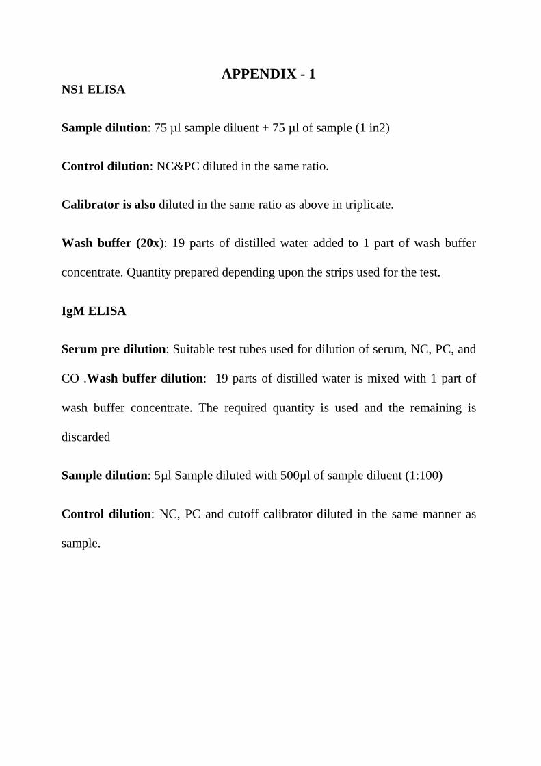
APPENDIX - 1 NS1 ELISA
Sample dilution: 75 µl sample diluent + 75 µl of sample (1 in2)
Control dilution : NC&PC diluted in the same ratio.
Calibrator is also diluted in the same ratio as above in triplicate.
Wash buffer (20x): 19 parts of distilled water added to 1 part of wash buffer
concentrate. Quantity prepared depending upon the strips used for the test.
IgM ELISA
Serum pre dilution: Suitable test tubes used for dilution of serum, NC, PC, and
CO .Wash buffer dilution : 19 parts of distilled water is mixed with 1 part of
wash buffer concentrate. The required quantity is used and the remaining is
discarded
Sample dilution: 5µl Sample diluted with 500µl of sample diluent (1:100)
Control dilution : NC, PC and cutoff calibrator diluted in the same manner as
sample.

IgG ELISA
Serum pre dilution:
1. Serum 10µl + serum diluent 1000µl(1:100 dilution) done
using suitable test tubes.
2. Positive control, negative control and calibrators diluted in the same
manner
Wash buffer dilution: One Part of buffer concentrate added to 19 parts of
distilled water. Total volume prepared as required for the procedure.

APPENDIX-2
Immunochromatography card Test (n=107)
Fever duration
NS1 alone
IgM alone
IgG NS1+IgM IgM+IgG NS1 +IgG
NS1+IgM+IgG
1-5 Days(60)
20 4 1 6
11
2
4
6 – 10 Days(36)
0 7
2 1
25
1 0
> 10 Days(11)
0 1 4
0 3
0 0
Total
20 12 7 7 39
3
4

LIST OF TABLES
1. Seropositivity of Dengue
2. Age and sex wise distribution of dengue positive cases
3. Seasonal distribution of dengue positive cases
4. Clinical presentation of dengue positive cases
5. Clinical classification based on WHO Guidelines
6. Platelet count in Dengue positive cases
7. WBC count of Dengue positive cases
8. Level of haematocrit in Dengue positive cases
9 Association of platelet, HCT and WBC count with DF,DHF and DSS
10. ALT and AST levels in Dengue positive cases
11. USG findings in Dengue positive cases
12. Serological Categorisation of Dengue positive cases
13. Primary Dengue based on duration of fever and serological markers.
14. Secondary dengue based on duration of fever and serological markers
15. Case detection by ICT and ELISA
15(a) Case detection by ICT and ELISA for individual parameters
16. Cross tabulation of ICT and ELISA for NS1
17. Cross tabulation of ICT and ELISA for IgM 18. Cross tabulation of ICT and ELISA for IgG

LIST OF CHARTS
1. Seropositivity of Dengue
2. Age and sex wise distribution of Dengue positive cases
3. Seasonal distribution of Dengue positive cases
4. Clinical presentation of Dengue positive cases
5. Clinical classification of Dengue positive cases
6. Association of platelet, HCT and WBC count with DF,DHF and DSS
7. Serological Categorisation of Dengue positive cases
8. Primary dengue based on duration of fever and serological markers
9 Secondary Dengue based on duration of fever and serological markers
10. Case detection by ICT and ELISA for NS1,IgM ,IgG
11. Sensitivity ,specificity ,PPV and NPV of ICT for NS1,IgM,IgG

LIST OF COLOUR PLATES
1. Dengue Duo combo card kit
2. Dengue NS1 antigen positive
3. Dengue NS1antigen with IgM antibody positive
4. Dengue NS1antigen with IgM & IgG antibody positive
5. Dengue NS1antigen with IgG antibody positive
6. NS1 antigen negative , IgM & IgG antibody positive
7. IgM antibody positive
8. IgG antibody positive
9. Dengue NS1 ELISA kit
10. Dengue IgM ELISA kit
11. Dengue IgG ELISA kit
12. ELISA reader
13. Antigen and antibody mixture of IgM &IgG ELISA
14. Microtitre plate with samples
15. Microtitre plate with samples after substrate addition
16. Samples after addition of stop solution

LIST OF ABBREVIATIONS
ADE- Antibody dependent enhancement
ALT- Alanine aminotransferase
AST- Aspartate aminotrnasferase
Bcell- B lymphocyte
C3a, C5a- complement factors
CFT-Complement fixation test
DEN –Dengue
DENV- Dengue virus
DF- Dengue fever
DHF- Dengue haemorrhagic fever
DSS- Dengue shock syndrome
ELISA- Enzyme linked immunosorbent assay
GB thickening- Gallbladder thickening
HCT- Haematocrit
HAI- Haemagglutination inhibition
HRP-Horse radish peroxidase
IgM- Immunoglobulin M
IgG- Immunoglobulin G
IFN- Interferon
IL- Interleukin
ICT- Immunochromatography
Mab- Monoclonal antibody

NASBA-Nucleicacid sequence based amplification
NT- neutralization
NS1- Nonstructural protein
NPV-Negative predictive value
PPV- Positive predictive value
PD-Primary dengue
RT-PCR- Reverse transcriptase polymerase chain reaction
RANTES- Regulated on activated normal T cell expressed and secreted
SPSS- Statistical package for social sciences
SD-Secondary dengue
T cell- T lymphocyte
TNF- Tumour necrosis factor
TMB- Tetramethylbenzidine
USG- Ultrasonogram

PROFORMA Date: Name of the patient: Age / sex: Ward: ID no: Occupation: Present address: Date of admission: Permanent address: H/o present illness: Fever:
o Onset
o Duration
o Type of fever
o Associated with rash/ myalgia/ arthralgia /abdominal pain/vomiting/headache
o Bleeding tendencies
H/O past illness: - Travel to endemic areas. - Previous h/o fever with hospitalization - Living in endemic areas. Any relevant investigations done outside: H/O treatment: Nature of specimen collected: Date of collection: Investigation done:

PROTOCOL
Selected Fever Cases
Blood samples collected & received in Laboratory
Rapid card test
ELISA
Dengue specific Antigen Dengue Specific IgM & IgG Antibodies
Dengue specific Antigen Dengue Specific IgM & IgG Antibodies
Hematological Markers 1. Platelet count 2. Packed cell volume 3. Total Leucocyte count
Biochemical Markers 1. SGOT 2. SGPT

WORKSHEET
Collect Blood Samples from selected Cases
Separate serum by Centrifugation
Subject to both ICT and ELISA
Categorise ELISA Positive cases into10 and 20 dengue
Compare Case detection by ELISA and ICT for serological markers
Calculate Sensitivity, specificity, PPV, NPV for ICT using ELISA as
Reference
Collect clinical datas, haematological and biochemical profile for
correlation

CONSENT FORM
Dr P.Malini, Post Graduate student in the Department of Microbiology,
Coimbatore Medical College, Coimbatore, is studying on ‘Multivariate Analysis
of Dengue in a Tertiary care Hospital’ regarding.
I am, ___________________________ of age _____ years, sex - M/F,
have been explained about the study. The venepuncture procedure, the risk
involved and complications of the procedure were explained to me in my mother
tongue. I have also been explained that the data obtained herein may be used for
research and publication. I whole heartedly give my consent to participate in the
study.
Place:
Date: Signature

S.No.
IP
/OP Name Age Sex Presenting Complaints Platelet WBC HCT ALT AST USG ELISA
NS1 IgM IgG
1 69212 Saraswathi 28 F
Fr - 10 days ,AbdPain 5
days,arthralgia,myalgia 20,000 3000 17.2 28 30 Normal N P P SEC DF
2 70201 Karthick 16 M
Fr - 4 days,
myalgia,headache 56,000 3000 38.7 27 29 Normal N P P SEC DF
3 73576 Rajkumar 33 M
Fr-12
days,Hemetemesis,Haem
aturia,myalgia,rash 23,000 3200 53.7 52 72 BILPE N P P SEC DHF
4 73482 Ganesh 5 mch Fr - 5days,vomiting,rash 33,000 2000 45.5 25 27 Normal N p P SEC DF
5 73486 Sangamithra 4 fch
Fr - 10 days ,Melena 2
days,headache 56000 3500 34.8 21 25 min PE GB thickening N p P SEC DHF
6 73561 Elan go 11 mch Fr - 2 days ,Arthralgia 43000 3000 24 25 27 Normal P N N PRI DF
7 73506 Vijaya 60 F
Fr -10days,
Alteredsensorium,arthral
gia,maylgia 89000 16100 32.7 45 89 Grade I MKD N p P PRI DF
8 113 Sumaiya 5 fch
Fr - 12DAYS, Melena 3
episodes,rash,epitaxis 43000 3400 25 28 30
GB wall thickning,min.R
PE N p P SEC DHF
9 351 Divyadharshni 21/2 fch
Fr-7
days,Headache,melena 13000 3600 27.3 97 142 hepatomegaly N p P SEC DHF
10 Sanfiya 8 fch
Fr-6
days,Myalgia,vomiting,ra
sh,epistaxis 91000 3000 23.3 31 32 Normal N p P SEC DF
11 795 Sanjay 8 mch
Fr-7 days,Abdmn
Pain,myalgia 19000 6000 24.7 33 35 GB wall thickening,R PE N p P SEC DHF
12 941 Abbas ali 14 M
Fr-1 week,Abdmn Pain 3
days,Vomiting 1
week,Bleeding gums 237000 3100 46 23 27 Mod.ascites N p P SEC DHF

13 548 Ajith 10 mch
Fr-
6days,Myalgia,bleeding
gums 47000 2800 27.6 37 39 Normal N p P SEC DF
14 2236 Mohammed Safix 14 M
Fr-3
days,Arthralgia,myalgia 52000 3500 43 41 43 Normal p p P SEC DHF
15 2767 Neelakandan 5 mch
Fr-5 days,AbdmnPain 3
days,Oliguria 40000 3000 45.5 43 45 GB thickening N p P SEC DHF
16 2512 Gowrav Kumar 11 mch
Fr-6 days,Adbdmn Pain 2
days,Oliguria 3 days 56000 3600 51.1 47 49 GB thickening N p P SEC DF
17 2805 Vanitha 18 F Fr-5 days, Arthralgia 84000 7000 52 51 53 GB thickening p p P SEC DHF
18 2930 Varsha 5 fch
Fr-6 days, Vomiting 3
episode,Abdmn
Pain,Oliguria 35000 3900 46 55 57 GB thickening,RPE, N p P SEC DSS
19 4738 Maran 75 M
Abdmn Pain, Fr-2
days,myalgia 136000 2500 46 59 60 Normal P p P SEC DF
20 4968 Vijayakumar 20 M Fr-3 days,myalgia 20000 3100 41.9 49 44 GB wall thickening,R PE p N N pri DHF
21 4144 Saraswathy 66 F
Fr-7
days,Arthralgia,myalgia 76000 3600 24 25 23 GB thickening N p P SEC DF
22 5765 Jayamani 47 F
Fr-5 days,
Melena,purpurahardpala
tebleeding 50000 3800 24.6 30 31 Normal P N N PRI DHF
23 6163 Samuel 22 M Fr-2 days,Arthralgia 35000 3950 42.2 27 30 R PE P N N PRI DHF
24 61653 Rajendran 27 M
Fr-4 days,Bleeding gums
2 days,myalgia 30000 3800 36.8 27 41 Normal N P P SEC DF
25 9143 Haridharshini 4 fch
Fr,Vomiting 6
days,oliguria 83000 2000 38.2 28 25 0 N p P SEC DF
26 18828 Ansiya 8 fch
Fr-11 days,Vomiting 1
day 53000 3100 38.8 29 22 GB thickening N P P SEC DHF

27 11348 Nuthun 26 M
Fr-5
days,Vomiting,Arthralgia,
Haematuria,Epitaxis 36000 2600 48.6 45 50 R minimal PE P N N PRI DHF
28 11943 Velmurugan 32 M
Fr-4
days,arthralgia,myalgia,b
leeding gums 39000 2000 45.7 22 24 Normal P N N PRI DF
29 68327 Irfanahmed 6 mch Fr-6 days,Myalgia 33000 3910 36 25 26 Thickened GB,,Bil.PE N P P SEC DHF
30 72407 Priyanka 8 fch Fr-5 days,arthralgia 31000 3700 38.5 41 43 - p N N PRI DF
31 Anandkumar 14 M
Fr-
5days,Hemetemesis,myal
gia 55000 3000 40 42 40 Normal P N N PRI DF
32 1E+05 Mansurali 24 M Fr-7 days,Haematuria 66000 3000 45.5 35 30 GB wall thickening
p p P SEC DHF
33 18433 Sagana 5 fch Fr-6 days,myalgia 82000 6000 46 25 27
minimal R pleural
effusion p p P SEC DHF
34 19258 Bathri 3 fch
Fr-11days, Edima 2
days,hemetemesis,rash 48000 3500 46.4 20 25 GB wall thickening N p P SEC DHF
35 9676 Karthick 3 fch Fr-4 days,abd.pain 74000 8000 48 58 55 diffuse GB thickening p N P SEC DHF
36 20502 Vignesh 6 mch
Fr-4
daysvomiting,maylgia 28000 8200 45.8 26 21
minimal R PE,GB wall
thickening p P N PRI DHF
37 20432 Rahul 3 mch Fr-6days,vomiting 27000 4900 37.5 20 25
minimal R PE,GB wall
thickening N p P SEC DHF
38 24426 Vinithkumar 3 mch
Fr-7 days, Abdmn
Pain,Nausea Vomiting 17000 3100 36 26 28 GB wall thickening N p P SEC DHF
39 24481 Prahalad 30 M Fr-6 days,myalgia 49000 4000 48 43 47 Normal N p P SEC DHF

40 26097 Harichander 2 mch Fr-8 days, Abdmn Pain, 60000 5000 33 35 30 - N p P SEC DF
41 2E+05 Ashok kumar 17 M
Fr-8
days,arthralgia,myalgia 37000 3500 42 37 30 GB thickening N p P SEC DHF
42 35207 Balu 36 M Fr-4 days, Arthralgia 45000 3800 33.1 43 45 Hepatomegaly P p P SEC DF
43 35906 Ramesh 38 M
Fr-7 days, Vomiting 1
episode,Myalgia,bleeding
gums 20000 10600 48.5 28 29 GB wall thickening P p P SEC DHF
44 32911 Manoba 9 mch
Fr-5 day Abdmn Pain,
Vomiting,headache,mele
na 37000 8200 41 25 27 GB thickening N p P SEC DHF
45 32910 Jeevasri 10 fch
Fr with Chills,Abdmn Pain
4days,myalgia 31000 2800 33.7 29 25 Bil.PE,min.FF abdomen P N N PRI DHF
46 32075 Meenambigai 6 fch
Fr-4
days,vomiting,myalgia 26000 3200 37.2 28 21 GB wall thcickened P p P SEC DHF
47 7815 Kumaravel 7 mch
Fr-7
days,vomiting,myalgia 49000 4500 32 27 28 Normal N P P SEC DHF
48 7816 Vetrivel 4 mch
Fr-7 days,Abdmn
pain,Vomiting 55000 3200 40 23 25 normal N P P SEC DF
49 33544 Arunvijay 15 M
Fr-11days,
abd.pain,myalgia 17000 3100 42 36 32 min.RPE,Asscites N P P SEC DHF
50 35493 Damodaran 12 M
Fr-12
days,Myalgia,Arthralgia 129000 2200 33.6 27 24 Gb wall edematous N P P SEC DF
51 35095 Saranya 16 F Fr-4 days,artralgia 17000 5700 32.2 45 48
GB edematous,Bil.PEFF
abdomen &pelvis P N N PRI DHF
52 35184 sathish 24 M
Fr-4
days,Myalgia,Arthralgia,h
eadache,vomiting 46000 5000 52.5 30 32 GB wall edematous p p N PRI DF

53 35240 Ashok kumar 8 M
Fr-6days,Vomiting 5
days,Melena,Abdmn Pain 36000 3200 33.7 35 30
GB wall edematous
,Bil.PE,ascites N p P SEC DHF
54 35375 Valliyammal 60 F Fr-5 days, Arthralgia 35000 7000 35 32 30 GB contracted, P N N PRI DF
55 35376 Mohammedhussain 15 M Fr-11days,myalgia 17000 3800 50.8 45 48 GB thickening,
N p P SEC DHF
56 31346 Murugan 32 M
Fr-4
days,dysuria,headache,ar
thralgia 61000 2600 42.5 29 30 - P p N PRI DF
57 35333 Navanethan 4 mch
Fr-11
days,vomiting,myalgia 45000 3500 40.2 28 30 GB thickened N P P SEC DF
58 35236 Selvakrishnan 1 mch Fr-4 days,headache 56000 14000 39.5 32 80 NA p P P SEC DF
59 33510 Logeswaran 19 M
Fr-7 days,
Myalgia,arthralgia 24000 5100 19 25 22 Normal N p P SEC DF
60 32336 Manikandan 32 M
Fr-1 weeks,Arthralgia 10
days 87000 3000 36 42 45 NA N p P SEC DF
61 34418 Shankar 26 M
Fr-2
days,Myalgia,arthralgia 7000 3000 49.4 24 26 Hepatomegaly p P P SEC DF
62 34579 Sivakumar 20 M
Fr-12 days,
Vomiting,Melena 3 days 14000 2900 45.6 28 30 minimal R PE ,Ascites N P P SEC DHF
63 36503 Dharshini 7 fch
Fr-5 days,Refusal of
feeds 3 days,melena 1
day 55000 13400 27.6 32 40 Hepatomegaly N N P SEC DF
64 36331 Jithin Samuel 10 mch Fr-4 days,arthralgia 87000 330000% 33 37 35
hepatomegaly, GB
contracted N N P SEC DF
65 36450 Divya 21 F
Fr-5days, rash, Abdmn
pain 88000 3600 45.9 35 32 GB thickened P N N PRI DHF
66 36394 Vijayaraghavan 1.5 mch
Fr-5 days,Abdmn Pain 4
days 360000 3300 48.8 42 43 GB contracted,
P N N PRI DHF

67 36436 Bhagyalakshmi 18 F
Fr-5 days, Vomiting 3
days,Adbmn pain 4 days 68000 3600 46.9 30 42 GB wall thickening
P N N PRI DHF
68 32482 Amirtha 17 F Fr-10 days,arthralgia 200000 6500 31 25 27 normal N P N PRI DF
69 34794 Amirtham 40 F
Fr-5
days,arthralgia,myalgia 100000 4000 42 43 45 NA P N N PRI DF
70 35040 Priyadharshini 12 fch Fr-5 days,arthralgia 111000 4800 12.7 25 27 hepatomegaly P N N PRI DF
71 37045 Deepika 17 F Fr-3 days,arthralgia 68000 6200 22.5 26 25 normal P N N PRI DF
72 33204 Kelvin 46 M
Fr,Abdmn pain 3
days,myalgia 70000 2500 32 23 25 - p N N PRI DF
73 35331 AyishaRithiga 23 F Fr-3 days,Arthralgia 100000 1500 12.7 25 25 Normal P N N PRI DF
74 34531 Munna 23 M
Fr- 6 days,vomiting,
abdmn pain,arthralgia 16000 6300 33.7 29 27 Normal N N P SEC DF
75 2E+05 Dharshan 1 mch
Fr-5 days, Abdmn
pain,hemetemesis 65000 2500 47 50 53
GB thickening,pleural
effusion,ascites. p N p SEC DSS
76 78957 Kamala 22 F Fr-10 days,Myalgia 27000 3400 32.8 26 21 Normal N P N PRI DF
77 73621 Amudhavalli 22 F
Fr-3
days,Myalgia,Arthralgia 13200 4800 36.9 25 28 NA P p P SEC DF
78 72846 Yamuna 6 fch Fr-5 days,vomiting 113000 5600 28 24 26 ,R PE
p P N PRI DHF
79 7973 Aravivel 12 mch
Fr-5 days,Myalgia,Abdmn
pain,Melena 45000 6000 34 42 43 GBthickening N p P SEC DHF
80 7862 Kalingaraj 13 M Fr-7 days,vomiting 39000 2400 46.6 35 37 GB thickening N p P PRI DHF

81 70488 Jaya 21 F Fr-3 days, 63000 1700 38.5 32 39 Normal N p N PRI DF
82 7389 Mani 34 M
Fr-6
days,Myalgia,Nausea,Vo
miting 14000 3000 35 29 27
-
N p P PRI DF
83 23275 Nitesesh 6 mch Fr-10 days 94000 6000 36 28 29 - N p N PRI DF
84 71821 Dinakaran 8 mch
Fr-15 days,Abdmn Pain 3
days 190,000 9800 30 25 27 Normal N p P PRI DF
85 62920 Poongodi 7 fch Fr-5 days,Abdmn pain, 82000 2300 38.6 27 29 Normal P N N PRI DF
86 7288 Maheswari 10 fch Fr-3 days,myalgia 70000 3000 49.7 28 30 hepatomegaly, PE
P p N PRI DHF
87 32266 Lakshmanan Babu 8 mch Fr-5 days 52000 2000 39 29 30 Normal P N N PRI DF
88 7173 Umadevi 9 fch
Fr-3 days,Melena 1 day,
Oliguria 2 days 34000 1700 48 25 27 FF abdomen P p P SEC DHF
89 31102 Priya 23 F Fr-3 days, 36000 3000 35 28 30 - P N N PRI DF
90 28551 lalitha 15 F Fr- 5 days, 72,000 2000 38 25 27 Normal P N N PRI DF
91 32394 Sathish Kumar 38 M Fr-5 days,Vomiting 60000 5200 46 30 42 minimal R PE P N N PRI DHF
92 67696 Sikinder 35 M
Fr-5 days,Abdmn Pain 2
days,arthralgia 37000 3000 29.4 25 27 Normal
P p N PRI DF
93 68299 Indurekha 13 F Fr - 3 days,arthralgia 57,000 7000 30 33 35 - P p N PRI DF
94 71223 Venkatprabu 8.12 mch
Fr-7 days,Melena 2
days,abdominal pain 29000 3000 45.3 25 29 bil.PE N p N PRI DHF

95 3320 Gopalakrishnan 10 mch
Fr-5 days,Vomiting 3
days, Melena 3 days 73000 3000 35 27 29 ascites
P N N PRI DHF
96 31647 Venkatachalam 30 M
Fr-4 days, Abdmn Pain,
Giddiness,arthralgia 15000 3000 40 29 30 - P N N PRI DF
97 30889 Bharath 10 mch
Fr-5 days, Abdmn
Pain,Melena 80000 3800 46 27 29 pleural effusion
P p P SEC DHF
98 3E+05 Karthick 11 M
Fever 5 days, Headache 5
day,Melena 4 days,
Abdmn Pain 32000 2600 37.5 30 35 bil.PE P p P SEC DHF
99 31346 Murugesan 32 M
Fr-6 days, Abdmn
Pain,Dysuria 4 days 61000 2600 45.5 31 35 pleural effusion
N p N PRI DHF
100 31344 Ashok 10 mch
Fr-4 days, Vomiting,Poor
appetite,Myalgia, Abdmn
pain 35000 3100 57.3 32 30
ascites
P N N PRI DHF
101 33233 Kaviraj 16 M
Fr-7 days Abdmn
pain,arthralgia 73000 2700 42 20 25 pleural effusion
N p P PRI DHF
102 34282 Dilfar 5 mch
Fr-7 days,Vomiting 2
days 22000 3500 46.5 25 27 minimal R PE N p N PRI DHF
103 28819 sakthivel 27 M Fr-6 days,Myalgia 48000 2900 39 23 25 - N p N PRI DF
104 33367 Amritha Iniya 7 fch
Fr-3 days, Vomiting,
Abdmn pain 21000 5600 39.8 43 47 ascites N N P SEC DHF
105 33739 Thangomani 40 F
Fr-2 days,Burning
Micturition,Myalgia 31000 2400 36 25 24 normal p p N PRI DF
106 33755 Subramani 62 M Fr-12days, Myalgia 57000 3000 33 25 27 normal N P P SEC DF
107 33855 NavaneethaKrishnan 12 mch
Fr-5 days,Myalgia,Abdmn
pain,Melena 28000 3500 48 29 30 ascites
N N P SEC DHF
Note: P-Positive, N - Negative, M-Male,F-Female,mch-MaleChild,fch-FemaleChild
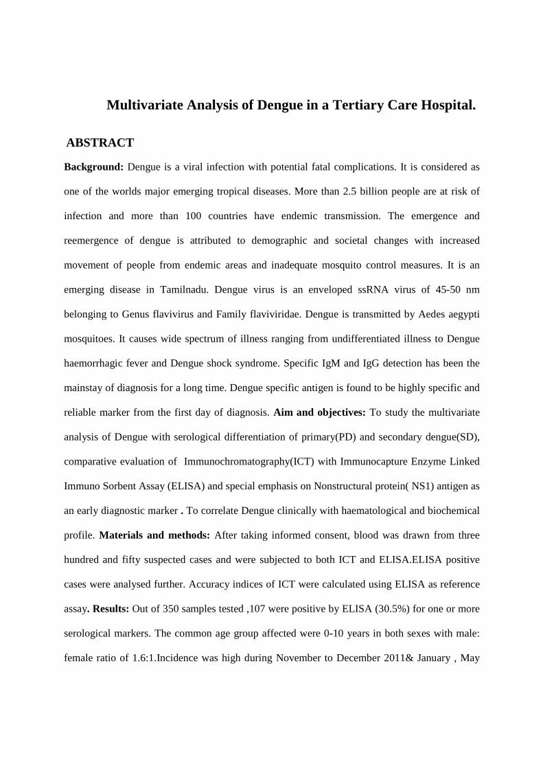
Multivariate Analysis of Dengue in a Tertiary Care Hospital.
ABSTRACT
Background: Dengue is a viral infection with potential fatal complications. It is considered as
one of the worlds major emerging tropical diseases. More than 2.5 billion people are at risk of
infection and more than 100 countries have endemic transmission. The emergence and
reemergence of dengue is attributed to demographic and societal changes with increased
movement of people from endemic areas and inadequate mosquito control measures. It is an
emerging disease in Tamilnadu. Dengue virus is an enveloped ssRNA virus of 45-50 nm
belonging to Genus flavivirus and Family flaviviridae. Dengue is transmitted by Aedes aegypti
mosquitoes. It causes wide spectrum of illness ranging from undifferentiated illness to Dengue
haemorrhagic fever and Dengue shock syndrome. Specific IgM and IgG detection has been the
mainstay of diagnosis for a long time. Dengue specific antigen is found to be highly specific and
reliable marker from the first day of diagnosis. Aim and objectives: To study the multivariate
analysis of Dengue with serological differentiation of primary(PD) and secondary dengue(SD),
comparative evaluation of Immunochromatography(ICT) with Immunocapture Enzyme Linked
Immuno Sorbent Assay (ELISA) and special emphasis on Nonstructural protein( NS1) antigen as
an early diagnostic marker . To correlate Dengue clinically with haematological and biochemical
profile. Materials and methods: After taking informed consent, blood was drawn from three
hundred and fifty suspected cases and were subjected to both ICT and ELISA.ELISA positive
cases were analysed further. Accuracy indices of ICT were calculated using ELISA as reference
assay. Results: Out of 350 samples tested ,107 were positive by ELISA (30.5%) for one or more
serological markers. The common age group affected were 0-10 years in both sexes with male:
female ratio of 1.6:1.Incidence was high during November to December 2011& January , May

,june & July 2012. Clinically seropositives were classified as Dengue fever, Dengue
haemorrhagic fever and Dengue shock syndrome. There was significant association of
thrombocytopenia, HCT and WBC count with Dengue fever, DHF and DSS. The107
seropositives by ELISA were categorized as primary and secondary dengue based on duration of
fever and serological markers. Out of 107, 27 were positive for NS1only, 9 were positive for
IgM alone, only 6 showed IgG alone. More than one marker was detected in the remaining 65
samples .In 48 primary dengue cases NS1 had higher positivity and in secondary dengue
IgM&IgG ratio >1 had higher positivity. Sensitivity, specificity, PPV and NPV of the ICT were
calculated using ELISA as reference for individual markers which showed moderate
performance. Haematological and biochemical profile also showed statistical significance in
association with Dengue fever, DHF and DSS. Conclusion: Inclusion of NSI in the routine
diagnosis of dengue increases the detection rate of cases significantly in the early period.
Irrespective of fever duration, all the three ELISA must be done to rule out Dengue infection like
NS1, IgM, and IgG. Immunocapture ELISA is reliable and sensitive than ICT even though it is
highly specific. Haematological and biochemical tests should also be given importance along
with serological markers for dengue diagnosis and management.
Keywords: Dengue, NS1, PD, SD, ICT, ImmunocaptureELISA.