murdoch research repository · page 2 of 26 accepted manuscript eimeria in l. calcarifer in vietnam...
TRANSCRIPT

MURDOCH RESEARCH REPOSITORY
This is the author’s final version of the work, as accepted for publication following peer review but without the publisher’s layout or pagination.
The definitive version is available at http://dx.doi.org/10.1016/j.vetpar.2011.04.040
Gibson-Kueh, S., Thuy, N.T.N., Elliot, A., Jones, J.B., Nicholls,
P.K. and Thompson, R.C.A. (2011) An intestinal Eimeria infection in juvenile Asian seabass (Lates calcarifer) cultured in Vietnam – A first report. Veterinary Parasitology, 181 (2-4). pp.
106-112..
http://researchrepository.murdoch.edu.au/5336/
Copyright: © 2011 Elsevier B.V.
It is posted here for your personal use. No further distribution is permitted.

Accepted Manuscript
Title: An intestinal Eimeria infection in juvenile Asian seabass(Lates calcarifer) cultured in Vietnam - a first report
Authors: S. Gibson-Kueh, N.T.N. Thuy, A. Elliot, J.B. Jones,P.K. Nicholls, R.C.A. Thompson
PII: S0304-4017(11)00301-3DOI: doi:10.1016/j.vetpar.2011.04.040Reference: VETPAR 5845
To appear in: Veterinary Parasitology
Received date: 11-1-2011Revised date: 27-4-2011Accepted date: 28-4-2011
Please cite this article as: Gibson-Kueh, S., Thuy, N.T.N., Elliot, A., Jones, J.B.,Nicholls, P.K., Thompson, R.C.A., An intestinal Eimeria infection in juvenile Asianseabass (Lates calcarifer) cultured in Vietnam - a first report, Veterinary Parasitology(2010), doi:10.1016/j.vetpar.2011.04.040
This is a PDF file of an unedited manuscript that has been accepted for publication.As a service to our customers we are providing this early version of the manuscript.The manuscript will undergo copyediting, typesetting, and review of the resulting proofbefore it is published in its final form. Please note that during the production processerrors may be discovered which could affect the content, and all legal disclaimers thatapply to the journal pertain.

Page 1 of 26
Accep
ted
Man
uscr
ipt
Eimeria in L. calcarifer in Vietnam Gibson-Kueh et al., 2011 Vetpar-D11-4695 Page 1
An intestinal Eimeria infection in juvenile Asian seabass (Lates calcarifer) cultured in 1
Vietnam - a first report 2
3
S. Gibson-Kueh a*
, N.T.N. Thuy b, A. Elliot
a, J.B. Jones
c, P.K. Nicholls
a, R.C.A. Thompson
a 4
5
a School of Veterinary and Biomedical Sciences, Murdoch University, South St., Murdoch, 6
Western Australia 6150, Australia 7
b Minh Hai Sub-Institute for Fisheries Research, 21-24 Phan Ngoc Hien, Group 3, Ward 6, Ca 8
Mau City, Ca Mau Province, Vietnam 9
c Fish Health Laboratory, Department of Fisheries, 3 Baron-Hay Court, South Perth, Western 10
Australia, 6151, Australia 11
12
13
__________________________________ 14
Correspondence: S. Gibson-Kueh, School of Veterinary and Biomedical Sciences, Murdoch 15
University, South St., Murdoch, Western Australia 6150, Australia. E-mail: 16
18

Page 2 of 26
Accep
ted
Man
uscr
ipt
Eimeria in L. calcarifer in Vietnam Gibson-Kueh et al., 2011 Vetpar-D11-4695 Page 2
ABSTRACT 19
This is the first report of an intestinal Eimeria infection in Asian seabass (Lates calcarifer) at 20
the histopathological and ultrastructural levels. The Eimeria infection was often associated 21
with severe pathology and significant mortality in the absence of other pathogens. This 22
showed that it is an important disease of juvenile L. calcarifer in small scale nurseries in 23
Vietnam. Heavy infection and high prevalence levels of the Eimeria infection are suspected to 24
be linked to the low daily water exchange rates practised in these nurseries. Although 25
systemic iridovirus infection was concurrently observed in some of the fish examined, it was 26
not as consistently present in diseased fish as the Eimeria infection. 27
28
Keywords: Lates calcarifer, Eimeria, systemic iridovirus, pathology 29
30

Page 3 of 26
Accep
ted
Man
uscr
ipt
Eimeria in L. calcarifer in Vietnam Gibson-Kueh et al., 2011 Vetpar-D11-4695 Page 3
1. Introduction 31
Asian seabass or barramundi (Lates calcarifer) is an aquaculture food fish of rapidly 32
growing importance in Australia and Asia. The culture of L. calcarifer is typically divided 33
into specialized operations in hatcheries, nurseries and grow-out farms. Farms are generally 34
small to medium scale though some larger grow out farms recently reported an annual 35
production of 300-400 tonnes (http://www.marineproduce.com/annual_reports.html; 36
http://www.seafoodsource.com/newsarticledetail.aspx?id=4294990500). Thailand and 37
Indonesia are currently the largest producers of cultured L. calcarifer at 15,700 and 4,417 38
tonnes, respectively (http://library.enaca.org/AquacultureAsia/Articles/april-june-2010/8-39
cage-culture-asia.pdf). The emerging L. calcarifer industry in Vietnam depends on the 40
grow-out of juvenile fish from small scale nurseries, from which the samples in this study 41
were taken. 42
Significant diseases limiting the culture of L. calcarifer include viral nervous necrosis 43
(Glazebrook et al., 1990; Munday et al., 1992; Maeno et al., 2004; Parameswaran et al., 44
2008), streptococcosis (Bromage et al., 1999; Creeper and Buller, 2006), flavobacteriosis or 45
tenacibaculosis (Carson et al., 1993; Avendano-Herrera et al., 2006; 46
http://aqua.intervet.com/news/2007-10-10.aspx), pot-belly disease (Gibson-Kueh et al., 2004) 47
and Neobenedenia melleni (Deveney et al., 2001; Ruckert et al., 2008). 48
Piscine apicomplexan parasites exhibiting epicytoplasmic development on host cells 49
may belong to the genera Cryptosporidium, Eimeria, Epieimeria or Goussia (Paperna 1995). 50
These parasitic infections have not been reported in L. calcarifer except for a brief mention of 51
Cryptospordium by Glazebrook and Campbell (1987). Cryptosporidium is typically 52
epicytoplasmic while Eimeria and Goussia may be either epicytoplasmic or intracytoplasmic 53
parasites (Molnar and Baska, 1986; Landsberg and Paperna, 1987; Molnar 1989; Lukes and 54
Dykova, 1990; Szekely and Molnar, 1992; Landsberg 1993; Benajiba et al., 1994; Paperna 55

Page 4 of 26
Accep
ted
Man
uscr
ipt
Eimeria in L. calcarifer in Vietnam Gibson-Kueh et al., 2011 Vetpar-D11-4695 Page 4
and Vilenkin, 1996; Alvarez-Pellitero et al., 1997; Baska 1997; Alvarez-Pellitero and Sitja-56
Bobadilla, 2002; Alvarez-Pellitero et al., 2004; Ryan et al., 2004; Murphy et al., 2009). These 57
apicomplexan parasites are distinguished by the morphology of their oocysts. Oocysts of 58
Eimeria and Epieimeria have four dizoic sporocysts each with a Stieda body or polar plug. In 59
Goussia, oocysts are characterized by four dizoic sporocysts each with a suture line. 60
Cryptosporidium oocysts have four naked sporozoites. (Davies and Ball, 1993; Molnar 2006) 61
Meronts, gamonts and oocysts of piscine Cryptosporidium with a size range of 3-5m 62
are much smaller than corresponding stages of Eimeria and Goussia, in the size range of 5-63
20m. The presence of invaginating feeder organelles at the attachment juncture of 64
Cryptosporidium distinguishes it from the other genera (Valigurova et al., 2008). The 65
attachment organelles of epicytoplasmic Eimeria and Goussia vary ultrastructurally from 66
monopodial to multiple finger-like attachment organelles (Paperna 1991; Benajiba et al. 67
1994; Alvarez-Pellitero et al. 1997; Lukes 1992; Lukes and Stary, 1992). 68
This is the first report of an intestinal Eimeria infection in juveniles of L. calcarifer, at 69
the histopathological and ultrastructural levels. This infection was often associated with 70
severe pathology even in the absence of other significant pathogens, and is therefore a 71
significant disease of L. calcarifer in small scale nurseries in Ca Mau, Vietnam. 72
73
2. Materials and methods 74
2.1 Background information on samples examined 75
Diseased juvenile L. calcarifer 2.5-7cm in body length were sampled from a total of 76
five nurseries in Ca Mau, Vietnam in Jan to Mar 2008, Mar and Dec 2009, and Nov to Dec 77
2010. Fixed tissue samples were sent to Murdoch University, Australia where they were 78
processed for histopathology (181 fish) and transmission electron microscopy (10 fish). 79

Page 5 of 26
Accep
ted
Man
uscr
ipt
Eimeria in L. calcarifer in Vietnam Gibson-Kueh et al., 2011 Vetpar-D11-4695 Page 5
Alcohol fixed oocysts obtained from discharged waste water from culture tanks were also 80
examined. 81
82
2.2 Light microscopy (LM) 83
Tissues were fixed in 10% phosphate buffered formalin for at least 24 h, dehydrated in 84
an ethanol-xylene series and embedded in paraffin wax. Formalin fixed bony tissues were 85
decalcified in 5% nitric acid overnight prior to dehydration and embedded in paraffin wax. 86
5µm tissue sections were dewaxed in xylene, rehydrated in an ethanol series and stained by 87
haematoxylin & eosin (H&E) or Giemsa. 88
89
2.3 Transmission electron microscopy (TEM) 90
Tissues were fixed in 5% glutaraldehyde in phosphate-buffered saline (PBS) at 4oC 91
overnight, washed in several changes of PBS and post-fixed in Dalton’s chrome osmic acid 92
for1 h at 4oC. Fixed tissues were dehydrated through a graded ethanol series to propylene 93
oxide followed by 1 h in 60:40 solution of propylene oxide/epoxy resin, overnight in pure 94
epoxy resin on a rotator and baked in an oven at 60oC for 24 h. Ultra-thin sections were 95
stained with uranyl acetate and lead citrate for viewing on a Philips CM100 Bio TEM. 96
97
3. Results 98
3.1 Field observations made on L. calcarifer nurseries sampled in this study 99
The L. calcarifer nurseries in Ca Mau, Vietnam were mainly small scale with less than 100
five ½- to 1-tonne tanks. These nurseries obtained their fry from hatcheries in Vung Tau or 101
Khanh Hoa Province in Vietnam, or as imported fry from Thailand. Fiber glass or cement 102
tanks were mainly used as holding facilities with static or closed recirculation systems. Less 103
Page 6 of 26
Accep
ted
Man
uscr
ipt
Eimeria in L. calcarifer in Vietnam Gibson-Kueh et al., 2011 Vetpar-D11-4695 Page 6
than 20-30% partial daily water exchange rates were practised. In earthen ponds which were 104
less commonly used, the fry were kept in nets suspended in the water column. Salinity of 105
rearing water ranged from 15 to 25 parts per thousand (ppt). Stocking density varied from 280 106
to 350 fish/m3 water. Fish were fed commercial feed pellets supplemented with coarsely 107
chopped trash fish. The trash fish fed consisted of wild caught fish from the sea. 108
Nurseries stocked 1-3 cm L. calcarifer fry obtained from hatcheries, and grow them 109
on to 7-9 cm body length fish to sell to grow-out farmers. Nursery reared 2.5 to 7.0 cm body 110
length L. calcarifer juveniles were reported to suffer low grade clinical disease soon after 111
stocking, with a cumulative mortality of up to 30% of stocked fish. Clinical signs included 112
fish preferentially hanging at water surface, inappetance, lethargy, darkened bodies, tail rot 113
and scales loss. 114
115
3.2 Histopathology 116
An Eimeria infection was observed in greater than 60% of diseased L. calcarifer 117
sampled from the five nurseries, often as early as the first week post stocking. Fish kept in 118
cement or fiber glass tanks or ponds in salinities of both 15 and 25 ppt were found to be 119
infected with this parasite. Concurrent systemic iridoviral disease was observed in 120
approximately 20% of diseased L. calcarifer examined. Low grade to heavy gill trichodinid 121
infection was sometimes observed but not associated with any significant pathology. 122
The primary infection site of the L. calcarifer Eimeria was the small intestine. Both 123
merogony and gamogony were mainly epicytoplasmic and occurred simultaneously (Figure 124
1). Infection levels varied from light to heavy, often with obliteration of the microvillous 125
brush border. Meronts were much smaller in size than gamonts, and had merozoites arranged 126
in rosettes or in parallel (Figures 1 and 2a). Intracytoplasmic meronts or unusually large 127
meronts with at least 18 merozoites were occasionally observed (Figure 2b). Macrogamonts 128

Page 7 of 26
Accep
ted
Man
uscr
ipt
Eimeria in L. calcarifer in Vietnam Gibson-Kueh et al., 2011 Vetpar-D11-4695 Page 7
with foamy cytoplasm due to the presence of amylopectin granules often outnumbered 129
microgamonts. Microgamonts were smaller than macrogamonts and had peripherally arranged 130
nuclei (Figures 1 and 2c). Mature microgamonts had numerous flagellated microgametes 131
(Figure 2c). Meronts measured 4.8 x 3.5 m (n=5), macrogamonts 13.1 x 7.6 m (n=10) and 132
microgamonts 8.1 x 6.0m (n=5). 133
Sporulated oocysts were very rarely observed in histological tissue sections, in fact in 134
only 1 out of 181 fish examined, and measured 18.5 x 12.3 µm (n = 5). These oocysts in 135
faecal materials within the intestinal lumen had four pairs of sporozoites held loosely within a 136
thin membranous oocyst wall (Figure 3a). Both unsporulated and sporulated oocysts were 137
readily observed in faeces collected from tank bottom and in waste water from rearing tanks 138
by wet mount microscopic examinations (Figures 3b and 3c). Nomarski interference 139
microscopy on alcohol fixed sporulated oocysts showed the absence of Stieda bodies and 140
suture lines. Alcohol fixed sporulated oocysts measured 36.6 x 22.8 µm (n=5). A residual 141
body was present in oocysts, and each pair of sporozoites was held together by a thin 142
sporocyst membrane (Figure 3c). 143
Squamous to cuboidal intestinal epithelium and low grade to severe mononuclear 144
inflammatory infiltrates in the lamina propria were frequently observed in association with the 145
Eimeria infection. The inflammation was sometimes extended into the mucosal epithelium. 146
There were often focal to extensive areas of intestinal mucosal degeneration, necrosis and 147
sloughed necrotic cells in intestinal lumen (Figure 4). Extra-intestinal parasite stages were not 148
commonly observed, except for two macrogamonts in renal tubules from 1 fish. Other 149
abnormalities observed included dermatitis in caudal peduncle, renal glomerular 150
degeneration, moderately reactive spleens with white pulps depleted of leucocytes, and 151
reduced levels of hepatic glycogen stores. 152
153

Page 8 of 26
Accep
ted
Man
uscr
ipt
Eimeria in L. calcarifer in Vietnam Gibson-Kueh et al., 2011 Vetpar-D11-4695 Page 8
3.3 Ultrastructural observations by TEM 154
Parasitic stages were observed within complete parasitophorous envelopes at 155
extracytoplasmic positions on the microvillous brush border of intestinal epithelium. 156
Shortening to loss of microvilli and necrosis of affected intestinal epithelium were often 157
observed. Both meronts and gamonts had finger-like attachment organelles that extended into 158
host cells but were limited to the extracytoplasmic cellular boundaries (Figures 5, 7a, 7b and 159
8). A large number of rodlet cells were often seen in association with these parasitic 160
infections, and sometimes within blood vessels in the intestine (Figure 6). 161
Meronts had merozoites with apical complexes in various stages of formation and up 162
to eight merozoites (Figures 7a and 7b). Macrogamonts had abundant amylopectin granules 163
(Figure 8) while microgamonts had flagellated microgametes with a large residual body 164
(Figure 5). 165
4. Discussions 166
This is the first report of a natural Eimeria infection in L. calcarifer. While it does not 167
supply all the answers, the high prevalence of the Eimeria infection in association with severe 168
pathology showed that it is a significant disease under nursery culture conditions in Ca Mau, 169
one which will need to be managed. Significant pathology was frequently reported in fish 170
with apicomplexan infections (Benajiba et al. 1994; Molnar 2006; Gjurcevic et al., 2008; 171
Morrison et al., 1993; Jendrysek et al., 1994; Hemmer et al., 1998). Although systemic 172
iridoviral disease was also observed, it was not as consistently observed as the Eimeria 173
infection in diseased fish. Nonetheless, iridovirus is a serious pathogen which can co-174
contribute to losses during the culture cycle (Gibson-Kueh et al. 2003). 175
The L. calcarifer Eimeria did not possess the feeder organelles typical of 176
Cryptosporidium but had finger-like attachment organelles very similar to epicytoplasmic 177

Page 9 of 26
Accep
ted
Man
uscr
ipt
Eimeria in L. calcarifer in Vietnam Gibson-Kueh et al., 2011 Vetpar-D11-4695 Page 9
species of Eimeria and Goussia. Although the sporocysts in oocysts examined in this study 178
lack Steida bodies, this was also the case in some previously described piscine Eimeria 179
(Upton et al., 1984; Landsberg and Paperna 1987; Paperna 1995; Molnar 2006). Therefore, 180
we will refer to this parasite as Eimeria until molecular analysis can be conducted. Since 181
sporulated oocysts were rarely observed in tissue sections of Eimeria infected L. calcarifer, it 182
is presumed that sporulation was mainly exogenous. Both unsporulated and sporulated 183
oocysts were readily observed in faecal materials collected from tank bottoms. The L. 184
calcarifer Eimeria oocysts in histological tissue sections were almost half the size of alcohol 185
fixed oocysts obtained from waste water, likely due to the dehydration process used in 186
histology. There is also the possibility that more than one species of Eimeria were involved. 187
A study on Goussia carpelli in common carp suggested the correlation of infection 188
rates to stress and immunosupression (Steinhagen et al., 1998). Depletion of splenic white 189
pulp of leucocytes in diseased L. calcarifer examined in this study is expected to have an 190
impact on their immunity, and may explain the heavy Eimeria infection often observed. The 191
diseased fish examined in this study were sampled during the initial post-stocking period 192
when the fish would be recovering from transport and acclimatization stress. 193
The origin of the Eimeria infection in L. calcarifer from nurseries in Ca Mau is 194
unknown, and warrants further study. The feeding of trash fish is a possible source of 195
infection. The stocking of fish in static or closed recirculation aquaculture systems with 196
relatively low daily water exchange rates (20-30%) would encourage the level of Eimeria 197
infection to build up to the high prevalence observed. Large scale L. calcarifer hatcheries and 198
nurseries in Indonesia, Singapore and Australia practised much higher water exchange rates 199
of 100 to 300% an hour (Schipp et al., 2007; personal observations). Whether the Eimeria 200
infection will persist in older fish as a chronic infection or were present in fish before being 201
stocked in nurseries in Vietnam remains to be elucidated and is vital information for its 202

Page 10 of 26
Accep
ted
Man
uscr
ipt
Eimeria in L. calcarifer in Vietnam Gibson-Kueh et al., 2011 Vetpar-D11-4695 Page 10
effective management. Experimental trials will complement what has been learnt from 203
examination of the naturally infected fish in this study. 204
There are currently no treatment options. The sequestering of these epicytoplasmic 205
parasites in parasitophorus envelopes away from the intestinal lumen and host cell cytoplasm 206
makes it resistant to currently available therapeutic drugs (Sterling 2000). Recent research 207
revealed that addition of proteins produced by Cryptosporidium competitively inhibited their 208
attachment to intestinal epithelium (Tzipori and Ward 2002). A similar approach could be 209
applied for this epicytoplasmic Eimeria. 210
211
Acknowledgement 212
Andrea Valigurova, Masaryk University, and Miloslav Jirku, Institute of Parasitology, Czech 213
Republic, for expert advice. Peter Fallon, EM unit, and Michael Slaven and Gerard Spoelstra, 214
Histology Laboratory, School of Veterinary Science and Biomedical Sciences, Murdoch 215
University, for technical help. 216
217
218

Page 11 of 26
Accep
ted
Man
uscr
ipt
Eimeria in L. calcarifer in Vietnam Gibson-Kueh et al., 2011 Vetpar-D11-4695 Page 11
References 219
Alvarez-Pellitero, P., Sitja-Bobadilla, A., 2002. Cryptosporidium molnari n. sp. 220
(Apicomplexa: Cryptosporidiidae) infecting two marine fish species, Sparus aurata L. 221
and Dicentrarchus labrax L. Int. J. of Parasitol. 32, 1007 - 1021. 222
Alvarez-Pellitero, P., Palenzuela, O., Sitja-Bobadilla, A., 1997. Ultrastructure and 223
cytochemistry study of Eimeria sparis (Protozoa: Apicomplexa) stages from the 224
intestine of gilthead sea bream Sparus aurata L. (Pisces: Teleostei). Parasitol. Res. 83, 225
126-136. 226
Alvarez-Pellitero, P., Quiroga, M.I., Sitjà-Bobadilla, A., Redondo, M.J., Palenzuela, O., 227
Padrós, F., Vázquez, S., Nieto, J.M., 2004. Cryptosporidium scophthalmi n. sp. 228
(Apicomplexa: Cryptosporidiidae) from cultured turbot Scophthalmus maximus. Light 229
and electron microscope description and histopathological study. Dis. Aquat. Organ. 62, 230
133-145. 231
Avendano-Herrera, R., Toranzo, A.E. & Magarinos, B. 2006. Tenacibaculosis infection in 232
marine fish caused by Tenacibaculum maritimum: a review. Dis. Aquat. Organ. 71, 233
255-266. 234
Baska, F., 1997. Epicellular and nodular coccidiosis in the intestine of barbel Barbus barbus. 235
Dis. Aquat. Organ. 29, 49-56. 236
Benajiba, M.H., Marques, A., Lom, J., Bouix, G., 1994. Ultrastructure and sporogony of 237
Eimeria (Syn Epieimeria) Anguillae (Apicomplexa) in the eel (Anguilla anguilla). J. 238
Eukaryotic Microbiol. 41, 215-222. 239
Bromage, E.S., Thomas, A., Owens, L., 1999. Streptococcus iniae, a bacterial infection in 240
barramundi Lates calcarifer. Dis. Aquat. Organ. 36, 177-181. 241

Page 12 of 26
Accep
ted
Man
uscr
ipt
Eimeria in L. calcarifer in Vietnam Gibson-Kueh et al., 2011 Vetpar-D11-4695 Page 12
Carson, J., Schmidtke, L.M., Munday, B.L.,1993. Cytophaga johnsonae: A putative skin 242
pathogen of juvenile farmed barramundi, Lates calcarifer Bloch. J. Fish Dis. 16, 209-243
218. 244
Creeper, J.H., Buller, N.B., 2006. An outbreak of Streptococcus iniae in barramundi (Lates 245
calcarifera) in freshwater cage culture. Aust. Vet. J. 84, 408-411. 246
Davies, A.J., Ball, S.J., 1993. The biology of fish coccidia. Adv. Parasitol. 32, 293-366. 247
Deveney, M.R., Chisholm, L.A., Whittington, I.D., 2001. First published record of the 248
pathogenic monogenean parasite Neobenedenia melleni (Capsalidae) from Australia. 249
Dis. Aquat. Organ. 46, 79-82. 250
Gibson-Kueh, S., Crumlish, M., Ferguson, H.W., 2004. A novel ‘skinny pot-belly’ disease in 251
Asian seabass fry, Lates calcarifer (Bloch). J. Fish Dis. 27, 731–735. 252
Gibson-Kueh, S., Netto, P., Ngoh-Lim, G.H., Chang, S.F., Ho L.L., Qin, Q.W., Chua, F.H.C., 253
Ng, M.L., Ferguson, H.W., 2003. The pathology of systemic iridoviral disease in fish. 254
J. Comp. Pathol. 129, 111-119. 255
Gjurcevic, E., Kozaric, Z., Bambir, S., Petrinec, Z., Kuzir, S., Andrea Gudan, A., Bazdaric, 256
B., 2008. Histological investigations of Eimeria infection in large-scaled gurnards, 257
Lepidotrigla cavillone (Lacepède, 1801) from the Novigrad Sea, Croatia. Acta 258
Parasitologica. 53, 81-84. 259
Glazebrook, J.S., Campbell, R.S.F., 1987. Diseases of Barramundi, (Lates calcarifer) in 260
Australia In: Copeland, J.W., Gray, D.L. (Eds.), Management of wild and cultured sea 261
bass/barramundi (Lates calcarifer). Australian Centre for International Agricultural 262
Research, Canberra, Australia, pp. 204-209. 263
Glazebrook, J.S., Heasman, M.P., de Beer, S.W., 1990. Picorna-like viral particles associated 264
with mass mortalities in larval barramundi, Lates calcarifer Bloch. J Fish Dis. 13, 265
245-249. 266

Page 13 of 26
Accep
ted
Man
uscr
ipt
Eimeria in L. calcarifer in Vietnam Gibson-Kueh et al., 2011 Vetpar-D11-4695 Page 13
Hemmer, N., Steinhagen, D., Drommer, W., Korting, W., 1998. Changes of intestinal 267
epithelial structure and cell turnover in carp Cyprinus carpio infected with Goussia 268
carpelli (Protozoa: Apicomplexa). Dis. Aquat. Organ. 34, 39-44. 269
Jendrysek, S., Steinhagen, D., Drommer, W., Korting, W., 1994. Carp coccidiosis: intestinal 270
histo- and cytopathology under Goussia carpelli infection. Dis. Aquat. Organ. 20, 171-271
182. 272
Landsberg, J.H., 1993. Two new species of coccidian parasites (Apicomplexa, Eimeriorina) 273
from red drum Sciaenops ocellatus. Dis. Aquat. Organ. 16, 83-90. 274
Landsberg, J.H., Paperna, I., 1987. Intestinal infections by Eimeria (s. l.) vanasi n. sp. 275
(Eimeriidae, Apicomplexa, Protozoa) in cichlid fish. Ann. Parasitol. Hum. Comp. 62, 276
283-293. 277
Lukes, J., 1992. Life cycle of Goussia pannonica (Molnar, 1989) (Apicomplexa, 278
Eimeriorina), an extracytoplasmic coccidium from the white bream Blicca bjoerkna.. J. 279
Eukaryotic Microbiol. 39, 484-494. 280
Lukes, J., Dykova, I., 1990. Goussia janae n. sp. (Apicomplexa, Eimeriorina) in dace 281
Leuciscus leuciscus and chub L. Cephalus. Dis. Aquat. Organ. 8, 85-90. 282
Lukes, J., Stary, V., 1992. Ultrastructure of the life cycles stages of Goussia janae 283
(Apicomplexa, Eimeriidae) with X-ray microanalysis of accompanying precipitates. 284
Can. J. Zool. 70, 2382-2397. 285
Maeno, Y., De la Pena, L.D., Cruz-Lacierda, E.R., 2004. Mass mortalities associated with 286
viral nervous necrosis in hatchery-reared sea bass Lates calcarifer in the Philippines. 287
JARQ 38, 69-73. 288
Molnar, K., 1989. Nodular and epicellular coccidiosis in the intestine of cyprinid fishes. Dis. 289
Aquat. Organ. 7, 1-12. 290

Page 14 of 26
Accep
ted
Man
uscr
ipt
Eimeria in L. calcarifer in Vietnam Gibson-Kueh et al., 2011 Vetpar-D11-4695 Page 14
Molnar, K., 2006. Phylum Apicomplexa. In: Woo, P.T.K. (Ed.), Fish Diseases and Disorders, 291
Volume 1, Protozoan and Metazoan Infections, 2nd
Edition. CAB International, UK, pp. 292
183-204. 293
Molnar, K., Baska, F. 1986. Light and electron microscopic studies on Epieimeria anguillae 294
(Leger and Hollande, 1922), a coccidium parasitizing the European eel, Anguilla 295
anguilla. J. Fish Dis. 9, 99-110. 296
Morrison, C.M., Leger, J.P., Morrison, C.A., 1993. Light and electron microscopic study of 297
the pathology and merogony of Goussia gadi (Apicomplexa: Coccidia) in the 298
swimbladder wall of haddock Melanogrammus aeglefinus. Dis. Aquat. Organ. 17, 113-299
125. 300
Munday, B.L., Langdon, J.S., Hyatt, A.D., Humphrey J.D., 1992. Mass mortality associated 301
with a viral-induced vacuolating encephalopathy and retinopathy of larval and juvenile 302
barramundi, Lates calcarifer. Aquaculture 103, 197–211. 303
Murphy, B.G., Bradway, D., Walsh, T., Sanders, G.E., Snekvik, K., 2009. Gastric 304
cryptosporidiosis in freshwater angelfish (Pterophyllum scalare). J. Vet. Diagn. Invest. 305
21, 722-727. 306
Paperna, I., 1991. Fine structure of Eimeria (s. l.) vanasi merogony stages in the intestinal 307
mucosa of cichlid fishes. Dis. Aquat. Organ. 10, 195-201. 308
Paperna, I., 1995. Ultrastructural and developmental affinities of piscine coccidian. Dis. 309
Aquat. Organ. 22, 67-76. 310
Paperna, I., Vilenkin, M., 1996. Cryptosporidiosis in the gourami Trichogaster leeri: 311
description of a new species and a proposal for a genus, Piscicryptosporidium, for 312
species infecting fish. Dis. Aquat. Organ. 27, 95-101. 313

Page 15 of 26
Accep
ted
Man
uscr
ipt
Eimeria in L. calcarifer in Vietnam Gibson-Kueh et al., 2011 Vetpar-D11-4695 Page 15
Parameswaran, V., Rajesh Kumar, S., Ishaq Ahmed, V.P., Sahul Hameed, A.S., 2008. A fish 314
nodavirus associated with mass mortalities in hatchery-reared Asian sea bass, Lates 315
calcarifer. Aquaculture 275, 366-369 316
Ruckert, S., Palm, H.W., Klimpel, S., 2008. Parasite fauna of seabass (Lates calcarifer) under 317
mariculture conditions in Lampung Bay, Indonesia. J. Appl. Ichthyol. 24, 321-327 318
Ryan, U., O’Hara, A., Xiao, L-.H., 2004. Molecular and biological characterization of a 319
Cryptosporidium molnari-like isolate from a guppy (Poecilia reticulata). Appl. 320
Environ. Microbiol., 3761–3765 321
Schipp, G., Bosmans, J., Humphrey, J., 2007. Barramundi Farming Handbook (3rd
Ed.). 322
Department of Primary Industry, Fisheries and Mines, Northern Territory Government, 323
Australia, pp. 23-24 324
Steinhagen, D., Hespe, K., Ellmer, B., Korting, W., 1998. Goussia carpelli (Portozoa: 325
Coccidia) infection in stressed and immunosuppressed common carp Cyprinus carpio. 326
Dis. Aquat. Organ. 34, 199-204 327
Sterling, C.S., 2000. Cryptosporidiosis: the treatment dilemma. Journal of Medical 328
Microbiology. 49, 207-208 329
Szekely, C., Molnar, K., 1992. Goussia trichogaster n. sp. (Apicomplexa: Eimeriidae) 330
infecting the aquarium-cultured golden gourami Trichogaster trichopterus trichopterus. 331
Dis. Aquat. Organ. 13, 79-81. 332
Tzipori, S., Ward, H., 2002. Cryptosporidiosis: biology, pathogenesis and disease. Microbes 333
Infect. 4, 1047-1058. 334
Upton, S.J., Reduker, D.W., Current, W.L., Duszynski, D.W., 1984. Taxonomy of North 335
American fish Eimeriidae. In: National Oceanic and Atmospheric Administration 336
(NOAA) Technical Report NMFS 11, U.S. Department of Commerce, 18 pp 337
Page 16 of 26
Accep
ted
Man
uscr
ipt
Eimeria in L. calcarifer in Vietnam Gibson-Kueh et al., 2011 Vetpar-D11-4695 Page 16
Valigurova, A., Jirku, M., Koudela, B., Gelnar, M., Modry, D., Slapeta, J., 2008. 338
Cryptosporidia: epicellular parasites embraced by the host cell membranes. Int. J. 339
Parasitol. 38, 913-922 340
341
342

Page 17 of 26
Accep
ted
Man
uscr
ipt
Eimeria in L. calcarifer in Vietnam Gibson-Kueh et al., 2011 Vetpar-D11-4695 Page 17
Legends for Figures 343
Figure 1. Eimeria infection on the brush border of intestinal mucosa in L. calcarifer from 344
Vietnam showed increased mononuclear infiltrate in lamina propria (Inf). Meronts (Me) were 345
smaller than gamonts (Ma, Mi), and commonly occurred simultaneously. Meronts often had 346
merozoites arranged in rosettes (Me). Macrogamonts (Ma) with foamy cytoplasm, 347
microgamonts (Mi) with peripherally arranged nuclei and darker basophilic stained 348
trophozoites (T). (H&E) 349
350
Figure 2a-c. Different developmental forms of meronts and microgamonts. (2a) Meronts with 351
merozoites arranged in parallel (Me) and merozoites apparently still within parasitophorus 352
envelopes (z). (Giemsa). (2b) An unusually large meront with at least 18 merozoites (arrows). 353
(H&E) (2c) Microgamonts with peripherally arranged nuclei (*) and microgametes (arrows). 354
(Giemsa) 355
356
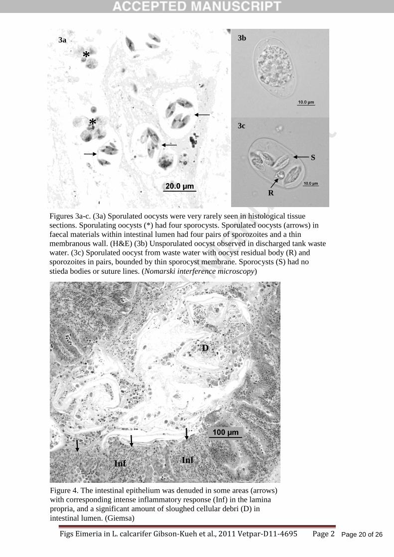
Figures 3a-c. (3a) Sporulated oocysts were very rarely seen in histological tissue sections. 357
Sporulating oocysts (*) had four sporocysts. Sporulated oocysts (arrows) in faecal materials 358
within intestinal lumen had four pairs of sporozoites and a thin membranous wall. (H&E) (3b) 359
Unsporulated oocyst observed in discharged tank waste water. (3c) Sporulated oocyst from 360
waste water with oocyst residual body (R) and sporozoites in pairs, bounded by thin sporocyst 361
membrane. Sporocysts (S) had no stieda bodies or suture lines. (Nomarski interference 362
microscopy) 363
364
Figure 4. The intestinal epithelium was denuded in some areas (arrows) with corresponding 365
intense inflammatory response (Inf) in the lamina propria, and a significant amount of 366
sloughed cellular debri (D) in intestinal lumen. (Giemsa) 367

Page 18 of 26
Accep
ted
Man
uscr
ipt
Eimeria in L. calcarifer in Vietnam Gibson-Kueh et al., 2011 Vetpar-D11-4695 Page 18
Figure 5. Macrogamonts (Ma) with amylopectin granules and microgamonts with 368
microgametes (MiG) at the microvillous brush border of intestines. ‘X’ was presumably a 369
microgamont from which microgametes had been released, thus giving a crenate appearance. 370
Parasitophorous envelopes (PE), residual body (Re) in microgamont. Figure 6. Rodlet cells 371
(Ro) often associated with response to parasitism in fish were observed within blood vessels 372
(bv) in intestines. One of the rodlet cells appeared to be in the process of exiting the blood 373
vessel (*). Vascular endothelial cells (E), fibroblast cells (F) that produced the collagen (C) of 374
blood vessel wall. 375
376
Figure 7a. Meront (Me) with finger-like attachment organelles (fao) and residual body (Re). 377
Merozoites (z) had apical complexes (A) at various stages of formation. Trophozoites (T) and 378
developing meront (dMe) in epicytoplasmic position. Figure 7b. Meront with at least 8 379
merozoites (z) and finger-like attachment organelles (fao). 380
381
Figure 8. Macrogamont with abundant amylopectin (A) granules and finger-like attachment 382
organelles (fao) extended into host cell but limited to the epicytoplasmic boundary. 383
Parasitophorus envelope (PE). 384

Page 19 of 26
Accep
ted
Man
uscr
ipt
Figs Eimeria in L. calcarifer Gibson-Kueh et al., 2011 Vetpar-D11-4695 Page 1
Figure 1. Eimeria infection on the brush border of intestinal mucosa in L.
calcarifer from Vietnam showed increased mononuclear infiltrate in lamina
propria (Inf). Meronts (Me) were smaller than gamonts (Ma, Mi), and
commonly occurred simultaneously. Meronts often had merozoites arranged in
rosettes (Me). Macrogamonts (Ma) with foamy cytoplasm, microgamonts (Mi)
with peripherally arranged nuclei and darker basophilic stained trophozoites
(T). (H&E)
Ma
Mi
Me
T Inf
2a
Me
z
2c
*
10m
2b
10m
Figure 2a-c. Different developmental forms of meronts and microgamonts. (2a)
Meronts with merozoites arranged in parallel (Me) and merozoites apparently
still within parasitophorus envelopes (z). (Giemsa). (2b) An unusually large
meront with at least 18 merozoites (arrows). (H&E) (2c) Microgamonts with
peripherally arranged nuclei (*) and microgametes (arrows). (Giemsa)
B&W Figures Vetpar-D11-4695
Page 20 of 26
Accep
ted
Man
uscr
ipt
Figs Eimeria in L. calcarifer Gibson-Kueh et al., 2011 Vetpar-D11-4695 Page 2
Figures 3a-c. (3a) Sporulated oocysts were very rarely seen in histological tissue
sections. Sporulating oocysts (*) had four sporocysts. Sporulated oocysts (arrows) in
faecal materials within intestinal lumen had four pairs of sporozoites and a thin
membranous wall. (H&E) (3b) Unsporulated oocyst observed in discharged tank waste
water. (3c) Sporulated oocyst from waste water with oocyst residual body (R) and
sporozoites in pairs, bounded by thin sporocyst membrane. Sporocysts (S) had no
stieda bodies or suture lines. (Nomarski interference microscopy)
3b
3a
*
*
3c
R
S
Figure 4. The intestinal epithelium was denuded in some areas (arrows)
with corresponding intense inflammatory response (Inf) in the lamina
propria, and a significant amount of sloughed cellular debri (D) in
intestinal lumen. (Giemsa)
Inf Inf
D

Page 21 of 26
Accep
ted
Man
uscr
ipt
Figs Eimeria in L. calcarifer Gibson-Kueh et al., 2011 Vetpar-D11-4695 Page 3
Figure 5. Macrogamonts (Ma) with amylopectin granules and microgamonts with microgametes (MiG)
at the microvillous brush border of intestines. ‘X’ was presumably a microgamont from which
microgametes had been released, thus giving a crenate appearance. Parasitophorous envelopes (PE),
residual body (Re) in microgamont. Figure 6. Rodlet cells (Ro) often associated with response to
parasitism in fish were observed within blood vessels (bv) in intestines. One of the rodlet cells appeared
to be in the process of exiting the blood vessel (*). Vascular endothelial cells (E), fibroblast cells (F)
that produced the collagen (C) of blood vessel wall.
Ma
X
MiG
PE
10m
Re
5
*
Ro
E
C C
E
F
bv
bv
C
5 µm
6

Page 22 of 26
Accep
ted
Man
uscr
ipt
Figs Eimeria in L. calcarifer Gibson-Kueh et al., 2011 Vetpar-D11-4695 Page 4
2 µm
z
z
fao
7b
Figure 7a. Meront (Me) with finger-like attachment organelles (fao) and residual body (Re).
Merozoites (z) had apical complexes (A) at various stages of formation. Trophozoites (T) and
developing meront (dMe) in epicytoplasmic position. Figure 7b. Meront with at least 8 merozoites (z)
and finger-like attachment organelles (fao).
A
A
Re
T
fao Me
z
z
z
z
T dMe
2m
A 7a
Figure 8. Macrogamont with abundant amylopectin (A) granules
and finger-like attachment organelles (fao) extended into host cell
but limited to the epicytoplasmic boundary. Parasitophorus envelope
(PE).
fao
A
A
2 µm
PE

Page 23 of 26
Accep
ted
Man
uscr
ipt
Figs Eimeria in L.calcarifer Gibson-Kueh et al., 2011 Vetpar-D11-4695 Page 1
Figure 1. Eimeria infection on the brush border of intestinal mucosa in L.
calcarifer from Vietnam showed increased mononuclear infiltrate in lamina
propria (Inf). Meronts (Me) were smaller than gamonts (Ma, Mi), and
commonly occurred simultaneously. Meronts often had merozoites arranged in
rosettes (Me). Macrogamonts (Ma) with foamy cytoplasm, microgamonts (Mi)
with peripherally arranged nuclei and darker basophilic stained trophozoites (T).
(H&E)
Mi
Me
Ma
T Inf
Figure 2a-c. Different developmental forms of meronts and microgamonts. (2a)
Meronts with merozoites arranged in parallel (Me) and merozoites apparently
still within parasitophorus envelopes (z). (Giemsa). (2b) An unusually large
meront with at least 18 merozoites (arrows). (H&E) (2c) Microgamonts with
peripherally arranged nuclei (*) and microgametes (arrows). (Giemsa)
Me
z
2a
2b
10 m
2c
10 m
*

Page 24 of 26
Accep
ted
Man
uscr
ipt
Figs Eimeria in L.calcarifer Gibson-Kueh et al., 2011 Vetpar-D11-4695 Page 2
Figures 3a-c. (3a) Sporulated oocysts were very rarely seen in histological tissue
sections. Sporulating oocysts (*) had four sporocysts. Sporulated oocysts (arrows) in
faecal materials within intestinal lumen had four pairs of sporozoites and a thin
membranous wall. (H&E) (3b) Unsporulated oocyst observed in discharged tank
waste water. (3c) Sporulated oocyst from waste water with oocyst residual body (R)
and sporozoites in pairs, bounded by thin sporocyst membrane. Sporocysts (S) had
no stieda bodies or suture lines. (Nomarski interference microscopy)
3b
*
*
3a
3c
R
S
Figure 4. The intestinal epithelium was denuded in some areas (arrows) with
corresponding intense inflammatory response (Inf) in the lamina propria, and a
significant amount of sloughed cellular debri (D) in intestinal lumen. (Giemsa)
Inf Inf
D

Page 25 of 26
Accep
ted
Man
uscr
ipt
Figs Eimeria in L.calcarifer Gibson-Kueh et al., 2011 Vetpar-D11-4695 Page 3
bv
Figure 5. Macrogamonts (Ma) with amylopectin granules and microgamonts with microgametes (MiG)
at the microvillous brush border of intestines. ‘X’ was presumably a microgamont from which
microgametes had been released, thus giving a crenate appearance. Parasitophorous envelopes (PE),
residual body (Re) in microgamont. Figure 6. Rodlet cells (Ro) often associated with response to
parasitism in fish were observed within blood vessels (bv) in intestines. One of the rodlet cells appeared
to be in the process of exiting the blood vessel (*). Vascular endothelial cells (E), fibroblast cells (F)
that produced the collagen (C) of blood vessel wall.
Ma
X
MiG
PE
10m
Re
5
*
Ro
E
C C
E
F
bv
bv
C
5 µm
6

Page 26 of 26
Accep
ted
Man
uscr
ipt
Figs Eimeria in L.calcarifer Gibson-Kueh et al., 2011 Vetpar-D11-4695 Page 4
Figure 8. Macrogamont with abundant amylopectin (A) granules
and finger-like attachment organelles (fao) extended into host cell
but limited to the epicytoplasmic boundary. Parasitophorus envelope
(PE).
fao
A
A
2 µm
PE
2 µm
z
z
fao
7b
Figure 7a. Meront (Me) with finger-like attachment organelles (fao) and residual body (Re).
Merozoites (z) had apical complexes (A) at various stages of formation. Trophozoites (T) and
developing meront (dMe) in epicytoplasmic position. Figure 7b. Meront with at least 8 merozoites (z)
and finger-like attachment organelles (fao).
A
A
Re
T
fao Me
z
z
z
z
T dMe
2m
A 7a