muscle stretch reflex - sites at penn...
TRANSCRIPT
MUSCLE STRETCH REFLEX: Components and Process Description
Introduction The muscle stretch reflex is an unconscious action caused by the collaboration between a person’s nervous and muscular systems. The reflex acts to prevent damage to muscles and maintain sensory input to the central nervous system. Often, these reflexes are tested during check-‐ups to make sure there are no problems with the patient’s nervous and muscular systems. The reflex happens when a muscle is stretched and causes an unconscious contraction of the stretched muscles to prevent injury. To describe the process, this description will be looking at the knee jerk reflex and will explain how muscle spindles regulate such a reflex. Muscle Spindle Location and Components
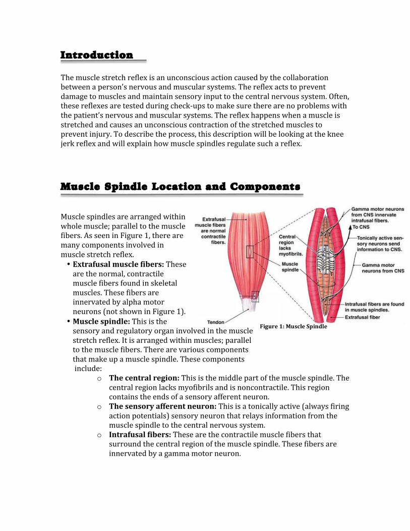
Muscle spindles are arranged within whole muscle; parallel to the muscle fibers. As seen in Figure 1, there are many components involved in muscle stretch reflex. • Extrafusal muscle fibers: These are the normal, contractile muscle fibers found in skeletal muscles. These fibers are innervated by alpha motor neurons (not shown in Figure 1).
• Muscle spindle: This is the sensory and regulatory organ involved in the muscle stretch reflex. It is arranged within muscles; parallel to the muscle fibers. There are various components that make up a muscle spindle. These components include:
o The central region: This is the middle part of the muscle spindle. The central region lacks myofibrils and is noncontractile. This region contains the ends of a sensory afferent neuron.
o The sensory afferent neuron: This is a tonically active (always firing action potentials) sensory neuron that relays information from the muscle spindle to the central nervous system.
o Intrafusal fibers: These are the contractile muscle fibers that surround the central region of the muscle spindle. These fibers are innervated by a gamma motor neuron.
Figure 1: Muscle Spindle
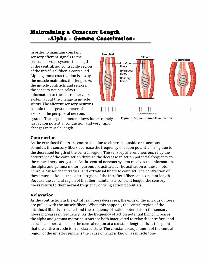
Maintaining a Constant Length -Alpha – Gamma Coactivation-
In order to maintain constant sensory afferent signals to the central nervous system, the length of the central, noncontractile region of the intrafusal fiber is controlled. Alpha-‐gamma coactivation is a way the muscle maintains this length. As the muscle contracts and relaxes, the sensory neuron relays information to the central nervous system about the change in muscle status. The afferent sensory neurons contain the largest diameter of axons in the peripheral nervous system. The large diameter allows for extremely fast action potential conduction and very rapid changes in muscle length. Contraction As the extrafusal fibers are contracted due to either an outside or conscious stimulus, the sensory fibers decrease the frequency of action potential firing due to the decreased length of the central region. The sensory afferent neurons relay the occurrence of the contraction through the decrease in action potential frequency to the central nervous system. As the central nervous system receives the information, the alpha and gamma motor neurons are activated. The activation of these motor neurons causes the intrafusal and extrafusal fibers to contract. The contraction of these muscles keeps the central region of the intrafusal fibers at a constant length. Because the central region of the fiber maintains a constant length, the sensory fibers return to their normal frequency of firing action potentials. Relaxation As the contraction in the extrafusal fibers decreases, the ends of the intrafusal fibers are pulled with the muscle fibers. When this happens, the central region of the intrafusal fiber is stretched and the frequency of action potentials in the sensory fibers increases in frequency. As the frequency of action potential firing increases, the alpha and gamma motor neurons are both inactivated to relax the intrafusal and extrafusal fibers and keep the central region at a constant length. It is at this point that the entire muscle is in a relaxed state. The constant readjustment of the central region of the muscle spindle is the cause of what is known as muscle tone.
Figure 2: Alpha-‐ Gamma Coactivation
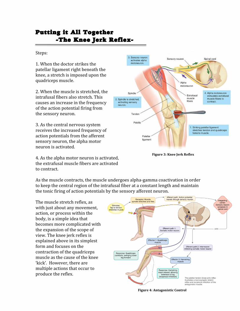
Figure 4: Antagonistic Control
Putting it All Together -The Knee Jerk Reflex- Steps: 1. When the doctor strikes the patellar ligament right beneath the knee, a stretch is imposed upon the quadriceps muscle. 2. When the muscle is stretched, the intrafusal fibers also stretch. This causes an increase in the frequency of the action potential firing from the sensory neuron. 3. As the central nervous system receives the increased frequency of action potentials from the afferent sensory neuron, the alpha motor neuron is activated. 4. As the alpha motor neuron is activated, the extrafusal muscle fibers are activated to contract. As the muscle contracts, the muscle undergoes alpha-‐gamma coactivation in order to keep the central region of the intrafusal fiber at a constant length and maintain the tonic firing of action potentials by the sensory afferent neuron. The muscle stretch reflex, as with just about any movement, action, or process within the body, is a simple idea that becomes more complicated with the expansion of the scope of view. The knee jerk reflex is explained above in its simplest form and focuses on the contraction of the quadriceps muscle as the cause of the knee ‘kick’. However, there are multiple actions that occur to produce the reflex.
Figure 3: Knee Jerk Reflex
The second component to the knee jerk reflex is the relaxation of the hamstring muscle. As seen in Figure 4, the quadriceps and the hamstring muscles can be described as antagonistic. This means that the two muscles work together, in opposition, to produce movement. When the quadriceps contracts, the hamstring must relax and vice verse. Therefore, as the quadriceps muscle is stretched by the doctor’s reflex hammer and the sensory afferent neuron sends the information to the spinal cord, the alpha motor neuron to the quadriceps is stimulated to contract the muscle. However, the alpha motor neuron to the hamstring muscle is inhibited, causing the hamstring to relax.
Muscle Stretch Reflex Overview
The entire purpose of the muscle stretch reflex is to prevent overstretching and tearing the muscle. The knee jerk reflex is just one example of a muscle stretch reflex. Every instance of the muscle stretch reflex is an intricate circuit that contains, at its simplest level, a stimulus, a sensory neuron, a motor neuron, and a target muscle. The body is constantly relaying information about its status and the muscle stretch reflex is just one way the body adjusts itself to maintain its desired state.
Resources Cited Information Gathered From: http://www.ncbi.nlm.nih.gov/books/NBK10809/ Silverthorn, Dee U. Human Physiology: An Integrated Approach 6th Edition Images From: Title Image: http://www.criticalbench.com/muscle_stretching.htm Figure 1: http://isaacsondianaphysiology.wikispaces.com/12+Muscle+Physiology Figure 2: http://matboule.com/you-are-a-muscle-spindle/ Figure 3: http://quizlet.com/10485742/physio-lab-u2-reflex-arc-flash-cards/ Figure 4: http://faculty.pasadena.edu/dkwon/PNS%20and%20propioception/peripheral%20nervous%20system%20and%20propioception_files/textmostly/slide49.html Audience and Scope
This purpose of this document is to provide the reader with a general understanding of how a msucle stretch reflex works. The document will focus on the purpose of muscle spindles, their integration into the muscular system, and their role in the innervation of the nervous and muscular systems. This document would appear in a textbook for biology students or kinesiology students who already have a general understanding of basic nervous system functions and processes. This document assumes that the reader has a general understanding of how neurons and action potentials work.