muscle tuning and preferred movement path – a paradigm shift
TRANSCRIPT

Muscle tuning and preferred movement path – a paradigm shiftBenno M. Nigg1, *, Maurice Mohr1 & Sandro R. Nigg1
1 Human Performance Laboratory, Faculty of Kinesiology, University of Calgary, Canada
* Corresponding author: Human Performance Laboratory, Faculty of Kinesiology, University of Calgary, 2500 University Drive NW, Calgary, Alberta, Canada T2N 1N4, Tel: +1 403 2203436, Email: [email protected]
TA R G E T A R T I C L E
Article History:Submitted 15th September 2017Accepted 5th October 2017Published 3rd November 2017
Handling Editor:Markus TilpUniversity of Graz, Austria
Editor-in-Chief:Martin KoppUniversity of Innsbruck, Austria
A B S T R AC T
In the last 40 years, the scientific debate around running injuries and running shoes has been domi-nated by two paradigms, the ‘impact’ and the ‘pronation’ paradigms. However, the development of running shoe technologies aimed at reducing impact forces and pronation has not led to a decline of running-related injuries. This article recommends to abandon the ‘impact’ and ‘pronation’ para-digms due to a lack of biomechanical and epidemiological evidence and instead suggests a shift to new paradigms: ‘Muscle tuning’ and the ‘preferred movement path’. These paradigms represent new approaches to understanding the biomechanical patterns of each individual runner and how they are controlled by the neuromuscular system. Experimental evidence in support of the ‘mus-cle tuning’ and ‘preferred movement path’ paradigms are presented and discussed regarding their relevance for running performance, injuries, and footwear. Finally, this paper proposes that the con-cept of ‘functional groups’ should be used and further developed to overcome the challenge that groups of individuals respond differently to footwear interventions. First, groups of individuals who behave similarly (functional groups) should be identified. Second, running shoes should be selected to match the characteristics of the identified functional groups in order to optimize the beneficial ef-fects of running shoes for improving running performance and reducing the risk of running injuries.
Keywords:Running injuries – running shoes – impact forces – pronation – functional groups
Introduction
In the last about 40 years running and running shoe discussions were dominated by two paradigms, the ‘impact’ and the ‘pro-nation’ paradigms. This paper will critically review these two paradigms and will suggest that they should be abandoned because there is not enough epidemiological and functional evidence to support them. In addition, this paper will also propose some new paradigms replacing the old paradigms of ‘cushioning’ and ‘pronation’, and a further suggestion for how to methodologically and conceptually investigate running per-formance and running injuries. Finally, this paper proposes that the concept of “functional groups” should be used and further
Citation:Nigg, B. M., Mohr, M. & Nigg, S. R. (2017). Muscle tuning and preferred movement path – a paradigm shift. Current Issues in Sport Science, 2:007. doi: 10.15203/CISS_2017.007
developed to understand running, running performance and running injuries.
The Impact force paradigm
An impact occurs as a result of a collision between two objects. In heel-toe running, an impact occurs because of the collision between the heel of the foot and the ground. In forefoot run-ning, the impact occurs because of the collision between the forefoot and the ground. In heel-toe running the impact force peak is a result of the deceleration of the foot and part of the leg. In forefoot running, the impact peak is a result of the decel-
Current Issues in Sport Science 2 (2017)
2017 I innsbruck university press, InnsbruckCurrent Issues in Sport Science I ISSN 2414-6641 I http://www.ciss-journal.org/Vol. 2 I DOI 10.15203/CISS_2017.007
OPEN ACCESS
B. M. Nigg, M. Mohr & S. R. Nigg Paradigm shift in running
CISS 2 (2017) November 2017 I Article 007 I 2
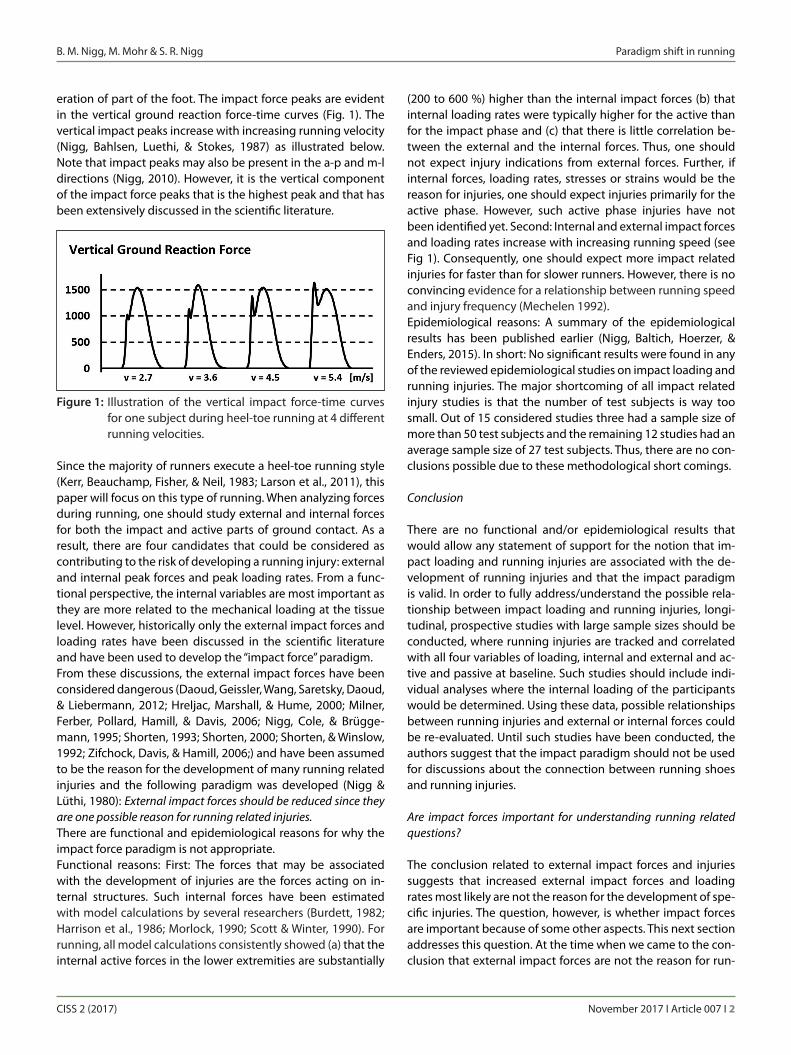
eration of part of the foot. The impact force peaks are evident in the vertical ground reaction force-time curves (Fig. 1). The vertical impact peaks increase with increasing running velocity (Nigg, Bahlsen, Luethi, & Stokes, 1987) as illustrated below. Note that impact peaks may also be present in the a-p and m-l directions (Nigg, 2010). However, it is the vertical component of the impact force peaks that is the highest peak and that has been extensively discussed in the scientific literature.
Since the majority of runners execute a heel-toe running style (Kerr, Beauchamp, Fisher, & Neil, 1983; Larson et al., 2011), this paper will focus on this type of running. When analyzing forces during running, one should study external and internal forces for both the impact and active parts of ground contact. As a result, there are four candidates that could be considered as contributing to the risk of developing a running injury: external and internal peak forces and peak loading rates. From a func-tional perspective, the internal variables are most important as they are more related to the mechanical loading at the tissue level. However, historically only the external impact forces and loading rates have been discussed in the scientific literature and have been used to develop the “impact force” paradigm.From these discussions, the external impact forces have been considered dangerous (Daoud, Geissler, Wang, Saretsky, Daoud, & Liebermann, 2012; Hreljac, Marshall, & Hume, 2000; Milner, Ferber, Pollard, Hamill, & Davis, 2006; Nigg, Cole, & Brügge-mann, 1995; Shorten, 1993; Shorten, 2000; Shorten, & Winslow, 1992; Zifchock, Davis, & Hamill, 2006;) and have been assumed to be the reason for the development of many running related injuries and the following paradigm was developed (Nigg & Lüthi, 1980): External impact forces should be reduced since they are one possible reason for running related injuries. There are functional and epidemiological reasons for why the impact force paradigm is not appropriate. Functional reasons: First: The forces that may be associated with the development of injuries are the forces acting on in-ternal structures. Such internal forces have been estimated with model calculations by several researchers (Burdett, 1982; Harrison et al., 1986; Morlock, 1990; Scott & Winter, 1990). For running, all model calculations consistently showed (a) that the internal active forces in the lower extremities are substantially
(200 to 600 %) higher than the internal impact forces (b) that internal loading rates were typically higher for the active than for the impact phase and (c) that there is little correlation be-tween the external and the internal forces. Thus, one should not expect injury indications from external forces. Further, if internal forces, loading rates, stresses or strains would be the reason for injuries, one should expect injuries primarily for the active phase. However, such active phase injuries have not been identified yet. Second: Internal and external impact forces and loading rates increase with increasing running speed (see Fig 1). Consequently, one should expect more impact related injuries for faster than for slower runners. However, there is no convincing evidence for a relationship between running speed and injury frequency (Mechelen 1992). Epidemiological reasons: A summary of the epidemiological results has been published earlier (Nigg, Baltich, Hoerzer, & Enders, 2015). In short: No significant results were found in any of the reviewed epidemiological studies on impact loading and running injuries. The major shortcoming of all impact related injury studies is that the number of test subjects is way too small. Out of 15 considered studies three had a sample size of more than 50 test subjects and the remaining 12 studies had an average sample size of 27 test subjects. Thus, there are no con-clusions possible due to these methodological short comings.
Conclusion
There are no functional and/or epidemiological results that would allow any statement of support for the notion that im-pact loading and running injuries are associated with the de-velopment of running injuries and that the impact paradigm is valid. In order to fully address/understand the possible rela-tionship between impact loading and running injuries, longi-tudinal, prospective studies with large sample sizes should be conducted, where running injuries are tracked and correlated with all four variables of loading, internal and external and ac-tive and passive at baseline. Such studies should include indi-vidual analyses where the internal loading of the participants would be determined. Using these data, possible relationships between running injuries and external or internal forces could be re-evaluated. Until such studies have been conducted, the authors suggest that the impact paradigm should not be used for discussions about the connection between running shoes and running injuries.
Are impact forces important for understanding running related questions?
The conclusion related to external impact forces and injuries suggests that increased external impact forces and loading rates most likely are not the reason for the development of spe-cific injuries. The question, however, is whether impact forces are important because of some other aspects. This next section addresses this question. At the time when we came to the con-clusion that external impact forces are not the reason for run-
Figure 1: Illustration of the vertical impact force-time curves for one subject during heel-toe running at 4 different running velocities.

B. M. Nigg, M. Mohr & S. R. Nigg Paradigm shift in running
CISS 2 (2017) November 2017 I Article 007 I 3
ning related injuries we went back and studied the movement of the lower extremities during landing. We knew at that time that running shoes that lead to different impact forces produce different comfort feelings (Miller, Nigg, Wen, Stefanyshyn, & Nurse, 2000). Thus we concluded that there should be differ-ences in kinematics, kinetics, muscle activity or some other running specific variables when running in different shoes. The first (surprising) result of this renewed approach was that the soft tissue compartments of the lower extremities did not vi-brate substantially as would be expected for a freely oscillating system. The soft tissue compartments are made up of muscles and other non-active materials. If they are vibrating less than expected, we suggested that they must be damped. Active damping, however, could only be provided through muscles and it has been demonstrated that muscles are quite good in doing this (Wilson, McGuigan, Su, & van den Bogert, 2001). Thus, we proposed that muscles are used to damp the unwant-ed and possibly excessive vibrations of soft tissue compart-ments. Experimental results showed that soft tissue compart-ments were vibrating systems, which, in a first approximation, could be characterized with a natural frequency and a damping coefficient (Wakeling & Nigg, 2001). Note that: (a) The natural frequencies and damping coefficients may typi-
cally be different (often small differences) in the three axis directions, which may produce beat effects in the move-ment of soft tissue compartments (superposition of two oscillations with close frequencies).
(b) The natural frequencies and damping coefficients are in-fluenced by the level of muscle activation. The differences between the natural frequencies and the damping coeffi-cients between a totally relaxed and a maximally contract-ed quadriceps and triceps surae were close to 100 %.
When studying the reaction of vibrating systems one often thinks of resonance phenomena. To analyze the question whether resonance plays a role during human locomotion one should consider mechanical model calculations as well as experimental results. The question to be answered is whether having shock input signals with a frequency close to the natu-ral frequency of a soft tissue compartment will affect the prepa-ration and execution of locomotion differently compared to an input frequency further away from the natural frequency of the soft tissue compartments.
Model considerations
Resonance occurs when a mechanical vibrating system is ex-posed to a continuous vibration input with the same frequency as the natural frequency of the vibrating system. However, it has been proposed, using a simple mechanical spring-damper model for a shock type input, that no resonance phenomena will take place (Kaiser, 2016). Thus, the author of this work sug-gests that muscle tuning doesn`t occur during heel-toe run-ning and that changes in EMG are rather an effect of changes in the landing geometry of the foot/shoe. Looking at the human
locomotor system as a purely mechanical system one can ar-gue that the impact related oscillations are completely damp-ened before the next shock occurs. In this case, resonance should not be a problem. In the situation, however, where this is not the case, where vibrations are still existing, one should expect resonance phenomena. That may be especially true for fast running and/or for subjects with a low muscle tonus. How-ever, to understand this question, the human body should be considered as a neuro-muscular control system as illustrated in the next paragraphs.
Experimental evidence
In an experiment using a vibration platform that produced a shock-type force signal this question has been addressed (Wakeling, Nigg, & Rozitis, 2002). In this experiment, the sub-ject was standing on the toes while exposed to specific force shock inputs (Fig 2).
The results of this experiment show that the input force sig-nal with a frequency slightly lower than the natural frequency (top signal) produces a different reaction (second signal from top) than the input force signal with a frequency substantially higher than the natural frequency of the soft tissue compart-ment (bottom two signals). The acceleration of the soft tissue compartment closer to the natural frequency of the soft tis-sue compartment is immediately damped while the accelera-tion for the input frequency farther away is not damped at all.
Figure 2: Hamstring acceleration as a function of a shock force input while standing on the toes on a vibra-tion platform. The natural frequency of the soft tis-sue compartment “hamstrings” was determined as 12.6 Hz. The force input signals were single dis-placements at frequencies of 10.0 Hz (signal 1) and 17.1 Hz (signal 3). The corresponding accelerations of the hamstring soft tissue compartment are just below the input acceleration signals of the vibration plate (signal 2 and 4). (Derived from data from Wakel-ing et al., 2002).

B. M. Nigg, M. Mohr & S. R. Nigg Paradigm shift in running
CISS 2 (2017) November 2017 I Article 007 I 4
• Theeffectsofmuscletuningshouldbeseenintheperfor-mance, fatigue, and comfort characteristics of specific im-pact/subject combinations.
Experimental evidence for “muscle tuning” for continuous oscil-lations in a quasi-static situation has been provided earlier (di Giminiani et al., 2015; Nigg, 2010; Perchthaler et al., 2013; Wake-ling et al., 2002;). The results show a high correlation between the frequency response and the muscle activity response, a result that would have been predicted based on the new para-digm.Experimental evidence for an actual running situation is more difficult to provide. It has been attempted earlier (Boyer & Nigg, 2004) and it was shown that muscle activity is in fact tuned in response to running conditions that produce different impact scenarios (e.g. shoes with different midsole hardness). How-ever, the results could be interpreted in different ways. One in-terpretation for the change in EMG activity could be that when running in shoes that lead to higher loading rates and the input signal frequency approaches the natural frequency, the muscle activity increases. Another interpretation of the results could be that when changing the shoe characteristics one changes the joint moments (especially for the ankle joint), which may demand a change in muscle activity. The current data don‘t support one or the other interpretation. More research is re-quired to answer this question.
The Cirque du Soleil story (from Nigg, 2010, p. 59-61)
In 1997, Cirque du Soleil had an injury problem with one of its touring troupes. At any time, about one quarter (25%) of its performing staff was injured and unable to perform. The typi-cal problems were tendon insertion injuries and the affected population was primarily supporting actors who had to run and jump frequently. The jumps and runs were moderate, and the landings were not after extreme performances. Boris Verkhovsky, the head coach of Cirque du Soleil, speculat-ed that the stage surface might be the source of these injuries and contacted us for help. We analyzed the problem and spent three days in California where this specific group of the Cirque du Soleil was stationed at the time.
As muscle activity is changing as a reaction of different input signals, these experimental results suggest that the human locomotor system assesses the frequency components of the input signal and reacts by damping when they are too close to the resonance frequency of the soft tissue compartment. These results are in agreement with more recent, similar experi-ments (Di Giminiani, Masedu, Padulo, Tihanyi, & Valenti, 2015; Perchthaler, Horstmann, & Grau, 2013; Pollock, Woledge, Mills, Martin, & Newham, 2010). In consequence, a purely mechanical consideration of the corresponding effects is not appropriate and that maybe the neuro-motor control aspect must be con-sidered together with the purely mechanical effect. However, it is also evident that there is much more research needed to un-derstand these phenomena completely. The experiments were quasi-static and the models were purely mechanical. Oscilla-tions can be influenced by changing the natural frequency or by changing (increasing) the damping. In all the published and not published results of our group (Boyer & Nigg, 2004; Enders et al., 2012; Nigg, 2010; Wakeling et al., 2002) the strategy to increase the damping was the preferred strategy when com-pared to shifting of the natural frequency. Thus, it is suggested that damping is one of the preferred strategies when dealing with unwanted oscillations of soft tissue compartments. In summary:• Softtissuecompartmentsofthehumanlocomotorsystem
are vibrating systems that can be described with a natural frequency and a damping characteristic.
• The damping of the soft tissue compartments is differ-ent for input signals close to compared to far away from the natural frequency of the soft tissue compartment. The damping is higher for input signals close to the natural fre-quency.
• Dampingisthepreferredstrategyforthereductionofsofttissue compartment oscillations as opposed to shifting the natural frequency.
• Dampingcanbeinfluencedbychangingtheactivationofthe involved muscles.
Muscle Tuning – A New Paradigm
Based on these considerations, a new paradigm for under-standing the reactions of the human locomotor system to re-petitive impact forces is proposed (from Nigg, 2010, p. 54):• Impact forces are an input signal characterizedby ampli-
tude, frequency, and time.• Thesesignalsaresensedand,ifnecessary,theCNSresponds
by adjusting (tuning) the activation of corresponding mus-cle groups.
• Tuningoccurstominimizesoft-tissuevibrations.• Theeffectsofmuscletuningarehighwhentheinputfre-
quency and natural frequency of a specific soft-tissue com-partment are close.
• Theeffectsaresubjectspecificanddependonthecharac-teristics of every single soft-tissue compartment.
Figure 3: Schematic construction of the stage with an illustra-tion of the possible deflection of the top surface.

B. M. Nigg, M. Mohr & S. R. Nigg Paradigm shift in running
CISS 2 (2017) November 2017 I Article 007 I 5
Pronation
Pronation: inwards rotation of the foot about its subtalar joint axis
Supination: outwards rotation of the foot about its subtalar joint axis
Eversion: inwards rotation of the foot about a longitudinal foot axis
Inversion: outwards rotation of the foot about a longitudinal foot axis
The subtalar joint axis is a functional axis associated with one anatomical joint, the subtalar joint. The longitudinal foot axis is a theoretically constructed axis not associated with one speci-fic anatomical joint. Experimentally, pronation and supination are difficult to determine (van den Bogert, Smith & Nigg, 1994). For this reason, experiments quantifying foot rotations have usually quantified eversion and inversion. For this paper the measured values discussed are always foot in- and eversion. Most studies concentrate on foot eversion, which is speculated to be a surrogate measure of foot pronation.“Pronation” is a variable that was of interest for foot orthopae-dics, podiatrists and orthotists for a long time. It was discussed long before the running boom and “excessive” pronation was typically considered as the reason for many injuries. This con-ceptual thinking was probably influenced by the fact that there is a movement coupling between the calcaneus and the tibia (Hicks, 1953; Hintermann, Nigg, Sommer, & Cole, 1994; Lund-berg, Svensson, Bylund, Goldie, & Selvik, 1989; Nawoczenski, Cook, & Saltzman, 1995; Nigg, Cole, & Nachbauer, 1993; Stacoff et al., 2000; Wright, Desai, & Henderson, 1964). Pronation of the foot is associated with internal rotation of the tibia and it was commonly assumed that large pronation would produce a high loading condition at the knee joint.Based on such considerations the “pronation paradigm” for running shoes was formulated (Nigg & Lüthi, 1980). It stated that foot pronation (foot eversion) should be minimized since it is a possible reason for running related injuries. There are several reasons why the “pronation” paradigm should be considered with caution: (a) It is difficult to quantify “prona-tion”, (b) “pronation” is a natural movement and (c) many epide-miological results don’t support the paradigm.
Problems with the quantification of foot eversion/pronation
Foot eversion has been determined in many static and dynamic ways. Static measures for foot eversion include (a) Rearfoot angle = angle between the calcaneus and the
ground (g), (b) Achilles tendon angle = angle between the calcaneus and
the lower leg (b) (c) FPI-6 index = a number based on 6 different assessments
of the foot (Redmond, Crosbie, & Ouvrier, 2006; Keenan, Redmond, Horton, Conaghan, & Tennant, 2007),
(d) Navicular drop
The stage surface (Fig. 3) was constructed of a frame of solid and stiff beams at about 35 cm on centre. The beams were cov-ered with a pliable material that allowed deflections of up to 2 cm when landing in the centre between the beams and deflec-tions of less than 0.1 cm when landing on a beam.At the time of the analysis, we had already developed our “muscle tuning paradigm.” Thus, we speculated that when the athletes/artists landed on the stage surface, they pre-activat-ed the muscles of the soft-tissue compartments of the lower extremities (e.g., triceps surae, quadriceps, and hamstrings). The pre-activation occurs based on the athlete’s expectation about the landing condition. One major goal of pre-activation is to minimize the vibration of the soft-tissue compartments of the lower extremities. If one cannot pre-activate the muscles properly, these soft-tissue packages may oscillate substantially, since resonance effects may occur. In resonance situations, the muscle-tendon units may be exposed to high forces, which may be the reason for possible insertion problems.Based on such considerations, we concluded that the non-uniform deflections of the stage surface produced a situation in which the artists could not prepare themselves for the land-ing by “tuning” their muscles to avoid excessive vibrations of the soft-tissue compartments. We proposed that the stage be changed to a much harder but uniform surface. The construc-tion was stiffened and the new surface was uniform (but hard) over the whole stage. This way, the artists knew what to expect for the landing and could prepare (tune) their muscles accord-ingly. The result was that the high number of injuries quickly returned to a normal level (2 to 3%), and the artistic work con-tinued as programmed.Although this story provides only anecdotal evidence, in terms of the muscle tuning paradigm, it is, in our view, stunning. It would be difficult to explain the results of this story with any-thing other than the muscle tuning concept.
Relevance for footwear
If the muscle tuning paradigm is correct this would suggest that running shoes can influence the muscle activity before and during ground contact. High muscle activity could mean (a) increased energy used during a running cycle and/or (b) less comfort during the locomotion activity. Thus, the main effects of this paradigm would not be with respect to running injuries but rather with respect to performance and comfort. Recently one sport shoe company decided to develop pro-ducts based on the paradigm of “muscle tuning”.
Research on the topic of muscle tuning is still in its infancy. Strategies to minimize muscle tuning activities are not well understood. The most obvious approach is to change the fre-quency of the input signal by changing (a) the material prop-erties of the midsole and/or (b) by changing the shape of the heel. However, there may be other approaches that have a positive effect that are not known right now.

B. M. Nigg, M. Mohr & S. R. Nigg Paradigm shift in running
CISS 2 (2017) November 2017 I Article 007 I 6
(e) Footprint analysis(f ) Subjective assessment of sales people in stores(g) Subjective assessment of clinicians in clinics
Dynamic measures for foot eversion include(h) Max. Rearfoot angle (gmax), (i) Change of Rearfoot angle in a defined time interval (Dg10,
Dgtot)(k) Max. Achilles tendon angle (bmax)(l) Change of Achilles tendon angle in a defined time interval
(Db10, Dbtot)(m) Footprint analysis(n) Inertial measurement unit (IMU) algorithms
To make the situation even more complicated, measurements can be done in shoes or barefoot.One can argue about the value of each of these variables. Some scientists suggest that the FPI-6 Index is a good assessment of pronation. Others prefer a dynamic assessment of pronation. However, a gold standard for the assessment of pronation/eversion does currently not exist. In addition, there seems to be little correlation between the different assessment variables. For instance, it has been shown (Stefanyshyn et al., 2003) that there is little correlation between subjective assessments in stores and assessments while running barefoot and/or running in shoes (Fig. 4). In the below example, of the 20 self-declared
male pronators, 14 were declared pronators by a store clerk, 6 were declared pronators based on a biomechanical assessment in shoes and 3 based on a biomechanical assessment barefoot. Furthermore, an analysis of previously collected data (Nigg, Vi-enneau, Smith, Trudeau, Mohr, & Nigg, 2017) demonstrated a lack of correlation between the Achilles tendon angle during standing and the change of the Achilles tendon angle from minimum to maximum during running for both a barefoot and a minimalist shoe condition (Fig. 5). Additionally, all other correlations between static and dynamic variables were small (all R2 < 0.2). Thus, there seems to be no significant correlation
between many of the used static and dynamic foot pronation/eversion variables. In other words, the variables used in most of the studies assessing “foot pronation” describe different as-pects of “foot pronation” and it is unknown whether they de-scribe foot pronation at all. Consequently, results from studies using different variables for assessing rearfoot eversion (“foot pronation”) should, conceptually, show different results with respect to type of injuries and/or injury frequencies which may or may not be related to these variables.
Natural movement and variability of runners
Another reason why the old “pronation” paradigm should be considered with caution is the fact that “pronation” is a natu-ral movement during gait (Shorten & Mientjes, 2011). This in-dicates that some pronation is healthy, natural, and necessary for locomotion, and the question should focus on the optimal amount of pronation instead of trying to reduce pronation to a minimum. The question of optimal pronation is also likely subject de-pendent as different subjects 1) have different ranges of pro-nation, and 2) have different kinematic adaptations to product interventions. An example of this was a study that investigated the occurrence of injuries in female runners, when exposed to different running shoe conditions (Ryan, Valiant, McDonald & Taunton, 2011). Regardless of foot posture type (neutral, pro-nated or highly pronated), one shoe type (motion control) re-ported the highest level of pain for runners. The investigators concluded that providing footwear interventions based on foot type, as is done in many shoe stores, may be both too sim-plistic and potentially cause unnecessary injuries.
Epidemiological results
Most epidemiological studies that discuss the association be-tween “pronation“ and running injuries have the same short-
Figure 4: Venn diagram (N=34) of the relationship between “self-declared pronators”, “barefoot pronators” and “shod pronators” (from Stefanyshyn et al., 2003, with permission).
Figure 5: Correlation coefficient for a comparison between a static and a dynamic pronation variable used in the literature based on previously collected data (Nigg et. al., 2017) for 34 subjects for a barefoot and a minimal-ist shoe condition.

B. M. Nigg, M. Mohr & S. R. Nigg Paradigm shift in running
CISS 2 (2017) November 2017 I Article 007 I 7
coming as the epidemiological studies related to impact load-ing: The sample sizes are too small. However, there are two epidemiological studies that have large sample sizes, which will be discussed in the following. The first study to be discussed in more detail (Nielsen et al., 2014) assessed foot posture of novice runners with a static measure-ment and grouped the 1854 feet of the 927 participants into very supinated (FPI6 < -3; N = 53), supinated (FPI6 = -3 to +1; N = 369), neutral (FPI6 +1 to +7; N = 1292), pronated (FPI6 7 to +10; N = 122) and very pronated (FPI >+10; N = 18). Their epidemio-logical results after a one year period of running showed sig-nificantly less injuries per 1000 km of running for the pronated group compared to the neutral group. Thus, the interpretation of this result would be that “pronation” as assessed with a static calcaneus position measure is not an injury predictor. Based on these results, one may even speculate that ‘pronation’ reduce the likelihood of sustaining running related injuries.A second notable finding of this study is that excessive prona-tors only made up about 1% of the study participants. For this group, the injury rates were the highest, but due to the small number of over-pronators (18 out of 1854), it was not a signifi-cant result. From these results, it can be concluded that 1) pro-nation may be a natural and healthy component of locomotion, 2) the number of “over-pronators” is actually very small, and is likely overestimated in running shoe stores, and 3) for this 1% of the population, the excessive pronation may be a mechanism for sustaining an injury. This is in the view of the authors the first epidemiological study on foot posture type and injuries with an adequate sample size. There are two critical comments about this study: The foot posture assessment was done statically, which is, in the view of the authors, not ideal. Secondly, subjects with orthotics were excluded from the study, which may have shifted the pro-supination distribution. However, the result is nevertheless interesting and contrary to all expectations.The second study to be discussed in more detail (Teyhen et al., 2013) analyzed the relationship between foot type and medi-cal costs associated with lower extremity musculo-skeletal in-juries in a military setting. They collected information from 668 military participants over a period of 31 months. Static foot posture was assessed using the FPI-6 index. The explicit and im-plicit results of this study showed (a) that the injury frequency was about the same (no significant differences) for all foot type groups with 49% for highly supinated, 55% for supinated, 48% for neutral, 51% for pronated and 51% for highly pronated feet (note, that these numbers have not been published in the paper but were calculated from information presented) and (b) that people with the highly pronated foot type (FPI-6 between +8 and +12) had significantly higher injury costs and health care utilization for injuries from the knee to the foot. The shortcom-ings of this study are that (a) it doesn’t quantify injury frequency (even though they have the data in Table 2) but rather injury costs, (b) it doesn’t deal with running but rather with a general mix of military exercises, and (c) like in the Nielsen study, the “pronation” assessment was done statically, not dynamically.In summary, there is epidemiological evidence that “pronation”
is not a good predictor of running injuries, except maybe in ex-treme cases (1% of population). The results demonstrate that the original pronation paradigm is likely incorrect with respect to injury development.
Conclusion
Based on these results, we have to conclude that currently, there is no variable that can be considered as the “gold stand-ard” to quantify foot pronation. Furthermore, the idea to mini-mize pronation is likely misleading, as an optimal amount of pronation is a necessary component of healthy locomotion. Most importantly, there is no conclusive epidemiological or functional evidence that pronation should be a reason for the development of running injuries and that the pronation para-digm is therefore valid. The authors suggest that the pronation paradigm should not be used for discussions about the devel-opment of running injuries for the majority of the population.
Skeletal reactions to changes in footwear
One of the possible reasons that kinematic measurements do not correlate well with the incidence of injuries is that most kinematic results are affected by errors. These errors are due to the fact that kinematic data obtained through the tracking of skin-mounted markers represent the actual movement of the skin and the underlying soft tissue. To avoid these soft tissue artefacts, we did a study using bone pins in the calcaneus, the tibia and the femur with markers on them to quantify the ac-tual skeletal movement of the lower extremities as a function of changes in footwear (Reinschmidt, van den Bogert, Murphy, Lundberg, & Nigg, 1997; Stacoff et al., 2000). The results of this study (Fig. 6) can be summarized as follows: The kinematic changes of the skeleton of the lower extremities for changes in footwear were small and not systematic.
Figure 6: Effects of changes in shoe inserts on the skeletal movement (foot eversion and tibial rotation) for five subjects using bone pins while running at a slow speed. (Stacoff et al., 2000).

B. M. Nigg, M. Mohr & S. R. Nigg Paradigm shift in running
CISS 2 (2017) November 2017 I Article 007 I 8
The preferred movement path – A new paradigm
The concept of the “preferred movement path” has been dis-cussed before (Nigg, 2001; Nigg, 2010; Nigg et al., 2017). The development of the concept was primarily influenced by three key publications. Wilson and coworkers (Wilson, Feikes, Zavat-sky, & Bayona, 1996) proposed a “minimal resistance movement path” for the lower extremity joints based on results from ca-daver experiments. Reinschmidt and colleagues (Reinschmidt et al., 1997) and Stacoff and colleagues (Stacoff, Nigg, Rein-schmidt, van den Bogert, & Lundberg, 2000) showed with bone pin studies that the skeletal movement in running changes lit-tle when changing the shoe/insert conditions.
Kinematic Dogma
The findings from the bone-pin studies contradicted the tradi-tional thinking concerning the functioning of sport shoes that shoes/inserts/orthotics should align the skeleton of the lower extremities. However, this assumption had little experimental support. Conversely, many different studies showed that the skeleton seems to change its path of movement only minimal-ly when exposed to a change in shoe, insert, and/or orthotic (summarized in Nigg, 2010, Tab. 3.2.). One could argue that the neuromuscular system seems to be programmed to avoid de-viation from this “path of least resistance.” Based on this line of thinking, one could propose that if a shoe/orthotic/insert in-tervention is used to produce a different skeletal movement, the locomotor system will typically activate appropriate mus-cles to keep the movement in a standard (preferred) path. This would be in agreement with the experimental observations that movement changes due to shoe/orthotic interventions are minimal.Experimentally, when collecting data, one doesn’t know whether a subject is in the preferred movement path or if neuromuscular adaptations are used to stay in the preferred path. The assumption is that when an intervention (e.g. shoe) supports the preferred movement path, the muscle activity is minimal. Contrary, we assume that when a shoe attempts to push the locomotor system out of the preferred movement path that muscles are activated to keep the locomotor system in the preferred movement path. Thus, in this case, the energy balance would not be optimal. These are, however, all specula-tions and more research is needed to support or reject these speculations. What has been found is that changing from one shoe condition to another may often not produce a change of the actual movement path. This has been documented re-cently (Nigg et al. 2017) in a comparison between three differ-ent shoes, a conventional running shoe (Mizuno Ryder, RY), a racing flat (Mizuno Universe, UN) and a new minimalist shoe (Mizuno BE). We determined the percentage of people not changing their ankle and knee kinematics more than 3 degrees when changing between these three shoe condition. In all three comparisons (RY-UN, RY-BE and UN-BE) and for all ankle and knee kinematic variables more than 80% of the subjects
stayed within an arbitrarily set threshold of 3 degrees. (The pa-per also provides information for 2 and for 5 degrees). The fact that three different shoe constructions did not change the low-er extremity kinematics of the majority of individuals, seems to support the notion that people try to stay in something like a “preferred movement path” when changing shoes.Note that the chosen (preferred) movement path is subject specific and depends on the current condition of the muscles and the locomotor control system. If a person, for instance, in-creases muscle strength due to strength training, the preferred movement path may change. If a person changes its training regime due to an injury, the preferred movement path may change. It may even be, that during a marathon the preferred movement path may change due to fatigue. These are all as-pects that still require further investigation.
Conclusion
Based on the current knowledge and speculation we propose that the paradigm of “foot pronation” should be replaced with the paradigm of the “preferred movement path”. Running shoes and other interventions should be constructed to facilitate the runners preferred movement path with the knowledge that the preferred movement path for an individual will contain some amounts of pronation. Such shoes would be energetically ad-vantageous, since muscle activity not related to propulsion would be minimized.
Relevance for footwear
If the preferred movement path paradigm is correct this would suggest that running shoes should influence the muscle ac-tivity before and during ground contact. High muscle activity could mean (a) increased energy used during a running cycle and/or (b) less comfort during the locomotion activity.There is one company that attempts to build shoes based on this paradigm (Brooks) and that performs research to improve the understanding of the preferred movement path in connec-tion with running shoes.Currently, research related to the proposed preferred move-ment path is in its infancy and strategies that minimize mus-cle activities due to the preferred movement path are not well understood. Further research is needed to facilitate progress in this direction.
Functional groups
Different sport shoes are liked or rejected by groups of athletes. The same intervention may produce different reactions by dif-ferent groups of athletes and be liked by some and disliked by others. Thus, when analysing the effects of different designs in sport shoes one will always find that the outcome depends on the subjects. The typical biomechanical conclusion is that the results are subject specific. This has to be taken into account

B. M. Nigg, M. Mohr & S. R. Nigg Paradigm shift in running
CISS 2 (2017) November 2017 I Article 007 I 9
when analyzing sport shoe interventions and when conduct-ing research in this area. The idea of “functional groups” should help in these situations and will be discussed in the next few paragraphs.
Definition
A functional group in sport shoe research is a group of subjects that reacts to a specific shoe/orthotic/insert intervention in a similar way.
Reactions to Interventions
When exposing a person to a shoe/orthotic/insert interven-tion, different groups of subjects react differently (Nigg, Ster-giou, Cole, Stefanyshyn, Mündermann, & Humble, 2003). For instance, when using a medial support, some runners shift the center of pressure medially, some move it laterally and some don’t change the location of the center of pressure at all (Fig. 7). Such interventions, however, influence the loading in the knee joint (Fig. 8) with substantial increases or decreases of the knee joint moments. If, for instance an orthotist prescribes and fab-ricates an orthotic, he/she may not know what the effect on a specific patient is and they may produce an outcome that may not be desired (e.g. high knee moments/loading). The same is true for the selection of a running shoe and the same is true when looking at all kinds of variables (muscle activity, kinetics, kinematics, pressure, etc.).
Identification of functional groups
Currently, there are many different construction features known for sport shoes (e.g. soft vs. hard midsoles; wide vs. narrow shoe lasts etc.). Additionally, there are many different characteristics known for subjects (e.g. high vs. low arch; flexible vs. stiff foot etc.). However, the connection between these two groups of characteristics is not well understood. Consequently, one has problems to determine the “right shoe” for a given athlete.It is suggested that research on sport shoes should concentrate on identifying functional groups. From a theoretical point of view, all measured data should be vectorized (Nigg, 2010). In vector representation, the measured data for each trial/sub-ject are represented by one point in a high dimensional vector space. This high dimensional vector space is populated by the mean movement patterns of these individuals, where ‘move-ment pattern’ includes many different variables. It is likely that groups of subjects who behave in a functionally similar way would be grouped/clustered in this vector space. Thus, one is interested in methods that can be used to identify such clusters of subjects with similar characteristics. Powerful approaches for analyzing data in vector space include (a) prin-ciple component analysis, and (b) various types of classifica-tion methods such as support vector machines. Both methods are excellent tools to extract information from signals in cases where the key elements are not yet known and the contribut-ing components are multifactorial.For example, we performed a vector-based analysis of lower extremity kinematics during running from 88 male and female subjects with varying ages (Hoerzer, van Tscharner, Jacob, & Nigg, 2015). The time-dependent kinematic data were vector-ized and clustered using an unsupervised learning algorithm (i.e. self-organizing maps) and support vector machines to identify groups of subjects with distinctive movement pat-terns. Eight groups with group-specific movement patterns were detected. While some of the groups differed in age and sex, other groups had similar age and sex distributions but dif-fered in their subjective comfort ratings with respect to three
Figure 7: Mediolateral shift, Δx, of the centre of pressure (COP) path during the initial stance phase due to interven-tion with full medial, full lateral, half medial, and half lateral shoe inserts. (Nigg et al., 2003). Changes are with respect to the neutral insert condition. A positive result indicates a shift toward the medial side, a nega-tive result indicates a shift toward the lateral side.
Figure 8: Relative changes in maximal knee abduction mo-ments due to intervention with full medial, full lateral, half medial, and half lateral shoe inserts. (Nigg et al., 2003).

B. M. Nigg, M. Mohr & S. R. Nigg Paradigm shift in running
CISS 2 (2017) November 2017 I Article 007 I 10
shoes with a different midsole hardness. This result shows that vector-based analyses can be useful in detecting groups of individuals with similar movement patterns but different re-sponses to certain running shoe characteristics. While these approaches are ideal for research projects, they are too complicated for a quick in store assessment. Consequently, a second group of research projects should be started to find simple methods for identifying functional groups in a sport shoe store. Correct selection of sport shoes will only be pos-sible when such solutions are provided.
Conclusion
The experimental data supports that specific groups of indi-viduals react differently to footwear related interventions. As a result, research that attempts to find the appropriate shoe for a runner should focus on groups of individual runners that behave similarly (functional groups) to a shoe intervention. The concept of functional groups is, therefore, a strategy for re-search to connect the characteristics of shoes with the charac-teristics of subjects and when combined with advanced analy-tics, can become a powerful tool for matching consumers with the appropriate products.
Final comments
This paper suggests several changes in our thinking about run-ning shoes, running injuries and running performance. Specifi-cally, this paper suggests:1. The commonly used paradigm concerning the association
between running injuries and impact loading does not have functional and/or epidemiological support. Unless large, prospective studies provide evidence for a relation-ship between impact loading and running injuries, the par-adigm should be dismissed.
2. The commonly used paradigm concerning the association between running injuries and foot pronation does not have functional and/or epidemiological support and should be dismissed.
3. It has been proposed that impact loading is important be-cause of soft tissue vibration and the corresponding muscle tuning. This new paradigm of muscle tuning may be related to injuries, performance and/or comfort. The experimental evidence for this new paradigm is, however, still weak and needs further research.
4. It has been proposed that foot kinematics are important be-cause of the preferred movement path paradigm. This new paradigm does not seem to be related to running injuries but rather to performance and/or comfort. The experimen-tal evidence for this new paradigm is, however, still weak and needs further research.
5. Different runners react to footwear interventions different-ly. Groups of runners that react in a similar way are called functional groups. These functional groups are extremely
important when research is performed to analyse running related questions.
Participants
Second-level subheadings should be indicated in italics (with-out a blank line after the subheading as “Participants” before this paragraph). In cases of articles presenting original research, the following second-level subheadings would be preferred: Participants, Apparatus, Procedure, Measures (or similar). If more than a single experiment is reported, these subheadings should appear on the third level as follows.This is a third-level subheading. Third-level subheading should be indicated in italics (as “This is a third-level subheading” at the beginning of this paragraph (without a blank line before the subheading and also without a return after the subhead-ing). Please use third-level subheadings as sparely as possible. Refrain from using fourth-level subheadings.
Funding
The authors have no funding or support to report.
Competing Interests
The authors have worked over about 40 years with more than 40 industry partners. Many of these contacts were paid for by these partners. Results and understanding from projects with these partners have influenced the development of the para-digms, presented in this paper. However, the authors declare that no competing interests exist.
Data Availability Statement
All relevant data are within the paper.
References
Boyer, K.A., & Nigg, B.M. (2004). Muscle activity in the leg is tuned in response to impact force characteristics. Journal of Biomechanics, 37 (10), 1583-1588.
Burdett, R.G. (1982). Forces predicted at the ankle during run-ning. Medicine & Science in Sports & Exercise, 14 (4), 308-316.
Daoud, A.I., Geissler, G.J., Wang, F., Saretsky, J., Daoud, Y.A., & Liebermann, D.E. (2012). Medicine & Science in Sports & Exer-cise, 44 (7), 1325-1334.
Di Giminiani, R., Masedu, F., Padulo, J., Tihanyi, J., & Valenti, M. (2015). The EMG activity–acceleration relationship to quan-tify the optimal vibration load when applying synchronous whole-body vibration. Journal of Electromyography and Ki-nesiology, 25 (6), 853-859.

B. M. Nigg, M. Mohr & S. R. Nigg Paradigm shift in running
CISS 2 (2017) November 2017 I Article 007 I 11
Enders, H., von Tscharner, V., Nigg, B.M. (2012). Analysis of damped tissue vibrations in time-frequency space: A wave-let-based approach. Journal of Biomechanics, 45 (16), 2855-2859.
Harrison, R.N., Lees, A., McCullagh, P.J.J., Rowe, W.B. (1986). A bioengineering analysis of human muscle and joint forces in the lower limbs during running. Journal of Sports Scienc-es, 4 (3), 201-218.
Hicks, J.H. (1953). The mechanics of the foot. I. Journal of Anat-omy, 87, 345-357.
Hintermann, b., Nigg, B.M., Sommer, C., & Cole, G.K. (1994). Transfer of movement between calcaneus and tibia in vitro. Clinical Biomechanics, 9 (6), 356-361.
Hoerzer, S., von Tscharner, V., Jacob, C., & Nigg, B.M. (2015). De-fining functional groups based on running kinematics us-ing Self-Organizing Maps and Support Vector Machines. Journal of Biomechanics, 48 (10), 2072-2079.
Hreljac, A., Marshall, R.N., & Hume, P.A. (2000). Evaluation of low-er extremity overuse injury potential in runners. Medicine & Science in Sports & Exercise, 32 (9), 1635-1641.
Kaiser, L. F. (2016). Muscle Tuning and comfort in running. Mas-ter Thesis, ETH Zurich.
Keenan, A.M., Redmond, A.C., Horton, M., Conaghan, P.G., & Tennant, A. (2007). The foot posture index: Rasch analysis of a novel, foot specific outcome measure. Archives of Physical Medicine and Rehabilitation, 88, 88-93.
Kerr, B.A., Beauchamp, L., Fisher, V., & Neil, R. (1983). Footstrike patterns in distance running. Biomechanical aspects of sport shoes and playing surfaces. Calgary, Canada: University Printing, p. 135-142.
Larson, P., Higgins, E., Kaminski, J., Decker, T., Preble, J., Lyons, D., McIntyre, K., & Normile, A. (2011). Foot strike patterns of recreational and sub-elite runners in a long-distance road race. Journal of Sports Sciences. 29 (15), 1665-1673.
Lundberg, A., Svensson, O.K., Bylund, C., Goldie, I., & Selvik, G. (1989). Kinematics of the Ankle/foot Complex - Part 2: Pro-nation and Supination. Foot and Ankle Interantional. 9, pp. 248-253.
Miller, J.E., Nigg, B.M., Wen, L., Stefanyshyn, D.J., & Nurse, M.A. (2000). Influence of Foot, Leg and Shoe Characteristics on Subjective Comfort. Foot & Ankle International. 21 ( 9), 759-767.
Milner, C.E., Ferber, R., Pollard, C.D., Hamill, J., & Davis, I.S. (2006). Biomechanical Factors Associated with Tibial Stress Frac-ture in Female Runners. Medicine & Science in Sports & Exer-cise, 38 (2), 323-328.
Morlock, M.M. (1990). A generalized 3-dimensional six-segment model of the ankle and the foot [PhD thesis]. Calgary: Univer-sity of Calgary.
Nawoczenski, D.A., Cook, T.M., & Saltzman, C.L. (1995). The ef-fect of foot orthotics on three-dimensional kinematics of the leg and rearfoot during running. Journal of Orthopaedic & Sports Physical Therapy, 21 (6), 317-327.
Nielsen, R.O., Buist, I., Palmer, E.T., Nohr, E.A., Sørensen, H., Lind, M., & Rasmussen, S. (2014). Foot pronation is not associated
with increased injury risk in novice runners wearing a neu-tral shoe: a 1-year prospective cohort study. British Journal of Sports Medicine, 48 (6), 440-447.
Nigg, B.M. (2001). The role of impact forces and foot pronation: a new paradigm. Clinical Journal of Sport Medicine, 11 (1), 2-9.
Nigg, B.M. (2010). Biomechanics of Sport Shoes. Calgary: Topline Printing, Calgary p. 173-194.
Nigg, B.M., Bahlsen, H.A., Luethi, S.M., & Stokes, S. (1987). The influence of running velocity and midsole hardness on ex-ternal impact forces in heel-toe running. Journal of Biome-chanics, 20 (10), 951-959.
Nigg, B., Baltich, J., Hoerzer, S., & Enders, H. (2015). Running shoes and running injuries: mythbusting and a proposal for two new paradigms: “preferred movement path” and “com-fort filter.” British Journal of Sports Medicine. 49, 1290–1294.
Nigg, B.M., Cole, G.K., & Brüggemann, G. (1995). Impact forces during heel-toe running. Journal of Applied Biomechanics, 4 (11), 407-432.
Nigg, B.M., Cole, G.K., & Nachbauer, W. (1993). Effects of arch height of the foot on angular motion of the lower extremi-ties in running. Journal of Biomechanics, 26 (8), 909-916.
Nigg, B.M., & Luethi, S. (1980). Bewegungsanalysen beim Lauf-schuh (Movement analysis for running shoes). Sportwissen-schaft, 3, 309-320.
Nigg, B.M., Stergiou, P., Cole, G., Stefanyshyn, D., Mündermann, A., & Humble, N. (2003). Effect of Shoe Inserts on Kinemat-ics, Center of Pressure, and Leg Joint Moments during Run-ning. Medicine & Science in Sports & Exercise, 35 (2), 314-319.
Nigg, B.M., Vienneau, J., Smith, A.C., Trudeau, M.B., Mohr, M., & Nigg, S.R. (2017). The Preferred Movement Path Paradigm: Influence of Running Shoes on Joint Movement. Medicine & Science in Sports & Exercise, 49 (8), 1641-1648.
Perchthaler, D., Horstmann, T., & Grau, S. (2013). Variations in neuromuscular activity of thigh muscles during whole-body vibration in consideration of different biomechanical variables. Journal of Sports Science and Medicine 12 (3), 439-446.
Pollock, R.D., Woledge, R.C., Mills, K.R., Martin, F.C., & Newham, D.J. (2010). Muscle activity and acceleration during whole body vibration: Effect of frequency and amplitude. Clinical Biomechanics, 25 (8), 840-846.
Redmond, A.C., Crosbie, J., & Ouvrier, R.A. (2006). Development and validation of a novel rating system for scoring standing foot posture: The foot posture index. Clinical Biomechanics, 21 (1), 89-98.
Reinschmidt, C., van den Bogert, A.J., Lundberg, A., Nigg, B.M., Murphy, N., Stacoff, A., & Stano, A. (1997). Tibiofemoral and tibiocalcaneal motion during walking: external vs. skeletal markers. Gait & Posture. 6 (2), 98-109.
Reinschmidt, C., van den Bogert, A.J., Murphy, N., Lundberg, A., & Nigg, B.M. (1997). Tibiocalcaneal motion during running, measured with external and bone markers. Clinical Biome-chanics, 12 (1), 8–16.

B. M. Nigg, M. Mohr & S. R. Nigg Paradigm shift in running
CISS 2 (2017) November 2017 I Article 007 I 12
ference of the Canadian Society of Biomechanics. Vancouver (Canada). Simon Fraser University. 194-195.
Wilson, A.M., McGuigan, M.P.; Su, A., & van den Bogert, A. (2001). Horses damp the spring in their step. Nature, 414, 895-899.
Wright, D.G., Desai, S.M., & Henderson, W.H. (1964). Action of the Subtalar and Ankle Joint Complex During the Stance Phase of Walking. The Journal of Bone and Joint Surgery, 46 (A), 361-375.
Zifchock, R.A., Davis, I., & Hamill J. (2006). Kinetic asymmetry in female runners with and without retrospective tibial stress fractures. Medicine & Science in Sports & Exercise, 39 (15), 2792-2797.
Ryan, M.B., Valiant, McDonald, K., & Taunton, J.E. (2011). The ef-fect of three different levels of footwear stability on pain outcomes in women runners: a randomized control trial. British Journal of Sports Medicine, 45 (9), 715-721.
Shorten, M.R. (1993). The energetics of running and running shoes. Journal of Biomechanics, 26, 41-51.
Shorten, M.R. (2000). Running shoe design: Protection and per-formance. In Marathon Medicine, London: Royal Society of Medicine Press Limited. p. 159-169.
Shorten, M.R., & Mientjes, M.I.V. (2011). The ‘heel impact’ force peak during running is neither ‘heel’ nor ‘impact’ and does not quantify shoe cushioning effects. Footwear Science, 3 (1), 41-58.
Shorten, M.R., & Winslow, D.S. (1992). Spectral analysis of im-pact shock during running. International Journal of Sport Biomechanics, 8 (4), 288-304.
Scott, S.H., & Winter, D.A. (1990). Internal forces of chronic run-ning injury sites. Medicine & Science in Sports & Exercise, 22 (3), 357-69.
Stacoff, A., Nigg, B.M., Reinschmidt, C., van den Bogert, A.J., & Lundberg, A. (2000). Tibiocalcaneal kinematics of bare-foot versus shod running. Journal of Biomechanics, 33 (11), 1387–1395.
Stacoff, A., Nigg, B.M., Reinschmidt, C., van den Bogert, A.J., Lundberg, A., Stüssi, E., & Denoth, J. (2000). Movement cou-pling at the ankle during the stance phase of running. Foot & Ankle International, 21 (3), 232-239.
Stacoff, A., Reinschmidt, C., Nigg, B.M., van den Bogert, A.J., Lundberg, A., Denoth, J., & Stüssi, E. (2000). Effects of foot orthoses on skeletal motion during running. Clinical Biome-chanics, 15 (1), 54-64.
Stefanyshyn, D.J., Stergiou, P., Nigg, B.M., Rozitis, A.I., Goepfert, B. (2003). Do pronators pronate?. In: Proceedings of the 6th Symposium on Footwear Biomechanics. Queenstown (New Zealand). 89-90.
Teyhen, D.S., Nelson, L.A., Koppenhaver, S.L., Honan, L.K., Mc-Kay, A.E., Young, A.R., & Christie, D.S. (2013). Impact of foot type on cost of lower extremity injury. NATO Science and Technology Symposium, Milan, Italy.
van den Bogert, A.J., Smith, G.D., & Nigg, B.M. (1994). In-vivo De-termination of the Anatomical Axes of the Ankle Joint Com-plex: An Optimization Approach. Journal of Biomechanics, 27 (12), 1477-1488.
van Mechelen, W. (1992). A review of the epidemiological litera-ture. Sports Medicine, 14 (5), 320-335.
Wakeling, J.M., & Nigg, B.M. (2001). Soft-tissue vibrations in the quadriceps measured with skin mounted transducers. Jour-nal of Biomechanics, 34 (4), 539-543.
Wakeling, J.M., Nigg, B.M., & Rozitis, A.I. (2002). Muscle activity damps the soft tissue resonance that occurs in response to pulsed and continuous vibrations. Journal of Applied Physiology, 93 (3), 1093-1103.
Wilson, D., Feikes, J., Zavatsky, A., & Bayona, F. (1996). The one degree of freedom nature of the human knee joint-basis for a kinematic model. In: Proceedings of the 9th Biennial Con-