muscles -
TRANSCRIPT

MALIGNANT HYPERTHERMIA
History
Ombrédanne (1929) described postoperative hyperthermia & pallor in children, with anassociated high mortality → Ombrédanne syndrome
Denborough & Lovell in 1960 first accurately described the familial nature of the syndromeat that time the mortality ~ 70-90%Briskey 1964 described pale soft exudative (PSE) porkthe term malignant hyperthermia was first used in print by WilsonHall et al. 1966 described porcine MH, in conjunction with rigidity, in suxamethonium-halothane
anaesthetised swine, and correlated the response with human MH1971 saw the first international symposiumHarrison, 1975 described the efficacy of dantrolene in treating porcine MHthe introduction of dantrolene in prevention and management was made in 1979the mortality from a full blown episode has presently decreased from ~ 80% to ~ 10%
Incidence
autosomal dominant metabolic defect, with reduced penetrance and variable expressionsome families do not follow this pattern of inheritancetherefore, ? transmitted by more than one gene & more than one alleleestimated incidence is,
a. fulminant MHi. total anaesthetics ~ 1:250,000ii. using suxamethonium ~ 1:62,000 (↑ ~ 4x)
b. suspected MHi. total anaesthetics ~ 1:16,000 (↑ ~ 4x)ii. using suxamethonium ~ 1:4,200 (↑ ~ 4x)
c. reported incidence in children ~ 1:3,000 - 1:15,000this large difference may be due to retrospective study design,or the age at which most surgery is performed
NB: suxamethonium ~ 4x ↑ incidencechildren ~ 15-80x ↑ incidence
Muscular Disorders
Pathophysiology
Skeletal Muscle
most of the study of the pathophysiology has been done in MHS swinethe metabolic defect is in the transport of Ca++ across the sarcoplasmic reticulumduring normal contraction, muscle membrane depolarization reaches the terminal sac of the SR
via the T-tubules and leads to release of Ca++ from the sarcolemma and the mitochondriathe normal resting cytosolic [Ca++] ~ 10-7 M → ↑ ~ 10x during depolarizationthe main defect lies in the inability of the SR to store Ca++, with the cytosolic [Ca++] ~ ↑'d 3-4xupon triggering MH this increases up to ~ 17x (~ 6x normal depolarization, 5 x 10⁻⁵ M)the large increase in intracellular [Ca++] leads to,
a. ATP'ase activation with conversion of ATP → ADP
b. inhibition of troponin, enabling muscle contraction
c. activates phosphorylase kinase & glycogenolysis → ATP & heat
d. increased mitochondrial [Ca++] & increased aerobic glycolysis
→ ATP depletion & increased muscle metabolismanaerobic metabolism & lactic acidosishigh MRO2 , VCO2 , heat productioneventual cellular breakdown & rhabdomyolysis
mitochondria effectively act as a secondary store for Ca++ and have deficient function in MHthis deficiency alone does not account for MH, and earlier theories regarding uncoupling of
oxidative phosporylation have been discounted
NB: the action of dantrolene, inhibiting Ca++ release from the SR, effectively localisesthe abnormality between the motor end-plate and the SR Ca++ release mechanism
mitochondrial deficiencies do not explain diminished aerobic responses in MH,
1. VO2 during exercise increases ≤ 10x
2. VO2 during MH consistently increases ~ 3x,given the metabolic derrangements this is inappropriately low
3. mitochondrial VO2 and ATP production appear to be limited in MH,i. binding of Ca++ - reserve function to supplement the SRii. intracellular acidosisiii. electrolyte abberationsiv. ? genetic disorder of function
the earliest abnormalities appear in the venous effluent from affected muscle,
1. decrease in pH & PO2, and
2. increase in PCO2, lactate, [K+] and temperature
NB: these occur prior to changes in HR, core temperature or circulating catecholamines
Muscular Disorders
2

lactate increases before there is evidence of tissue hypoxia (↓ PvO2)the increased demand for ATP alters the ratio → ↑ NAD+/NADH forcing an increase in lactate
production
heat dissipation & muscle energy requirements will initially be met but later blood flow will beshunted away from the skin in order to increase muscle blood flow
→ dramatic rise in core body temperature
heat production derives from,i. aerobic metabolismii. anaerobic metabolismiii. neutralisation of acidiv. hydrolysis of high energy phosphate compoundsv. muscle fibre contraction/relaxation
later, muscles swell with rhabdomyolysis and efflux of Ca++, K+ and CPKthe resulting hyperkalaemia is the major source of mortalityearly increases in K+ are due to sympathetic stimulation & hepatic efflux
Calcium Entry Blockers
Ca++ channel blockers have contradictory effects in MH,
1. they generally affect smooth muscle more than skeletal
2. block contractures in affected human muscle in vitro
3. are associated with elevations of K+ and increased mortality when used in vivo,especially in conjunction with dantrolene
4. in conjunction with dantrolene may result in hypotension
5. they do not prevent or effectively treat MH in swine
NB: therefore, their use is contraindicated in the management of MH
Muscular Disorders
3

Ryanodine Receptor
large protein, 5032 AA, which spans the T-tubule & sarcoplasmic reticulum → Ca++ channel
1. all strains of MHS swine have a defective ryanodine receptor→ RYRI 615 Arg → Cys
2. molecular genetic studies on some affected families have shown a defect on the longarm of C19 (C19 q 13.1), which codes for thr RYRI receptor in humans
3. several studies have shown abnormal calcium induced calcium release CICR,possibly related to abnormalities of the ryanodine receptor in humans
normal release of calcium from the SR being by depolarisation induced calcium release DICR
NB: CICR may represent an abnormal pathway for Ca++ release once MH is triggered,and is blocked by dantrolene at 37°C
therefore the disease in swine is believed to be a point mutation in the coding of RYRIhowever, the human aetiology is more complicated
1. only 2 of over 90 human families tested have the point mutation found in swine
2. MHS is associated with other muscle disorders which are not near the RYRI gene
3. other linkage studies is humans do not map to C19 q 13.1
4. MHS is swine is a recessive trait, cf. the autosomal dominant disease in humans
it would therefore appear that MH is a heterogeneous group of disordersother potential causes include,
1. other defects on the RYRI receptor
2. IP3 metabolism is abnormal in human MHS
3. FFA metabolism is abnormal in MHS patients
4. Na+ channels are also defective in MHS
NB: all of these share the final common pathway of increased cytoplasmic Ca++
studies of proposed abnormal enzymes which have been negative or inconclusive includes,
1. adenylate kinase
2. adenylate cyclase
3. glutathione peroxidase
Muscular Disorders
4
The Heart
function is severely affected in porcine and human MH,
a. initially tachyacrdia & arrhythmias
b. later hypotension and cardiac arrest
porcine data suggests 2° myocardial involvementincreased myocardial MRO2 is 2° to sympathetic stimulation, without lactate production or K+
efflux suggestive of a 1° MH response
human myocardial involvement was suggested due to,
1. the high incidence of sudden death in members of susceptible families
2. occurrence of non-specific cardiomyopathy & abnormal thallium scans in affectedpatients
NB: however, myocardial biopsies have demonstrated only artifactual changes,evidence suggests myocardial dysfunction only during acute episodes
cardiac effects cannot be attributed to altered function of,
1. α / β receptors
2. adenosine receptors
3. cholinergic receptors
cardiac arrhythmias are frequent and relate to sympathetic overactivity & biochemicalabnormalities, cf. the CNS below
Central Nervous System
changes appear to be 2° to,
1. hypoxia, hypercapnia and acidosis
2. hyperthermia
3. hyperkalaemia
4. autonomic hyperactivity
the extreme picture of coma, areflexia, unresponsiveness, and fixed, dilated pupils is suggestive ofacute cerebral oedema and raised ICP
recovery is variable and related to the severity and duration of the episodesevere hyperthermia itself, > 42.5°C, may result in a virtually flat EEG & comaearly CNS involvement is unlikely as CMRO2 & lactate production are not elevated in swine
Muscular Disorders
5

Sympathetic Nervous System
1. stress and sympathetic overactivity can trigger MH in susceptible swine
2. signs of sympathetic overactivity occur in both human and porcine MH
3. circulating levels of adrenaline & noradrenaline are markedly elevated in MH (30+x)
however, these changes are likely to be 2°, and are not essential to MH as,
1. the catecholamine response is not required for the development of porcine MH
2. total spinal blockade & denervation, plus circulating catecholamine blockade do notaffect the onset, development or charteristics of halothane induced MH
induction of porcine MH 2° to sympathetic overactivity probably relates to an indirect effect ofmuscle vasoconstriction, with decreased heat-loss and local tissue hypoxia
sympathetic hyperactivity probably produces the hyperglycaemia and a major portion of the earlyhyperkalaemia, via hepatic efflux
sympathetic antagonists may offer some protection by enhancing temperature loss and modifyingacid-base changes, though, demonstration of this has been quite variable,
a. α-antagonists - increase heat loss by cutaneous vasodilatation- potentially increase muscle perfusion
b. β-antagonists - attenuate metabolism & fever* no improvement in survival
Other Systems
if the patient survives the acute episode, other problems may occur,
1. consumption coagulopathyvarious causes postulated, most likely due to release of tissue thromboplastin andthe gross alteration of membrane permeability throughout the body
2. renal failure occurs as a 2° phenomenon,i. hypotension, hypoperfusion, tissue hypoxiaii. haemolysis & haemoglobinuria ± ATNiii. myoglobinaemia & myoglobinuria ± ATN
3. pulmonary changes appear to be 2° and include,i. tachypnoea, hyperventilationii. V/Q mismatchiii. increased PaCO2 and ETCO2
iv. decreased PaO2
v. pulmonary oedema
a deficiency of plasma pseudocholinesterase, and an increased incidence of the fluoride resistentgene have occasionally been associated with MH
smooth muscle does not respond abnormally in MH susceptible swine
Muscular Disorders
6

Cause of Death
1. early - hoursi. VFii. hyperkalaemia
2. intermediate - following resuscitationi. pulmonary oedemaii. cerebral oedemaiii. coagulopathyiv. acid-base/electrolyte imbalance
3. late - daysi. MOSFii. renal failureiii. brain damage
NB: the height of the fever does not correlate with outcome
Muscular Disorders
7
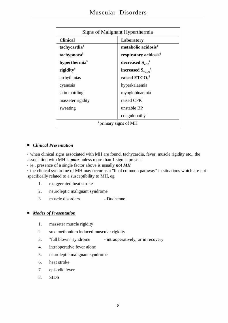
Signs of Malignant HyperthermiaClinical Laboratorytachycardia§ metabolic acidosis§
tachypnoea§ respiratory acidosis§
hyperthermia§ decreased SvO2§
rigidity§ increased SvCO2§
arrhythmias raised ETCO2§
cyanosis hyperkalaemia
skin mottling myoglobinaemia
masseter rigidity raised CPK
sweating unstable BP
coagulopathy§ primary signs of MH
Clinical Presentation
when clinical signs associated with MH are found, tachycardia, fever, muscle rigidity etc., theassociation with MH is poor unless more than 1 sign is present
ie., presence of a single factor above is usually not MHthe clinical syndrome of MH may occur as a "final common pathway" in situations which are not
specifically related to a susceptibility to MH, eg,
1. exaggerated heat stroke
2. neuroleptic malignant syndrome
3. muscle disorders - Duchenne
Modes of Presentation
1. masseter muscle rigidity
2. suxamethonium induced muscular rigidity
3. "full blown" syndrome - intraoperatively, or in recovery
4. intraoperative fever alone
5. neuroleptic malignant syndrome
6. heat stroke
7. episodic fever
8. SIDS
Muscular Disorders
8
Masseter Spasm
the occasional patient will develop trismus as the earliest sign of MH2 USA retrospective studies found an incidence of trismus ~ 1% following halothane & SChmost often in children given a mask halothane induction, followed by SCh, therefore either,
1. MH susceptibility occurs more frequently than previously thought, or,
2. trismus may occur in normal subjects
NB: due to the comparitively low incidence of MH, the later is more likely
tachycardia, occasional PVC's and mild metabolic acidosis usually occurthe implication of this is uncertain, as the definition of masseter muscle spasm/rigidity varies with
the investigatormasseter muscles have an atypical fibre type which responds with slow tonic contractions
→ there is a range of response for the masseter muscles following SCh,
1. subclinical "jaw stiffness" ≡t normal response- only demonstrable with strain devices- virtually none of these are MH prone
2. "jaw tightness interfering with intubation"~ 1% of children- a small undetermined percentage are at risk of MH
3. "extreme jaw rigidity, unable to open the mouth"= masseter muscle rigidity, MMR~ 50% are biopsy determined at risk for MH
the last group is that often quoted as showing ~ 50% positive for MH with a contracture testthis association is frequently quoted for the second group, which would lead to an expected MH
susceptibility frequency of ~ 0.5% !!the actual incidence of true MMR is uncertain and Miller suggests the incidence of MMR is
actually less than the quoted 1%, and there is a need for formal prospective studies the problem is then of how to manage the child who displays MMR,
1. Rosenburg (1988) - stop anaesthesia- administer dantrolene- monitor for rhabdomyolysis (CPK, myoglobinuria)* muscle biopsy
2. Gronert (1988) - continue with safe agents- monitor for rhabdomyolysis- monitor ETCO2, temp., etc.* muscle biopsy
3. Littleford (1991) - continue (triggering) anaesthesia- look for other rigidity- monitor ETCO2, temp., etc.* muscle biopsy
NB: the later is clearly the most controversial and it would seem unwise to continuewhen equally effective, safe alternatives are available
Muscular Disorders
9

recommendations by Kaplan, ASA 1992,
1. jaw stiffnessable to be opened with firm manual pressurenormal response and usual anaesthetic may be continued
2. diminished mouth opening ~ 1:100 childrenmouth cannot be fully opened, despite firm manual pressureinterfers with intubationmore suggestive of MH ? incidence unknownswitch to non-triggering agents, monitor carefully & continue anaesthetic
3. masseter muscle rigidityjaw cannot be budged, "jaw of steel"may well be the beginning of MH episode ~ 50% children
~ 25% adultsstop anaesthetic & monitor carefully
NB: #2/3 → monitor temperature, HR, BP in recovery for 4 hoursobtain postoperative serum CK's q6h x 4urine for myoglobinmonitor in hospital for 24 hoursif CK > 20,000 then assume MHS positivecounsel with family regarding muscle biopsy
Triggering
1. a genetic predisposition
2. the absence of inhibiting factors
3. the presence of triggering factors
depolarisation may be a significant factor, either "awake" or anaesthesia induced,
a. mechanical threshold is lower cf. "normal", therefore predisposed to contractures
b. SCh & carbachol trigger MH susceptible muscle
c. electrical stimulation triggers MH susceptible muscle
d. non-depolarising muscle relaxants delay the appearance of MH
NB: however, 4-aminopyridine does not trigger MHS swine ? why
volatile induced MH may be triggered by,
a. perturbation of the surface membranehalothane → surface or internal membranes of the fibrilSCh → end-plate effects
b. ? effects on the SR or mitochondira in vivoeffects on isolated preparations imply these are too small to trigger MH
Muscular Disorders
10
succinylcholine has a number of variant responses which may occur in isolation or combination,
a. muscle contracture
b. altered membrane permeability without contractureresulting in release of myoglobin, CPK, and K+
this occurs to a small extent in "normal" individualsenhanced by the presence of halothane and reduced by curare
c. an increase in metabolismas for MH, is usually associated with altered permeability and contracture
nitrous oxide has been proposed as a weak trigger, however there is minimal evidence for this
amide local anaesthetics were previously thought to trigger MH, but have since been exoneratedanimal data showing Ca++ release from the SR require mM concentrations not achieved clinically
muscle relaxants block the effects of SCh in triggering MH and delay or attenuate the effects ofthe volatile agents
dTC has been associated with greater lactate production in porcine MH & does producecontracture in denervated muscle, indicating it may have some depolarising action not normallyclinically evident
however, it has not been shown to trigger porcine MHreversal of NMJ blockade with antiacetylcholinesterase agents could theoretically trigger MHhowever, 4-aminopyridine which increases ACh does not, and reversal has been performed in
susceptible patients without untoward effects
the youngest reported episode was in utero, immediately prior to birth, at LUSCS under GAthe father was known MH susceptibledelayed onset of MH may represent depressed MH responses, 2° to drugs, or to proloned
anaesthetic stresses
awake triggering occurs readily in the porcine model 2° to heat stress, exercise, anoxia,apprehension and excitement
these relate to muscle activity or increase temperature, as suggested by,
1. MHS swine increase MRO2 & lactate production in response to,i. heat > 41°C or carbacholine, butii. not α/β sympathetic agonists
2. these abnormal responses are blocked or delayed by neuromuscular blockers
factors which suggest non-anaesthetic triggering in humans include,
1. increased incidence of unexplained sudden death in affected families
2. these families develop a non-specific cardiomyopathy
3. there are a series of case reports relating heat stroke, unusual stress & fatigue, andmyalgias to possible awake MH episodes
Muscular Disorders
11

Differential Diagnosis Raised ETCO2
a. increased CO2 production - fever- sepsis, sepsis syndrome- light anaesthesia- pregnancy- thyrotoxicosis- obesity- drugs
b. decreased ventilationi. increased anaesthetic depth - SVii. machine related - ↓ FGF, disconnect, leakiii. ventilator related - setting, malfunction
- decreased driving pressure- decreased patient compliance (pressure cycled)
iv. breathing circuitMapleson - ↓ FGF, disconnect, obstructioncircle - valve malfunction
- absorbant (depletion, channeling or bypass)- obstruction, leak, disconnect
v. pulmonary - upper airway obstruction- mainstem intubation- secretions, blood, aspiration- asthma, ARDS- CCF- pneumothorax, haemothorax
vi. extrathoracic - ↑ abdominal muscle tone- retractors with ↓ pulmonary compliance- ascites- pregnancy- morbid obesity
c. monitor error - calibration drift- moisture in measuring chamber
d. multifactorial - pregnancy, obesity, children, etc.
Muscular Disorders
12
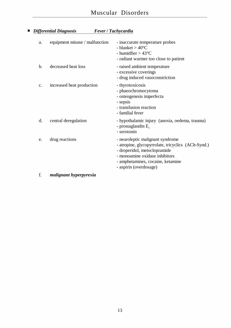
Differential Diagnosis Fever / Tachycardia
a. equipment misuse / malfunction - inaccurate temperature probes- blanket > 40°C- humidfier > 43°C- radiant warmer too close to patient
b. decreased heat loss - raised ambient temperature- excessive coverings- drug induced vasoconstriction
c. increased heat production - thyrotoxicosis- phaeochromocytoma- osteogenesis imperfecta- sepsis- transfusion reaction- familial fever
d. central deregulation - hypothalamic injury (anoxia, oedema, trauma)- prostaglandin E1
- serotonin
e. drug reactions - neuroleptic malignant syndrome- atropine, glycopyrrolate, tricyclics (ACh-Synd.)- droperidol, metoclopramide- monoamine oxidase inhibitors- amphetamines, cocaine, ketamine- aspirin (overdosage)
f. malignant hyperpyrexia
Muscular Disorders
13
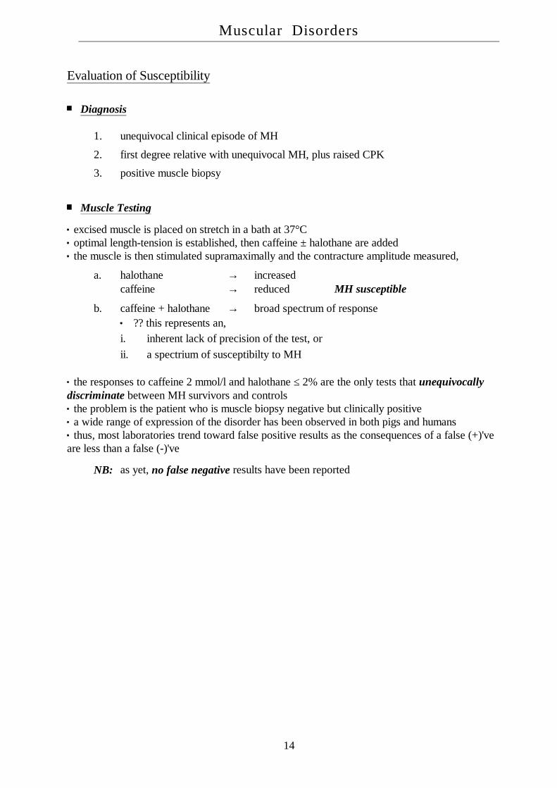
Evaluation of Susceptibility
Diagnosis
1. unequivocal clinical episode of MH
2. first degree relative with unequivocal MH, plus raised CPK
3. positive muscle biopsy
Muscle Testing
excised muscle is placed on stretch in a bath at 37°Coptimal length-tension is established, then caffeine ± halothane are addedthe muscle is then stimulated supramaximally and the contracture amplitude measured,
a. halothane → increasedcaffeine → reduced MH susceptible
b. caffeine + halothane → broad spectrum of response?? this represents an,
i. inherent lack of precision of the test, orii. a spectrium of susceptibilty to MH
the responses to caffeine 2 mmol/l and halothane ≤ 2% are the only tests that unequivocallydiscriminate between MH survivors and controls
the problem is the patient who is muscle biopsy negative but clinically positivea wide range of expression of the disorder has been observed in both pigs and humansthus, most laboratories trend toward false positive results as the consequences of a false (+)'ve
are less than a false (-)'ve
NB: as yet, no false negative results have been reported
Muscular Disorders
14
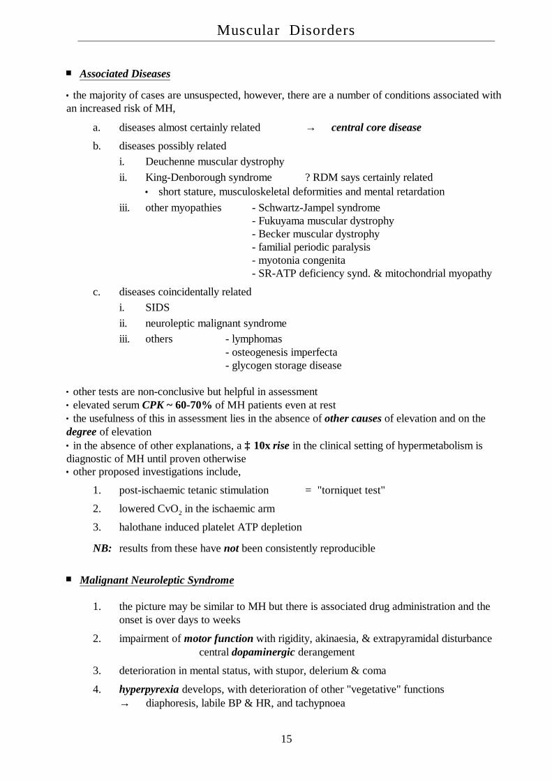
Associated Diseases
the majority of cases are unsuspected, however, there are a number of conditions associated withan increased risk of MH,
a. diseases almost certainly related → central core disease
b. diseases possibly relatedi. Deuchenne muscular dystrophyii. King-Denborough syndrome ? RDM says certainly related
short stature, musculoskeletal deformities and mental retardationiii. other myopathies - Schwartz-Jampel syndrome
- Fukuyama muscular dystrophy- Becker muscular dystrophy- familial periodic paralysis- myotonia congenita- SR-ATP deficiency synd. & mitochondrial myopathy
c. diseases coincidentally relatedi. SIDSii. neuroleptic malignant syndromeiii. others - lymphomas
- osteogenesis imperfecta- glycogen storage disease
other tests are non-conclusive but helpful in assessmentelevated serum CPK ~ 60-70% of MH patients even at restthe usefulness of this in assessment lies in the absence of other causes of elevation and on the
degree of elevationin the absence of other explanations, a ≥ 10x rise in the clinical setting of hypermetabolism is
diagnostic of MH until proven otherwiseother proposed investigations include,
1. post-ischaemic tetanic stimulation = "torniquet test"
2. lowered CvO2 in the ischaemic arm
3. halothane induced platelet ATP depletion
NB: results from these have not been consistently reproducible
Malignant Neuroleptic Syndrome
1. the picture may be similar to MH but there is associated drug administration and theonset is over days to weeks
2. impairment of motor function with rigidity, akinaesia, & extrapyramidal disturbance∝ central dopaminergic derangement
3. deterioration in mental status, with stupor, delerium & coma
4. hyperpyrexia develops, with deterioration of other "vegetative" functions→ diaphoresis, labile BP & HR, and tachypnoea
Muscular Disorders
15

Management - Acute
1. stop all triggering agents immediately* continue with safe agents if surgery cannot be immediately ceased
2. hyperventilate with 100% O2
* use new soda-lime & change to "clean MH-machine", when available
3. administer dantrolene 2-3 mg/kg immediately20 mg vials with - NaOH to a pH ~ 9-10
- mannitol to maintain isotonicitya 70 kg adult will require 7 vials initially, and may need up to 35 vials in total
i. repeat dose every 5 minutes until vital signs normaliseii. total dose up to 10-20 mg/kgiii. continue with 1 mg/kg q6h ó/IV postoperatively for 48-72 hrs
RDM states 15 hourly as this is the approximate half-life
4. bicarbonate 1-2 mmol/kg stat, then follow AGA's
5. initiate cooling - iced saline, cooling blanket, body cavity lavage- extracorporeal circulation* cease at ~ 38-39°C to prevent hypothermia
6. manage hyperkalaemia - control MH by giving dantrolene- NaHCO3
- insulin & dextrose* CaCl for life-threatening arrhythmias* hypokalaemia frequently follows treatment
7. manage arrhythmias - procainamide 3 mg/kg if persistent- to maximum of 15 mg/kg
8. manage DIC - maintain tissue perfusion, IVT- decrease temperature- 1° treatment of MH
9. monitoring - AGA's, ETCO2, SpO2, ECG, core T°- U&E's, Ca++, CK, myoglobin- APTT/PT, platelets, FDP's
10. maintain urine output - IVT ± mannitol/frusemide
11. transfer to ICU - observe for 24-48 hours
12. counsel family ± investigate
NB: no anaesthetic should be given without access to 36 vials of dantrolene& a clean anaesthetic machine (Kaplan)
Gronert (Miller) states it is no longer necessary to provide a non-contaminated anaesthesiamachine by flushing with O2 for several hours
removal of the vaporisers, replacement of the fresh gas outlet hose, use of a disposable circlewith a flush of O2 for 6 minutes is sufficient
Muscular Disorders
16
Protocol For Management Royal Hobart
1. recognitioni. masseter spasm following SChii. unexplained tachycardiaiii. tachypnoea in unparalysediv. rising ETCO2
v. rising temperaturevi. cyanosis / arterial desaturation
2. immediate action following recognition of acute MHi. announce life-threatening emergency & conclude surgery ASAPii. send for skilled anaesthetic/ICU assistanceiii. enlist the immediate assistance of at least 4 experienced nursesiv. anaesthetist in charge simultaneously coordinates 5 tasks,
reconstitution & administration of dantroleneremoval of precipitating causesmonitoringresuscitationactive cooling
Dantrolene
i. 20 mg vials + 60 ml sterile waterii. final pH ~ 9.5, ∴ large bore central line preferableiii. poor solubility & difficult to prepare, may occupy several nursesiv. 2-3 mg/kg bolus, then 1 mg/kg 5 minutely prn, to maximum 20 mg/kgv. dantrolene takes ~ 6 minutes to have any effect
the actions of dantrolene include,
a. decreases release of Ca++ from the SR, without affecting re-uptake
b. ? antagonises the effects of Ca++ at the actin/myosin - troponin/tropomyosin level
c. muscular weakness, which may potentiate NMJ blockade~ 5-15 mg/kg produces significant muscular relaxation
d. there is no effect on NMJ transmission
e. up to 15 mg/kg there is no significant effect on the CVS
f. up to 30 mg/kg there is no significant effect on respiration
NB: there is no evidence of toxicity when administered acutely
Muscular Disorders
17
reducted muscle rigidity results in rapid normalisation of serum biochemistry, especiallyhyperkalaemia, and cardiac function
cardiac arrhythmias & arrest are almost always 2° to hyperkalaemia/acidosis, the myocardium isnot directly involved in MH pathology
these can usually be managed by treating the 1° disturbanceCaCl2 can be used as a last resort for hyperkalaemiaCa++-channel blocking agents should not be used as they may result in cardiovascular collapse in
the presence of dantrolene
Precipitating Causes
1. high priorityi. remove all inhalational agents & known trigger agents,
remove vapourisers from Boyle's machineii. hyperventilate with O2 > 10 l/min FGF
2. lower priorityi. soda lime is not a significant reservoir for volatile, but will require replenishing
due to rapid exhaustionii. replace rubber hoses
1 minute at 10 l/min O2 → [halothane] < 100 ppm~ 100 x less than expired gas
Monitoring / Tests
1. SpO2 / ETCO2 / BP / ECG
2. temperature probes - rectal & oespohageal
3. IABP - for serial AGA's initially- pressure monitoring is lower priority
4. baseline biochemistry - ECU & AGA's initially
5. IV accessi. large peripheral line - fluids
- initial administration of dantroleneii. CVC line - EJV / IJV preferable due to risk of coagulopathy
- administration of dantrolene- pressure monitoring
6. urinary catheter > 2 ml/kg/hr target urine output- sample for myoglobinuria
7. repeat testsi. AGA's ~ 10 minutelyii. ECU ~ hourlyiii. Coag's ~ hourly
Muscular Disorders
18
Resuscitation
1. paralyse with pancuronium
2. intubate & hyperventilate with 100% O2 > 2-3x MV- as guided by ETCO2 / AGA's
3. administer HCO3- as per AGA's ? initial bolus per RDM
4. management of arrhythmiasi. treat MH with dantroleneii. propranolol - 1 mg boluses prniii. procainamide ~ 3-5 mg/kg slowly
≤ 20 mg/kg maximum doseiv. calcium channel blockers are contraindicated
5. management of hyperkalaemiai. treat MH with dantroleneii. correction of acidosis with HCO3
-
iii. Actrapid 10U / Dextrose 50% 50 mliv. RHH states do not use CaCl2, cf. ASA lectures say OK if hyperkalaemia severev. avoid resonium - action too slow
- hypokalaemia common in recovery phase of MH
6. renal protectioni. saline diuresisii. mannitol ± frusemideiii. maintain urine output > 2 ml/kg/hr
7. management of coagulopathyi. treat MH with dantroleneii. maintain tissue perfusion - CVP/MAP
- IVT fluids ± inotropesiii. decrease temperatureiv. FFP/platelets if clinical bleeding
Active Cooling
1. commence immediatelyi. fanning, cool spongesii. ice packs to groins, axillae, neck, popliteal & cubital fossae & abdomeniii. gastric, peritoneal, bladder, rectal or pleural lavage with cool saline
2. reduce theatre temperature if possible
3. cease active cooling at core temperature < 38.5°C
Muscular Disorders
19
Follow-Up Immediate
1. admit to ICU
2. intensive monitoring for 24-48 hoursi. temperature- core & peripheralii. ECG, IABP, CVP ± PAOPiii. biochemistry - ECU, CK's, myoglobinuria
- AGA's- Coag's
iv. urine outputv. neuromuscular status *rigidity
3. dantrolene~ 1mg/kg q6h for 24 hourshigher doses prn - ↑ rigidity / temperature
- ↓ pH, PaO2 / ↑ PaCO2
may be given enterally if GIT functioning (price ~ 1000x less)
Follow-Up Late
1. this is essential
2. counsel patient & family
3. screening CK's on all family members
4. suggest muscle biopsies if CK normal
5. medi-alert bracelets, letters etc.
Muscular Disorders
20

Management - Elective
Assessment
1. history from patient or relatives, previous anaesthetic records, etc.
2. detailed informed consent from patient / guardian
3. bias for regional anaesthesia if practicable
4. schedule operation during "working hours" when staff available
Conduct of General Anaesthesia
a. preparation of theatre personnel
b. prepare a "clean" anaesthetic machineMiller, Kaplan and others state separate machine no longer requiredremove vaporisers from machine & flush for > 10 minutes with 10 l/min O2
replace all rubber hoses with new rubber or plastic hosesfit new rubber belows to the ventilator
c. MH carti. drugs - dantrolene 36 vials + 2000 ml sterile H2O
- NaHCO3
- dextrose 50%- mannitol 25%, frusemide- procainamide- chlorpromazine
ii. equipment - T° probes- NG tubes- urinary catheters- disposable breathing circuit- soda lime for circle- blood collection tubes- syringes/needles, AGA syringes- CVC cannulation equipment
d. use of safe anaesthetic agents
e. monitoring - ETCO2 , SpO2 , ECG, NIBP, FiO2
- T° core & peripheral± urinary catheter
f. adequate recovery ≥ 4 hrs duration → ward or home
g. alert back-up support - anaesthetic staff- local ICU
Muscular Disorders
21
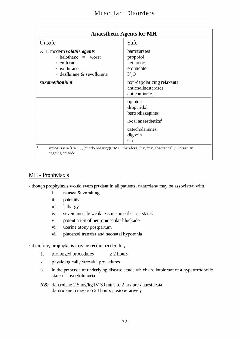
Anaesthetic Agents for MH
Unsafe SafeALL modern volatile agents
halothane = worstenfluraneisofluranedesflurane & sevoflurane
barbituratespropofolketamineetomidateN2O
suxamethonium non-depolarizing relaxantsanticholinesterasesanticholinergics
opioidsdroperidolbenzodiazepines
local anaesthetics1
catecholaminesdigoxinCa++
1 amides raise [Ca++]ICF but do not trigger MH, therefore, they may theoretically worsen anongoing episode
MH - Prophylaxis
though prophylaxis would seem prudent in all patients, dantrolene may be associated with,i. nausea & vomitingii. phlebitisiii. lethargyiv. severe muscle weakness in some disease statesv. potentiation of neuromuscular blockadevi. uterine atony postpartumvii. placental transfer and neonatal hypotonia
therefore, prophylaxis may be recommended for,
1. prolonged procedures ≥ 2 hours
2. physiologically stressful procedures
3. in the presence of underlying disease states which are intolerant of a hypermetabolicstate or myoglobinuria
NB: dantrolene 2.5 mg/kg IV 30 mins to 2 hrs pre-anaesthesiadantrolene 5 mg/kg ó 24 hours postoperatively
Muscular Disorders
22
Management - Family
most important is adequate information and supportbiopsy of the patient is reasonable after an appropriate interval (> 6 weeks)biopsy of the remaining family members is not essential, as most anaesthetists will treat them as
susceptible irrespective of the biopsy resultbiopsy at the time of incidental surgery is therefore logicalprior to biopsy dantrolene & droperidol should be avoided as they "normalise" the abnormal
responses of MH susceptible individualsthose refusing biopsy should have their plasma CPK checked, if elevated this may be taken as
evidence of MH susceptibility in a close relative
Muscular Disorders
23

MYOPATHIES
Classification
1. hereditaryi. muscular dystrophiesii. myotoniasiii. congenital myopathiesiv. glycogen storage diseasesv. glycolytic defectsvi. lipid metabolism disordersvii. familial periodic paralysis
2. acquiredi. neuromuscular junctionii. autoimmuneiii. endocrine & metaboliciv. toxic myopathiesv. alcoholvi. infectivevii. infiltrativeviii. disuse atrophyix. rhabdomyolysis
Hereditary Myopathies
a. muscular dystrophies - Duchene- Becker's - limb girdle, F-S-H, etc.
b. myotonias - dystrophica myotonica- myotonia congenita- paramyotonia
c. congenital myopathies - central core- nemaline- microtubular- congenital fibre disproportion
d. glycogen storage diseases - types II, III, IV, V
e. glycolytic defects - types VII, IX, X, XI
f. lipid metabolism disorders - carnitine deficiency- carnitine palmityltransferase deficiency
g. familial periodic paralysis
Muscular Disorders
24
Acquired
a. neuromuscular junction - myasthenia gravis- Eaton-Lambert- organophosphates
b. autoimmune - SLE, RA- polymyositis / dermatomyositis- polymyalgia rheumatica
c. endocrine - diabetes- thyrotoxic (apathetic), hypothyroidism- hypo / hyperparathyroid- hypopituitarism- Cushings, ? Addison's
d. metabolici. hypo - glycaemia / K+ / Ca++ / HPO4
=
ii. hyper - Mg++ / K+
iii. nutritional - vitamin E & D deficiencyiv. systemic disorders - renal & hepatic failure
- malignancy
e. toxic myopathiesi. focal (IMI) - pentazocine, pethidineii. generalised - chloroquine, clofibrate, colchicine
- steroids, D-penicillamine- propranolol, perhexiline, labetalol
iii. rhabdomyolysis - alcohol, heroin, amphetamines, PCP, cocaineiv. malignant hyperthermia * see table
f. alcohol * multifactorial
g. infectivei. viral - influenza A & B, adenovirus, EBV, herpes
- Coxsackie B5
- dengue, measlesii. bacterial - brucella
- legionella- Staphlococcal- leptospirosis
iii. fungaliv. protozoal - toxoplasmosis, trichinosis, worms
h. infiltrative - amyloid, tumour, fibrositis
i. disuse atrophy
j. rhabdomyolysis - traumatic, toxic, MH
Muscular Disorders
25

MYASTHENIA GRAVIS
Def'n: a neuromuscular disorder resulting in weakness and fatiguability of skeletalmuscle, due to an autoimmune mediated decrease in the number, andfunctional integrity of ACh receptors at the neuromuscular junction;"the prototype of antibody mediated autoimmune disease"
i. degradation of AChR's at an accelerated rate due to cross-linkingii. effective junctional blockade due to receptor occupancy by antibodiesiii. damage to the postsynaptic membrane due to complement activation
Essential Features
a. muscular weaknessexternal ophthalmoplegia ≥ 90%facial weaknessbulbar muscle involvement * risk of aspirationrespiratory failure
b. easy fatigability
c. recovery with rest or anticholinesterases
Myasthenia Grades§
I extraocular muscle involvement onlygood response to anticholinesterases
IIA generalised mild muscle weaknessno respiratory involvementgood response to anticholinesterases and steroids
IIB generalised moderate muscle weakness, and/or bulbar dysfunctionmore severe, rapidly progressivemay involve respiratory muscles
III acute, fulminating presentation, and/or respiratory dysfunctionrapid deterioration over ≤ 6 monthshigh mortality
IV late, severe, generalised myasthenia gravisincidence: 1:20,000females > males80% > 20 yrsprogression from types I & II
§ Osserman and Genkins (1971)
Muscular Disorders
26

Presentation
a. transient neonatal myasthenia~ 15-20% of neonates born to myasthenic motherspregnancy may result in remission or exaccerbation of maternal myastheniano correlation between the severity of maternal disease and neonatal occurrenceno correlation between the level of maternal AChR-Ab and neonatal occurrencespontaneous remission usually in 2-4 weeks
b. congenital or infantile myasthenianot autoimmune, possibly autosomal recessive inheritancerare in the absence of maternal myastheniacomprises a number of genetically determined abnormalities of the AChR or thepost-synaptic membrane
c. juvenile myasthenia~ 4% onset before 10 years and ~ 24% before age 20 yearsmarked female predominance ~ 4:1pathologically identical to the adult disease, though, thymoma is not a feature
d. adult myastheniaprevalence ~ 1:20,000 * F:M ~ 3:2 overall
- F:M ~ 2:1 < 50 years- F:M ~ 1:1 > 50 years
males tend to have more severe & rapidly progressing diseasehyperplasia of the thymus in > 70%, thymoma in 10-15%distribution, severity & outcome are determined by the course within the first 2-3years following onset, suggesting most ACh receptor damage occurs early~ 14% remain localised to the extraocular muscles, 86% becoming generalised
Anti-ACh-Receptor Ab's
NB: * virtually diagnostic if present
i. all grades ~ 85-90% (+)'veii. grade I ~ 50% (+)'veiii. AChR-Ab (-)'ve patients have mild or localised myastheniaiv. IgG predominantly against the α-subunit of the endplate receptorsv. individual patients have heterogenous populations of AChR antibodiesvi. there is limited sharing of idiotypes between patientsvii. T-cells become sensitised against thymic myoid cell AChR's during maturationviii. T-cell dependent B-cell antibody production results in circulating Ab's
Muscular Disorders
27

Complications
a. myasthenic crisis - severe life-threatening relapse
b. cholinergic crisis
c. respiratory failure - aspiration, infection, weakness
d. "Mary Walker phenomenon"
→ acute muscle weakness following exercise due to lactic acidosis
e. cardiomyopathy
f. associated diseases making weakness worse - hyper / hypothyroidism- SLE, RA, polymyositis
Differential Diagnosis
i. myasthenic syndrome - Eaton-Lambertii. neurastheniaiii. hyperthyroidismiv. botulinismv. intracranial mass lesions
Eaton-Lambert Syndrome
i. acquired disorder of quantal release of ACh from motor nerve terminalii. usually males, aged 50-70 yearsiii. disease predominantly of the limb girdle musclesiv. high association with small cell carcinoma of the lungv. ? IgG-Ab to the presynaptic voltage-dependent Ca++ channelsvi. ACh content and acetyltransferase activity are normalvii. decreased quantal release decreases MEPP frequencyviii. dysautonomia may occur, with dry mouth, impaired accomodation, urinary
hesitancy and constipationix. EMG → "characteristic"
incremental responseimprovement with exercise / tetanic stimulationmarked deficit with "normal" clinical strength§
x. weakness is not reliable reversed with anti-AChE agents,however, 3,4-diaminopyridine increases ACh release
xi. patients are sensitive to both deplarising and non-depolarising relaxants
NB: § this is in contrast to myasthenia, where the EMG abnormality is mild in thepresence of marked clinical weakness
Muscular Disorders
28

Myasthenic Crisis
Def'n: sudden, severe life-threatening relapse
a. may last weeks-months
b. risk factors - introduction of steroids- age- pregnancy- infection- surgery, trauma
c. drugs - aminoglycosides, tetracyclines- class Ia antiarrhythmics- narcotics, volatile anaesthetics- muscle relaxants
Clinical Features
a. rapid deterioration
b. positive tensilon test
c. NM stimulation - tetanic fade- post-tetanic facilitation
Cholinergic Crisis
a. excessive doses of anticholinesterases
b. risk factors - recovery phase from any "stress"- following response to steroids- thymectomy- plasmapheresis- immunosuppressives
c. differentiation from myasthenic crisis
Clinical Features
a. negative Tensilon test
b. NM stimulationi. depressed single twitchii. absent fade & absent post-tetanic facilitation
Muscular Disorders
29

Tensilon Test
edrophonium is commonly used due to rapid onset (< 30s) and short duration of action (~ 5m),resulting from freely reversible binding with ACh-E
objective assessment of one of the unequivocally weak groups of muscles,
a. initial dose 2-3 mg IV
b. improvement (+)'ve - test is terminated
c. no improvement (-)'ve - further dose of 8 mg
d. small initial dose due to unpleasant side-effectsnausea, diarrhoea, salivation, fasciculations and rarely syncopeatropine (0.6 mg) should be available for administration
e. false positives - amyotrophic lateral sclerosis- placebo-reactors
some cases may be better assessed with a long acting anticholinesterase agents, such asneostigmine
Treatment
a. anticholinesteraseslittle benefit in severe cases with respiratory muscle involvementanimal studies show long term administration results in changes in the AChR similarto those seen in myastheniapatient education regarding overdose (cholinergic) vs. underdose (myasthenic)
i. neostigmine 15 mg qid ~ 0.5 mg IV~ 1.5 mg IM
ii. pyridostigmine 60 mg 6-8 hrly
b. immunosupressioni. prednisolone 50-100 mg/day → increases muscle strengthii. cyclophosphamide, azathioprine
c. plasmapheresisevery 2-3 days for 2 wks → ~ 45% show marked improvement or remissionhowever, this only lasts 4 days to 12 weeksindications
i. myasthenic crisis, especially with respiratory failureii. respiratory failureiii. preoperative (for thymectomy)iv. refractory to drug therapy (steroids & anticholinesterases)
Muscular Disorders
30

Thymectomy
NB: should be performed on all adult patients with generalised disease,especially between puberty & 55 years;there is also unanimity regarding resection of thymomas,although, disease remission is less frequent
a. removal of thymoma ~ 10% of cases, most are benign- resection to prevent local spread
b. therapeutic thymectomy ≤ 85% of patients improve~ 35% achieve drug-free remission
thymus is abnormal in ~ 75% (65% hyperplasia + 10% thymoma)improvement may begin up to 1-10 years post-surgery !!there is no evidence that removal in childhood results in immunodeficiencyoperation is usually recommended for patients with only extraocular diseasethe anterior, trans-sternal approach is superior, as even small remnants left during the
transcervical approach will limit success
Anaesthetic Management
use regional or local anaesthesia whenever possible
a. preoperative evaluation - age, sex, onset & duration of disease- presence or absence of thymoma- bulbar involvement, aspiration risk- CAL
b. optimisation of condition - steroids ± azathioprine (age > 15)- plasmapheresis? anticholinesterases
the use of anticholinesterases is debatedthey potentiate vagal responses & require the use of atropinedecrease the metabolism of suxamethonium and ester local anaesthetics
c. premedication - avoid respiratory depressants? atropine IM ± benzodiazepines
d. induction / maintenance - deep inhalational anaesthesia- balanced anaesthesia with muscle relaxants
abnormal response to both depolarizing (↓) & non-depolarizing (↑) relaxantsthese responses are seen during remission & with localised extraocular diseasethe ED95 for SCh in myasthenia may be 2-2.5 x normal, however type II blockade isreadily producedconversely, the ED95 for the non-depolarising agents may be 10% of normalatracurium & vecuronium have short enough half-lives to allow titration to effect
Muscular Disorders
31

e. postoperative managementneuromuscular monitoring should be continued into the postoperative phasefew studies correlate tests of NMJ function with adequacy of ventilation
NB: the differential responses seen between peripheral versus bulbar muscles is furtherexaggerated in the myasthenic patient !
Elective Postoperative VentilationFactor Points
long history of myasthenia > 6 yrs 12
moderate to severe CAL - not 2° to MG 10
high pyridostigmine dose > 750mg/day 8
diminished vital capacity < 2.9 l< 40 ml/kg
4
NB: total score > 10 points = post-operative ventilation for > 3 hours
following transcervical thymectomy ~ 7.4% of patients require prolonged (> 3 hrs) ventilation
Outcome
a. thymectomy benefits ~ 96% of patients, irrespective of preoperative statusi. ~ 46% develop complete remissionii. ~ 50% are asymptomatic or improve on therapyiii. ~ 4% remain the same
b. thymectomy does not always result in a decrease the anti-AChR-Ab titre
NB: the anti-AChR sensitised T-cells survive long after thymectomy
Muscular Disorders
32

MUSCULAR DYSTROPHIES
Duchenne Muscular Dystrophy
a. X-linked recessive disorder, affecting almost exclusively males
b. incidence ~ 13-33:100,000~ 1:3,000-8,000
c. progressive, symmetrical weakness of the pelvic & shoulder girdles,i. onset by age 5 yearsii. leg braces by 8-10iii. non-ambulatory by 12 yearsiv. survival beyond 25 years rare
d. associated problems - tendon and muscle contractures- progressive kyphoscoliosis- impaired pulmonary function- cardiomyopathy- intellectual impairment (~ 33%)
e. palpable enlargement of some muscles, resulting initially from hypertrophy and laterfrom replacement with fat and connective tissue
f. laboratory findingsi. CK, aldolase - massive & early elevations
- MM & MB bands- not BB (cancer, heart trauma, CPB, CT disorders)
ii. EMG - myopathic patterniii. ECG - tall R in V1 , deep Q in precordial leadsiv. biopsy - necrotic fibres, phagocytosis, fatty replacement
g. carrier detectioni. CK ~ 50% of female carriers show elevationii. DNA probes - abnormal gene coding for dystrophin
- restriction fragment length polymorphisms (RFLP's)
h. complicationsi. respiratory - respiratory failure
- recurrent infectionsii. CVS - cardiomyopathy in almost all patients
- CCF occurs rarely, only with major stress- arrhythmias occur but also uncommon* cardiac death is rare
iii. GIT - acute gastric dilatation (may be fatal)- aspiration syndromes
Muscular Disorders
33

Myotonic Dystrophy Dystrophica Myotonica
a. autosomal dominant ~ 1:10,000
b. onset - typically 2nd or 3rd decade- affected individuals may remain asymptomatic
c. congenital myotonic dystrophyoccurs in infants of affected mothers with severe facial and bulbar palsyneonatal respiratory insufficiency may occur but is usually self-limiting
d. clinical featuresmanifests as an inability to relax muscles following strong contractioninitially muscles of face, neck and distal extremitiescharacteristic "hatchet" face
ptosis, temporal wasting, drooping of the lower lip and sagging of the jawcardiac involvement usually affects conducting tissue
1st degree heart block is present in the majorityCHB may dictate pacemaker insertionsudden death may occur, tachyarrhythmias & CCF are less frequent
respiratory muscle weakness may be severe with minimal limb involvementimpaired ventilatory drive & extreme sensitivity to opioids etc.central & peripheral sleep apnoea with chronic hypoxia may lead to cor pulmonaleand this is the usual cause of CCF in these patients
e. characteristic facial featuresi. ptosis ii. posterior subcapsular cataracts iii. atrophy of facial muscles and sternomastoidiv. frontal baldness v. hyperostosis frontalis
f. laboratory studiesi. CK - normal or mildly elevatedii. EMG - characteristic myotonia & myopathic featuresiii. ECG - 1st degree HB ± CHBiv. biopsy - distinctive type I fibre atrophyv. genetics - mutant gene long arm of C19
* antenatal diagnosis possible
g. general managementcondition is seldom so disabling as to require treatmentphenytoin is drug of choiceantimyotonia agents, quinidine & procainamide, may worsen cardiac conduction
h. treatment of myotonic contractures - hydrocortisone- procainamide- dantrolene
Muscular Disorders
34
Myotonic Contracture Triggers
i. cold, shivering, stressii. trauma, exercise, mechanical stimulationiii. tourniquets, hyperkalaemiaiv. drugs - suxamethonium
- halothane- anticholinesterases
Other Complications
i. respiratory muscle weakness - respiratory failureii. myotonic contracture - chest wall rigidity
- difficult to ventilateiii. cardiomyopathy ± cor pulmonaleiv. endocrinopathy - hypothyroidism
- diabetes mellitusv. gastrointestinal disease - pharyngeal weakness
- aspiration riskvi. gonadal atrophyvii. intellectual impairmentviii. hypersomnia / sleep apnoea syndromeix. possible association with MH * abnormality on C19
x. drugs - contractures- respiratory depression
Myotonia Congenita
a. occurs as autosomal dominant and autosomal recessive forms
b. those with the recessive form may develop slight weakness,those with the dominant form do not
c. there is no other significant organ involvement
d. respond well to antimyotonia agents - quinine, procainamide, tocainide- phenytoin- acetazolamide
Muscular Disorders
35

Miscellaneous Muscular Dystrophies
1. oculopharyngeal dystrophy
2. congenital muscular dystrophy
3. distal muscular dystrophy
4. scapuloperoneal dystrophy
Congenital Myopathies
NB: 1. these are rare disorders, distinguished from the muscular dystrophies by thepresence of specific histochemical & structural abnormalities in muscle2. a non-progressive course is common but not invariable3. pectus excavatum, kyphoscoliosis, hip dislocation & pes cavum are common
Central Core Disease
the first congenital myopathy described, by Shy & Magee in 1956autosomal dominant inheritance but sporadic cases occurweakness of muscles of the face & legs is usually mildserum CK and EMG may be normaldiagnostic biopsy with "central cores" in fibres, devoid of oxidative enzymesalmost definite association with malignant hyperpyrexia
Nemaline Myopathy
usually autosomal dominant, may be recessive or sporadicinfantile hypotonia is present & striking leading to respiratory failureserum CK may be normal, EMG usually shows myopathy
Myotubular Myopathy
multiple patterns of inheritance plus sporadic casessimilar to above but distinguished by external ophthalmoplegiaCK is normal or slightly elevated, the EMG abnormal
Congenital Fibre Disproportion
hypotonia, weakness, delayed motor milestones, skeletal deformities as abovebiopsy shows increased number of small type I fibres, with normal or hypertrophied type II fibres
Muscular Disorders
36