muscles of mastication
TRANSCRIPT

MUSCLES OF MASTICATION

Mastication is a harmonious and skillful activity which requires the presence and co ordination of not only the muscles of mastication but also the supra infra-hyoid muscles, and the facial muscles
• BASIC MUSCLES:
TemporalisMasseterMedial PterygoidLateral Pterygoid


• ACCESSORY MUSCLES
Muscles of tongue, lip & cheek
Suprahyoid muscles
MylohyoidGeniohyoidStylohyoidDigastric muscle
(anterior belly)
Infrahyoid muscles:
SternothyroidThyrohyoidOmohyoidSternohyoid

DEVELOPMENT• The basic muscles of mastication
develop from the mesoderm of the first pharyngeal arch.

• So they receive all their innervations from the mandibular branch of the trigeminal nerve, all from the anterior division except the medial pterygoid which gets its nerve supply from the main trunk.

MOVEMENTS OF MANDIBLE
• Movements that the mandible can undergo are:
1. Depression: As in opening the mouth.
2. Elevation: As in closing the mouth.
3. Protraction: Horizontal movement of the mandible anteriorly.
4. Retraction: Horizontal movement of the mandible posteriorly.
5. Rotation: The anterior tip of the mandible is “slewed” from side to side.

• These movements of mandible are performed by various muscles involved in it. So, functionally, the muscles of mastication are classified as:
• Jaw elevators: • Masseter• Temporalis• Medial pterygoid
• Jaw depressors:• Lateral pterygoid• Anterior digastric• Geniohyoid• Mylohyoid

TEMPORALISIt is the largest among all the
mastication muscles and is a fan shape muscle.
It has been divided into 2 heads: Deep head (anterior, middle and posterior
fibers) Superficial head (much smaller)

Origin: From the inferior temporal line , floor of the temporal fossa and from the overlying temporal fascia of the side of the skull.
Insertion: Superior border and medial tip of the coronoid process.

• Action:• Elevation
(anterior fibers)• Retraction
(posterior fibers)
• Nerve supply:• Anterior division
of the mandibular nerve
(by two deep temporal nerves)

•Blood Supply•This is furnished by the middle & deep temporal arteries. The middle temporal artery is a branch of the superficial temporal artery & the deep temporal arteries are branches of the maxillary artery
PALPATION
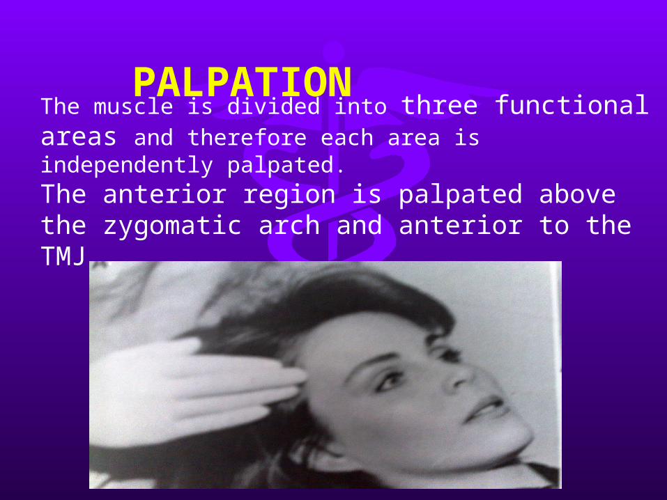
The muscle is divided into three functional areas and therefore each area is independently palpated.
The anterior region is palpated above the zygomatic arch and anterior to the TMJ .

The middle region is palpated directly above the TMJ and superior to the zygomatic arch .

• The posterior region is palpated above and behind the ear.
• If uncertainty arises regarding the proper finger placement. The patient is asked to clench the teeth together so that the temporal muscle contracts and the fibers should be felt beneath the finger tips.

MASSETERIt consist of three overlapping layers:
The origin of the whole muscle is mainly from the zygomatic process which consists of :
The superficial layer
The middle layer
The deep layer

SUPERFICIAL LAYER
It is the largest component that arises from the anterior two thirds of the lower border of the zygomatic arch.
Its fibers run downwards and backwards and inserts into lower half of the ramus including angle of the mandible.

The middle layer takes its origin from the medial surface of the anterior two-thirds and the lower border of posterior one third of the arch.
The fibers run more directly downwards to be inserted into lateral surface of the middle part of the ramus.
MIDDLE LAYER

The deep layer arises from the whole length of medial surface of the zygomatic arch.
The fibers pass downwards to attach to the upper part of the mandible ramus.
DEEP LAYER

• Action of masseter is mainly to elevate the mandible (antigravity action) and also helps in protrusive movement.
• It is the main powerful muscle involved in the elevation of the mandible

•Nerve supply•Supplied by Masseteric nerve a branch of anterior division of Mandibular nerve
•Blood Supply•Supplied by masseteric artery branch of maxillary artery

PALPATION
• The patient is asked to clench their teeth and, using both hands, the practitioner palpates the masseter muscles on both sides extraorally, making sure that the patient continues to clench during the procedure.
• Palpate the origin of the masseter bilaterally along the zygomatic arch and continue to palpate down the body of the mandible where the masseter is attached.

D-Palpation of the masseter muscles
E-Bimanual palpation ofthe masseter muscles

MEDIAL PTERYGOID
• It is also called as the Pterygoideus internus (Internal pterygoid muscle).
• It consist of Two heads which differ in origin:
The superficial head
The deep head

•
• SUPERFICIAL HEAD
The superficial head originates from the maxillary tuberosity & pyramidal process of palatine bone

DEEP HEAD
The deep head originates from the medial surface of lateral pterygoid plate of the sphenoid bone.

• Action:
1. Elevates the mandible .
2. Protrusion of the mandible (lateral & medial pterygoid on one side protrude the mandible to the opposite side).
3. Side to side movement (these lateral movements are achieved by lateral & medial pterygoid on both sides acting together to produce side to side movements).

•Nerve Supply•The never supplying the medial pterygoid muscles is the medial pterygoid nerve branch of the mandibular nerve
•Blood Supply•The artery supplying the medial pterygoid muscles is a branch of the maxillary artery
PALPATION It can be palpated by placing the
finger on the lateral aspect of the pharyngeal wall of the throat, this palpation is difficult and sometimes uncomfortable for the patient.
• Functional manipulation is done when the muscle becomes fatigued and symptomatic.
The muscle contracts as the teeth are coming in contact Also stretches when the mouth is open wide.

G- Palpation of the medial pterygoid
muscle

LATERAL PTERYGOID
• Also called as the Pterygoideus externus (External pterygoid muscle).
• It is a short conical muscle, having 2 heads: upper and lower.

• Upper head:• Origin: infra-temporal surface & crest of
the greater wing of sphenoid

• Lower head:• Origin: Lateral surface of the lateral
pterygoid plate

• Actions of lateral pterygoid:
• Depression of the mandible .• Side to side movement
(lateral movement) .• Protrusion of the mandible.
• If the Pterygoid muscles of one side act, the other side of the mandible is drawn forward while the same condyle remains comparatively fixed.

•Nerve Supply•The nerve to the lateral pterygoid muscle branches off from the masseteric or buccal nerve, which is the branch of the anterior trunk of the mandibular nerve
•Blood Supply•Pterygoid vessels from Maxillary artery

PALPATION
The patient is asked to protrude the mandible against resistance & Clench on maximum intercuspation
For Inferior lateral pterygoid :

• For Superior lateral pterygoid muscle:
The muscle contracts and stretches on clenching.
In order to differentiate pain arising from elevator muscle, the patient is asked to open the jaw wide.

MASTICATORY MUSCLE DISORDERS
Some of the common masticatory muscle disorders involve:
• Congenital hyperplasia/ hypoplasia• Hypermobility/ hypomobility of the
muscle• Muscle pains• MPDS• Myositis ossificans etc.

CONGENITAL HYPOPLASIA/ HYPERPLASIA
• It occurs very rarely, and is more common in masseter and orbicularis oris.
• Its oral symptoms include enlargement or decreased size of the affected muscle, which may show an asymmetric facial pattern and stiffness in the temporo-mandibular joint.
• It may or may not be associated with hypermobility/ hypomobility of the muscles.

MUSCLE HYPERMOBILITY/ HYPOMOBILITY
• This disorder involves extreme or diminished activity of the masticatory muscles.
• Its etiology includes various factors such as:
• Decreased/ increased threshold potential of neural activity.
• Parkinsonism• Facial paralysis• Nerve decompression• Secondary involvement of systemic
diseases.

MPDS
• Pain disorder in which unilateral pain is referred from trigger points in myofacial structure
• Constant pain, dull acheLaskin’s Cardinal Signs• Muscle tenderness• Pain• Clicking or propping noise in
TMJ• Limited Jaw Movement

TEMPORAL TENDONITIS
• Chronic strain from temporalis muscle pulling on tendon that attaches to mandible
• Causes sharp headaches in temple just to side of the eyes

CONCLUSION
• The masticatory muscles include a vital part of the orofacial structure and are important both functionally and structurally.
• The proper management and periodical self-examination of the muscles may provide a greater chance of catching the disease process at an early stage which may be useful for its better prognosis.
