muscles of mastication · and anterior border of the ramus of the mandible action: temporalis is a...
TRANSCRIPT

Muscles of mastication
Temporal and Infratemporal
fossae
Dr. Heba Kalbouneh
Associate Professor of Anatomy and Histology

Dr.
Heb
a K
albouneh

Depression
Dr.
Heb
a K
albouneh

Elevation
Dr.
Heb
a K
albouneh

Protraction
Dr.
Heb
a K
albouneh

Retraction
Dr.
Heb
a K
albouneh

Temporalis muscle
Origin: from the bony surfaces of the temporal
fossa
Insertion: coronoid process
And anterior border of the ramus of the
mandible
Action: Temporalis is a powerful elevator
of the mandible, closing jaws
Retraction of the mandible
Temporalis is innervated by deep
temporal nerves (mandibular nerve)
Dr.
Heb
a K
albouneh

Masseter muscle
Origin: the zygomatic arch, maxillary
process of zygomatic bone.
Insertion: into the lateral surface of the
ramus of the mandible
The masseter is innervated by
the masseteric nerve from the
mandibular nerve [V3]
Action: elevation of the
mandible, closing jaws
The masseter muscle is
quadrangular in shape
Dr.
Heb
a K
albouneh

Medial pterygoid
Origin: medial surface of the lateral plate
of the pterygoid process
Insertion: medial surface of the ramus of
mandible near the angle
The medial pterygoid is innervated by the
nerve to medial pterygoid from the
mandibular nerve [V3].
Action: The medial pterygoid mainly
elevates the mandible, closing jaws
The medial pterygoid muscle is
quadrangular in shape and has deep
and superficial heads
Note: the fibers of the medial
pterygoid are oriented almost
vertically
Dr.
Heb
a K
albouneh
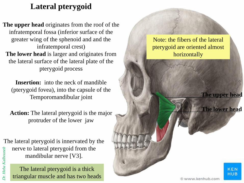
Lateral pterygoid
The upper head originates from the roof of the
infratemporal fossa (inferior surface of the
greater wing of the sphenoid and and the
infratemporal crest)
The lower head is larger and originates from
the lateral surface of the lateral plate of the
pterygoid process
Insertion: into the neck of mandible
(pterygoid fovea), into the capsule of the
Temporomandibular joint
The lateral pterygoid is innervated by the
nerve to lateral pterygoid from the
mandibular nerve [V3].
The lateral pterygoid is a thick
triangular muscle and has two heads
Note: the fibers of the lateral
pterygoid are oriented almost
horizontally
Action: The lateral pterygoid is the major
protruder of the lower jaw
Dr.
Heb
a K
albouneh
The upper head
The lower head

Infratemporal crest
lateral pterygoid plate
Lateral
Dr.
Heb
a K
albouneh

Pterygoid fovea D
r. H
eba
Kal
bouneh

Lateral pterygoid
Dr.
Heb
a K
albouneh
The upper head
The lower head

Medial pterygoid
Dr.
Heb
a K
albouneh

Masseter/ Superficial part
Dr.
Heb
a K
albouneh

Masseter/ deep part
Dr.
Heb
a K
albouneh


Temporal fossa
Pterygo-palatine fossa
Lies below the apex of the orbit
Temporal and infratemporal
fossae are interconnected spaces
on the lateral side of the head
Temporal fossa is superior to the
infratemporal fossa above the
zygomatic arch
Dr.
Heb
a K
albouneh

Of the four muscles of mastication (masseter, temporalis,
medial pterygoid, and lateral pterygoid) that move the lower
jaw at the TMJ
One (masseter) is lateral to the infratemporal fossa
Two (medial and lateral pterygoid) are in the infratemporal
fossa
One fills the temporal fossa (temporalis)
Dr.
Heb
a K
albouneh
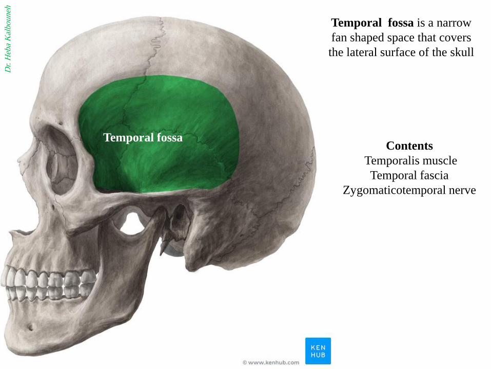
Temporal fossa
Temporal fossa is a narrow
fan shaped space that covers
the lateral surface of the skull
Contents
Temporalis muscle
Temporal fascia
Zygomaticotemporal nerve
Dr.
Heb
a K
albouneh

The infratemporal fossa is an
irregularly shaped cavity, situated
below the zygomatic arch, deep to
the ramus of the mandible
The infratemporal fossa
acts as a pathway for
neurovascular structures
passing to and from the
cranial cavity,
pterygopalatine fossa and
temporal fossa.
It also contains some of the
muscles of mastication
Dr.
Heb
a K
albouneh

Infra temporal fossa
Anterior wall: back of the
maxilla
Medial wall: lateral pterygoid
plate
Roof: greater wing of
sphenoid bone
Lateral wall: ramus of
mandible
Communications
Temporal fossa: through the
gap deep to the zygomatic
arch
Orbit: through the inferior
orbital fissure
Pterygo-palatine fossa:
through the pterygo-maxillary
fissure
Middle cranial fossa: through
foramen ovale and spinosum
Note: the infratemporal fossa lies
between the ramus of the mandible
laterally and the wall of pharynx
medially
Dr. Heba Kalbouneh

lateral pterygoid plate
greater wing of sphenoid bone
Back of the maxilla
Dr.
Heb
a K
albouneh

Infratemporal fossa

Note:
The foramen ovale and foramen spinosum open
on its roof
Foramen ovale
(Mandibular nerve)
Foramen spinosum
(Middle meningeal
artery)
Inferior orbital fissure
The roof of Infratemporal fossa
Inferior view of the skull
Dr.
Heb
a K
albouneh

Pterygo-maxillary fissure
Communication with
Pterygo-palatine fossa
Inferior orbital
fissure
Communication
with the orbit
Pterygo-palatine fossa
The medial and anterior walls of Infratemporal fossa
Dr.
Heb
a K
albouneh
Maxilla Lat
Pterygoid
plate

Lateral wall is formed by
the medial surface of the
ramus of mandible
Which contains the
mandibular foramen
Mandibular foramen: an opening to the mandibular
canal
Transmits inferior alveolar nerve (mandibular nerve)
Dr.
Heb
a K
albouneh
The lateral wall of
Infratemporal fossa
Lateral pterygoid
plate
Greater wing of sphenoid

Contents of infratemporal fossa
Lateral pterygoid muscle
Medial pterygoid muscle
Sphenomandibular ligament
Maxillary artery (and its
branches:
☺Middle meningeal artery
Deep temporal arteries
Buccal artery
Inferior alveolar artery
Mandibular nerve (and its
branches:
Auriculotemporal nerve
Buccal nerve
Lingual nerve
Inferior alveolar nerve
Chorda tympani
Pterygoid venous plexus
Maxillary vein
Middle meningeal vein
Otic ganglion
Dr.
Heb
a K
albouneh

Lateral pterygoid muscle
Maxillary artery
Medial pterygoid muscle
Temporalis
Masseter
Buccinator
Dr.
Heb
a K
albouneh

Maxillary artery
the terminal branch of
the external carotid artery.
It travels through the
infratemporal fossa.
Within the fossa, it gives
rise to the middle
meningeal artery, which
passes through the
foramen spinosum.
Foramen
spinosum
Dr.
Heb
a K
albouneh

Middle
meningeal
artery
Maxillary
artery
External carotid
artery
Infraorbital
artery
Deep
temporal
arteries
Buccal
artery
Masseteric
artery
Superficial
temporal
artery
Dr.
Heb
a K
albouneh
Inferior alveolar
artery

Mandibular Nerve (V3)
The mandibular nerve is both
motor and sensory
The sensory root leaves the
trigeminal ganglion and passes out
of the skull through the foramen
ovale to enter the infratemporal
fossa.
The motor root of the trigeminal
nerve also leaves the skull through
the foramen ovale and joins the
sensory root to form the trunk of
the mandibular nerve
Then divides into a small anterior
and a large posterior division
.
foramen ovale
Dr.
Heb
a K
albouneh

Branches From the Main Trunk of the
Mandibular Nerve:
1- Meningeal branch
2- Nerve to the medial pterygoid muscle
Branches From the Anterior Division of
the Mandibular Nerve
1- Masseteric nerve (to masseter muscle)
2- Deep temporal nerves (to temporalis
muscle)
3- Nerve to the lateral pterygoid muscle
4- Buccal nerve
Branches From the Posterior Division of
the Mandibular Nerve
1- Auriculotemporal nerve
2- Lingual nerve
3- Inferior alveolar nerve
Dr. Heba Kalbouneh

Anterior Division Posterior Division
Meningeal branch Nerve to the medial pterygoid
Auriculotemporal nerve
Lingual nerve
Inferior alveolar nerve
Masseteric nerve
Deep temporal nerve
Nerve to the lateral pterygoid
Buccal nerve
Dr. Heba Kalbouneh
Foramen ovale Foramen
spinosum

Meningeal branch
Masseteric nerve
Buccal nerve
Deep temporal nerves
Auriculotemporal nerve
Lingual nerve
Inferior
Alveolar
nerve
Dr.
Heb
a K
albouneh

The buccal nerve is the only sensory
branch of the anterior division of the
mandibular nerve.
Note: The buccal nerve of mandibular nerve is
SENSORY and does not supply the buccinator
muscle (which is supplied by buccal branch of
facial nerve MOTOR)
Nerve to the medial pterygoid muscle supplies:
1- The medial pterygoid muscle
2- The tensor veli palatini muscle
3- The tensor tympani muscle
Buccal nerve supplies the skin and the mucous
membrane of the cheek
Lingual nerve
It supplies the mucous membrane of the anterior two thirds of the tongue and the floor of the
mouth (general sensations)
It is joined by the chorda tympani nerve
It gives off preganglionic parasympathetic secretomotor fibers to the submandibular ganglion.
It carries taste information from the anterior two thirds of the tongue, via the chorda tympani
Auriculotemporal nerve conveys postganglionic
parasympathetic secretomotor fibers from the otic ganglion to
the parotid salivary gland.
Dr.
Heb
a K
albouneh

The inferior alveolar nerve
(inferior dental nerve) is a branch
of the mandibular nerve
It supplies sensation to the lower
teeth, lower lip and chin
Mental nerve is a branch of
inferior alveolar nerve to supply
the skin and mucous membrane of
the lower lip and chin
(Passes through mental foramen)
Dr.
Heb
a K
albouneh

Sphenomandibular ligament is
an extra-capsular ligament of TMJ
.
It runs between the spine of
sphenoid and the lingula of the
mandible
Lingula
Dr.
Heb
a K
albouneh

Spine of sphenoid
Spine of sphenoid
Foramen spinosum
Greater
wing
Dr.
Heb
a K
albouneh

Sphenomandibular
ligament
It is the primary passive support
of the mandible, along with the
muscles of mastication.
Temporomandibular
joint
Dr.
Heb
a K
albouneh

Superior and inferior
ophthalmic veins
Facial vein
Pterygoid venous
plexus
Cavernous
sinus
deep facial vein
Pterygoid venous plexus
It is a valveless venous plexus of
considerable size, and is situated
on the lateral aspect of medial
pterygoid within the infratemporal
fossa
It drains the eye and is directly
connected to the cavernous sinus.
It provides a potential route by
which infections of the face can
spread intracranially.
It receives tributaries
corresponding with the branches of
the maxillary artery
It forms the maxillary vein
Dr.
Heb
a K
albouneh

Facial vein
Superficial temporal
vein
Maxillary vein
Retromandibular
vein
Post.
division of
Retro-
mandibular
vein
Ant. division of
retromandibular
vein
Posterior auricular
vein
External
jugular
vein Internal jugular vein
Common facial vein
The maxillary vein consist
of a short trunk
It is formed by a confluence
of the veins of the pterygoid
plexus
It unites with the superficial
temporal vein to form the
retromandibular vein
Dr.
Heb
a K
albouneh

Note:
Pterygoid venous plexus lies
around lateral pterygoid
muscle
Deep facial vein
Facial
vein
Pterygoid
venous plexus
Retromandibular
vein
Superficial
temporal
vein
Maxillary
vein

The preganglionic parasympathetic fibers
originate in the glossopharyngeal nerve,
and they reach the ganglion via the lesser
petrosal nerve
The postganglionic parasympathetic
(secretomotor) fibers reach the parotid
salivary gland via the auriculotemporal
nerve.
The otic ganglion is a small
parasympathetic ganglion located
immediately below the foramen ovale
in the infratemporal fossa and on the
medial surface of the mandibular
nerve.
It is functionally associated with the
glossopharyngeal nerve and innervates
the parotid gland for salivation.
Dr.
Heb
a K
albouneh

The lesser petrosal nerve is a branch from
glossopharyngeal nerve (CN IX), carrying
parasympathetic preganglionic fibers from
the tympanic plexus to the parotid gland. It
synapses in the otic ganglion, from where the
postganglionic fibers emerge
Glossopharyngeal
nerve IJV
Tympanic
nerve
Dr.
Heb
a K
albouneh

Medial wall
Glossopharyngeal
nerve IJV
Tympanic
nerve
Lesser petrosal nerve
Dr.
Heb
a K
albouneh

Man
dib
ula
r n
erv
e
Lesser p
etrosal
nerv
e
Auriculotemporal
nerve
Foramen
ovale
Dr.
Heb
a K
albouneh
Foramen
spinosum
Otic ganglion
Mid
dle
men
ing
eal
arte
ry
Auri
culo
tem
pora
l ner
ve
Mandibular nerve
Lesser petrosal nerve
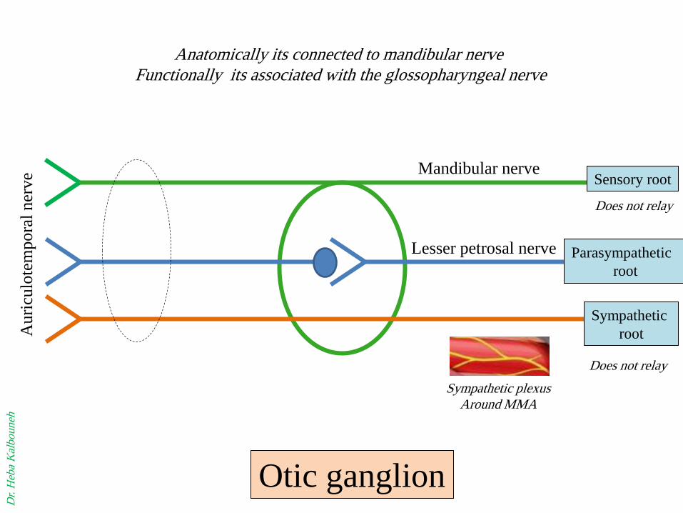
Anatomically its connected to mandibular nerve
Functionally its associated with the glossopharyngeal nerve
Dr.
Heb
a K
albouneh
Sensory root
Parasympathetic
root
Sympathetic
root
Does not relay
Does not relay
Sympathetic plexus Around MMA
Otic ganglion

Foramen ovale transmits:
Mandibular nerve
Accessory meningeal artery
Lesser petrosal nerve
Emissary vein
MALE
Dr. Heba Kalbouneh

Pterygoid venous plexus in
Infratemporal fossa
Foramen ovale
Dr. Heba Kalbouneh
Emissary vein
Cavernous sinus