muscular system chapter 6 muscles l are muscles organs? l yes, each individual muscle is an organ...
TRANSCRIPT

MUSCULAR SYSTEMMUSCULAR SYSTEM
CHAPTER 6

MusclesMuscles Are muscles organs? Yes, each individual muscle is an organ
– Why? Muscles are composed of more than just muscle
tissue.– Example: Skeletal muscles are composed of skeletal
muscle tissue, nervous tissue, blood, and connective tissue.
3 types:– Skeletal (We will concentrate mostly on this one.)– Smooth– Cardiac

Skeletal muscleSkeletal muscle
Cells - long, cylindrical, multinucleated, striated
Voluntary - controlled by the nervous system– When are they not voluntary?
Speed of contraction varies (fast to slow) Location?

Cardiac muscleCardiac muscle
Cells - branching, intercalated discs, striated Involuntary - controlled by its own
pacemaker and hormones Slow rhythmic contractions

Smooth muscleSmooth muscle
Cells - no striations, appear smooth and “stringy” in microscope
Involuntary Very slow contraction
– Can be rhythmic Location


Functions of Skeletal MusclesFunctions of Skeletal Muscles Produce Movement
– Muscles cross joints and work in opposite forces to move the bones
Maintain Posture– Always working against gravity
Stabilize joints– Many joints don’t fit together well, muscles help reinforce
those (ex. shoulder) Generate Heat
– Heat is a by product of muscle activity

Rules of Skeletal MusclesRules of Skeletal Muscles
1. All muscles cross at least one joint A few minor exceptions
2. Typically, the bulk of the muscle is going to be proximal to the joint crossed
3. All muscles have at least two attachment points: the origin and insertion
4. Muscles can only pull, they cannot push 5. During contraction, the insertion point moves
toward the origin.
Structure of Skeletal MusclesStructure of Skeletal Muscles Muscles have multiple layers
– Cylinders within cylinders Levels of muscles, largest to smallest: Muscle fascicle muscle fiber myofibril
filaments Each level of a muscle is enclosed in a layer of
connective tissue Fascia - connective tissue that covers and is
intertwined with the muscles. Has three purposes
– DQ – What do you think those might be?

Structure of Skeletal MusclesStructure of Skeletal Muscles
1. Separate Muscle levels from one another Endomysium – separates each muscle fiber Perimysium – separates each fascicles
2. Separate muscles from one another Epimysium – holds the group of fascicles together
3. Hold muscles in place– Tendons - connective tissue that connects muscles to the
bone. It is an area beyond the end of the muscle where the fascia gets very
dense and compact.– Aponeuroses - associated with sheet-like muscles, connects
muscle to muscle.


Skeletal Muscle FibersSkeletal Muscle Fibers
DQ - What is the purpose of all the levels? Help with contraction, all of them can work
together– Muscle contracts at the smallest level, many small
contractions add up to one big one Muscle fiber – individual muscle cell that
contracts in response to stimulation. These have different names for the cell
membrane and cytoplasm– Cell membrane = sarcolemma– Cytoplasm = sarcoplasm

Skeletal Muscle FibersSkeletal Muscle Fibers
Each muscle fiber contains numerous threadlike myofibrils that lie parallel to one another.
Myofibrils contain two kinds of protein filaments.– Myosin – “Thick” filaments
Cross bridges for contraction
– Actin - “Thin” filaments Active sites for contraction
These filaments overlap, what type of appearance does this create in skeletal muscle?– Striated

Skeletal Muscle FibersSkeletal Muscle Fibers
Striation pattern parts: I band - (light bands) where only actin
filaments are. Z – line – where actin filaments meet A bands - (dark bands) where both myosin
and actin are overlapping H zone – where only myosin is From Z line to Z line is one sarcomere.

Structure of Skeletal MuscleStructure of Skeletal Muscle Within the muscle fiber there are networks of
tubes:– Sarcoplasmic reticulum - run parallel to each
myofibril
– Transverse tubules – perpendicular to SR and run in between myofibrils (a.k.a. T tubules)
DQ - What would these structures be used for?
Activate entire fiber when contraction occurs


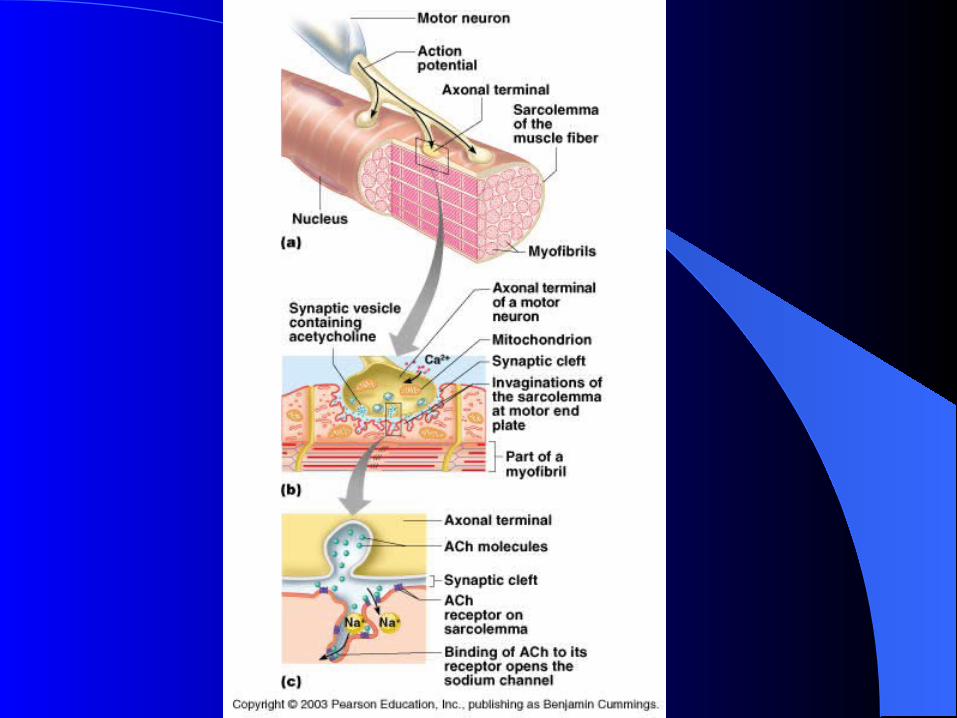
Muscle activationMuscle activation DQ - How are muscles activated to
contract? The neuromuscular junction (pg 185) Each skeletal muscle fiber is connected to an
extension of a motor neuron. The nerve fiber and the muscle meet at the
neuromuscular junction.– The two do not actually touch – Synaptic cleft
– Then how does the nerve stimulate the fiber? Neurotransmitters (Acetylcholine)

Motor UnitsMotor Units
DQ - Does each nerve only control one muscle fiber?
NO, we have motor units. (pg 184)– consists of a motor neuron and all of the muscle
fibers it controls. The number of muscle fibers in a motor unit
varies considerably. DQ - Why would that be? The fewer muscle fibers in the motor units, the
finer the movement.– Examples – hands vs legs


Muscle ResponseMuscle Response When the nerve receives an impulse, the neuron
releases the neurotransmitter into the synaptic cleft. DQ - What will this cause? All muscle fibers in the motor unit contract
– All or none response DQ - Does this mean entire muscle
contracts? NO, just the motor unit
– DQ - How do we control force of muscles? More motor units = More force

Skeletal Muscle ContractionSkeletal Muscle Contraction
The sliding filament theory is the most widely accepted theory on how muscle contraction works.
It says that the head of a myosin cross-bridge can attach to an actin binding site and bend slightly– What will this cause?
The actin to move (or slide) with it The head can then release, straighten itself, and
combine with another binding site farther down the actin filament. – Example, arms with a meter stick
This can happen several times causing the muscle fiber to shorten dramatically

- Shortening ofa sarcomere

Skeletal Muscle ContractionSkeletal Muscle Contraction
All of this binding and changing of shape will take energy, where will it come from?
ATP DQ – How is ATP made? (3 ways) (pg 190) Direct Phosphorylation
– Fastest, Lasts ~15 sec No O2 needed
Anaerobic glycolysis– Lasts ~ 30 – 60 sec Causes lactic acid, No O2
Aerobic respiration– Slowest, Lasts hours, Requires O2

Skeletal Muscle ContractionSkeletal Muscle Contraction
Two types of contractions Isotonic contractions
– Contractions that shorten the muscle.– This is the more common one
Isometric contractions – Tension in the muscle but no change in
length.

Steps to muscle contractionSteps to muscle contraction
1. Nerve impulse arrives at the neuromuscular junction
2. Acetylcholine diffuses across the gap at the neuromuscular junction
3. T Tubules carry acetlycholine to sarcoplasmic reticulum, which causes cell membrane to be permeable to Ca2+
4. Ca2+ binds to troponin

Steps to muscle contractionSteps to muscle contraction
5. Conformation shift - troponin pulls tropomyosin off active binding sites
6. One ATP per myosin Head releases and activates myosin
7. Actin and myosin filaments form linkages8. Myosin cross-bridges pull actin filaments
inward9. Muscle fiber shortens and contracts

Muscle RelaxingMuscle Relaxing DQ - What happens when a muscle
does not need to contract anymore? – Think about how it all started
The acetylcholine needs to be removed the Ach is rapidly decomposed by action of an
enzyme called acetylcholinesterase. (Ach-ase)– This enzyme is present at the neuromuscular junction
The Ach-ase stops a single nerve impulse from continuously stimulating the muscle fiber.

Muscle RelaxingMuscle Relaxing As Ach is broken down, the calcium pump
quickly moves Ca2+ back into the sarcoplasmic reticulum.
DQ - What does this do? The linkages will break– the troponin and tropomyosin return to the normal
conformation, blocking the binding sites.
The muscle fiber relaxes.

Muscle FatigueMuscle Fatigue Muscle fatigue most often occurs because of oxygen
debt.– Causing?
Anaerobic respiration – accumulation of lactic acid. Oxygen debt is “paid back” after activity
– Heavy breathing DQ - How are people able to “train” to
prolong activity before muscle fatigue? Exercise stimulates new capillaries to grow within the
muscles and it also causes an increase in the number of mitochondria

Rigor MortisRigor Mortis What is it? Stiffening of joints and locking into place after
death– Why does it happen?
As the cells begin to shut down, the sarcoplasmic reticulum becomes permeable to Ca2+
– Causing what? Sets in place within a few hours of death
– Lasts ~72 hrs

Fast and Slow MusclesFast and Slow Muscles
There are two types of muscle fibers relating to the speed of contraction.– Fast and Slow
The speed of contraction is related to the specialized function of a muscle.– Example: Eye muscles that blink contract
ten times faster than the muscles involved in posture.

Fast and Slow MusclesFast and Slow Muscles Slow-contracting (slow twitch) fibers Often called red muscles
– Why?
The fibers contain red, oxygen-storing myoglobin.
Well supplied with blood. What would these be useful for?

Fast and Slow MusclesFast and Slow Muscles
Fast-contracting (fast twitch) muscle fibers
Also called white muscles. – Why?
They contain less myoglobin and have a poorer blood supply than red muscles.
What would these be used for? Shown to have a very well developed
sarcoplasmic reticulum?– Why is that important?

Fast and Slow MusclesFast and Slow Muscles
Research has discovered an intermediate fiber.
How would you classify these?

Fast and Slow twitchFast and Slow twitch
How would you describe the ratio of fast twitch to slow twitch for Usain Bolt? Ryan Hall?

Muscle HypertrophyMuscle Hypertrophy
Hypertrophy – increasing in size Occurs when more proteins are produced, not
more muscle fibers/muscle cells. Occurs from working muscles hard or isometric
exercises When worked hard the muscle becomes
damaged, which causes an increased number of nuclei.– DQ - Why would increased nuclei cause increase
in muscle mass (think transcription and translation)?

Muscle AtrophyMuscle Atrophy
Atrophy – decrease in size Caused by lack of use
– Proteins broken down

Muscle interactionsMuscle interactions
Prime movers – responsible for major movements Antagonists – muscles that oppose or reverse a
movement (examples?) Synergists - help prime movers by helping with
movement or stabilizing unwanted movement. (Examples?)
Fixators – specialized synergists, stabilize bones (Ex. rhomboideus major)

DIDN’T USEDIDN’T USE
ANY SLIDES AFTER THIS POINT, I DID NOT USE, BUT KEPT IN CASE I WANTED TO.

Skeletal Muscle ContractionSkeletal Muscle Contraction
In the presence of calcium ions, the myosin cross-bridges react with actin filaments and form linkages with them.
This reaction between the myosin and actin filaments provides the force that shortens myofibrils during muscle contraction.

Skeletal Muscle ContractionSkeletal Muscle Contraction
Actin accounts for about 1/4 of the total protein in skeletal muscle.
Actin molecules, arranged together in a double twisted strand, form an actin filament.
Tropomyosin & troponin are two proteins associated with actin filaments.

Skeletal Muscle ContractionSkeletal Muscle Contraction
The tropomyosin-troponin complex blocks the binding sites on the actin molecules when the muscle is at rest.
If a high concentration of calcium ions is present, the calcium ions bind to the troponin, and this modifies the position of the tropomyosin.

Skeletal Muscle ContractionSkeletal Muscle Contraction
The tropomyosin molecules move, exposing the binding sites on the actin filaments, and the linkages form between the actin and myosin filaments.

Skeletal Muscle ContractionSkeletal Muscle Contraction
The sliding filament theory of muscle contraction suggests that the head of a myosin cross-bridge can attach to an actin binding site and bend slightly, pulling the actin filament with it.
Then the head can release, straighten itself, and combine with another binding site farther down the actin filament.

Skeletal Muscle ContractionSkeletal Muscle Contraction
The enzyme ATPase causes the breakdown of ATP to supply energy for these actions.

Stimulus for ContractionStimulus for Contraction
Acetylcholine (ACh) is a neurotransmitter that is synthesized in the cytoplasm of the motor neuron and is stored in vesicles.
A nerve impulse reaches the end of the axon, some of these vesicles release ACh into the gap between the nerve and the motor end plate.

Stimulus for ContractionStimulus for Contraction
The ACh diffuses rapidly across the gap, combines with certain protein molecules in the sarcolemma, and thus stimulates the muscle fiber membrane.
This stimulus causes a muscle impulse that passes in all directions over the surface of the sarcolemma.

Stimulus for ContractionStimulus for Contraction
It also travels through the sarcoplasmic reticulum and the transverse tubules.
The sarcoplasmic reticulum contains a high concentration of calcium ions compared to the sarcoplasm.

Stimulus for ContractionStimulus for Contraction
In response to a muscle impulse, the membranes of the cisternae become more permeable to these ions and the calcium ions diffuse into the sarcoplasm of the muscle fiber.

Stimulus for ContractionStimulus for Contraction
When a relatively high concentration of calcium ions is present in the sarcoplasm, linkages form between the actin and myosin filaments, and a muscle contracts.

Energy Sources of Energy Sources of ContractionContraction
The energy for muscle contractions comes from ATP.
The muscle has enough ATP to contract briefly. Therefore, when a fiber is active, ATP must be regenerated.
Creatine phosphate supplies the energy to change ADP back to energy rich ATP

Oxygen Supply and Cellular Oxygen Supply and Cellular RespirationRespiration
Oxygen is transported by the red blood cells. It is loosely bound to molecules of hemoglobin.
The hemoglobin releases the oxygen in areas of the body that are low in oxygen content

Oxygen Supply and Cellular Oxygen Supply and Cellular RespirationRespiration
Myoglobin, in the muscle cells, can store oxygen temporarily.
This reduces a muscle’s need for a continuous blood supply during a contraction.

Oxygen DebtOxygen Debt
During strenuous exercise, the available oxygen supply may be used up. The body then relies on anaerobic respiration creating an oxygen debt.
Anaerobic respiration builds up lactic acid. The liver converts lactic acid back to glucose, but it takes several hours to complete the conversion.

Muscle CrampsMuscle Cramps
Cramps occur when the muscle contracts spasmodically, but does not relax completely.
The condition is due to a lack of ATP needed to move calcium or other ions, can also be caused by a lack of those ions
How would a lack of ATP be a problem? ATP required to release cross bridges Exercise stimulates new capillaries to grow
within the muscles and it also causes an increase in the number of mitochondria.

Heat ProductionHeat Production
Since muscle tissue represents a large proportion of the total body mass, it is a major source of heat.
About 25% of the energy released in cellular respiration is available for use in metabolic processes.

Heat ProductionHeat Production
Active muscles release large amounts of heat.
Blood transports this heat to other tissues to help maintain body temperature.
Smooth MuscleSmooth Muscle
Smooth muscle characteristics: Shorter than the fibers of skeletal
muscle. Single, central nucleus. Cells are elongated with tapering ends. Filaments are more randomly arranged
than skeletal muscle.

Smooth MuscleSmooth Muscle
The two types of smooth muscle are multiunit and visceral.
Multiunit muscles are less organized and occur as separate fibers.
Multiunit are found in the irises of the eyes and walls of blood vessels.
They contract through motor nerve impulse or hormone action.

Smooth MuscleSmooth Muscle
Visceral smooth muscle is composed of sheets of spindle shaped cells held together by gap junctions.
Visceral smooth muscles are found in walls of hollow organs (ex. Stomach, intestines)
There usually will be two muscle layers, a longitudinal and a circular layer.

Smooth MuscleSmooth Muscle
When one fiber is stimulated, the impulse will also excite adjacent cells causing a rhythmic contraction.
Peristalsis - wavelike contraction that occurs in tubular organs.

Smooth Muscle ContractionSmooth Muscle Contraction
Smooth muscle contraction is similar to skeletal muscle contraction.
Smooth muscle lacks troponin. Instead it uses a protein called calmodulin to bind calcium ions.
Calcium diffuses into the cell from the extracellular fluid.

Smooth Muscle ContractionSmooth Muscle Contraction
Smooth muscle reacts to the neurotransmitter ACh and norepinephrine.
Some hormones cause smooth muscle contraction. (Ex. Childbirth)
Stretching smooth muscle can cause contractions. (Ex. Digesting food in the stomach)

Smooth Muscle ContractionSmooth Muscle Contraction
Smooth muscle is slower to contract and slower to relax.
Smooth muscle can contract for a longer time with the same amount of ATP.
Smooth muscles stretch without changing tautness while a hollow organ fills.

Cardiac MuscleCardiac Muscle
Found only in the heart. Striated cells joined end to end forming
interconnecting branched three-dimensional network.
Each cell contains a single nucleus. Well developed sarcoplasmic reticulum
and transverse tubules.
Cardiac MuscleCardiac Muscle
Sarcoplasmic reticulum stores less calcium but the enlarged transverse tubules store extra calcium.
The extra calcium allows cardiac muscle to maintain a contraction longer than skeletal muscles.

Cardiac MuscleCardiac Muscle
Intercalated disks separate opposing ends of cardiac cells.
The disks help hold adjacent cells together and transmit the force of contraction from cell to cell.
Cardiac MuscleCardiac Muscle
When one portion of the cardiac network is stimulated, the impulse passes throughout the network causing the whole structure to contract as a unit.

Muscle ActionMuscle Action
The main muscle is the prime mover. Muscles that contract along with the
prime mover are called synergists. Muscles opposing the prime mover are
called antagonists.