musculoskeletal examination v - rheum · the screening musculoskeletal examination1 the screening...
TRANSCRIPT
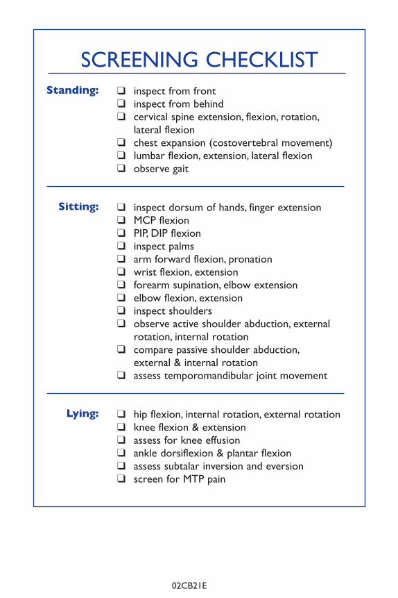
SCrEEnIng ChECklIST
Standing:
Sitting:
Lying:
q inspect from front
q inspect from behind
q cervical spine extension, flexion, rotation,
lateral flexion
q chest expansion (costovertebral movement)
q lumbar flexion, extension, lateral flexion
q observe gait
q inspect dorsum of hands, finger extension
q MCP flexion
q PIP, DIP flexion
q inspect palms
q arm forward flexion, pronation
q wrist flexion, extension
q forearm supination, elbow extension
q elbow flexion, extension
q inspect shoulders
q observe active shoulder abduction, external
rotation, internal rotation
q compare passive shoulder abduction,
external & internal rotation
q assess temporomandibular joint movement
q hip flexion, internal rotation, external rotation
q knee flexion & extension
q assess for knee effusion
q ankle dorsiflexion & plantar flexion
q assess subtalar inversion and eversion
q screen for MTP pain
The MusculoskeletalScreening Examination
John M. Thompson MD FRCPC
and Ainsley Walton
02CB21E
MSK Book ENG CVR 94193_HSE COX C00011-Musculoske.49454 12-06-21 11:47 AM Page 1
Contents
Introduction . . . . . . . . . . . . . . . . . . . . . . . . . . . . . . . . . . . . . . . . . . . . . p.1
Patient Standing . . . . . . . . . . . . . . . . . . . . . . . . . . . . . . . . . . . . . . . . . . p.3
Patient Sitting . . . . . . . . . . . . . . . . . . . . . . . . . . . . . . . . . . . . . . . . . . . . p.12
Patient lying . . . . . . . . . . . . . . . . . . . . . . . . . . . . . . . . . . . . . . . . . . . . . p.26
Written By
John M. Thompson MD FrCPC
Division of rheumatology
Department of Medicine
Faculty of Medicine
University of Western Ontario
St. Joseph’s hospital
268 grosvenor Street
london, Ontario n6A 4V2
Illustrated By
Ainsley S. Walton
©1998 by John M. Thompson
Unauthorized duplication of these materials is strictly prohibited.
Dedicated To
hugh little MD FrCPC
mentor, colleague and friend
The Financial assistance of
The Arcangelo Rea Family Foundation
is gratefully acknowledged
Supported by an educational grant
from Pfizer Canada Inc.
Thanks to
karen and John hueston of the Aylmer Express
Anne lyddiatt
and my colleagues in the Division of rheumatology
MSK Book ENG CVR 94193_HSE COX C00011-Musculoske.49454 12-06-21 11:47 AM Page 2
1The Screening Musculoskeletal Examination
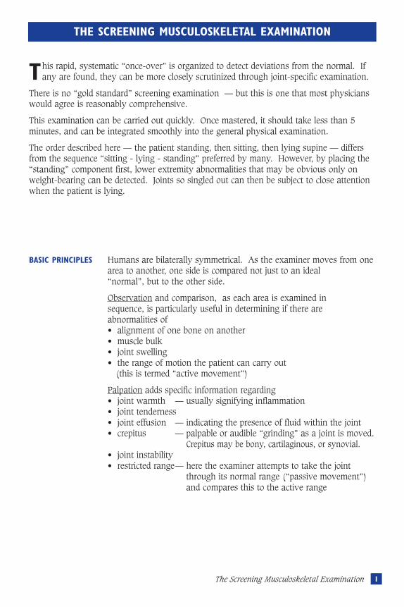
THE SCREENING MUSCULOSKELETAL EXAMINATION
This rapid, systematic “once-over” is organized to detect deviations from the normal. Ifany are found, they can be more closely scrutinized through joint-specific examination.
There is no “gold standard” screening examination — but this is one that most physicianswould agree is reasonably comprehensive.
This examination can be carried out quickly. Once mastered, it should take less than 5minutes, and can be integrated smoothly into the general physical examination.
The order described here — the patient standing, then sitting, then lying supine — differsfrom the sequence “sitting - lying - standing” preferred by many. However, by placing the“standing” component first, lower extremity abnormalities that may be obvious only onweight-bearing can be detected. Joints so singled out can then be subject to close attentionwhen the patient is lying.
BASIC PRINCIPLES Humans are bilaterally symmetrical. As the examiner moves from onearea to another, one side is compared not just to an ideal “normal”, but to the other side.
Observation and comparison, as each area is examined in sequence, is particularly useful in determining if there are abnormalities of• alignment of one bone on another• muscle bulk• joint swelling• the range of motion the patient can carry out
(this is termed “active movement”)
Palpation adds specific information regarding• joint warmth — usually signifying inflammation• joint tenderness• joint effusion — indicating the presence of fluid within the joint• crepitus — palpable or audible “grinding” as a joint is moved.
— Crepitus may be bony, cartilaginous, or synovial.• joint instability• restricted range— here the examiner attempts to take the joint
— through its normal range (“passive movement”) — and compares this to the active range
Musculoskeletal revisec.57105 5/16/05 1:59 PM Page 1
2 The Screening Musculoskeletal Examination
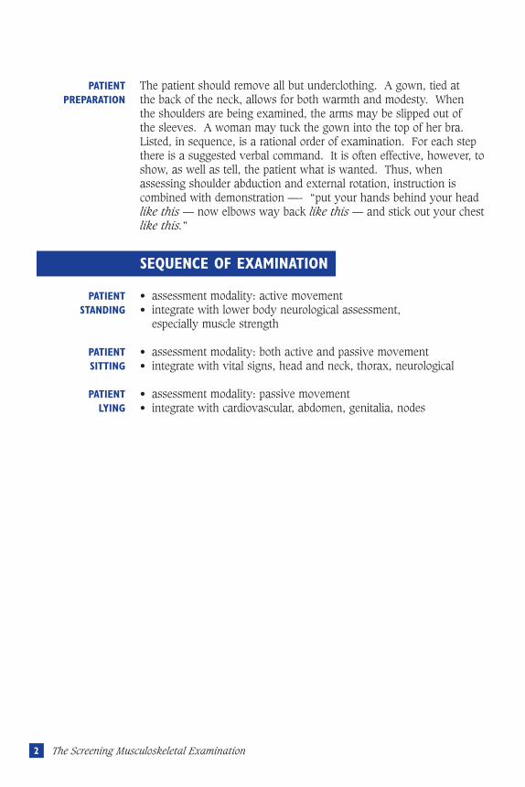
SEQUENCE OF EXAMINATION
PATIENT The patient should remove all but underclothing. A gown, tied at PREPARATION the back of the neck, allows for both warmth and modesty. When
the shoulders are being examined, the arms may be slipped out of the sleeves. A woman may tuck the gown into the top of her bra.Listed, in sequence, is a rational order of examination. For each stepthere is a suggested verbal command. It is often effective, however, toshow, as well as tell, the patient what is wanted. Thus, whenassessing shoulder abduction and external rotation, instruction iscombined with demonstration —- “put your hands behind your headlike this — now elbows way back like this — and stick out your chestlike this.”
PATIENT • assessment modality: active movementSTANDING • integrate with lower body neurological assessment,
• especially muscle strength
PATIENT • assessment modality: both active and passive movementSITTING • integrate with vital signs, head and neck, thorax, neurological
PATIENT • assessment modality: passive movementLYING • integrate with cardiovascular, abdomen, genitalia, nodes
Musculoskeletal revisec.57105 5/16/05 1:59 PM Page 2
3I Patient Standing
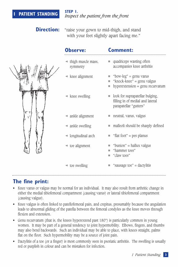
I PATIENT STANDING STEP 1.Inspect the patient from the front
Direction: “raise your gown to mid-thigh, and stand with your feet slightly apart facing me.”
Observe:
thigh muscle mass, symmetry
knee alignment
knee swelling
ankle alignment
ankle swelling
longitudinal arch
toe alignment
toe swelling
Comment:
■ quadriceps wasting often accompanies knee arthritis
■ “bow-leg” = genu varus■ “knock-knee” = genu valgus■ hyperextension = genu recurvatum
■ look for suprapatellar bulging, filling in of medial and lateral parapatellar “gutters”
■ neutral, varus, valgus
■ malleoli should be sharply defined
■ “flat foot” = pes planus
■ “bunion” = hallux valgus■ “hammer toes”■ “claw toes”
■ “sausage toe” = dactylitis
The fine print:• Knee varus or valgus may be normal for an individual. It may also result from arthritic change in
either the medial tibiofemoral compartment (causing varus) or lateral tibiofemoral compartment(causing valgus).
• Knee valgus is often linked to patellofemoral pain, and crepitus, presumably because the angulationleads to abnormal gliding of the patella between the femoral condyles as the knee moves throughflexion and extension.
• Genu recurvatum (that is, the knees hyperextend past 180°) is particularly common in youngwomen. It may be part of a general tendency to joint hypermobility. Elbows, fingers, and thumbsmay also bend backwards. Such an individual may be able to place, with knees straight, palmsflat on the floor. Such hypermobility may be a source of joint pain.
• Dactylitis of a toe (or a finger) is most commonly seen in psoriatic arthritis. The swelling is usuallyred or purplish in colour and can be mistaken for infection.
Musculoskeletal revisec.57105 5/16/05 1:59 PM Page 3
4 The Screening Musculoskeletal Examination
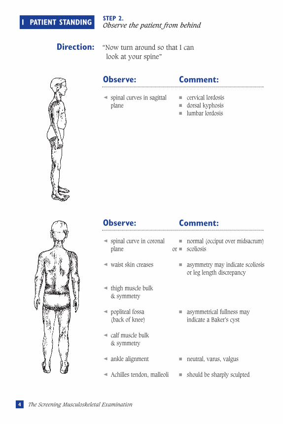
I PATIENT STANDING STEP 2.Observe the patient from behind
Direction: “Now turn around so that I can look at your spine”
Observe:
spinal curves in sagittalplane
Comment:
■ cervical lordosis■ dorsal kyphosis■ lumbar lordosis
Observe:
spinal curve in coronalplane
waist skin creases
thigh muscle bulk & symmetry
popliteal fossa (back of knee)
calf muscle bulk & symmetry
ankle alignment
Achilles tendon, malleoli
Comment:
■ normal (occiput over midsacrum)■ scoliosis
■ asymmetry may indicate scoliosis or leg length discrepancy
■ asymmetrical fullness may indicate a Baker’s cyst
■ neutral, varus, valgus
■ should be sharply sculpted
or
Musculoskeletal revisec.57105 5/16/05 1:59 PM Page 4

5I Patient Standing
The fine print:• Scoliosis may be more obvious when the patient demonstrates forward flexion (step 3)
• A difference in leg length, either actual or factitious (due to incomplete extension of either the hipor knee) may be assessed by palpating the iliac crests in the mid-axillary line. With an index fingeron each side, look to see if they line up horizontally, or if there is a pelvic tilt.
• In roughly a third of all patients, a communication exists between the knee joint space and thepopliteal bursa. In the presence of a knee effusion, joint fluid will drain from the high pressureknee to low pressure bursa, resulting in bursal swelling (a Baker’s cyst). If the swelling comes onrapidly, the bursa may rupture, with calf pain and ankle bruising, simulating a deep veinthrombosis of the calf. If the swelling is chronic, it may expand into the calf beneath thegastrocnemius, causing calf asymmetry.
• A unilaterally thickened Achilles tendon, often red and tender, is a hallmark of seronegativearthritis.
• The normal concavity beneath the malleoli may be “filled in” in the case of tenosynovitis involvingthe posterior tibial tendon (medially) or peroneus longus tendon (laterally)
Musculoskeletal revisec.57105 5/16/05 1:59 PM Page 5
6 The Screening Musculoskeletal Examination
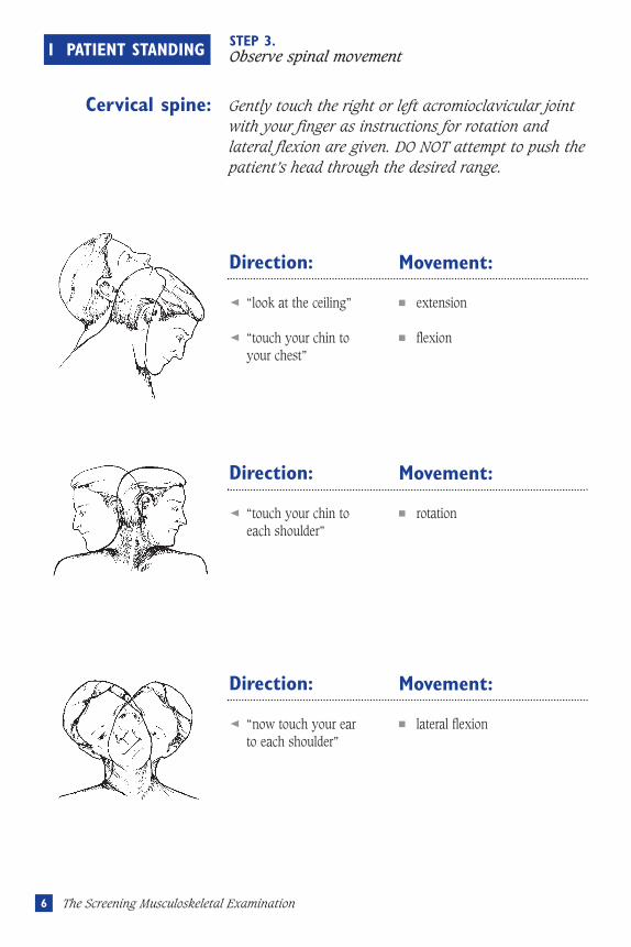
I PATIENT STANDING STEP 3.Observe spinal movement
Direction:
“look at the ceiling”
“touch your chin to your chest”
Movement:
■ extension
■ flexion
Direction:
“touch your chin to each shoulder”
Movement:
■ rotation
Direction:
“now touch your ear to each shoulder”
Movement:
■ lateral flexion
Cervical spine: Gently touch the right or left acromioclavicular joint with your finger as instructions for rotation and lateral flexion are given. DO NOT attempt to push the patient’s head through the desired range.
Musculoskeletal revisec.57105 5/16/05 1:59 PM Page 6
7I Patient Standing
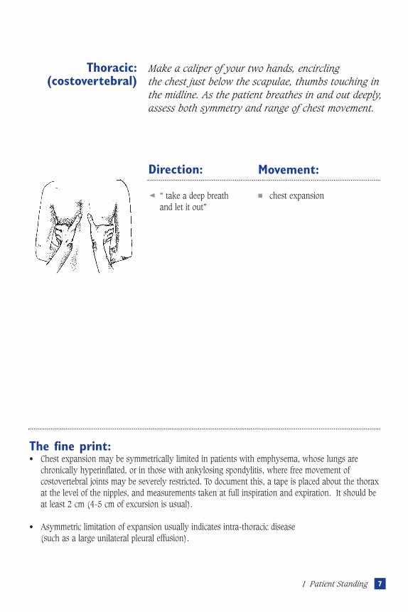
Direction:
“ take a deep breathand let it out”
Movement:
■ chest expansion
Thoracic: Make a caliper of your two hands, encircling (costovertebral) the chest just below the scapulae, thumbs touching in
the midline. As the patient breathes in and out deeply,assess both symmetry and range of chest movement.
The fine print:• Chest expansion may be symmetrically limited in patients with emphysema, whose lungs are
chronically hyperinflated, or in those with ankylosing spondylitis, where free movement ofcostovertebral joints may be severely restricted. To document this, a tape is placed about the thoraxat the level of the nipples, and measurements taken at full inspiration and expiration. It should beat least 2 cm (4-5 cm of excursion is usual).
• Asymmetric limitation of expansion usually indicates intra-thoracic disease (such as a large unilateral pleural effusion).
Musculoskeletal revisec.57105 5/16/05 1:59 PM Page 7
8 The Screening Musculoskeletal Examination
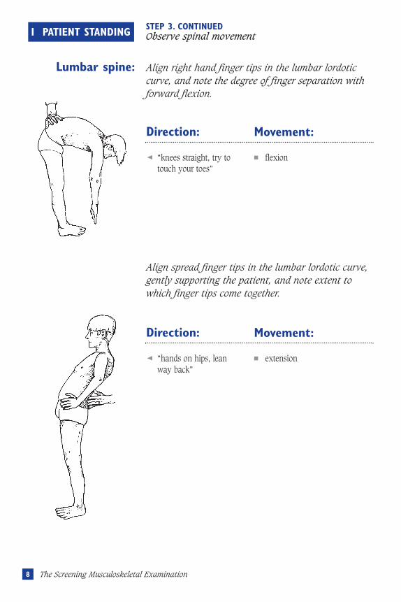
I PATIENT STANDING STEP 3. CONTINUED Observe spinal movement
Direction:
“knees straight, try totouch your toes”
Movement:
■ flexion
Lumbar spine: Align right hand finger tips in the lumbar lordotic curve, and note the degree of finger separation with forward flexion.
Direction:
“hands on hips, leanway back”
Movement:
■ extension
Align spread finger tips in the lumbar lordotic curve, gently supporting the patient, and note extent to which finger tips come together.
Musculoskeletal revisec.57105 5/16/05 1:59 PM Page 8
9I Patient Standing
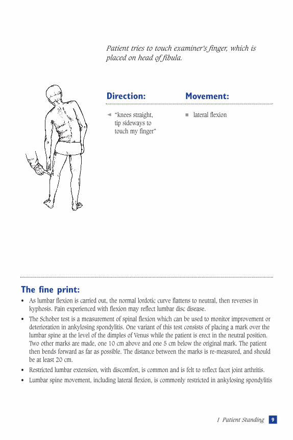
Direction:
“knees straight, tip sideways to touch my finger”
Movement:
■ lateral flexion
Patient tries to touch examiner’s finger, which is placed on head of fibula.
The fine print:• As lumbar flexion is carried out, the normal lordotic curve flattens to neutral, then reverses in
kyphosis. Pain experienced with flexion may reflect lumbar disc disease.
• The Schober test is a measurement of spinal flexion which can be used to monitor improvement ordeterioration in ankylosing spondylitis. One variant of this test consists of placing a mark over thelumbar spine at the level of the dimples of Venus while the patient is erect in the neutral position.Two other marks are made, one 10 cm above and one 5 cm below the original mark. The patientthen bends forward as far as possible. The distance between the marks is re-measured, and shouldbe at least 20 cm.
• Restricted lumbar extension, with discomfort, is common and is felt to reflect facet joint arthritis.
• Lumbar spine movement, including lateral flexion, is commonly restricted in ankylosing spondylitis
Musculoskeletal revisec.57105 5/16/05 1:59 PM Page 9
10 The Screening Musculoskeletal Examination
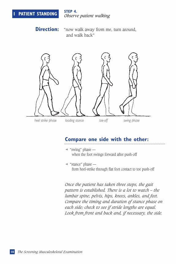
I PATIENT STANDING STEP 4.Observe patient walking
Direction: “now walk away from me, turn around, and walk back”
Compare one side with the other:
“swing” phase — when the foot swings forward after push-off
“stance” phase — from heel-strike through flat foot contact to toe push-off
Once the patient has taken three steps, the gaitpattern is established. There is a lot to watch – thelumbar spine, pelvis, hips, knees, ankles, and feet.Compare the timing and duration of stance phase oneach side; check to see if stride lengths are equal.Look from front and back and, if necessary, the side.
heel strike phase loading stance toe-off swing phase
Musculoskeletal revisec.57105 5/16/05 1:59 PM Page 10
The fine print:• The antalgic (antalgic = “against pain”) gait may reflect a painful focus anywhere from the foot up
to the pelvis. The patient attempts to “unload” the painful side. Consequently, the stance phase onthat side is briefer, and the swing phase on the opposite (normal) side is shorter – making for ashorter stride.
• The arthrogenic (stiff knee or hip) gait is a consequence of joint stiffness, and is not necessarilypainful. Because there is a limited hip/knee flexion on the affected side, there is exaggerated footplantar flexion on the “good” side (the patient goes up higher on the toes), whereas the “bad” legis circumducted during the swing phase to allow toe clearance.
• Trendelenburg’s Gait results from unilateral hip abductor muscle dysfunction. This may be aconsequence of neurological weakness, or of hip joint dysfunction (e.g. hip osteoarthritis) thatwould be aggravated by adductor contraction (which can double the weight-bearing load on thathip). There are two components to the Trendelenburg gait. The thorax is thrust laterally to placethe center of gravity over the “bad” leg during the stance phase, and simultaneously the pelvissags on the contralateral side (because the ipsilateral hip adductors do not contract to hold thepelvis level).
11I Patient Standing
Musculoskeletal revisec.57105 5/16/05 1:59 PM Page 11
12 The Screening Musculoskeletal Examination
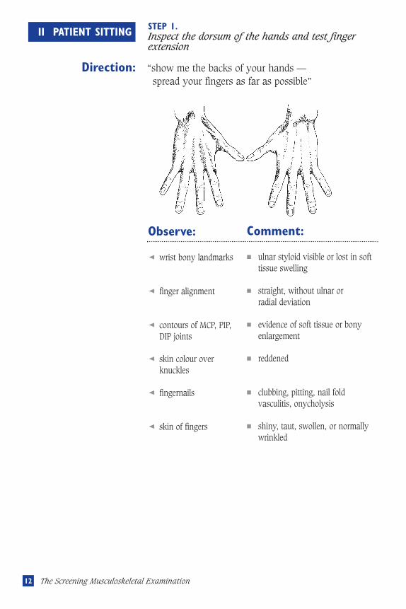
II PATIENT SITTING STEP 1.Inspect the dorsum of the hands and test fingerextension
Direction: “show me the backs of your hands —spread your fingers as far as possible”
Observe:
wrist bony landmarks
finger alignment
contours of MCP, PIP,DIP joints
skin colour overknuckles
fingernails
skin of fingers
Comment:
■ ulnar styloid visible or lost in softtissue swelling
■ straight, without ulnar or radial deviation
■ evidence of soft tissue or bonyenlargement
■ reddened
■ clubbing, pitting, nail foldvasculitis, onycholysis
■ shiny, taut, swollen, or normallywrinkled
Musculoskeletal revisec.57105 5/16/05 1:59 PM Page 12
13II Patient Sitting
The fine print:• Ulnar deviation of the fingers, due to rheumatoid arthritis, occurs only after several years of active
disease.
• If the fingers cannot be fully extended, there is probably a finger flexion contracture. This can beconfirmed by the “prayer sign” — ask the patient to oppose palms and digits as if praying. Thereshould be no visible gap between palmar surfaces of the fingers.
• The most common cause of enlargement of the DIP joint is nodal osteoarthritis. These bony bumpsare known as Heberden’s nodes (their counterparts in the PIP joints are known as Bouchard’snodes). Early in their development they may be quite tender and even inflamed, but not asinflamed as the swollen, red, tender DIP joints seen in some patients with psoriatic arthritis.
• Redness of knuckles, denoting inflammation, is seldom seen in the commonest form ofinflammatory arthritis, rheumatoid arthritis — but it is a feature of gouty arthritis, infection,psoriatic arthritis, and even in some cases of nodal osteoarthritis (see above).
• Clubbing may be a clue to an underlying malignancy or endocarditis. Pitting of the fingernails, orseparation of the nail from the underlying tissue (onycholysis), may provide the essential clue to adiagnosis of psoriatic arthritis. Nail fold vasculitis is seen in lupus and rheumatoid arthritis.
• Taut skin over the fingers may be a clue to scleroderma.
Musculoskeletal revisec.57105 5/16/05 1:59 PM Page 13
14 The Screening Musculoskeletal Examination
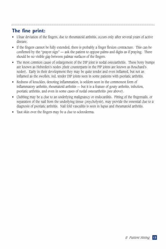
II PATIENT SITTING STEP 2.Assess MCP joint flexion
Direction: “Make a fist”
Observe:
knuckles flex to almost90°
troughs between MCPsare concave
Comment:
■ “filled in” troughs may be due tosubcutaneous tissue (“pudgyhands”) or to MCP joint capsulardistension.
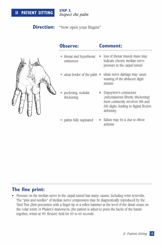
The fine print:• Inflammation of the MCP joints, evidenced by capsular distension, is
one of the hallmarks of rheumatoid arthritis. If suspected, the MCPjoints should be palpated, using both thumbs (see illustration). Tryto feel the joint lines.
• A quick “screen” for MCP arthritis is the manoeuvre shown(compression of the MCPs from the side). A similar “screen” iscarried out on the MTP joints of the feet.
Musculoskeletal revisec.57105 5/16/05 1:59 PM Page 14
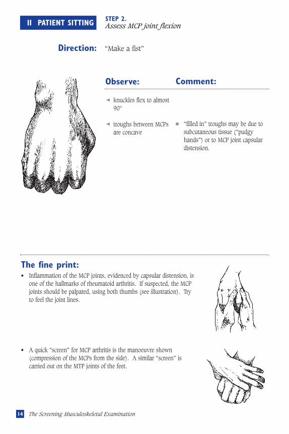
STEP 3.Inspect the palm
Direction: “Now open your fingers”
Observe:
thenar and hypothenareminences
ulnar border of the palm
puckering, nodularthickening
palms fully supinated
Comment:
loss of thenar muscle mass mayindicate chronic median nervepressure in the carpal tunnel
ulnar nerve damage may causewasting of the abductor digitiminimi
Dupuytren’s contracture(subcutaneous fibrotic thickening)most commonly involves 4th and5th digits, leading to digital flexiondeformity
failure may be a clue to elbowarthritis
The fine print:• Pressure on the median nerve in the carpal tunnel has many causes, including wrist synovitis.
The “pins and needles” of median nerve compression may be diagnostically reproduced by theTinel Test (firm percussion with a finger tip or a reflex hammer at the level of the distal crease onthe volar wrist) or Phalen’s manoeuvre (the patient is asked to press the backs of the handstogether, wrists at 90o flexion) held for 45 to 60 seconds.
Musculoskeletal revisec.57105 5/16/05 1:59 PM Page 16
II Patient Sitting
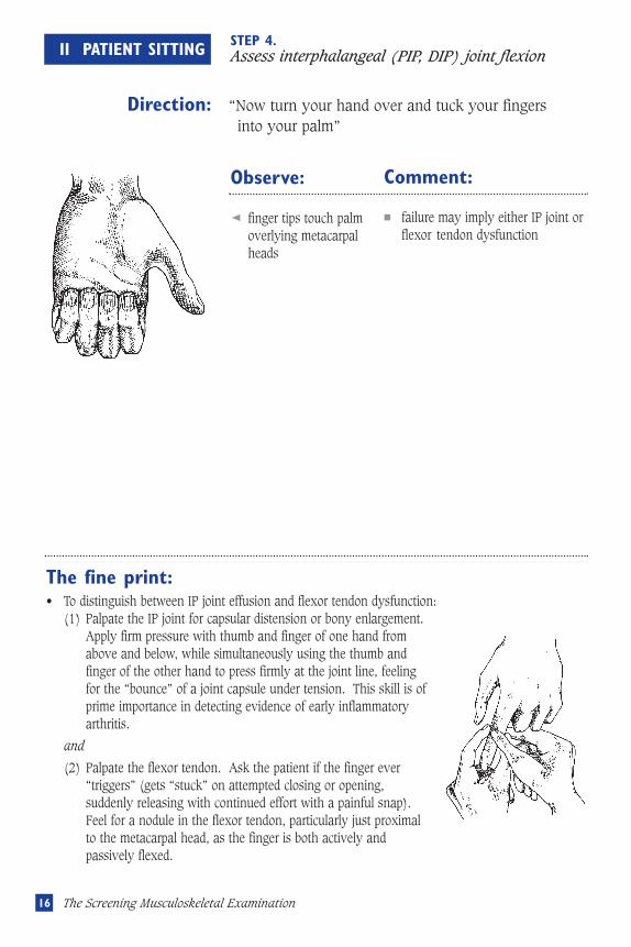
STEP 4.Assess interphalangeal (PIP, DIP) joint flexion
Direction: “Now turn your hand over and tuck your fingers into your palm”
Observe:
finger tips touch palmoverlying metacarpalheads
Comment:
failure may imply either IP joint orflexor tendon dysfunction
The fine print:• To distinguish between IP joint effusion and flexor tendon dysfunction:
(1) Palpate the IP joint for capsular distension or bony enlargement. Apply firm pressure with thumb and finger of one hand from above and below, while simultaneously using the thumb and finger of the other hand to press firmly at the joint line, feeling for the “bounce” of a joint capsule under tension. This skill is of prime importance in detecting evidence of early inflammatory arthritis.
and
(2) Palpate the flexor tendon. Ask the patient if the finger ever “triggers” (gets “stuck” on attempted closing or opening, suddenly releasing with continued effort with a painful snap). Feel for a nodule in the flexor tendon, particularly just proximal to the metacarpal head, as the finger is both actively and passively flexed.
Musculoskeletal revisec.57105 5/16/05 1:59 PM Page 15
The Screening Musculoskeletal Examination
17II Patient Sitting
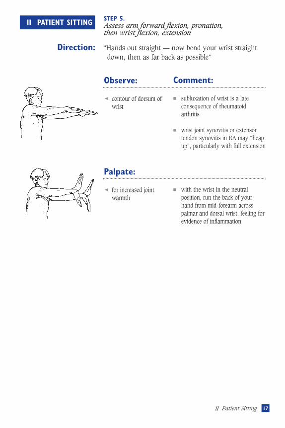
II PATIENT SITTING STEP 5.Assess arm forward flexion, pronation, then wrist flexion, extension
Direction: “Hands out straight — now bend your wrist straight down, then as far back as possible”
Observe:
contour of dorsum ofwrist
Comment:
■ subluxation of wrist is a lateconsequence of rheumatoidarthritis
■ wrist joint synovitis or extensortendon synovitis in RA may “heapup”, particularly with full extension
Palpate:
for increased jointwarmth
■ with the wrist in the neutralposition, run the back of yourhand from mid-forearm acrosspalmar and dorsal wrist, feeling forevidence of inflammation
Musculoskeletal revisec.57105 5/16/05 1:59 PM Page 17
18 The Screening Musculoskeletal Examination
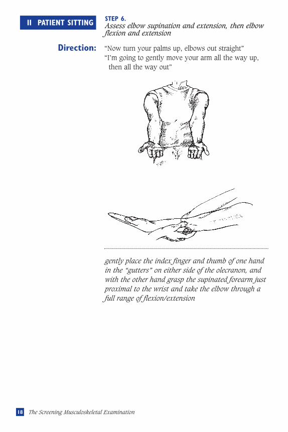
II PATIENT SITTING STEP 6.Assess elbow supination and extension, then elbowflexion and extension
Direction: “Now turn your palms up, elbows out straight”“I’m going to gently move your arm all the way up,
then all the way out”
Observe:
gently place the index finger and thumb of one handin the “gutters” on either side of the olecranon, andwith the other hand grasp the supinated forearm justproximal to the wrist and take the elbow through afull range of flexion/extension
Musculoskeletal revisec.57105 5/16/05 1:59 PM Page 18
19II Patient Sitting
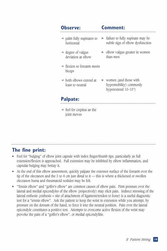
Observe:
palm fully supinates tohorizontal
degree of valgusdeviation at elbow
flexion so forearm meetsbiceps
both elbows extend atleast to neutral
Comment:
■ failure to fully supinate may besubtle sign of elbow dysfunction
■ elbow valgus greater in womenthan men
■ women (and those withhypermobility) commonlyhyperextend 10-15°)
Palpate:
feel for crepitus as thejoint moves
The fine print:• Feel for “bulging” of elbow joint capsule with index finger/thumb tips, particularly as full
extension/flexion is approached. Full extension may be inhibited by elbow inflammation, andcapsular bulging may betray it.
• At the end of this elbow assessment, quickly palpate the extensor surface of the forearm over thetip of the olecranon and the 3 to 4 cm just distal to it — this is where a thickened or swollenolecranon bursa and rheumatoid nodules may be felt.
• “Tennis elbow” and “golfer’s elbow” are common causes of elbow pain. Firm pressure over thelateral and medial epicondyles of the elbow (respectively) may elicit pain. Indirect stressing of thelateral enthesis (enthesis = site of attachment of ligament/tendon to bone) is a useful diagnostictest for a “tennis elbow”. Ask the patient to keep the wrist in extension while you attempt, bypressure on the dorsum of the hand, to force it into the neutral position. Pain over the lateralepicondyle constitutes a positive test. Attempts to overcome active flexion of the wrist mayprovoke the pain of a “golfer’s elbow”, or medial epicondylitis.
Musculoskeletal revisec.57105 5/16/05 1:59 PM Page 19
20 The Screening Musculoskeletal Examination
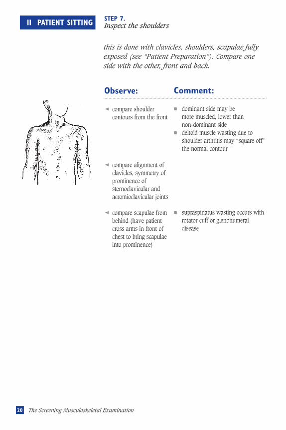
II PATIENT SITTING STEP 7.Inspect the shoulders
this is done with clavicles, shoulders, scapulae fully exposed (see “Patient Preparation”). Compare one side with the other, front and back.
Observe:
compare shouldercontours from the front
compare alignment ofclavicles, symmetry ofprominence ofsternoclavicular andacromioclavicular joints
compare scapulae frombehind (have patientcross arms in front ofchest to bring scapulaeinto prominence)
Comment:
■ dominant side may be more muscled, lower than non-dominant side
■ deltoid muscle wasting due toshoulder arthritis may “square off”the normal contour
■ supraspinatus wasting occurs withrotator cuff or glenohumeraldisease
Musculoskeletal revisec.57105 5/16/05 1:59 PM Page 20
21II Patient Sitting
The fine print:• Palpation, particularly to elicit pain and/or crepitus in patients with shoulder pain, can be added to
observation, and takes very little additional time.
• Both the sternoclavicular and acromioclavicular joints may be checked for pain and palpablecrepitus by asking the patient to shrug while the respective joints are firmly palpated. Using thetips of the fingers of each hand, each pair can be assessed at the same time.
• The bicipital tendon, located in the bicipital groove between the greater and lesser tuberosities, canbe palpated for tenderness, as can the subdeltoid bursa (an extension of the subacromial bursa) inthe groove at the tip of the acromion.
Musculoskeletal revisec.57105 5/16/05 1:59 PM Page 21
22 The Screening Musculoskeletal Examination
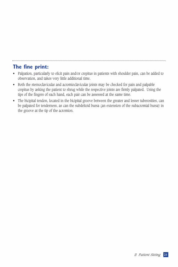
II PATIENT SITTING STEP 8.Observe shoulder range of motion (active)
Abduction Direction:
“Bring your arms from your side straight overhead, palms touching; now bring them slowly down to your side again.”
External rotation Direction:
“Put your hands behind your head, bring elbows way back, and stick out your chest”
Internal rotation (and extension)Direction:
“Now put one hand behind your back and touch the opposite shoulder blade. Now do the same with the other hand”
Movements should be smooth, symmetrical, and simultaneous
Musculoskeletal revisec.57105 5/16/05 1:59 PM Page 22
23II Patient Sitting
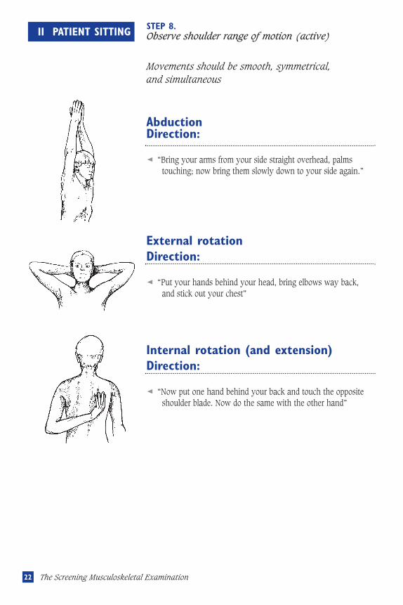
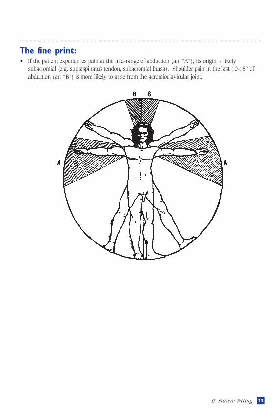
The fine print:• If the patient experiences pain at the mid-range of abduction (arc “A”), its origin is likely
subacromial (e.g. supraspinatus tendon, subacromial bursa). Shoulder pain in the last 10-15° ofabduction (arc “B”) is more likely to arise from the acromioclavicular joint.
Musculoskeletal revisec.57105 5/16/05 1:59 PM Page 23
24 The Screening Musculoskeletal Examination
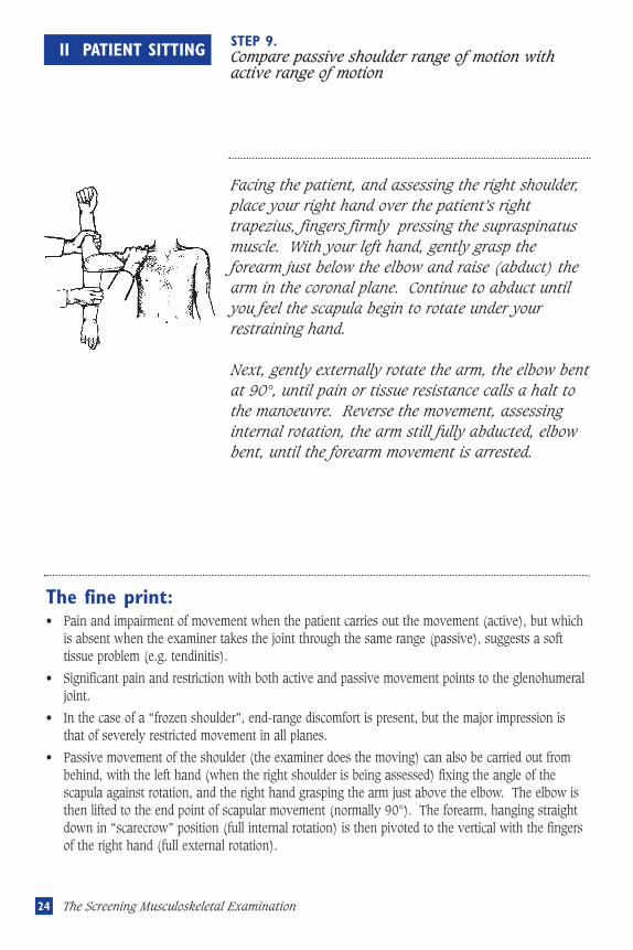
II PATIENT SITTING STEP 9.Compare passive shoulder range of motion withactive range of motion
Facing the patient, and assessing the right shoulder,place your right hand over the patient’s righttrapezius, fingers firmly pressing the supraspinatusmuscle. With your left hand, gently grasp theforearm just below the elbow and raise (abduct) thearm in the coronal plane. Continue to abduct untilyou feel the scapula begin to rotate under yourrestraining hand.
Next, gently externally rotate the arm, the elbow bentat 90°, until pain or tissue resistance calls a halt tothe manoeuvre. Reverse the movement, assessinginternal rotation, the arm still fully abducted, elbowbent, until the forearm movement is arrested.
The fine print:• Pain and impairment of movement when the patient carries out the movement (active), but which
is absent when the examiner takes the joint through the same range (passive), suggests a softtissue problem (e.g. tendinitis).
• Significant pain and restriction with both active and passive movement points to the glenohumeraljoint.
• In the case of a “frozen shoulder”, end-range discomfort is present, but the major impression isthat of severely restricted movement in all planes.
• Passive movement of the shoulder (the examiner does the moving) can also be carried out frombehind, with the left hand (when the right shoulder is being assessed) fixing the angle of thescapula against rotation, and the right hand grasping the arm just above the elbow. The elbow isthen lifted to the end point of scapular movement (normally 90°). The forearm, hanging straightdown in “scarecrow” position (full internal rotation) is then pivoted to the vertical with the fingersof the right hand (full external rotation).
Musculoskeletal revisec.57105 5/16/05 1:59 PM Page 24
25II Patient Sitting
The fine print:• In the case of a TMJ arthritis there may be crepitus and tenderness elicited as the jaw moves. The
central incisors may initially deviate toward the painful side.
• Childhood arthritis may result in growth restriction of the mandible (“micrognathia”) with amarked overbite
• Patients with TMJ arthritis, and patients with scleroderma, may havemarked restriction and be unable to pass the “three finger test”. If suchrestriction is suspected, ask the patient to attempt to place three fingers,vertically, between the upper and lower teeth (see illustration)
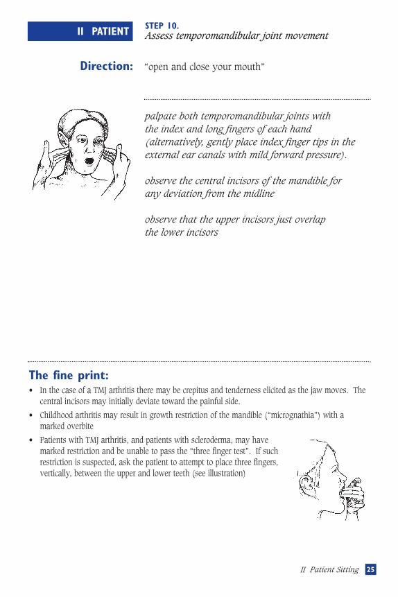
II PATIENT STEP 10.Assess temporomandibular joint movement
Direction: “open and close your mouth”
Observe:
palpate both temporomandibular joints with the index and long fingers of each hand(alternatively, gently place index finger tips in theexternal ear canals with mild forward pressure).
observe the central incisors of the mandible for any deviation from the midline
observe that the upper incisors just overlap the lower incisors
Musculoskeletal revisec.57105 5/16/05 1:59 PM Page 25
26 The Screening Musculoskeletal Examination
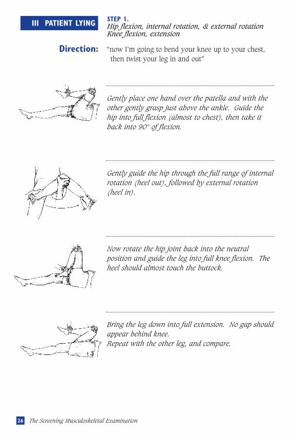
III PATIENT LYING STEP 1.Hip flexion, internal rotation, & external rotationKnee flexion, extension
Direction: “now I’m going to bend your knee up to your chest, then twist your leg in and out”
Gently place one hand over the patella and with theother gently grasp just above the ankle. Guide thehip into full flexion (almost to chest), then take itback into 90° of flexion.
Bring the leg down into full extension. No gap shouldappear behind knee.Repeat with the other leg, and compare.
Now rotate the hip joint back into the neutralposition and guide the leg into full knee flexion. Theheel should almost touch the buttock.
Gently guide the hip through the full range of internalrotation (heel out), followed by external rotation(heel in).
Musculoskeletal revisec.57105 5/16/05 1:59 PM Page 26
27III Patient Lying
The fine print:• Watch the opposite leg when assessing full hip flexion. If it lifts off the examining table, there is
probably a flexion contracture on that side.
• The earliest finding in hip disease is usually restriction of internal rotation (in the shoulder, it isusually restriction of external rotation).
• By palpating the patella as the range is assessed passively, knee joint crepitus may be detected. If the patello-femoral articulation is the culprit, this may be confirmed by grasping the patella andmoving it side-to-side — you may feel crepitus and the patient may show, in facial expression,displeasure (the “apprehension sign”).
• If there appears to be quadriceps muscle bulk asymmetry (common in knee arthritis), mark theskin on each leg a fixed distance above the upper patellar poles (e.g. 6 cm), measure thecircumference and compare the two sides
• A rough assessment of leg length inequality may be made by measuring each leg from anteriorsuperior iliac spine to medial malleolus (you can also check to see how the heels line up with thelegs fully extended on the examining table).
Musculoskeletal revisec.57105 5/16/05 1:59 PM Page 27
28 The Screening Musculoskeletal Examination
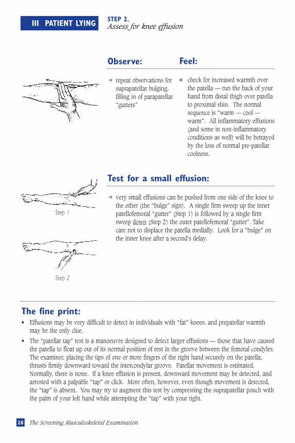
III PATIENT LYING STEP 2.Assess for knee effusion
Observe:
repeat observations forsuprapatellar bulging,filling in of parapatellar“gutters”
Feel:
■ check for increased warmth overthe patella — run the back of yourhand from distal thigh over patellato proximal shin. The normalsequence is “warm — cool —warm”. All inflammatory effusions(and some in non-inflammatoryconditions as well) will be betrayedby the loss of normal pre-patellarcoolness.
Test for a small effusion:
very small effusions can be pushed from one side of the knee tothe other (the “bulge” sign). A single firm sweep up the innerpatellofemoral “gutter” (Step 1) is followed by a single firmsweep down (Step 2) the outer patellofemoral “gutter”. Takecare not to displace the patella medially. Look for a “bulge” onthe inner knee after a second’s delay.
The fine print:• Effusions may be very difficult to detect in individuals with “fat” knees, and prepatellar warmth
may be the only clue.
• The “patellar tap” test is a manoeuvre designed to detect larger effusions — those that have causedthe patella to float up out of its normal position of rest in the groove between the femoral condyles.The examiner, placing the tips of one or more fingers of the right hand securely on the patella,thrusts firmly downward toward the intercondylar groove. Patellar movement is estimated.Normally, there is none. If a knee effusion is present, downward movement may be detected, andarrested with a palpable “tap” or click. More often, however, even though movement is detected,the “tap” is absent. You may try to augment this test by compressing the suprapatellar pouch withthe palm of your left hand while attempting the “tap” with your right.
Step 1
Step 2
Musculoskeletal revisec.57105 5/16/05 1:59 PM Page 28
29III Patient Lying
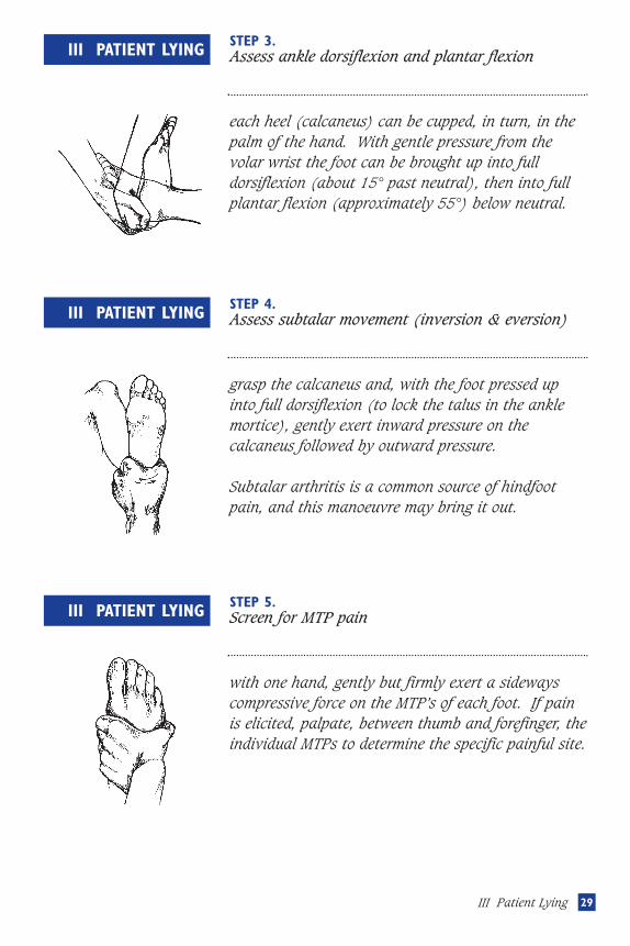
III PATIENT LYING STEP 3.Assess ankle dorsiflexion and plantar flexion
each heel (calcaneus) can be cupped, in turn, in thepalm of the hand. With gentle pressure from thevolar wrist the foot can be brought up into fulldorsiflexion (about 15° past neutral), then into fullplantar flexion (approximately 55°) below neutral.
III PATIENT LYING STEP 4.Assess subtalar movement (inversion & eversion)
grasp the calcaneus and, with the foot pressed upinto full dorsiflexion (to lock the talus in the anklemortice), gently exert inward pressure on thecalcaneus followed by outward pressure.
Subtalar arthritis is a common source of hindfootpain, and this manoeuvre may bring it out.
III PATIENT LYING STEP 5.Screen for MTP pain
with one hand, gently but firmly exert a sidewayscompressive force on the MTP’s of each foot. If painis elicited, palpate, between thumb and forefinger, theindividual MTPs to determine the specific painful site.
Musculoskeletal revisec.57105 5/16/05 1:59 PM Page 29

Notes
Musculoskeletal revisec.57105 5/16/05 1:59 PM Page 30

Notes
Musculoskeletal revisec.57105 5/16/05 1:59 PM Page 31

Notes
Musculoskeletal revisec.57105 5/16/05 1:59 PM Page 32

Contents
Introduction . . . . . . . . . . . . . . . . . . . . . . . . . . . . . . . . . . . . . . . . . . . . . p.1
Patient Standing . . . . . . . . . . . . . . . . . . . . . . . . . . . . . . . . . . . . . . . . . . p.3
Patient Sitting . . . . . . . . . . . . . . . . . . . . . . . . . . . . . . . . . . . . . . . . . . . . p.12
Patient lying . . . . . . . . . . . . . . . . . . . . . . . . . . . . . . . . . . . . . . . . . . . . . p.26
Written By
John M. Thompson MD FrCPC
Division of rheumatology
Department of Medicine
Faculty of Medicine
University of Western Ontario
St. Joseph’s hospital
268 grosvenor Street
london, Ontario n6A 4V2
Illustrated By
Ainsley S. Walton
©1998 by John M. Thompson
Unauthorized duplication of these materials is strictly prohibited.
Dedicated To
hugh little MD FrCPC
mentor, colleague and friend
The Financial assistance of
The Arcangelo Rea Family Foundation
is gratefully acknowledged
Supported by an educational grant
from Pfizer Canada Inc.
Thanks to
karen and John hueston of the Aylmer Express
Anne lyddiatt
and my colleagues in the Division of rheumatology
MSK Book ENG CVR 94193_HSE COX C00011-Musculoske.49454 12-06-21 11:47 AM Page 2
SCrEEnIng ChECklIST
Standing:
Sitting:
Lying:
q inspect from front
q inspect from behind
q cervical spine extension, flexion, rotation,
lateral flexion
q chest expansion (costovertebral movement)
q lumbar flexion, extension, lateral flexion
q observe gait
q inspect dorsum of hands, finger extension
q MCP flexion
q PIP, DIP flexion
q inspect palms
q arm forward flexion, pronation
q wrist flexion, extension
q forearm supination, elbow extension
q elbow flexion, extension
q inspect shoulders
q observe active shoulder abduction, external
rotation, internal rotation
q compare passive shoulder abduction,
external & internal rotation
q assess temporomandibular joint movement
q hip flexion, internal rotation, external rotation
q knee flexion & extension
q assess for knee effusion
q ankle dorsiflexion & plantar flexion
q assess subtalar inversion and eversion
q screen for MTP pain
The MusculoskeletalScreening Examination
John M. Thompson MD FRCPC
and Ainsley Walton
02CB21E
MSK Book ENG CVR 94193_HSE COX C00011-Musculoske.49454 12-06-21 11:47 AM Page 1