mutagenesis of the ns2b-ns3-mediated cleavage site in the
TRANSCRIPT

JOURNAL OF VIROLOGY,0022-538X/99/$04.0010
Oct. 1999, p. 8083–8094 Vol. 73, No. 10
Copyright © 1999, American Society for Microbiology. All Rights Reserved.
Mutagenesis of the NS2B-NS3-Mediated Cleavage Sitein the Flavivirus Capsid Protein Demonstrates a
Requirement for Coordinated ProcessingSEAN M. AMBERG AND CHARLES M. RICE*
Department of Molecular Microbiology, Washington University School of Medicine,St. Louis, Missouri 63110-1093
Received 11 March 1999/Accepted 6 July 1999
Analysis of flavivirus polyprotein processing has revealed the presence of a substrate for the virus-encodedNS2B-NS3 protease at the carboxy-terminal end of the C (capsid or core) protein. Cleavage at this site has beenimplicated in the efficient generation of the amino terminus of prM via signal peptidase cleavage. Yellow fevervirus has four basic residues (Arg-Lys-Arg-Arg) in the P1 through P4 positions of this cleavage site. Multiplealanine substitutions were made for these residues in order to investigate the substrate specificity andbiological significance of this cleavage. Mutants were analyzed by several methods: (i) a cell-free transprocessing assay for direct analysis of NS2B-NS3-mediated cleavage; (ii) a trans processing assay in BHK-21cells, using a C-prM polyprotein, for analysis of prM production; (iii) an infectivity assay of full-lengthtranscripts to determine plaque-forming ability; and (iv) analysis of proteins expressed from full-lengthtranscripts to assess processing in the context of the complete genome. Mutants that exhibited severe defectsin processing in vitro and in vivo were incapable of forming plaques. Mutants that contained two adjacent basicresidues within the P1 through P4 region were processed more efficiently in vitro and in vivo, and transcriptsbearing these mutations were fully infectious. Furthermore, two naturally occurring plaque-forming revertantswere analyzed and shown to have restored protein processing phenotypes in vivo. Finally, the efficient pro-duction of prM was shown to be dependent on the proteolytic activity of NS3. These data support a model oftwo coordinated cleavages, one that generates the carboxy terminus of C and another that generates the aminoterminus of prM. A block in the viral protease-mediated cleavage inhibits the production of prM by the signalpeptidase, inhibits particle release, and eliminates plaque formation.
Yellow fever virus (YF) is a member of the Flavivirus genusof the Flaviviridae, a family of small enveloped positive-strandRNA viruses which also includes the Pestivirus and hepatitis Cvirus genera. The flavivirus genome encodes a single long openreading frame which yields a long polyprotein. This polypro-tein is processed by a combination of host and viral proteasesto generate the mature viral proteins (for a review, see refer-ence 32). The three structural proteins are located within theN-terminal one-quarter of the polyprotein, in the order C-prM-E, where C is the capsid protein, prM is a glycosylatedprecursor of the viral envelope-bound M protein, and E is theenvelope protein. The nonstructural proteins (NS1-NS2A-NS2B-NS3-NS4A-NS4B-NS5) are located in the remainingportion of the polyprotein. The amino termini of prM, E, NS1,and NS4B are generated by signal peptidase cleavage withinthe lumen of the endoplasmic reticulum (ER). A trypsin-likeserine protease residues within the N-terminal one-third ofNS3; the activity of this protease requires the presence ofNS2B as a cofactor. Several cleavages within the nonstructuralregion are mediated by this protease complex, namely the2A/2B, 2B/3, 3/4A, and 4B/5 cleavages. In addition, the NS2B-NS3 protease is also responsible for mediating cleavages at theC terminus of the C protein and the C terminus of NS4A, aswell as alternative cleavages within NS2A and NS3.
Several lines of evidence support a significant role for theNS2B-NS3-mediated cleavage at the C terminus of the C pro-
tein. The preferred substrate for the viral protease is a pair ofbasic amino acids in the P2 and P1 positions, followed usuallyby a serine or glycine (sometimes alanine or threonine) in theP19 position (nomenclature as per reference 4). For YF, sucha motif is located 20 amino acids N terminal to the eventualamino terminus of prM. This motif is a conserved featureamong the flaviviruses, suggesting a functional role. Cleavageat this location has been demonstrated for YF with a cell-freetrans-processing assay in a manner dependent on NS2B andthe protease domain of NS3 (2). A similar assay for a relatedflavivirus, West Nile virus, suggests cleavage at the analogouslocation (44). The C proteins from two flaviviruses, Kunjinvirus (40) and West Nile virus (31), have been shown to containpairs of basic amino acids at their C termini. Interestingly,efficient generation of the prM protein is dependent on thepresence of the viral protease; in the absence of the protease,a C-prM polyprotein is commonly detected and release ofstructural proteins is impaired (2, 22, 37, 45). This has led to amodel of two coordinated cleavages, one on either side of theER membrane (22). A hydrophobic domain between these twosites serves to direct translocation of prM into the lumen,where efficient signal peptidase cleavage occurs only aftercleavage at the upstream dibasic site. More direct evidence forthis model comes from the demonstration of posttranslationalsignal peptidase cleavage following trypsin digestion of thecytoplasmic portion of a C-prM polyprotein (41).
Despite the evidence that NS2B-NS3-mediated cleavage ofthe C protein plays an important role in the processing of thestructural protein, it remains to be demonstrated that thisprocessing plays an essential role in the viral replication cycle.To investigate the substrate specificity of the capsid dibasic-site
* Corresponding author. Mailing address: Department of MolecularMicrobiology, Washington University School of Medicine, Box 8230,660 S. Euclid Ave., St. Louis, MO 63110-1093. Phone: (314) 362-2842.Fax: (314) 362-1232. E-mail: [email protected].
8083
on February 12, 2018 by guest
http://jvi.asm.org/
Dow
nloaded from

cleavage and to examine the importance of this cleavage forthe replication of the virus, a number of site-directed muta-tions were made around the NS2B-NS3-mediated cleavagesite. An assay developed previously allowed the direct analysisof NS2B-NS3-dependent cleavage at the dibasic site (2), whilethe YF infectious clone was used to investigate the plaque-forming ability of mutant transcripts (5, 33). In addition, atransient-expression assay was used to express mutant C-prMpolyproteins in the presence or absence of the viral protease. Itwas also possible to analyze protein processing directly in cellstransfected with full-length transcripts, since protein expres-sion levels for both plaque-forming and non-plaque-formingmutants were similar. Viral protease-mediated processing atthe conserved dibasic site was found to correlate with properprocessing of the C-prM polyprotein and with the ability togenerate plaques on two cell lines.
MATERIALS AND METHODS
Cell lines, virus, and antisera. Growth of YF stocks and BHK-21 and SW-13monolayers has been summarized previously (9). A BHK-21 derivative (BHK-21/J) was kindly provided by Paul Olivo (Washington University, St. Louis, Mo.)and was used for experiments involving full-length YF transcripts. Antiseraspecific for prM, E, NS2B, and NS3 have been described previously (8), and arabbit antiserum raised against a fusion protein containing residues 1 to 94 of theYF C protein was kindly provided by M. Bouloy (Institute Pasteur, Paris,France).
Plasmid constructions. Standard recombinant DNA techniques (3) were usedfor plasmid constructions. The construction of the pTM3-NS2B, pTM3-NS3181,and pTM3-NS2B-3181 plasmids has been described previously (10). pTM3-NS3181(S3A) has also been described previously (17). pTM3-C-prM was con-structed through a three-step process: (i) the NcoI site of pTM3 (28) wasremoved by digestion with NcoI, treatment with mung bean nuclease to generateblunt ends, and religation; (ii) the C-prM cassette from pBS/C-prM (2) wascloned into this pTM3 derivative by using the EcoRI and SstI sites of each; and(iii) a 41-bp region preceding the translation initiation site for the C-prMpolyprotein was removed by digestion with EcoRI and BstEII, generation ofblunt ends with mung bean nuclease, and religation. The regions around thedeleted NcoI site and the 41-bp deletion were verified by sequencing.
Mutagenesis. Site-directed mutagenesis was performed by a modification ofthe Kunkel method (15, 18) with either pBS-anchC (2) or pBS-YFS (whichcontains a cassette encoding the YF structural proteins [14a]) as a template.Restriction sites were introduced by using silent mutations for easy identification.The 78-bp MscI-BsmI fragment from each mutant was substituted into pBS-anchC.3 (2) for use in the in vitro assay; the identity of the subcloned fragmentwas verified by sequencing. Mutants were initially subcloned into pYF5939IV (33)by a three-piece ligation: a 3,868-bp SalI-MscI fragment from pYF5939IV plus a2,960-bp SalI-partial BsmI fragment from pYF5939IV plus the 78-bp MscI-BsmIfragment from each pBS-anchC or pBS-YFS mutant. Mutant derivatives of afull-length YF cDNA clone, pACNR-FLYF (5), were then constructed by sub-stitution of a 1,903-bp EagI-NsiI fragment from the appropriate pYF5939IVmutant. The pTM3-C-prM mutant derivatives were constructed by substitutionof the 394-bp NcoI-MscI fragments from the pYF5939IV mutants into pTM3-C-prM.
Cell-free trans-processing assay. The protease assay has been described pre-viously (2). The small peptides produced in this reaction were resolved by usingthe Tricine system of sodium dodecyl sulfate-polyacrylamide gel electrophoresis(SDS-PAGE) (38) with 16.5% acrylamide in the separating portion of the geland an acrylamide/bisacrylamide ratio of 21:1. Tricine gels were fixed for 30 minin 10 volumes of 25% ethanol–10% acetic acid, washed twice with distilled H2O(15 min per wash), and dried for 4 h under a vacuum at 60°C, followed byvisualization with a Bio-Rad Molecular Imager System (model GS-363). Quan-titation of protein bands was performed with Bio-Rad software. The proportionof cleavage was defined as the ratio of radioactivity in the two product bands tothe total radioactivity present in all three bands (substrate and two products).
Transient expression. The vaccinia virus-T7 system was used to transientlyexpress transfected plasmids (14). BHK-21 monolayers in 35-mm-diameterdishes were infected with vTF7-3 (a vaccinia virus recombinant expressing T7RNA polymerase) at a multiplicity of infection of 10 in a total volume of 0.2 mlof phosphate-buffered saline plus 1% fetal bovine serum (FBS). The inoculumwas kept on the monolayers for 0.5 h at 37°C and then replaced with 0.5 ml ofminimal essential medium (MEM; Life Technologies, Inc.) containing 12 mg ofLipofectamine (Gibco-BRL) and 1 mg of each of the relevant plasmids. Follow-ing a 2.5-h transfection period at 37°C, cells were labeled for 4.5 h at 37°C with30 mCi of [35S]methionine (ICN Translabel)/ml in MEM containing 1/40th of thenormal concentration of methionine and 2% FBS. After being labeled, mono-layers were washed twice with MEM and lysed with 0.3 ml of a lysis buffercontaining 0.5% SDS, 50 mM Tris-Cl (pH 7.5), 150 mM NaCl, and 1 mM EDTA.
One-sixth of each SDS lysate was immunoprecipitated with the appropriateantiserum as described previously (9) and separated by conventional SDS-PAGEusing 13% acrylamide (3). Gels were treated for fluorography as describedelsewhere (2) and exposed to X-ray film (Kodak X-Omat) at 280°C.
For the transient-expression experiment (see Fig. 5), the above procedure wasmodified as follows. Only 10 ng of pTM3-NS2B-3181 was used for each transfec-tion, although pTM3-C-prM derivatives were added at 1 mg per transfection.Following transfection, the transfection mix was replaced with MEM plus 2%FBS. After 50 min at 37°C, cells were labeled for 20 min with methionine-freeMEM plus 100 mCi of [35S]methionine/ml and then lysed as described above.
Specific infectivity measurements. YF transcripts were synthesized with SP6RNA polymerase (33), using XhoI-linearized pACNR-FLYF (5) as a template.Incorporation of trace [5,6-3H]UTP was assayed by adsorption to DE81 (What-man) filter paper as a means of quantitating RNA yields (35), and transcriptintegrity was confirmed by electrophoresis in agarose gels followed by ethidiumbromide staining (1 mg/ml for 20 min). For electroporation, 3 mg of each tran-script was mixed with 8 3 106 (BHK-21) or 4 3 106 (SW-13) cells in a phosphate-buffered solution (137 mM NaCl, 2.7 mM KCl, 10 mM Na2HPO4, 1.8 mMKH2PO4) and pulsed in 2-mm-gap electroporation cuvettes (BTX) with an elec-troporator (BTX Electro Square Porator model T820) set for 5 pulses at 960 Vwith a pulse length of 99 ms (BHK-21) or for 3 pulses at 800 V with a pulse lengthof 60 ms (SW-13). After a 10-min recovery phase at room temperature, 35-mm-diameter dishes were seeded with serial 10-fold dilutions of transfected cells inthe presence of untransfected cells (6 3 105 BHK-21 cells or 8 3 105 SW-13 cellsfor each 35-mm-diameter dish). Cell medium was replaced with an agaroseoverlay (MEM with 0.6% agarose and 2% FBS) after allowing 4 to 6 h forattachment. Cells were incubated for 4 (BHK-21) or 5 (SW-13) days at 37°C inan atmosphere of 5% CO2, fixed with 7% formaldehyde, and stained with 1%crystal violet in 5% ethanol. Data presented in Tables 1 and 2 are the averagesof values from two or three experiments, except for the SW-13 results in Table1, which are from a single experiment. However, this particular experiment wasperformed several times via Lipofectin-mediated transfection with comparableresults, although with lower overall efficiency.
Protein analysis of cells transfected with full-length YF transcripts. BHK-21cells were transfected as described above, diluted in MEM plus 10% FBS, and
TABLE 1. Multiple alanine substitutions introduced at the capsiddibasic cleavage site and the resultant phenotypes
TranscriptAmino acid sequence
around the capsiddibasic cleavage site
% ofsubstratecleavedin vitroa
Specific infectivity oftranscript (PFU/mg)
in cell line:
BHK-21 SW-13
2WTb S S R K R R S H D 38 1.0 3 106 1.6 3 105
291 . . A A . . . . . 24 1.0 3 106 1.2 3 105
292 . . A . A . . . . ,3 ,10 ,10293 . . A . . A . . . 10 5.6 3 105 1.8 3 105
294 . . . A A . . . . ,3 ,10 14295 . . . A . A . . . ,3 ,10 ,10296 . . . . A A . . . 16 2.7 3 105 1.5 3 105
297 . . A A A . . . . ,3 ,10 ,10298 . . A . A A . . . ,3 ,10 ,10299 . . A A A A . . . ,3 ,10 ,10
a As calculated from Fig. 2.b WT, wild type.
TABLE 2. Comparison of infectivities of naturally occurringrevertants and their parents
TranscriptAmino acid sequence
around the capsid dibasiccleavage site
Specific infectivity oftranscript (PFU/mg)
in cell line:
BHK-21 SW-13
2WTa S R K R R S H D 5.4 3 105 8.2 3 104
292 S A K A R S H D ,10 ,102R S A K V R S H D 3.5 3 102 1.0 3 105
294 S R A A R S H D ,10 144C S R A V R S H D 6.9 3 102 9.2 3 104
a WT, wild type.
8084 AMBERG AND RICE J. VIROL.
on February 12, 2018 by guest
http://jvi.asm.org/
Dow
nloaded from

used to seed 35-mm-diameter dishes (106 cells per dish). Cells were labeledbeginning at 18 h (see Fig. 8) or 19 h (see Fig. 7) postelectroporation, using 120(see Fig. 8) or 150 (see Fig. 7) mCi of [35S]methionine (ICN Translabel)/ml inMEM containing 1/40th of the normal concentration of methionine and 2% FBS.At 24 h postelectroporation, cells were lysed and subjected to immunoprecipi-tation as described above, using 1/6 (see Fig. 7) or 1/15 (see Fig. 8) of the totallysate from each 35-mm-diameter dish. For one of the experiments (see Fig. 8),the labeling medium was harvested and clarified (16,000 3 g for 2 min) imme-diately prior to cell lysis. One-sixth of the total harvested medium was denaturedby adding SDS to a final concentration of 0.5% and heating at 75°C for 5 min;immunoprecipitation was performed as described above.
RT-PCR. Viral stocks of mutant or potentially revertant populations wereprecipitated with one-fourth volume of 40% polyethylene glycol 8000 in TNE(100 mM NaCl, 10 mM Tris-Cl [pH 8], 1 mM EDTA) for 2 h on ice, pelleted (30min at 4°C and 16,000 3 g), and resuspended in 40 ml of 2% SDS containing 0.1mg of proteinase K/ml. This mixture was incubated at 37°C for 30 min, extractedtwice with phenol and twice with chloroform, precipitated, and resuspended inTE (10 mM Tris-Cl [pH 8.0], 0.1 mM EDTA). The resulting RNA preparationserved as a template for cDNA synthesis with SuperScript II reverse transcrip-tase (Gibco-BRL) and a YF genome-specific primer (CMR#45, which is theminus-strand complement to YF nucleotides 1050 to 1065 [59-ACACACTTGTCTTGCT-39]). Amplification of cDNA by PCR was done with two additionalYF-specific primers (PCL#8108, which corresponds to YF nucleotides 1 to 18[59-AGTAAATCCTGTGTGCTA-39] and CMR#44, which is the minus-strandcomplement to YF nucleotides 836 to 851 [59-ATCTCTCAATCTTTTG-39]) andKlenTaq LA DNA polymerase (supplied by Wayne Barnes, Washington Uni-versity, St. Louis). Reverse transcription (RT)-PCR fragments were sequencedwith a Thermo-Sequenase [33P]ddNTP Cycle Sequencing Kit (Amersham). A78-bp MscI-BsmI fragment derived from the RT-PCR fragments for populations2R and 4C was cloned into pBS-anchC.3 (2), verified by sequencing, and furthersubcloned into pYF5939IV, pACNR-FLYF, and pTM3-C-prM as describedabove.
RESULTS
Mutagenesis of the capsid dibasic cleavage site. Mutagene-sis around the dibasic cleavage sites of the YF nonstructuralregion has demonstrated little substrate specificity beyond theP2, P1, and P19 residues (11, 18, 30). However, it is likely thatthere are important structural determinants beyond this motif,since several regions of the polyprotein contain a similar se-quence and are not known to be cleaved. In addition, sometolerance to nonconservative substitution has been observed inthe P1 position of the 3/4A and 4B/5 sites (18), the P19 positionof the 2B/3 site (11), and the P2 position of the 2B/3 and 4B/5sites (11, 18). The predicted dibasic cleavage site of the Cprotein is especially basic, with typically four or five basicresidues located within the P1 through P6 positions; cleavagesites in the nonstructural region of the polyprotein are gener-ally less basic (7). For YF, the site of the capsid cleavage hasbeen experimentally determined in vitro (2). The amino acidsin the P6 through P1 positions are Ser-Ser-Arg-Lys-Arg-Arg(Fig. 1). The presence of multiple basic residues has severalimplications for mutagenesis. First, it is possible that this playsa role in substrate determination. For example, a minimum ofthree or four basic amino acids may be a requirement for transcleavage, in contrast to the probable cis cleavages of the non-structural portion of the polyprotein. Second, this feature maybe involved in some other aspect of viral replication, such asgenome packaging. Finally, amino acid substitutions in thisregion might ablate the original cleavage site while permittingcleavage at a secondary site. One example of this possibility isthe introduction of a serine in the P1 position, which results ina sequence predicted to be a good substrate for the viralprotease (Arg-Lys-Arg-Ser-Ser). This consideration made itnecessary to focus on all four basic residues preceding theNS2B-NS3-mediated cleavage site.
Figure 1 depicts the substrate used for the cell-free assay ofcapsid cleavage. Multiple alanine substitutions were intro-duced in the P1 through P4 positions of the capsid cleavage site(Table 1), and these mutants were subcloned into pBS-anchC.3(2). These constructs were linearized with XbaI, transcribed
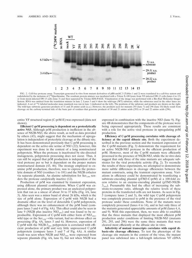
with T7 RNA polymerase, and translated with rabbit reticulo-cyte lysate; the in vitro translation products were incubatedwith Triton X-100 lysates of YF-infected SW-13 cells and sep-arated by SDS-PAGE in the presence of Tricine. Figure 2 is aphosphorimage of such a gel. Cleavage products are detectablefor only three of the nine mutants tested: 291, 293, and 296(Fig. 2 and Table 1). Each of these three mutants contain twoadjacent basic residues, and these are the only three mutantswith this feature. Based on the substrate specificity of theNS2B-NS3 protease (32), this suggests that the cleavage sitecan be shifted by one (mutant 293) or two (mutant 296) resi-dues. It should be pointed out that with this gel system, themigration of small peptides is quite sensitive to amino acidcomposition. The overall charge of the peptide seems to havethe most dramatic effect on migration, since substrates withidentical charges migrate similarly (the presence of fewer basicresidues is associated with faster migration).
Processing of the C-prM polyprotein. The efficient produc-tion of prM is thought to be dependent on prior cleavage at thedibasic cleavage site (22). However, this hypothesis is not uni-versally accepted since it has been reported that efficient prMproduction can occur with mutants that do not cleave at thedibasic site (43). The production of prM was examined in thecontext of the dibasic cleavage site mutations by subcloning the291 to 299 panel into a vector expressing the two structuralproteins C and prM (pTM3-C-prM). Transient expression inthe absence of the viral NS2B-NS3 protease yielded primarilyan unprocessed C-prM molecule, although small amounts ofprM were also visible. This was true for the wild-type constructas well as for each of the mutants (Fig. 3). Pulse-chase exper-iments demonstrate a very slow accumulation of prM over time(data not shown), indicating that the viral protease is not es-sential for the signal peptidase cleavage that generates prM.Expression of C-prM in the presence of the NS2B-NS3 pro-tease was initially performed by transfecting an equimolar ratioof plasmids encoding C-prM and NS2B-NS3. Surprisingly, prM(Fig. 3A) and C (Fig. 3B) were produced quite efficiently fornearly all of the mutants, with the exception of 298 (AKAA)and 299 (AAAA). A similar result was obtained when the
FIG. 1. Schematic diagram of the substrate used in the cell-free trans-pro-cessing assay. The YF polyprotein is shown at the top; the locations of thestructural proteins (C, prM, and E) and the nonstructural proteins (NS1 throughNS5) are indicated. Below the YF polyprotein is an expanded view of the C-prMcassette; the glycosylation sites (p), the signal peptidase cleavage site (}), and thecapsid dibasic cleavage site (2) are noted, and the hydrophobic regions of theC-prM polyprotein are shaded. The substrate used for the protease assay, de-noted anchC.3, is 51 amino acids in length. Its size and position relative to theC-prM polyprotein are shown. The sequence around the cleavage site is indi-cated, using the one-letter amino acid code.
VOL. 73, 1999 MUTAGENESIS OF THE NS2B-NS3 CAPSID CLEAVAGE SITE 8085
on February 12, 2018 by guest
http://jvi.asm.org/
Dow
nloaded from
entire YF structural region (C-prM-E) was expressed (data notshown).
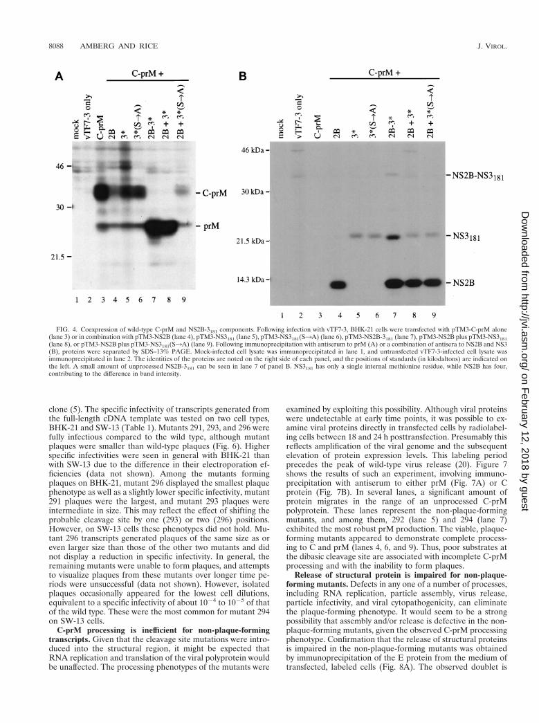
Efficient C-prM processing is dependent on a proteolyticallyactive NS3. Although prM production is inefficient in the ab-sence of NS2B-NS3, the above result, as well as data providedby others (43), might suggest that the mechanism of upregu-lation is independent of proteolytic cleavage at the dibasic site.It has been demonstrated previously that C-prM processing isdependent on the active-site serine of NS3 (22); however, thisexperiment was done in the context of a long nonstructuralpolyprotein. When the protease is inactivated by site-directedmutagenesis, polyprotein processing does not occur. Thus, itcan still be argued that prM production is independent of theviral protease per se but is dependent on the proper maturenonstructural domain (43, 46). The strategy employed to ex-amine prM production, therefore, was to express the proteo-lytic domain of NS3 (residues 1 to 181) and the NS2B cofactorvia separate plasmids. An alanine substitution for Ser138 ren-ders the protease catalytically inactive (12).
Production of prM was examined by transient expression,using different plasmid combinations. When C-prM was ex-pressed alone, the primary product was an uncleaved polypro-tein that ran as a smear of about 34 to 37 kDa (Fig. 4A, lane3). Also seen was a minor band of about 26 kDa, which is thesize of prM alone. Expression of C-prM with NS2B had adramatic effect on the level of detectable C-prM polyprotein,although there was no enhancement of the prM band (com-pare lanes 3 and 4 of Fig. 4A). This effect could also be seen inlane 9 (NS2B plus inactive NS3181) and was found to be re-producible. Expression of C-prM with either form of NS3181,wild type or the Ser1383Ala variant, had no obvious effect onprocessing (Fig. 4A, lanes 5 and 6). However, expression ofC-prM with the NS2B-3181 polyprotein is correlated with effi-cient production of prM and very little unprocessed C-prMpolyprotein (compare lanes 3 and 7 of Fig. 4A). A similarresult was seen when NS2B and NS3181 were expressed fromseparate plasmids (Fig. 4A, lane 8), but not when NS2B was
expressed in combination with the inactive NS3 (lane 9). Fig-ure 4B demonstrates that the components of the protease werebeing expressed appropriately. These results are consistentwith a role for the active viral protease in upregulating prMproduction.
Efficiency of C-prM processing correlates with cleavage ef-ficiency at the capsid dibasic site. Both the experiment de-scribed in the previous section and the transient expression ofthe C-prM mutants (Fig. 3) demonstrate the requirement foran active NS2B-NS3 protease in the efficient production ofprM. However, most of the C-prM mutants were efficientlyprocessed in the presence of NS2B-NS3, while the in vitro datasuggest that only three of the nine mutants are adequate sub-strates for the viral proteolytic activity (Fig. 2). To reconcilethe results of these experiments, we attempted to demonstratemore subtle differences in cleavage efficiencies between themutant constructs, using the transient expression assay. Vari-ations in efficiency could be demonstrated by transfecting asubstrate-encoding plasmid (pTM3-C-prM) at a 100-fold ex-cess relative to an enzyme-encoding plasmid (pTM3-NS2B-3181). Presumably this had the effect of increasing the sub-strate-to-enzyme ratio, although the relative levels of theseproteins in the transfected cells are not known. As seen in Fig.5 (compare lanes 2 and 3), the wild type C-prM polyproteinwas completely processed to prM in the presence of the viralprotease under these conditions. None of the mutants werecompletely processed (lanes 4 to 14), and in fact only three ofthe mutants generated appreciable amounts of prM (lanes 4, 6,and 9). With this experiment, then, we were able to establishthat the three mutants that displayed the most efficient prMproduction under conditions of limiting NS2B-NS3 (mutants291, 293, and 296) were the same three mutants that werecleaved most efficiently at the dibasic site in vitro.
Infectivity of mutant transcripts correlates with capsid di-basic-site cleavage efficiency. To test the phenotype of thecleavage site mutants in the context of the virus, the mutantpanel was subcloned into a full-length infectious YF cDNA
FIG. 2. Cell-free protease assay. Transcripts generated in vitro from mutant derivatives of pBS-anchC.3 (Tables 1 and 2) were translated in a cell-free system andradiolabeled by the inclusion of [35S]methionine. The resultant protein mixture was incubated with a Triton X-100 lysate from YF-infected SW-13 cells (lanes 4 to 15)or from mock-infected SW-13 cells (lane 3) and then separated by Tricine-SDS-PAGE. Visualization of the image was performed with a Bio-Rad Molecular ImagerSystem. RNA was omitted from the translation mixture in lane 2. Lanes 3 and 4 show the wild-type (WT) substrate, while the substrates used in the other lanes areindicated. A set of 14C-labeled molecular mass standards was run in lane 1 (indicated on the left). The positions of the substrate and products are shown on the right.The wild-type substrate generates products of 31 and 20 amino acids (a.a.). However, the products seen for mutants 293 (lane 7) and 296 (lane 10) likely result fromcleavage on the carboxy-terminal side of the basic pair of residues that generate products of 30 and 21 amino acids (293) or 29 and 22 amino acids (296).
8086 AMBERG AND RICE J. VIROL.
on February 12, 2018 by guest
http://jvi.asm.org/
Dow
nloaded from

FIG. 3. Coexpression of C-prM mutants and NS2B-3181. Expression of the transfected plasmids was driven by a vaccinia virus recombinant that expresses T7 RNApolymerase (vTF7-3). The C-prM cassettes contained the mutations at the dibasic capsid site that are listed in Table 1. SDS lysates were immunoprecipitated withantiserum to either prM (A) or C (B). Mock infection, vTF7-3 alone, and pTM3-NS2B-3181 without pTM3-C-prM are shown in the first three lanes. Wild-type (WT)and mutant C-prM are indicated, either without (2) or with (1) cotransfection of pTM3-NS2B-3181. Positions of molecular mass standards ([14C]MW markers) areshown, with sizes in kilodaltons noted on the left (the 12.5- and 14.3-kDa markers comigrate on SDS–13% PAGE). The positions of C, prM, and the C-prM polyproteinare indicated on the right.
8087
on February 12, 2018 by guest
http://jvi.asm.org/
Dow
nloaded from
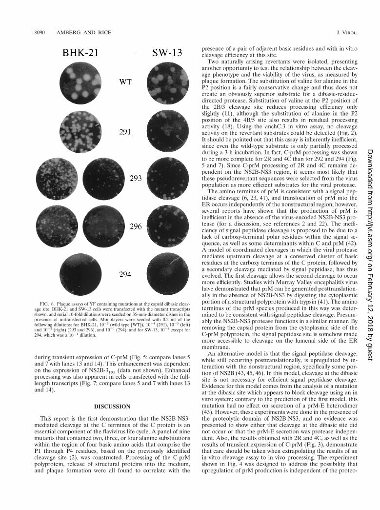
clone (5). The specific infectivity of transcripts generated fromthe full-length cDNA template was tested on two cell types,BHK-21 and SW-13 (Table 1). Mutants 291, 293, and 296 werefully infectious compared to the wild type, although mutantplaques were smaller than wild-type plaques (Fig. 6). Higherspecific infectivities were seen in general with BHK-21 thanwith SW-13 due to the difference in their electroporation ef-ficiencies (data not shown). Among the mutants formingplaques on BHK-21, mutant 296 displayed the smallest plaquephenotype as well as a slightly lower specific infectivity, mutant291 plaques were the largest, and mutant 293 plaques wereintermediate in size. This may reflect the effect of shifting theprobable cleavage site by one (293) or two (296) positions.However, on SW-13 cells these phenotypes did not hold. Mu-tant 296 transcripts generated plaques of the same size as oreven larger size than those of the other two mutants and didnot display a reduction in specific infectivity. In general, theremaining mutants were unable to form plaques, and attemptsto visualize plaques from these mutants over longer time pe-riods were unsuccessful (data not shown). However, isolatedplaques occasionally appeared for the lowest cell dilutions,equivalent to a specific infectivity of about 1024 to 1025 of thatof the wild type. These were the most common for mutant 294on SW-13 cells.
C-prM processing is inefficient for non-plaque-formingtranscripts. Given that the cleavage site mutations were intro-duced into the structural region, it might be expected thatRNA replication and translation of the viral polyprotein wouldbe unaffected. The processing phenotypes of the mutants were
examined by exploiting this possibility. Although viral proteinswere undetectable at early time points, it was possible to ex-amine viral proteins directly in transfected cells by radiolabel-ing cells between 18 and 24 h posttransfection. Presumably thisreflects amplification of the viral genome and the subsequentelevation of protein expression levels. This labeling periodprecedes the peak of wild-type virus release (20). Figure 7shows the results of such an experiment, involving immuno-precipitation with antiserum to either prM (Fig. 7A) or Cprotein (Fig. 7B). In several lanes, a significant amount ofprotein migrates in the range of an unprocessed C-prMpolyprotein. These lanes represent the non-plaque-formingmutants, and among them, 292 (lane 5) and 294 (lane 7)exhibited the most robust prM production. The viable, plaque-forming mutants appeared to demonstrate complete process-ing to C and prM (lanes 4, 6, and 9). Thus, poor substrates atthe dibasic cleavage site are associated with incomplete C-prMprocessing and with the inability to form plaques.
Release of structural protein is impaired for non-plaque-forming mutants. Defects in any one of a number of processes,including RNA replication, particle assembly, virus release,particle infectivity, and viral cytopathogenicity, can eliminatethe plaque-forming phenotype. It would seem to be a strongpossibility that assembly and/or release is defective in the non-plaque-forming mutants, given the observed C-prM processingphenotype. Confirmation that the release of structural proteinsis impaired in the non-plaque-forming mutants was obtainedby immunoprecipitation of the E protein from the medium oftransfected, labeled cells (Fig. 8A). The observed doublet is
FIG. 4. Coexpression of wild-type C-prM and NS2B-3181 components. Following infection with vTF7-3, BHK-21 cells were transfected with pTM3-C-prM alone(lane 3) or in combination with pTM3-NS2B (lane 4), pTM3-NS3181 (lane 5), pTM3-NS3181(S3A) (lane 6), pTM3-NS2B-3181 (lane 7), pTM3-NS2B plus pTM3-NS3181(lane 8), or pTM3-NS2B plus pTM3-NS3181(S3A) (lane 9). Following immunoprecipitation with antiserum to prM (A) or a combination of antisera to NS2B and NS3(B), proteins were separated by SDS–13% PAGE. Mock-infected cell lysate was immunoprecipitated in lane 1, and untransfected vTF7-3-infected cell lysate wasimmunoprecipitated in lane 2. The identities of the proteins are noted on the right side of each panel, and the positions of standards (in kilodaltons) are indicated onthe left. A small amount of unprocessed NS2B-3181 can be seen in lane 7 of panel B. NS3181 has only a single internal methionine residue, while NS2B has four,contributing to the difference in band intensity.
8088 AMBERG AND RICE J. VIROL.
on February 12, 2018 by guest
http://jvi.asm.org/
Dow
nloaded from

the result of incompletely reduced E protein (data not shown).No significant differences in the level of intracellular E proteinwere observed in SDS lysates prepared from the cell monolay-ers (Fig. 8B). Immunoprecipitation with an antiserum to anonstructural protein, NS3, also shows equivalent intracellularexpression levels across the mutant panel (Fig. 8C). Lysateswere tested to confirm that the amount of protein was notsaturating (data not shown). Although a defect in RNA repli-cation cannot be formally ruled out, the fact that equivalentlevels of radiolabeled viral proteins were detected between 18and 24 h postinfection makes this unlikely.
Mutants 291, 293, and 296 secrete E protein, although atlevels well below what is seen for the wild-type transcript.Although the proportion of E that is in the form of infectiousparticles is unknown, these results suggest that even theplaque-forming mutants are impaired with respect to particlerelease. This would be consistent with the reduced processingefficiency at the capsid dibasic site (Fig. 2 and Table 1), less-efficient C-prM processing (Fig. 5), and the smaller-plaquephenotype (Fig. 6). In fact, the level of E detected in Fig. 8Acorrelates nicely with plaque size on BHK-21 monolayers(291 . 293 . 296).
Characterization of revertants. The three plaque-formingmutants could be passaged on either SW-13 or BHK-21 cells,with titers approaching wild-type levels, while maintaining theoriginal cleavage site mutation (data not shown). Although thespecific infectivities of the non-plaque-forming mutant tran-scripts were very low, it was sometimes possible to isolatestocks of plaque-forming virus from cells transfected with thesetranscripts. This was accomplished through a combination ofextended incubation of the transfected cells and passaging ofthe virus by using the medium of transfected cells as an inoc-
ulum for untransfected cells. RT-PCR fragments derived fromthese resulting virus populations were sequenced across theregion coding for the capsid dibasic cleavage site. In one case,a virus stock derived from the 292 parent (P4 to P1 5 Ala-Lys-Ala-Arg) contained a single nucleotide change(GCC3GTC) resulting in a valine substitution in the P2 po-sition (Table 2). The same substitution was observed in apopulation recovered from transfection with the 294 parentmutation. In addition, several independently derived virus pop-ulations that did not contain reversions or additional mutationsin the structural-region coding sequences were obtained fromthe 294 transcripts. Presumably these populations contain anadditional mutation(s) in either the noncoding sequences orthe nonstructural protein coding sequence which allows thevirus to overcome the original defect. These populations dis-played one of at least three distinct plaque phenotypes, sug-gesting that they are not genetically identical.
To verify that the substitutions detected in the revertantpopulations of 292 and 294 (denoted 2R and 4C, respectively)were alone sufficient for the plaque phenotypes exhibited, afull-length cDNA clone containing this region within a wild-type genetic background was constructed. When tested on thecell line from which the revertants were derived (SW-13), thespecific infectivities of the 2R and 4C transcripts were similarto that of the wild type (Table 2). However, on BHK-21 cells,the infectivities of the revertant transcripts were dramaticallyreduced and plaques were barely discernible. The nature ofthis host-specific difference is unknown.
No cleavage products were detected when the 2R and 4Csequences were tested with the in vitro cleavage assay (Fig. 2,lanes 14 and 15). However, 2R and 4C displayed enhancedproduction of prM in comparison to the parent sequences
FIG. 5. Coexpression of C-prM mutants and NS2B-3181 following transfection of plasmids at a 100:1 ratio. One microgram of pTM3-C-prM derivative and 10 ngof pTM3-NS2B-3181 were transfected for each 35-mm-diameter dish (lanes 3 to 14). SDS lysates were immunoprecipitated with prM antiserum and separated bySDS–13% PAGE. Lanes: 1, vTF7-3 alone; 2 and 3, wild-type (WT) pTM3-C-prM with (lane 3) or without (lane 2) pTM3-NS2B-3181; 15, pTM3-NS2B-3181 in theabsence of pTM3-C-prM plasmid. The positions of protein standards (in kilodaltons) are shown on the left, and the positions of prM and the C-prM polyprotein areindicated on the right.
VOL. 73, 1999 MUTAGENESIS OF THE NS2B-NS3 CAPSID CLEAVAGE SITE 8089
on February 12, 2018 by guest
http://jvi.asm.org/
Dow
nloaded from
during transient expression of C-prM (Fig. 5; compare lanes 5and 7 with lanes 13 and 14). This enhancement was dependenton the expression of NS2B-3181 (data not shown). Enhancedprocessing was also apparent in cells transfected with the full-length transcripts (Fig. 7; compare lanes 5 and 7 with lanes 13and 14).
DISCUSSION
This report is the first demonstration that the NS2B-NS3-mediated cleavage at the C terminus of the C protein is anessential component of the flavivirus life cycle. A panel of ninemutants that contained two, three, or four alanine substitutionswithin the region of four basic amino acids that comprise theP1 through P4 residues, based on the previously identifiedcleavage site (2), was constructed. Processing of the C-prMpolyprotein, release of structural proteins into the medium,and plaque formation were all found to correlate with the
presence of a pair of adjacent basic residues and with in vitrocleavage efficiency at this site.
Two naturally arising revertants were isolated, presentinganother opportunity to test the relationship between the cleav-age phenotype and the viability of the virus, as measured byplaque formation. The substitution of valine for alanine in theP2 position is a fairly conservative change and thus does notcreate an obviously superior substrate for a dibasic-residue-directed protease. Substitution of valine at the P2 position ofthe 2B/3 cleavage site reduces processing efficiency onlyslightly (11), although the substitution of alanine in the P2position of the 4B/5 site also results in residual processingactivity (18). Using the anchC.3 in vitro assay, no cleavageactivity on the revertant substrates could be detected (Fig. 2).It should be pointed out that this assay is inherently inefficient,since even the wild-type substrate is only partially processedduring a 3-h incubation. In fact, C-prM processing was shownto be more complete for 2R and 4C than for 292 and 294 (Fig.5 and 7). Since C-prM processing of 2R and 4C remains de-pendent on the NS2B-NS3 region, it seems most likely thatthese pseudorevertant sequences were selected from the viruspopulation as more efficient substrates for the viral protease.
The amino terminus of prM is consistent with a signal pep-tidase cleavage (6, 23, 41), and translocation of prM into theER occurs independently of the nonstructural region; however,several reports have shown that the production of prM isinefficient in the absence of the virus-encoded NS2B-NS3 pro-tease (for a discussion, see references 2 and 22). The ineffi-ciency of signal peptidase cleavage is proposed to be due to alack of carboxy-terminal polar residues within the signal se-quence, as well as some determinants within C and prM (42).A model of coordinated cleavages in which the viral proteasemediates upstream cleavage at a conserved cluster of basicresidues at the carboxy terminus of the C protein, followed bya secondary cleavage mediated by signal peptidase, has thusevolved. The first cleavage allows the second cleavage to occurmore efficiently. Studies with Murray Valley encephalitis virushave demonstrated that prM can be generated posttranslation-ally in the absence of NS2B-NS3 by digesting the cytoplasmicportion of a structural polyprotein with trypsin (41). The aminoterminus of the prM species produced in this way was deter-mined to be consistent with signal peptidase cleavage. Presum-ably the NS2B-NS3 protease functions in a similar manner. Byremoving the capsid protein from the cytoplasmic side of theC-prM polyprotein, the signal peptidase site is somehow mademore accessible to cleavage on the lumenal side of the ERmembrane.
An alternative model is that the signal peptidase cleavage,while still occurring posttranslationally, is upregulated by in-teraction with the nonstructural region, specifically some por-tion of NS2B (43, 45, 46). In this model, cleavage at the dibasicsite is not necessary for efficient signal peptidase cleavage.Evidence for this model comes from the analysis of a mutationat the dibasic site which appears to block cleavage using an invitro system; contrary to the prediction of the first model, thismutation had no effect on secretion of a prM-E heterodimer(43). However, these experiments were done in the presence ofthe proteolytic domain of NS2B-NS3, and no evidence waspresented to show either that cleavage at the dibasic site didnot occur or that the prM-E secretion was protease indepen-dent. Also, the results obtained with 2R and 4C, as well as theresults of transient expression of C-prM (Fig. 3), demonstratethat care should be taken when extrapolating the results of anin vitro cleavage assay to in vivo processing. The experimentshown in Fig. 4 was designed to address the possibility thatupregulation of prM production is independent of the proteo-
FIG. 6. Plaque assays of YF containing mutations at the capsid dibasic cleav-age site. BHK-21 and SW-13 cells were transfected with the mutant transcriptsshown, and serial 10-fold dilutions were seeded on 35-mm-diameter dishes in thepresence of untransfected cells. Monolayers were seeded with 0.2 ml of thefollowing dilutions: for BHK-21, 1025 (wild type [WT]), 1024 (291), 1023 (left)and 1024 (right) (293 and 296), and 1021 (294); and for SW-13, 1024 except for294, which was a 1021 dilution.
8090 AMBERG AND RICE J. VIROL.
on February 12, 2018 by guest
http://jvi.asm.org/
Dow
nloaded from

lytic activity of NS3. This experiment demonstrates that NS2Balone is insufficient to upregulate prM production, although itappears to have an impact on the amount of unprocessedC-prM that is detectable. The nature of this effect is unknown,although possibly NS2B has a destabilizing influence on thestructural region in the absence of proper processing. It isdifficult to completely disprove the alternative, NS2B-mediated
model, since NS2B is a necessary component of and is gener-ated by the active protease. However, no prM upregulation wasdetected when NS2B was expressed with a mutant NS3181containing an alanine at the active-site serine. The upregula-tion of prM was restored by using a wild-type NS3181. Wecannot rule out the possibility that NS2B has a function otherthan its role as a cofactor for the viral protease. For example,
FIG. 7. Analysis of C-prM processing in cells transfected with full-length mutant transcripts of YF cDNA. Transfected BHK-21 cells were labeled with [35S]me-thionine for 5 h (19 to 24 h postelectroporation), and SDS lysates were immunoprecipitated with prM (A) or C (B) antiserum. As a control, cells were infected withYF at a multiplicity of infection of 10 and labeled under the same conditions (16 to 21 h postinfection). Immunoprecipitation of YF-infected cell lysates was performedwith only one-half the volume used for transfected-cell lysates. Lane 1, YF control; lane 2, cells electroporated with no RNA (TE only); lane 3, electroporation ofwild-type (WT) transcript; lanes 4 to 14, mutant transcripts (as denoted). The positions of C, prM, and the C-prM polyprotein are indicated on the left, and those ofthe molecular mass standards (in kilodaltons) are shown on the right.
VOL. 73, 1999 MUTAGENESIS OF THE NS2B-NS3 CAPSID CLEAVAGE SITE 8091
on February 12, 2018 by guest
http://jvi.asm.org/
Dow
nloaded from

NS2B is likely to be involved in the membrane association ofthe active protease, and it may in fact be involved in directinteractions with the structural region. The observation thatexpression of NS2B alone had a significant effect on the ob-servable level of C-prM polyprotein may reflect such an inter-
action. However, our data demonstrate that the efficient pro-duction of prM requires an enzymatically active viral protease.
It is interesting that processing of C-prM can occur in mu-tants with drastically altered dibasic substrates (Fig. 3). Thisprobably indicates that important structural determinants be-yond the primary amino acid sequence are preserved in thesemutants. Despite the efficiency of C-prM processing seen withthis experiment, the processing is much less efficient in thesame mutants when the entire genome is expressed (Fig. 7).This could be a consequence of the high levels of expressionthat are induced by T7 RNA polymerase in the transient-expression assay. Alternatively, it may reflect different specificactivities of two forms of the protease. Only the N-terminalone-third of NS3 is expressed from pTM3-NS2B-3181, whereaswithin infected cells, the full-length NS3 protein would beexpected to be a component of the replication complex.
The partial processing of C-prM observed for some of thenon-plaque-forming mutants (Fig. 7) suggests that some lowlevel of particle assembly and release is possible. Certainlythere must be a limit to the rate of infectious-particle releasebelow which it becomes very difficult to resolve plaques by thestandard assay. It is noteworthy that the two mutants fromwhich revertants were derived, 292 and 294, showed higherlevels of C-prM processing than the other non-plaque-formingmutants (Fig. 7A, lanes 5 and 7). Mutant 294 in particular wasthe easiest mutant from which to derive revertant populations.This may reflect a less-severe defect to overcome genetically,or it may be a consequence of low levels of cell-to-cell spreadand, hence, more replication cycles in which mutations couldarise.
The correlation seen among cleavage at the dibasic site,C-prM processing, release of structural protein, and plaqueformation indicates a critical role for the mechanism of coor-dinated cleavages. In addition, substitutions which optimizethe prM signal sequence and allow efficient signal peptidasecleavage independent of NS2B-NS3 also eliminate plaque for-mation (16). One conspicuous explanation for this mechanismof coordinated cleavages is that it functions as a regulatorydevice. Processing intermediates may have functions distinctfrom their terminal products, such that the local concentrationof active NS2B-NS3 protease plays an important regulatoryrole. For example, perhaps the capsid dibasic-site cleavage isdelayed until the levels of NS2B-NS3 reach a certain threshold,thereby preventing or delaying nucleocapsid formation untilsufficient levels of viral genome are available for encapsidation.Another possibility is that the uncleaved C-prM blocks prema-ture transport out of the ER.
By any of these scenarios, the envelopment of assembling orassembled nucleocapsids might be coordinated by the twocleavages and thus might account for the inability to detect anucleocapsid intermediate in flavivirus-infected cells (29). Thelocal concentration of viral RNA might also be involved in thisregulatory scheme. Interaction of the capsid protein with RNAmight make the dibasic site more accessible to cleavage; thus,the capsid molecules most likely to be cleaved by NS2B-NS3would be complexed with RNA. This could be in addition to orinstead of regulation by the concentration of NS2B-NS3. It hasbeen demonstrated that prM and E can form empty, subviralparticles in the absence of C (1, 26, 39); perhaps the tetheringof the core protein to the prM-E complex is a mechanism toensure the envelopment of genome-containing core particles,particularly given that prM and E are predicted to contain veryfew residues on the cytoplasmic side of the ER membrane withwhich to interact with C.
Another possible regulator of cleavage at the dibasic site isthe specific activity of the protease, rather than its concentra-
FIG. 8. Analysis of envelope protein distribution in cells transfected withfull-length mutant transcripts of YF cDNA. (A) Transfected BHK-21 cells werelabeled with [35S]methionine for 6 h (18 to 24 h postelectroporation); media washarvested after the labeling period, solubilized with SDS, and immunoprecipi-tated with antiserum to the E protein. (B) SDS lysates of the cell monolayersfrom the above experiment were prepared and immunoprecipitated with Eantiserum. (C) SDS lysates of the cell monolayers from the above experimentwere prepared and immunoprecipitated with NS3 antiserum. Immunoprecipita-tion of medium or of lysate of cells transfected with no RNA is shown in the firstlane of each panel. The positions of E and NS3 are indicated on the right side oftheir respective panels, and the positions of protein standards (in kilodaltons) areshown on the left. WT, wild type.
8092 AMBERG AND RICE J. VIROL.
on February 12, 2018 by guest
http://jvi.asm.org/
Dow
nloaded from

tion. The protease is likely to be a part of the replicationcomplex, and it is conceivable that rearrangements of the com-plex result in altered specific activity for cleavage at the capsiddibasic site. Thus, a signal for cleavage of the capsid proteinmight be a modification of the replication complex preceding ahypothetical shift from synthesis of minus strands to synthesisof plus strands.
A situation that parallels the two coordinated cleavages inthe structural region of the polyprotein appears to exist in thenonstructural region (17). NS2B-NS3-mediated cleavage at thecarboxy terminus of NS4A, denoted the 4A/2K site, appears tobe a necessary precursor to the signal peptidase cleavage thatgenerates the amino terminus of NS4B (at the 2K/4B site).This raises the question of why there might be two such coor-dinated cleavages in the same polyprotein. Sequence align-ments indicate that these cleavages are completely conservedacross the Flavivirus genus. Perhaps there is some interactionbetween the two transmembrane signal sequences. Possiblythis is a means of synchronizing two distinct aspects of repli-cation; for example, the simultaneous upregulation of signalpeptidase cleavage within the structural and nonstructural re-gions would be a way of coordinating the initiation of theassembly process and a shift from the synthesis of minusstrands to plus strands. Finally, either or both of the cleavedsignal sequences may have some function independent of reg-ulation. The fate of these peptides, 20 and 23 amino acids inlength for YF, is unknown. If they remain integrated in themembrane, either or both could conceivably be incorporatedinto virions. The prM signal sequence in particular would likelybe localized at the site of viral assembly. Although there hasnever been a report of these peptides being found in virions orinfected cells, they could easily go undetected due to theirsmall size. Among the flaviviruses, the distance between thecapsid dibasic site and the prM signal peptidase site rangesfrom 14 to 22 amino acids, while the 2K fragment precedingthe amino terminus of NS4B invariably contains 23 aminoacids. Reports on the fate of eukaryotic signal peptide frag-ments have recently become available (24); they can be de-tected in the cytosol, can bind to major histocompatabilitycomplex class I molecules in the ER lumen, can be furtherprocessed by an unidentified signal peptide peptidase, and canbind to calmodulin. The signal sequence of human immuno-deficiency virus type 1 p-gp160 has been shown to be releasedinto the cytoplasm following cleavage and to interact withcalmodulin, although no function for this interaction is known(25).
The flaviviruses have evolved a coordinated processingscheme that exploits the host cell signal peptidase. Analysis ofstructural-protein processing has been complicated by thepresence of two neighboring cleavages. With the use of a cell-free trans-processing assay, we were able to directly observecleavage at the dibasic site. These data were correlated withplaque formation and prM production to demonstrate that thisprocessing is a critical step in the viral life cycle. This particularmechanism, in which the viral protease plays a central role,appears to be unique to the Flavivirus genus. However, the Cprotein of the mature hepatitis C virus particle is also trun-cated, in a delayed fashion, with respect to the amino terminusof E1 by an unknown enzyme, possibly a signal peptidase (21,36, 47). In addition, delayed signal peptidase cleavages alsoappear to play a role in the processing of the E2-p7-NS2 regionof hepatitis C virus (13, 19, 27) and in cleavage of the E0-E1polyprotein of pestiviruses (34). It remains to be seen whetherthe delayed cleavages observed in this family reflect any com-mon function. Such a determination will require a better un-derstanding of the role of these processing cascades.
ACKNOWLEDGMENTS
We thank Brett Lindenbach and Keril Blight for careful reviews ofthe manuscript.
This work was supported in part by a grant from the Public HealthService (AI31501). Much of this work was conducted while S.M.A. wasa predoctoral candidate supported by the Division of Biology andBiomedical Sciences at Washington University and by NRSA grants5T32 GM 07067 and 5T32 AI 07172.
REFERENCES
1. Allison, S. L., K. Stadler, C. W. Mandl, C. Kunz, and F. X. Heinz. 1995.Synthesis and secretion of recombinant tick-borne encephalitis virus proteinE in soluble and particulate form. J. Virol. 69:5816–5820.
2. Amberg, S. M., A. Nestorowicz, D. W. McCourt, and C. M. Rice. 1994.NS2B-3 proteinase-mediated processing in the yellow fever virus structuralregion: in vitro and in vivo studies. J. Virol. 68:3794–3802.
3. Ausubel, F. M., R. Brent, R. E. Kingston, D. D. Moore, J. G. Seidman, J. A.Smith, and K. Struhl (ed.). 1998. Current protocols in molecular biology.John Wiley & Sons, New York, N.Y.
4. Barrett, A. J. 1994. Classification of peptidases. Methods Enzymol. 244:1–15.5. Bredenbeek, P. J., E. Kooi, B. D. Lindenbach, M. Lucassen, N. Huijkman,
W. J. M. Spaan, and C. M. Rice. Unpublished data.6. Castle, E., T. Nowak, U. Leidner, G. Wengler, and G. Wengler. 1985. Se-
quence analysis of the viral core protein and the membrane-associated pro-teins V1 and NV2 of the flavivirus West Nile virus and of the genomesequence for these proteins. Virology 145:227–236.
7. Chambers, T. J., C. S. Hahn, R. Galler, and C. M. Rice. 1990. Flavivirusgenome organization, expression, and replication. Annu. Rev. Microbiol.44:649–688.
8. Chambers, T. J., D. W. McCourt, and C. M. Rice. 1990. Production of yellowfever virus proteins in infected cells: identification of discrete polyproteinspecies and analysis of cleavage kinetics using region-specific polyclonalantisera. Virology 177:159–174.
9. Chambers, T. J., D. W. McCourt, and C. M. Rice. 1989. Yellow fever virusproteins NS2A, NS2B, and NS4B: identification and partial N-terminalamino acid sequence analysis. Virology 169:100–109.
10. Chambers, T. J., A. Nestorowicz, S. M. Amberg, and C. M. Rice. 1993.Mutagenesis of the yellow fever virus NS2B protein: effects on proteolyticprocessing, NS2B-NS3 complex formation, and viral replication. J. Virol.67:6797–6807.
11. Chambers, T. J., A. Nestorowicz, and C. M. Rice. 1995. Mutagenesis of theyellow fever virus NS2B/3 cleavage site: determinants of cleavage site spec-ificity and effects on polyprotein processing and viral replication. J. Virol.69:1600–1605.
12. Chambers, T. J., R. C. Weir, A. Grakoui, D. W. McCourt, J. F. Bazan, R. J.Fletterick, and C. M. Rice. 1990. Evidence that the N-terminal domain ofnonstructural protein NS3 from yellow fever virus is a serine protease re-sponsible for site-specific cleavages in the viral polyprotein. Proc. Natl. Acad.Sci. USA 87:8898–8902.
13. Dubuisson, J., H. H. Hsu, R. C. Cheung, H. B. Greenberg, D. G. Russell, andC. M. Rice. 1994. Formation and intracellular localization of hepatitis C virusenvelope glycoprotein complexes expressed by recombinant vaccinia andSindbis viruses. J. Virol. 68:6147–6160.
14. Fuerst, T. R., E. G. Niles, F. W. Studier, and B. Moss. 1986. Eukaryotictransient-expression system based on recombinant vaccinia virus that syn-thesizes bacteriophage T7 RNA polymerase. Proc. Natl. Acad. Sci. USA83:8122–8126.
14a.Grakoui, A., and C. M. Rice. Unpublished data.15. Kunkel, T. A. 1985. Rapid and efficient site-specific mutagenesis without
phenotypic selection. Proc. Natl. Acad. Sci. USA 82:488–492.16. Lee, E., C. E. Stocks, S. M. Amberg, C. M. Rice, and M. Lobigs. Submitted
for publication.17. Lin, C., S. M. Amberg, T. J. Chambers, and C. M. Rice. 1993. Cleavage at a
novel site in the NS4A region by the yellow fever virus NS2B-3 proteinase isa prerequisite for processing at the downstream 4A/4B signalase site. J. Vi-rol. 67:2327–2335.
18. Lin, C., T. J. Chambers, and C. M. Rice. 1993. Mutagenesis of conservedresidues at the yellow fever virus 3/4A and 4B/5 dibasic cleavage sites: effectson cleavage efficiency and polyprotein processing. Virology 192:596–604.
19. Lin, C., B. D. Lindenbach, B. Pragai, D. W. McCourt, and C. M. Rice. 1994.Processing in the hepatitis C virus E2-NS2 region: identification of p7 andtwo distinct E2-specific products with different C termini. J. Virol. 68:5063–5073.
20. Lindenbach, B. D., and C. M. Rice. 1997. trans-complementation of yellowfever virus NS1 reveals a role in early RNA replication. J. Virol. 71:9608–9617.
21. Liu, Q., C. Tackney, R. A. Bhat, A. M. Prince, and P. Zhang. 1997. Regulatedprocessing of hepatitis C virus core protein is linked to subcellular localiza-tion. J. Virol. 71:657–662.
VOL. 73, 1999 MUTAGENESIS OF THE NS2B-NS3 CAPSID CLEAVAGE SITE 8093
on February 12, 2018 by guest
http://jvi.asm.org/
Dow
nloaded from

22. Lobigs, M. 1993. Flavivirus premembrane protein cleavage and spike het-erodimer secretion requires the function of the viral proteinase NS3. Proc.Natl. Acad. Sci. USA 90:6218–6222.
23. Markoff, L. 1989. In vitro processing of dengue virus structural proteins:cleavage of the pre-membrane protein. J. Virol. 63:3345–3352.
24. Martoglio, B., and B. Dobberstein. 1998. Signal sequences: more than justgreasy peptides. Trends Cell Biol. 8:410–415.
25. Martoglio, B., R. Graf, and B. Dobberstein. 1997. Signal peptide fragmentsof preprolactin and HIV-1 p-gp160 interact with calmodulin. EMBO J.16:6636–6645.
26. Mason, P. W., S. Pincus, M. J. Fournier, T. L. Mason, R. E. Shope, and E.Paoletti. 1991. Japanese encephalitis virus-vaccinia recombinants produceparticulate forms of the structural membrane proteins and induce high levelsof protection against lethal JEV infection. Virology 180:294–305.
27. Mizushima, H., H. Hijikata, S.-I. Asabe, M. Hirota, K. Kimura, and K.Shimotohno. 1994. Two hepatitis C virus glycoprotein E2 products withdifferent C termini. J. Virol. 68:6215–6222.
28. Moss, B., O. Elroy-Stein, T. Mizukami, W. A. Alexander, and T. R. Fuerst.1990. New mammalian expression vectors. Nature (London) 348:91–92.
29. Murphy, F. A. 1980. Togavirus morphology and morphogenesis, p. 241–316.In R. W. Schlesinger (ed.), The togaviruses: biology, structure, replication.Academic Press, New York, N.Y.
30. Nestorowicz, A., T. J. Chambers, and C. M. Rice. 1994. Mutagenesis of theyellow fever virus NS2A/2B cleavage site: effects on proteolytic processing,viral replication and evidence for alternative processing of the NS2A protein.Virology 199:114–123.
31. Nowak, T., P. M. Farber, G. Wengler, and G. Wengler. 1989. Analyses of theterminal sequences of West Nile virus structural proteins and of the in vitrotranslation of these proteins allow the proposal of a complete scheme of theproteolytic cleavages involved in their synthesis. Virology 169:365–376.
32. Rice, C. M. 1996. Flaviviridae: the viruses and their replication, p. 931–960.In B. N. Fields, D. M. Knipe, P. M. Howley, et al. (ed.), Fields virology, 3rded., vol. 1. Lippincott-Raven Publishers, Philadelphia, Pa.
33. Rice, C. M., A. Grakoui, R. Galler, and T. J. Chambers. 1989. Transcriptionof infectious yellow fever virus RNA from full-length cDNA templates pro-duced by in vitro ligation. New Biol. 1:285–296.
34. Rumenapf, T., G. Unger, J. H. Strauss, and H.-J. Thiel. 1993. Processing ofthe envelope glycoproteins of pestiviruses. J. Virol. 67:3288–3294.
35. Sambrook, J., E. F. Fritsch, and T. Maniatis. 1989. Molecular cloning: a
laboratory manual, 2nd ed. Cold Spring Harbor Laboratory, Cold SpringHarbor, N.Y.
36. Santolini, E., G. Migliaccio, and N. La Monica. 1994. Biosynthesis andbiochemical properties of the hepatitis C virus core protein. J. Virol. 68:3631–3641.
37. Sato, T., C. Takamura, A. Yasuda, M. Miyamoto, K. Kamogawa, and K.Yasui. 1993. High-level expression of the Japanese encephalitis virus Eprotein by recombinant vaccinia virus and enhancement of its extracellularrelease by the NS3 gene product. Virology 192:483–490.
38. Schagger, H., and G. von Jagow. 1987. Tricine-sodium dodecyl sulfate-polyacrylamide gel electrophoresis for the separation of proteins in the rangeof 1 to 100 kDa. Anal. Biochem. 166:368–379.
39. Schalich, J., S. L. Allison, K. Stiasny, C. W. Mandl, C. Kunz, and F. X.Heinz. 1996. Recombinant subviral particles from tick-borne encephalitisvirus are fusogenic and provide a model system for studying flavivirus enve-lope glycoprotein functions. J. Virol. 70:4549–4557.
40. Speight, G., and E. G. Westaway. 1989. Carboxy-terminal analysis of nineproteins specified by the flavivirus Kunjin: evidence that only the intracellu-lar core protein is truncated. J. Gen. Virol. 70:2209–2214.
41. Stocks, C. E., and M. Lobigs. 1995. Posttranslational signal peptidase cleav-age at the flavivirus C-prM junction in vitro. J. Virol. 69:8123–8126.
42. Stocks, C. E., and M. Lobigs. 1998. Signal peptidase cleavage at the flavivirusC-prM junction: dependence on the viral NS2B-3 protease for efficientprocessing requires determinants in C, the signal peptide, and prM. J. Virol.72:2141–2149.
43. Yamshchikov, V. F., and R. W. Compans. 1995. Formation of the flavivirusenvelope: role of the viral NS2B-NS3 protease. J. Virol. 69:1995–2003.
44. Yamshchikov, V. F., and R. W. Compans. 1994. Processing of the intracel-lular form of the West Nile virus capsid protein by the viral NS2B-NS3protease: an in vitro study. J. Virol. 68:5765–5771.
45. Yamshchikov, V. F., and R. W. Compans. 1993. Regulation of the late eventsin flavivirus protein processing and maturation. Virology 192:38–51.
46. Yamshchikov, V. F., D. W. Trent, and R. W. Compans. 1997. Upregulation ofsignalase processing and induction of prM-E secretion by the flavivirusNS2B-NS3 protease: roles of protease components. J. Virol. 71:4364–4371.
47. Yasui, K., T. Wakita, K. Tsukiyama-Kohara, S.-I. Funahashi, M. Ichikawa,T. Kajita, D. Moradpour, J. R. Wands, and M. Kohara. 1998. The nativeform and maturation process of hepatitis C virus core protein. J. Virol.72:6048–6055.
8094 AMBERG AND RICE J. VIROL.
on February 12, 2018 by guest
http://jvi.asm.org/
Dow
nloaded from