mutation detection with muth, mutl, and mismatch repair proteins
TRANSCRIPT

Proc. Natl. Acad. Sci. USAVol. 93, pp. 4374-4379, April 1996Genetics
Mutation detection with MutH, MutL, and MutS mismatchrepair proteinsJANE SMITH* AND PAUL MODRICH*t:*Department of Biochemistry and tHoward Hughes Medical Institute, Duke University Medical Center, Durham, NC 27710
Contributed by Paul Modrich, January 4, 1996
ABSTRACT Escherichia coli methyl-directed mismatchrepair is initiated by MutS-, MutL-, and ATP-dependentactivation of MutH endonuclease, which cleaves at d(GATC)sites in the vicinity of a mismatch. This reaction provides anefficient method for detection of mismatches in heterodu-plexes produced by hybridization of genetically distinct se-quences after PCR amplification. Multiple examples of tran-sition and transversion mutations, as well as one, two, andthree nucleotide insertion/deletion mutants, have been de-tected in PCR heteroduplexes ranging in size from 400 bp to2.5 kb. Background cleavage ofhomoduplexes is largely due topolymerase errors that occur during amplification, and theMutHLS reaction provides an estimate of the incidence ofmutant sequences that arise during PCR.
A number of molecular methods for mutation detection havebeen described. Several rely upon differential resolution ofDNA fragments in polyacrylamide gels due to sequence het-erogeneity. The single-strand conformation polymorphismprocedure depends upon conformational differences betweengenetically distinct sequences (1), while denaturing gradientgel electrophoresis exploits the differential melting behavior ofheteroduplex and homoduplex DNAs (2, 3). These methodsare typically used to screen DNA fragments 200-500 bp long,and some mutations fail to induce a conformational transitionthat is detectable by these procedures.
Chemical approaches to mutation detection utilize reagentsthat modify bases in single-stranded DNA. Such reagents oftenreact with mismatched bases due to breathing of the helix inthe vicinity of mispairs (4). Carbodiimide derivatizes unpaireddeoxyguanylate and deoxythymidylate, resulting in retardedelectrophoretic mobility of DNA fragments containing thesemodifications (4). Hydroxylamine and osmium tetroxide mod-ify deoxycytidylate, or deoxycytidylate and deoxythymidylateresidues, respectively, rendering the polynucleotide backbonelabile to cleavage by strong base at the position of the modifiedresidue (5). These methods are limited by the propensity ofthese reagents to react with anyDNA region with single-strandcharacter. This results in high background activity in early-melting regions of the helix.Enzymatic methods for mutation detection have also been
described. Ribonuclease A cleaves DNA-RNA duplexes andRNA-RNA duplexes at mispaired ribonucleotides (6, 7). Theefficacy of mismatch detection by this procedure is highlydependent upon the nature of the mispair and its sequencecontext; consequently, the detection sensitivity is only 30-70%(6, 8). MutY is a glycosylase that excises adenine from G.Amismatches and at much reduced efficiency from A-C mispairs(9, 10). The enzyme is highly specific and sensitive but onlyrecognizes a subset of the possible mismatches (9-11). Themismatch binding activity of the MutS protein has also beenexploited for mutation detection (12-14), but in our hands thismethod is limited by the relatively modest affinity of MutS for
The publication costs of this article were defrayed in part by page chargepayment. This article must therefore be hereby marked "advertisement" inaccordance with 18 U.S.C. §1734 solely to indicate this fact.
heteroduplex DNA (15, 16). The bacteriophage resolvases T4endonuclease VII and T7 endonuclease I can cleave DNAheteroduplexes in the vicinity of a mismatch (17, 18), but theseenzymes fail to recognize some mispairs and incise perfectlypaired DNA yielding background signals that obscure productsderived from incision at mispairs.The Escherichia coli methyl-directed DNA mismatch repair
system identifies and repairs base-base mispairs (19) and one,two, and three nucleotide insertion/deletion mismatches (20).Four nucleotide heterologies are weakly repaired, whereasinsertion/deletion mismatches larger than four nucleotidesand C.C mispairs do not appear to be recognized. Repair isinitiated by binding of MutS to the mispair (21). Binding ofMutL to this complex (22) results in activation of MutH, whichincises the heteroduplex at d(GATC) sequences in the vicinityof the mispair (23). The data of table IV of Au et al. (23)demonstrate the exquisite specificity of the MutHLS initiationreaction for a mismatched base pair. For each d(GATC)cleavage provoked by a G-T mispair, less than one spuriousd(GATC) cleavage occurs per 300 kb of perfectly pairedWatson-Crick helix. We describe the efficacy of this system formismatch detection in heterohybrids produced by denatur-ation and reannealing of PCR products derived from geneticvariants.
MATERIALS AND METHODSPlasmids Used as Templates in PCR. Wild-type and mutant
lacI sequences were amplified from the plasmid clones ofMatteson et al. (24). Phage fl gene VII sequences wereamplified from the plasmid templates of Ivey-Hoyle andSteege (25). In each case PCR products consisted of the genefragments of interest as well as the surrounding vector se-quence. One to three-nucleotide insertion/deletion mutantswere amplified from derivatives of the replicative form ofphage flMR1 into which synthetic oligonucleotide duplexeshad been inserted (26). Phage flMR22 contains one extranucleotide relative to phage flMR21, phage flMR24 containstwo extra nucleotides relative to phage flMR23, and phageflMR26 contains three extra nucleotides relative to phageflMR25 (see Table 1).PCR Amplification of DNAs. Selection of PCR primers (21
nucleotides long with 5'-OH termini; Oligos Etc., Guilford,CT) was based on known template sequences. Unless other-wise noted, reactions (100 ,ul) contained 20 mM Tris-HCl (pH8.2), 10mM KCI, 6mM (NH4)2SO4,4mM MgCl2, 0.1% TritonX-100, 10 tag of bovine serum albumin per ml, 1 mM eachdNTP (Pharmacia), 100 pmol of each primer, 5 ,tg of T4 gene32 protein (Boehringer Mannheim), 100 ng template DNA,and 2.5 units of native Pfu DNA polymerase (Stratagene).Reactions in which synthetic products were uniformly labeledalso contained 70 A,Ci of [a-32p]dCTP (3000 Ci/mmol; 1 Ci =37 GBq; DuPont/New England Nuclear). Reactions in whichsynthetic products were end-labeled contained 100 pmol of theappropriate primer labeled with T4 polynucleotide kinase
*To whom reprint requests should be addressed.
4374

Proc. Natl. Acad. Sci. USA 93 (1996) 4375
(Amersham) and [y-32p]ATP (3000 Ci/mmol, DuPont/NewEngland Nuclear) as described (27). PCRs (15 cycles) were
performed using a Perkin-Elmer Gene Amp 9600 thermocy-cler with incubations at 94°C for 15 sec, 60°C for 15 sec, and72°C for 90 sec, 3 min, 4 min, or 6 min for amplification of 400bp, 1.3 kb, 1.7 kb, and 2.5 kb, sequences respectively.
Comparative amplification with Pfu, Vent (New EnglandBiolabs), and Taq (Amersham) polymerases (see Fig. 3B) usedbuffer conditions recommended by the manufacturer. Thesereactions (100 tpl) contained Ix buffer supplied with eachpolymerase as well as 200 txM of each dNTP, 100 pmol of eachprimer, 5 gg T4 gene 32 protein, 15 ng template DNA, and 2.5units of polymerase. Amplification was for 25 cycles, with eachcycle consisting of 15 sec at 94°C, 15 sec at 55°C, and 30 sec at72°C.To avoid introduction of contaminating DNA into PCRs,
buffer components were made fresh daily and reactions wereassembled in a laminar flow hood using filtered pipette tips.Products were extracted with phenol and ether, precipitatedwith ethanol, and quantitated by an ethidium bromide dotmethod. Samples (0.5 l\ of an appropriate dilution) and DNAsof known concentration were added to 8 ptl of 1 ptg ethidiumbromide per ml and spotted onto plastic wrap. UV-inducedfluorescence was measured using a Photometrics (Tucson,AZ) cooled charge-coupled device imager. The concentrationof PCR products was determined by comparison to the fluo-rescence of the standards.MutHLS Reactions. Denaturation/reannealing reactions
(20 IJl) contained 2.5 tg of unlabeled PCR product, 0.5 txg ofuniformly 32P-labeled PCR product, 10 mM NaCI, 1 mMEDTA, and 50mM Hepes-KOH (pH 8.0). Freshly prepared 10M NaOH (0.6 pl) was added to a final concentration of 300mM, and the mixture was incubated at room temperature for5 min. The solution was neutralized by addition of acetic acidto a final concentration of 300 mM, KC1 to 100 mM, andpotassium phosphate (pH 7.4) to 100 mM, and the DNA was
hybridized at 65°C for 30 min followed by 30 min at 37°C.Reactions were then bound to a silica matrix spin column(Pierce Xtreme DNA purification columns) and eluted withdistilled H20 to remove PCR primers, dNTPs, and salts.
Reactions (10 pl) (23) contained 50 mM Hepes-KOH (pH8.0), 20 mM KC1, 4 mM MgCI2, 1 mM dithiothreitol, 50 tgbovine serum albumin per ml, 2 mM ATP, -10,000 cpm ofPCR DNA, 250 ng MutS (21), 600 ng MutL (22), and 0.9 ngMutH (28). DNA and buffer components were preincubatedat 37°C for 8 min; the reactions were initiated by adding a
+
m
+ ++ ++
+ + + m
Mix-
aue
PCK15-25 Cycles
m
Mix, denature,& reanneal
m + m m
m m m m
premixed solution of MutH, MutL, and MutS and incubationcontinued for 15 min at 37°C. After addition of 0.5 tl of 0.5 MEDTA and 20 ILl of deionized formamide containing 0.05%bromophenol blue and 0.05% xylene cyanol, DNA productswere analyzed by electrophoresis through 6% polyacrylamidein 89 mM Tris/89 mM boric acid/2 mM EDTA (final pH 8.5)and 8 M urea. DNA species were visualized by autoradiogra-phy and quantitated using a Molecular Dynamics Phosphor-Imager.
RESULTSDetection of Mutations in PCR-Amplified Gene Fragments.
Fig. 1 illustrates the mismatch-dependent d(GATC) cleavageassay used here for mutation detection. Regions of the E. colilacId and phage fl gene VII genes containing known pointmutations were subject to 15 cycles of PCR amplification fromplasmid clones. Sequences of 390 bp, 1360 bp, and 2502 bpwere amplified from lacI-containing plasmids and 432-bp and1169-bp sequences were amplified from fl gene VII-con-taining plasmids. Heterohybrids were prepared by denaturingand reannealing a mixture of PCR products obtained frommutant and wild-type DNA (see Materials and Methods).Background d(GATC) cleavage not attributable to the pres-ence of a known mispair was assessed using homohybridcontrol molecules prepared by denaturing and reannealing aPCR product to itself.Four examples of each of the possible base substitution
heteroduplexes were examined, and in each case MutS-dependent d(GATC) cleavage was observed (Table 1 and Fig.2). The fraction of both heterohybrid and homohybrid mole-cules incised by MutH increased with increasing PCR productlength. However d(GATC) cleavage of heterohybrids rangedfrom 17-fold (SE = 1.6, n = 8) to 40-fold (SE = 8.3, n = 8)above that observed for the corresponding homohybrids for390-bp and 432-bp products, respectively. Signal-to-noise val-ues decreased to 10 (SE = 0.65, n = 8), 9.1 (SE = 0.80, n =
8), and 5.7 (SE = 0.67, n = 8) as PCR products increased inlength to 1169 bp, 1360 bp, and 2502 bp, respectively (Table 1).We have also used this method to detect small insertion/
deletion mutations of one, two, and three nucleotides. DNAfragments of 1750 bp were amplified from derivatives of thereplicative form of phage flMR1 (15) and from mutant phagecontaining one, two, and three nucleotide insertion mutations.Each of the heterohybrids containing nucleotide insertions wascleaved in a MutS-dependent fashion at its single d(GATC)
++ +m
MutS, MutL,MutH
m + m m
FIG. 1. Method for mutation detection using the MutH, MutL, and MutS proteins.
Genetics: Smith and M~odrich

4376 Genetics: Smith and Modrich
Table 1. MutS- and MutL-dependent MutH endonuclease activity on PCR-amplified gene fragments containingknown mutations
Size of PCR % cleaved
Heteroduplexes Gene Sequence context product, bp Heterohybrid HomohybridG/T + A/C lacI GTAGGCC 390 34 1.7
lacI
lad
fl gene VII
lad
fl gene VII
fl gene VII
fl gene VII
lad
fl gene VII
fl gene VII
fl gene VII
lacI
lacI
lacI
fl gene VII
flMR21, flMR22
flMR23, flMR24
flMR25, flMR26
AACGAGG
AAAGAGT
GTTTCGG
GTGAAAC
CTATGTA
GATATCG
ATTACGG
ATAGGAT
TTCGGGC
TAACTAA
GGACCAG
GTAGGCC
CGGAGGG
GTGAAAA
CTCTTTCA
AAA/ \AAGTG
GCC / \TGTCTG
CCT / \GCT
136025023901360250239013602502432116939013602502432116943211694321169390136025024321169432116943211693901360250239013602502390136025024321169
1754, 1755
1754, 1756
1755, 1758
51673845882538842244343169182915391252244367136110469.4
44323858242962223975165724
17
15
7.8141.75.4161.53.98.70.34.22.03.0
130.52.80.54.80.56.71.93.4
210.46.90.54.90.43.41.36.7121.82.7121.84.3110.24.74.9
2.5
1.5
MutS- and MutL-dependent MutH endonuclease activity on products containing known mutations obtained after 15 cyclesof amplification using PCR. Sequence shown is that of the sense strand with the mutation indicated in boldface type. To confirmthat cleavage occurred at d(GATC) sites, hybrid molecules were digested with Dpn II, which cleaves at unmethylated d(GATC)sites (data not shown). Products 390 bp and 432 bp long have a single d(GATC) site '80 bp from the 3' end of the sense strand(Fig. 2B). Products 1169 bp and 1360 bp long have seven d(GATC) sites. Point mutations are clustered in the center of thesemolecules flanked by three d(GATC) sites on the 5' side and four sites on the 3' side with respect to the sense strand. The2502-bp products have 17 d(GATC) sites. Point mutations in these molecules are clustered in the 3' one-third of the moleculewith respect to the sense strand, with four d(GATC) sites on the 3' side of the mutations and 13 sites on the 5' side of themutations.
site to a degree 5- to 10-fold greater than the homohybrid "
controls (Table 1).Thus, multiple transition and transversion mutations in
differing sequence contexts in PCR products ofvarious lengthshave been detected by MutS-dependent d(GATC) cleavage ashave one, two, and three nucleotide insertion/deletion mu-tants.
Polymerase Errors During Amplification Largely Account ford(GATC) Cleavage of Homohybrid Products. During their char-
acterization of the MutHLS reaction, Au et al. (23) observed lowbut detectable levels of MutS-dependent d(GATC) cleavage ofputative homohybrid control DNAs. However, since homohybridcleavage depended on denaturation and reannealing, this effectwas attributed to DNA damage incurred during this step or tonatural genetic variation within the phage DNA population usedfor homohybrid construction. To determine whether MutS-dependent d(GATC) cleavage ofhomohybrids observed herewasdue to damage incurred during DNA preparation or to genetic
G/T + A/C
G/T + A/C
G/T + A/C
A/A + T/T
A/A + T/T
A/A + T/T
A/A + T/T
G/G + C/C
G/G + C/C
G/G + C/C
G/G + C/C
G/A + T/C
G/A + T/C
G/A + T/C
G/A + T/C
A, T idl
TG, CA idl
CTG, CAG idl
Proc. Natl. Acad. Sci. USA 93 (1996)

Proc. Natl. Acad. Sci. USA 93 (1996) 4377
A AT TA (G(iC TA--I(;C '(I'(A C (;' IA (;(' IA ('( 'TA
--~.--w4W-4
UAA2 UAG12 UAG26 UAA39 GATCv__v V v
UAG7 UAG17 UAA36 UAG60
20 bpi I
FIG. 2. Detection of mutations in the lacI gene. (A) Wild-type and mutant lacI gene sequences (390 bp) were amplified from plasmid templatesfor 15 cycles using the PCR. Unlabeled PCR products containing base substitution mutations were denatured and reannealed to radiolabeledwild-type product or to the corresponding labeled mutant product to create heterohybrids and homohybrids, respectively. Hybrids were treatedwith MutH, MutL, MutS, and cofactors, subjected to electrophoresis through a 6% sequencing gel, and DNA bands were visualized byautoradiography. The first two lanes in each set show products obtained from heterohybrids in the presence and absence of MutS, respectively.The third and fourth lanes in each set show products produced from homohybrids, with and without MutS, respectively. The two control lanesindicate products obtained from wild-type homohybrid sequences with and without MutS. Arrows indicate cleavage products. (B) Map of thePCR-amplified DNA fragments used as substrates inA indicating the d(GATC) site and point mutations designated by the RNA sequence of thenonsense codon followed by the number of the lacI codon in which they occur.
variation introduced during PCR amplification, the dependenceof the level of such cleavage on PCR conditions was examined.Phage fl gene VII sequences of 1169 bp were amplified for
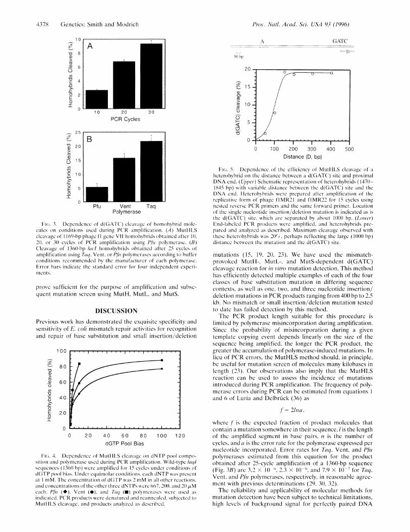
10, 20, or 30 cycles using Pfu polymerase, and homohybridmolecules were prepared by a denaturation and reannealingstep. The fraction of homohybrids cleaved by MutH increasedwith the number of cycles (Fig. 3A), a finding consistent witheither the damage or genetic variation hypothesis. Howeverthe degree of homohybrid cleavage was also found to dependon the polymerase used for PCR amplification. Thus, cleavageof amplified lacI homohybrids was highest with Taq polymer-ase, intermediate with Vent polymerase, and lowest with Pfupolymerase (Fig. 3B). These results parallel the error rates forthese polymerases, with the lower fidelity of Taq polymerasedue to absence of a 3' to 5' editing exonuclease (29-31).Although we cannot exclude a low level of template damageassociated with thermal cycling, these findings indicate that themajority of the homohybrid background signal is due topolymerase errors occuring during amplification. In light ofthese results, we have used the high fidelity reaction conditions(4 mM MgCl2, 1 mM each dNTP) of Eckert and Kunkel (32)for subsequent experiments.Use of dNTP Pool Bias During PCR Amplification to
Determine Detectability of Nucleotide Substitution Errors. AdNTP pool imbalance leads to an increased error rate duringin vitro synthesis by DNA polymerases (33, 34). We haveexploited this observation to test the utility of the MutHLSreaction for detection of PCR errors. For these experimentswild-type lacI sequences 1360 bp long were amplified usingPfu, Vent, or Taq polymerases under conditions of dGTP pool
imbalance. As shown in Fig. 4, d(GATC) cleavage of homo-hybrids was dependent on the dGTP concentration bias.Homohybrids derived from amplification using Taq polymer-ase were subject to MutHLS-dependent d(GATC) cleavage toa greater degree than homohybrids amplified under the sameconditions using Pfu and Vent polymerases. Negligible PCRproduct was obtained in reactions using Taq polymerase inwhich dGTP was present in a 100-fold molar excess over theother dNTPs. Since the enzyme lacks a 3' exonuclease activity,high levels of misincorporated dNTPs induce chain termina-tion (35). Likewise, homohybrids amplified using Vent poly-merase were cleaved to a greater extent than homohybridsamplified using Pfu polymerase (Fig. 4). Polymerase misin-corporation errors are therefore readily detectable by MutS-dependent d(GATC) cleavage.Dependence of the Efficiency of Cleavage by Activated
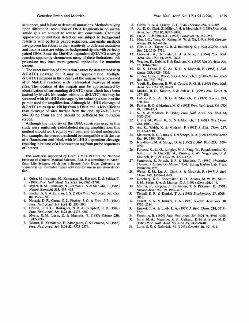
MutH on the Distance Between a d(GATC) Site and the Endof a DNA Heterohybrid. Although highly sensitive to mis-matched base pairs, the MutHLS reaction can only be used formutation screen if the sequence of interest contains ad(GATC) site. To determine the feasibility of introducing ad(GATC) site into a PCR primer to screen sequences lackingsuch a site, we have evaluated the dependence of the reactionon the distance of a d(GATC) site from a DNA end. As shownin Fig. 5, the efficiency of mismatch-provoked cleavage in-creased with increasing distance of the d(GATC) site from theproximal end in the range of 50-150 bp reaching a maximumat the latter distance. These results suggest that a PCR primerwith a d(GATC) site 50-100 nucleotides from an end would
Genetics: Smith and Mcodrich
('tri
4378 Genetics: Smith and Modrich
-D0
0a)
>
(-
-o.C0
E0
10
8
6
4
2
0
0 -,;
-u 20
r)0)0 l")-o
10>0
0 50I
0
A
10 20 30
PCR Cycles
A GATC
-D-50 bp
20
1a)
I 1
0aDO
--
B0 100 200 300 400 500
Distance (D, bp)
Pfu VentPolymerase
Taq
Fi(;. 3. Dependence of d(GATC) cleavage of homohybirid mole-cules on conditions used during PCR amplification. (A) MutHLScleavage of 1169-bp phage fl gcne VII homohybrids obtained after 1().20(). or 3() cycles of PCR amplification using Pf/i polymcrasc. (B)Cleavage of 1360-bp ladl homohybrids obtained after 25 cycles ofamplification using Taq, Vent, or Pf/i polymcrascs according to bufferconditions rccommcndcd by the manufacturer of each polymcrasc.Error bars indicate the standard error ffoiour independent cxpcri-llents.
prove sufficient for the purpose of amplification and subse-quent mutation screen using MutH, MutL, and MutS.
DISCUSSIONPrevious work has demonstrated the exquisite specificity andsensitivity of E. coli mismatch repair activities for recognitionand repair of base substitution and small insertion/deletion
100
80
60
40,
20
00 20 40 60 80 100 120
dGTP Pool Bias
Fi(;. 4. Dependence (of MutHLS cleavage on dNTP pool compo-sition and polymcrasc used during PCR amplification. Wild-type (laqsequences (1361) bp) wcir amplified for 15 cycles under conditioins ofdGTP pool bias. Under equimolar conditions, each dNTP was presentat I mM. The concentration of dGTP wvas 2 mM in all other reactions,and concentrations of the other three dNTPs were 667, 200()(). and 20 .rMeach. Pfi (*), Vent (0), and 7tiq (-) polymcrascs were used as
indicated. PCR products wcrc dcnatured and rcanncalcd. subjected toMutIHLS cleavage, and products analyzed as described.
Fi(i. 5. Dependence of the efficiency of MutItLS cleavage of a
hlictcroliybrid on the distance between a d(GATC) site and proximalDNA end. (Upper) Schematic representation of heterohybrids (147()-1845 bp) with variable distance between the d(GATC) site and theDNA end. lletcrohybrids were prepared after amplification of thereplicative form of phage flMR21 and flMR22 for 15 cycles usingnested rcverse PCR primers and the same forward primer. Locationof the single nucleotide insertion/deletion mutation is indicated as isthe d(GATC) site, which are separated by about 1()()0 bp. (Lower)End-labeled PCR produtcts were amplified, and heterohybiids pre-pared and analyzed as described. Maximum cleavage observed withthese heterohybrids was 20()%, perhaps reflecting the large (1()0)() bp)distance between the mutation and the d(GATC) site.
mutations (15, 19, 20, 23). We have used the mismatch-provoked MutH-, MutL-, and MutS-dependent d(GATC)cleavage reaction for in vitro mutation detection. This methodhas efficiently detected multiple examples of each of the fourclasses of base substitution mutation in differing sequencecontexts, as well as one, two, and three nucleotide insertion/deletion mutations in PCR products ranging from 400 bp to 2.5kb. No mismatch or small insertion/deletion mutation testedto date has failed detection by this method.The PCR product length suitable for this procedure is
limited by polymerase misincorporation during amplification.Since the probability of misincorporation during a giventemplate copying event depends linearly on the size of thesequence being amplified, the longer the PCR product, thegreater the accumulation of polymerase-induced mutations. Inlieu of PCR errors, the MutHLS method should, in principle,be useful for mutation screen of molecules many kilobases inlength (23). Our observations also imply that the MutHLSreaction can be used to assess the incidence of mutationsintroduced during PCR amplification. The frequency of poly-merase errors during PCR can be estimated from equations Iand 6 of Luria and Delbrdick (36) as
f/= 2na,
where / is the expected fraction of product molecules thatcontain a mutation somewhere in their sequence, / is the lengthof the amplified segment in base pairs, n is the number ofcycles, and a is the error rate for the polymerase expressed pernucleotide incorporated. Error rates for Taq, Vent, and P/thpolymerases estimated from this equation for the productobtained after 25-cycle amplification of a 1360-bp sequence(Fig. 3B) are 3.2 x 10 ", 2.3 x 10 (, and 7.9 x 10 7 for Taq,Vent, and Pfi polymcrases, respectively, in reasonable agree-ment with previous determinations (29, 30, 32).The reliability and applicability of molecular methods for
mutation detection have been subject to technical limitations,high levels of background signal for perfectly paired DNA
0OxI
")
-Q
0
I
Pr·oc·. Nadr. Acrrl-l. Sci. U/SA 93 (1996)
,TI
Proc. Natl. Acad. Sci. USA 93 (1996) 4379
sequences, and failure to detect all mutations. Methods relyingupon differential resolution of DNA fragments in polyacryl-amide gels are subject to severe size constraints. Chemicalapproaches to mutation detection are subject to backgroundreactivity with perfectly paired sequences. Enzymatic methodshave proven less robust in their sensitivity to different mutationsand in some cases are subject to background signals with perfectlypaired DNA. Since the MutHLS-dependent d(GATC) cleavagereaction apparently circumvents many of these limitations, thisprocedure may have more general application for mutationscreens.The exact location of a mutation cannot be determined with
d(GATC) cleavage but it may be approximated. Multipled(GATC) incisions in the vicinity of the mispair were observedafter MutHLS reaction, with preferential cleavage of somesites. The location of the mispair may be approximated byidentification of surrounding d(GATC) sites which have beenincised by MutH. Molecules without a d(GATC) site may bescreened with MutHLS cleavage by introducing a site into theprimer used for amplification. Although MutHLS cleavage ofd(GATC) sites up to 150 bp from a DNA end is less efficientthan cleavage of sites further from the end, d(GATC) sites50-100 bp from an end should be sufficient for mutationscreen.Although the majority of the DNA substrates used in this
study were uniformly radiolabeled during amplification, thismethod should work equally well with end-labeled molecules.For example, the procedure should be compatible with the useof a fluorescent end-label, with MutHLS-dependent cleavageresulting in release of a fluorescence tag from probe sequencesof interest.
This work was supported by Grant GM23719 from the NationalInstitute of General Medical Sciences. P.M. is a consultant to Amer-sham Life Science, which has a license from Duke University tocommercialize mismatch repair activities for use in mutation detec-tion.
1. Orita, M., Iwahana, H., Kanazawa, H., Hayashi, K. & Sekiya, T.(1989) Proc. Natl. Acad. Sci. USA 86, 2766-2770.
2. Myers, R. M., Lumelsky, N., Lerman, L. S. & Maniatis, T. (1985)Nature (London) 313, 495-498.
3. Fischer, S. G. & Lerman, L. S. (1983) Proc. Natl. Acad. Sci. USA80, 1579-1583.
4. Novack, D. F., Casna, N. J., Fischer, S. G. & Ford, J. P. (1986)Proc. Natl. Acad. Sci. USA 83, 586-590.
5. Cotton, R. G. H., Rodrigues, N. R. & Campbell, R. D. (1988)Proc. Natl. Acad. Sci. USA 85, 4397-4401.
6. Myers, R. M., Larin, Z. & Maniatis, T. (1985) Science 230,1242-1246.
7. Winter, E., Yamamoto, F., Almoguera, C. & Perucho, M. (1985)Proc. Natl. Acad. Sci. USA 82, 7575-7579.
8. Gibbs, R. A. & Caskey, C. T. (1987) Science 236, 303-305.9. Au, K. G., Clark, S., Miller, J. H. & Modrich, P. (1989) Proc. Natl.
Acad. Sci. USA 86, 8877-8881.10. Lu, A.-L. & Hsu, I.-C. (1992) Genomics 14, 249-255.11. Hsu, I.-C., Yang, Q., Kahng, M. W. & Xu, J.-F. (1994) Carcino-
genesis 15, 1657-1662.12. Ellis, L. A., Taylor, G. R. & Baumberg, S. (1994) Nucleic Acids
Res. 22, 2710-2711.13. Lishanski, A., Ostrander, E. A. & Rine, J. (1994) Proc. Natl.
Acad. Sci. USA 91, 2674-2678.14. Wagner, R., Debbie, P. & Radman, M. (1995) Nucleic Acids Res.
23, 3944-3948.15. Su, S., Lahue, R. S., Au, K. G. & Modrich, P. (1988) J. Biol.
Chem. 263, 6829-6835.16. Jiricny, J., Su, S., Wood, S. G. & Modrich, P. (1988) NucleicAcids
Res. 16, 7843-7853.17. Youil, R., Kemper, B. W. & Cotton, R. G. H. (1995) Proc. Natl.
Acad. Sci. USA 92, 87-91.18. Mashal, R. D., Koontz, J. & Sklaar, J. (1995) Nat. Genet. 9,
177-183.19. Lahue, R. S., Au, K. G. & Modrich, P. (1989) Science 245,
160-164.20. Parker, B. 0. & Marinus, M. G. (1992) Proc. Natl. Acad. Sci. USA
89, 1730-1734.21. Su, S. & Modrich, P. (1986) Proc. Natl. Acad. Sci. USA 83,
5057-5061.22. Grilley, M., Welsh, K., Su, S. & Modrich, P. (1989) J. Biol. Chem.
264, 1000-1004.23. Au, K., Welsh, K. & Modrich, P. (1992) J. Biol. Chem. 267,
12142-12148.24. Matteson, R. J., Biswas, S. J. & Steege, D. A. (1991) NucleicAcids
Res. 19, 3499-3506.25. Ivey-Hoyle, M. & Steege, D. A. (1992) J. Mol. Biol. 224, 1039-
1054.26. Parsons, R., Li, G., Longley, M. J., Fang, W., Papadopoulos, N.,
Jen, J., de la Chapelle, A., Kinzler, K. W., Vogelstein, B. &Modrich, P. (1993) Cell 75, 1227-1236.
27. Sambrook, J., Fritsch, E. F. & Maniatis, T. (1989) MolecularCloning: A Laboratory Manual (Cold Spring Harbor Lab. Press,Plainview, NY).
28. Welsh, K. M., Lu, A., Clark, S. & Modrich, P. (1987) J. Biol.Chem. 262, 15624-15629.
29. Lundberg, K. S., Shoemaker, D. D., Adams, M. W. W., Short,J. M., Sorge, J. A. & Mathur, E. J. (1991) Gene 108, 1-6.
30. Mattila, P., Korpela, J., Tenkanen, T. & Pitkanen, K. (1991)Nucleic Acids Res. 19, 4967-4973.
31. Tindall, K. R. & Kunkel, T. A. (1988) Biochemistry 27, 6008-6013.
32. Eckert, K. A. & Kunkel, T. A. (1990) Nucleic Acids Res. 18,3739-3744.
33. Kunkel, T. A. & Loeb, L. A. (1979) J. Biol. Chem. 254, 5718-5725.
34. Fersht, A. R. (1979) Proc. Natl. Acad. Sci. USA 76, 4946-4950.35. Innis, M. A., Myambo, K. B., Gelfand, D. H. & Brow, M. D.
(1988) Proc. Natl. Acad. Sci. USA 85, 9436-9440.36. Luria, S. E. & Delbriick, M. (1943) Genetics 28, 491-511.
Genetics: Smith and Muodrich