mutation induction in zamioculcas … of contents acknowldegements iii abslract iv . table of...
TRANSCRIPT

UNIVERSITY OF HAWAII LIBRARY
THE IDENTIFICATION OF A SUITABLE IRRADIATION DOSAGE FOR
MUTATION INDUCTION IN ZAMIOCULCAS ZAMlIFOLIA (LODD.)
AND THE POL YPLOIDIZATION OF
Z ZAMlIFOLIA AND MARSDENIA FLORI BUNDA
A THESIS SUBMITIED TO THE GRADUATE DMSION OF THE UNIVERSITY OF HAW AI'I IN PARTIAL FULFILLMENT
OF THE REQUIREMENTS FOR THE DEGREE OF
MASTER OF SCIENCE
IN
HORTICULTURE
DECEMBER 2006
By Susana D. Vanzie-Canton
Thesis Committee:
Kenneth Leonhardt, Chairperson Richard CrUey Yoneo Sagawa
Kheng-Tuan Cheah

We certify that we have read this thesis and that, in our opinion, it is satisfactory in scope
and quality as a thesis for the degree of Master of Science in Horticulture.
THESIS COMMITIEE
ii

ACKNOWLDEGEMENTS
I am grateful to my committee chairman Dr. Kenneth Leonhardt for his support
His knowledge, experiences, and technical assistance were of great value throughout the
entire process of this research project. I would also like to express my sincere
appreciation to Drs. Richard CriIey, Yoneo Sagawa, and Kbeng-Tuan Cheah who served
on my thesis committee.
I had great help from all Magoon Research Facility personnel, namely Craig
Okazaki and Ronald Matsuda. In addition, my appreciation extends to Susan Takahashi,
Shirley Ishihara, and Elsie Sun in the Department office, who have always been kind and
helpful. I wish to express my sincere appreciation to Dr. Mark Wright, who provided
statistical support; Dr. Ania Wieczorek, Mr. Jaco Le Roux, and Ms. Carol Tran who
kindly provided assistance when needed; and Dr. Karen Selph for her time and helpful
suggestions during flow cytometric analysis. Sandy Kasman (Hawaiian Sunshine
Nursery) also contributed to my knowledge of Zamioculcas zamiiJolia. I would also like
to thank my labmates, namely Emily Teng for her help, and Tilden Miguel for his helpful
suggestion of the callus inducing media recipe utilized in Chapter 2.
Finally, special thanks are expressed to my husband for his patience, emotional
support, encouragement, and understanding throughout my graduate education. Thanks
to my parents, my brothers and my sisters for all their words of encouragement.
iii

ABSTRACT
Radiation mutation has been successfully used to create a great variety of
ornamental crop cultivars by supplementing existing germplasm and improving existing
cultivars, and chemical mutagens such as colchicine and oryza1in have been used to
create new plant cultivars by doubling the chromosome number of the treated plant
material to produce tetraploids. The main objective of this thesis research was to develop
protocols to create tetraploid plants of Zamioculcas zamii/olia (Lodd.) Engl., an
important foliage plant, and Marsdenia jloribunda (Brongn.), an important lei flower
plant, and to determine the LDso of ZZ leaflets. ZZ leaflets and M. jloribunda seeds were
treated with colchicine at various concentrations and durations in order to induce ploidy
changes and regenerate polyploids. Five ZZ tetraploids and one M. jloribunda tetraploid
were produced using colchicine. A tissue culture protocol was also developed for the
oryza1in treatment of ZZ callus for the in vitro polyploidy induction of ZZ. The LDso of
ZZ leaflets irradiated with x-rays was calculated as 20±1 Gy. A ZZ germplasm
collection was also initiated to provide ZZ plant material for use in future breeding
studies.
iv

TABLE OF CONTENTS
ACKNOWLDEGEMENTS ............................................................................................... iii ABSlRACT ........................................................................................... iv . TABLE OF CONTENTS .................................................................................................... v LIST OF TABLES ............................................................................................................ vii LIST OF FIGURES ......................................................................................................... viii LIST OF ABBREVIATIONS ............................................................................................. x LITERATURE REVIEW ................................................................................................... 1
SECTION 1- ZAMIOCULCAS ZAMIIFOLIA .................................................................... 6 Introduction ................................................................................................................. 7
CHAPTER 1: COLCHICINE TREATMENT OF ZZ LEAFLETS IN VIVO ............ 9 Background Information ......................................................................................... 9 Materials and Methods ............................................................................................ 9 Results and Discussion ......................................................................................... 13 Tables and Figures ................................................................................................ 21
CHAPTER 2: POL YPLOIDIZATION OF ZAMIOCULCAS ZAMIIFOLIA VIA ORYZALlN TREATMENT OF CALLUS .............................................................. 31
Background Information ....................................................................................... 31 Materials and Methods .......................................................................................... 32 Results and Discussion ......................................................................................... 36 Figures ................................................................................................................... 39
CHAPTER 3: IDENTIFICATION OF A SUITABLE IRRADIATION DOSAGE FOR ZAMIOCULCAS ZAMIIFOLIA ........................................................................ 41
Background Infonnation ....................................................................................... 41 Materials and Methods .......................................................................................... 42 Results and Discussion ......................................................................................... 43 Tables and Figures ................................................................................................ 46
CHAPTER 4: GERMPLASM COLLECTION OF ZAMIOCULCAS ZAMIIFOLIA50 Figures ................................................................................................................... 53 .
SECTION 2 - MARSDENIA FLORI BUNDA .................................................................... 63
CHAPTER 5: POL YPLOIDIZATION OF MARSDENIA FLORIBUNDA .............. 64 Background Information ....................................................................................... 64 Materials and Methods .......................................................................................... 65 Results and Discussion ......................................................................................... 68 Tables and Figures ................................................................................................ 71
v

APPENDIX. SASS DATA OUTPUT FOR PROBIT ANALySIS ......................... 77 LITERATURE CITED ..................................................................................................... 81
vi
LIST OF TABLES
1.1 Guard cell measurements of colchicine treated ZZ leaflets before rhizome development ......................................•...........•........... 21
1.2 Guard cell measurements of colchicine treated ZZ leaflets after rhizome development ........................•.............•........................ 21
1.3 Number of ZZ tetraploids, mixoploids, and DNA aneuploids identified via flow cytometIy ..........................................•............•..... 22
3.1 Number ofleaflets irradiated and dosages used to identifY LDloo and LDso ..................................................•...............•................................... 46
3.2 Results of ZZ leaflets irradiated in March 2006 .....................•..............•... 46
3.3 Results of ZZ leaflets irradiated in June 2006 .......................................... 47
3.4 Results ofZZ leaflets irradiated in August 2006 ...................................... .47
5.1 Number of seedHngs and seedHng height of M. j/oribunda after colchicine treatment of seeds .....................................•................. 67
5.2 Guard cell measurements of M. j/oribunda seedHngs visually identified as possible polyploids ......................................................... 68
vii

LIST OF FIGURES
Figure ~
1.1 Vegetative propagation of ZZ via leaflet cuttings ..................................... 23
1.2 Actively growing ZZ root tips harvested for chromosome counts ........................................................................ 23
1.3 Sample preparation for flow cytometric analysis ...................................... 24
1.4 ZZ leaflet morphology of diploid and suspected polyploids ......................... 25
1.5 Stained ZZ chromosomes isolated from single cells .................................. 26
1.6 Distribution of DNA content of diploid ZZ ............................................. 27
I. 7 Distribution of DNA content of tetraploid ZZ .......................................... 28
1.8 Distribution of DNA content ofmixoploid ZZ ......................................... 29
1.9 Distribution of DNA content of a ZZ DNA aneuploid ................................ 30
1.10 Identified debris in samples to be analyzed by flow cytometry ............................. 30
2.1 Tissue culture protocol developed for callus induction followed by plantiet regeneration of ZZ ................................................ 39 .
2.2 Protocol developed for In vitro polyploidy induction of ZZ .......................... 40
3.1 Irradiation of ZZ leaflets ................................................................. .48
3.2 Irradiated ZZ leaflets ...................................................................... .48
3.3 Relationship between dosage and percent death of x-ray irradiated ZZ leaflets ....................................................................... 49
3.4 ZZ plant regenerated from leaflet irradiated at 20Gy .................................. 49
4.1 ZZ: Hawaiian Sunshjne Nursery ......................................................... 53
4.2 ZZ: National Botanic Garden of Belgium (species) ................................... 54 viii

LIST OF FIGURES (CONTD.)
Figure ~
4.3 ZZ: National Botanic Garden of Belgium (variegata) ................................. 55
4.4 ZZ: National Botanic Garden of Belgium (variegata) ................................. 56
4.5 ZZ: National Botanic Garden of Belgium (variegata) ................................. 57
4.6 ZZ: National Botanic Garden of Belgium (variegata) ................................. 58
4.7 ZZ: Waimea Valley Audubon Center ................................................... 59
4.8 ZZ: Harold L. Lyon Arboretum .......................................................... 60
4.9 ZZ: Nong Nooch Tropical Botanical Garden ........................................... 61
4.10 ZZ: Nong Nooch Tropical Botanical Garden ........................................... 62
5.1 Mjloribunda seedlings 5 weeks after colchicine treatment .......................... 69
5.2 Percentage seedling emergence after colchicine treatment of M jloribunda seeds ..................................................................... 69
5.3 Guard cell measurements of M jloribunda ........................................ ..... 70
5.4 Distribution of DNA content ofmixoploid Mjlorlbunda ........................... 71
5.5 Distribution of DNA content of diploid M jlorlbunda ................................ 71
5.6 Distribution of DNA content of tetraploid M jlorlbunda ............................. 72
ix

LIST OF ABBREVIATIONS
2,4-D 2,4-dichlorophenoxyacetic acid
2-ip 6-y-y-dimethylaminopurine
ADC analog-to-digital convertors
BA 6-benzyladenine
CV coefficients of variation
DI DNA index
DMSO dimethyl sulfoxide
Gy Gray
HCl hydrochloric acid
FCM flow cytometry
IAA indole-3-acetic acid
LDx 1etha1 dosage
MS Murashige and Skoog basal salts
MSo Murashige and Skoog basal salts with no plant growth regulators
PGRs plant growth regulators
PI propidium iodide
ZZ Zamioculcas zamii/olia
x

LITERATURE REVIEW
The 200S wholesale value of floriculture crops in the US is estimated at $S.4
billion, with Hawaii ranking fourth in both the foliage and cut flower/cut greens
commodities (USDA, 2006). The introduction of new ornamentals for production can
help increase the value of Hawaii's ornamental plant industry. For example, Florida is
consistently the greatest producer of foliage plants year after year, accounting for over
SS% of the nation's wholesale value. Florida's success has been pattially attributed to
the continuous introductions of new cultivars and genera into the Florida foliage market
(Chen, Henny, and McConnell, 2002).
The main objective of my Master's research program is to develop protocols and
systems to create new cultivars of Zamioculcas zamiijolia (Lodd.) Engl., an important
foliage plant, and Marsdeniafloribunda (Brongn.), an important lei flower plant formerly
known as Stephanotis floribunda. Both chemical mutagens and radiation mutation were
employed in order to achieve the objective.
New plant cultivars contribute significantly to the ornamental plant industry.
Among the various ways to bring about improvements in crops, genetic improvements
are more desirable than non-genetic crop improvements such as the use of growth
retardants. For example, growth retardants have been shown to reduce vegetative growth
in trees and other ornamental crops to produce a more compact plant growth (Manriquez,
200S; Feng, 2004; Criley 2000). The changes induced, however, are not permanent and
require repeated inputs. Genetic improvements, on the other hand, are passed on to
progeny or to new clones produced by vegetative propagation. It is for this reason that
1

the use of chemical and physical mutagens was elected as the method of choice for the
production of new ZZ and M jloribunda cultivars, rather than using other methods that
induce a non-permanent change on morphological growth.
Radiation mutation has been successfully used to create a great variety of
ornamental crop cu1tivars by supplementing existing germplasm and improving cu1tivars
(Yamaguchi, 1988; Kaicker and Dhyani, 1986). In ornamental plants, irradiation has
produced improved attributes such as earlier flowering, compact growth habits, the
production oflarger flowers, changes in flower color (Micke et al., 1987), changes in leaf
color, and changes in leaf shape (Matsubara, 1982).
The energy radiated from physical mutagens induces chemical reactions within
the plant cell resulting in a structural change of the DNA. lfthese changes are then
maintained during DNA replication, they are manifested as mutations. Mutations in
somatic tissues are usually induced by treating leafbuds with mutagens or adventitious
buds are induced immediately following the mutagenic treatment (Lapins, 1983). The
vegetative propagation of ZZ via leaflet cuttings lends itself to irradiation via the latter
method. The advantage of this method is that adventitious buds arising from leaves and
petioles are thought to arise from a single cell, so that the formation of a chimera is
minimized and the irradiation results almost exclusively in solid, non-chimeric mutants
and normal, unmutated plants (Broerges, Haccius, and Weidlich, 1968; Broerges and
Keen, 1980). Though some scientists dispute the origins of adventitious meristems
(single vs. multiple cells), Zalewska (2001) concluded that irrespective of their formation,
adventitious meristems may give rise to plants with stable genetic changes.
2

Chemical mutagens such as colchicine (N-acetyltrlmethylcolchinic acid), an
alkaloid, and oryzalin [(4-(Dipropylamino)-3, 5-dinitrobenzene-sulfonamide], a
dinitroaniline herbicide, have been used to create new plant cultivars by doubling the
chromosome number of the treated plant material. Colchicine, for example, has proven
to be a useful tool in many breeding programs aimed at regaining fertility, preventing
hybrid sterility (Kamemoto et oJ., 1997; Kamemoto, 1985), producing superior cultivars
(Vainola, 2000; Tambong and Garton 1998), and producing sterile triploids by breeding
induced tetraploids with diploids (Blakesley et oJ., 2002; St. Marseille and Grant 1997).
Similarly, oryzalin has been shown to double the chromosome numbers in Nepeta
(Mitrofanova et oJ., 2003), AZocasio (Thao et aZ., 2003), Rhododendron hybrids (Vainola,
2000), and Lilium (Tuyl, Meijer, and Van Dien, 1990).
Research has shown that colchicine and oryzalin have a similar mode of action at
the molecular level: both chemicals were found to inhibit elongation, produce swelling in
the elongation zone, depolarize cell enlargement, and disrupt cell differentiation and
polyploidization in maize seedling roots (Upadhyaya and Nooden, 1976). Both agents
bind to plant microtubules, which are subcellular structures that are mainly composed of
the protein tubulin, and are involved in chromosome migration. Colchicine is thought to
depolymerize the microtubules (Morejohn et al., 1987), while oryzalin binds to free
tubulin in the plant cell, forming a dinitroaniline-tubulin complex that is incapable of
polymerizing into plant microtubules. Consequently, both agents disrupt mitosis by
inhibiting spindle fiber formation at metaphase (Strachan and Hess, 1983), resulting in an
increase in the chromosome number of the daughter cells. Chemical mutagens are most
effective when applied to cells that are in a high state of cell division (Eigsti and Dustin,
3

1955); therefore, callus tissue, seeds, and freshly cut leaflets were utilized in this research
project in order to create polyploids.
Researchers have discovered that oryzalin can disrupt mitosis at a lower
concentration than colchicine due to its stronger binding affinity to plant tubulins.
Ramulu, Verhoeven, and Dijkbula (1991) showed that in potato, oryzalin was more
efficient for chromosome doubling than colchicine. Tuyl et aI., (1990) and Thao et al .•
(2003), reported similar results in Lilium and Alocasia respectively. Contrastingly,
colchicine has been found to be more effective in chromosome doubling for plants such
as Miscanthus sinensis. a perennial grass (petersen, Hagberg and Kristiansen, 2003) and
two leguminous tree species of Acacia (Blakesley, et aI., 2002). The variation in the
mutagenic effectiveness of both agents indicates that the efficiency of each chemical to
double the chromosome number is plant specific, and should be determined on a plant by
plant basis. Accordingly, both chemicals were used in attempts to induce chromosome
doubling in the subject plants of this research project.
In the past, screening for ploidy level changes was limited to measurement of
stomatal guard cells of the treated plant material or by root tip chromosome counts of
identified suspects. Measuring stomatal guard cells requires producing an imprint of the
abaxial side of the leaf of interest. The imprint is then viewed under the microscope and
the size of the guard cells is measured. The theory behind this method of screening for
changes in ploidy levels is simple. The cell volume of a plant is directly proportional to
the amount of DNA present in the cell, so that doubling the amount of DNA, which
occurs when diploids are converted to tetraploids, causes the cell to double its volume.
Doubling of the volume allows an increase in the size of the cell in anyone dimension by
4

1.25, and comparisons of guard cells among a batch of chemically treated plants allows
for the identification of possible polyploids (Russell, 2004). Typically, the ploidy level
of the suspected plants identified using guard cell measurements is then verified by
performing root tip chromosome counts, a laborious and time consuming process.
Today, more researchers rely on flow cytometry (FCM) for the early screening of
treated plant material and ploidy level confirmation. FCM allows for the quantification
of plant nuclear DNA, subsequently providing the user with the ploidy level of the
samples screened. A typical flow cytometer contains several components: a light source,
a flow chamber and optical assembly, photodetectors and processors to convert light
signals into analog electrical impulses, analog-to-digital convertors (ADCs) and a
computer system for the analysis and storage of digitized data. Essentially, the plant
nuclei is extracted from plant tissue using an extraction buffer and labeled with a
fluorescent dye. The sample is then loaded into the cytometer and illuminated, causing
the dye to absorb the illuminating light and fluoresce. The emitted light is then converted
to electric current pulses, which are fed to amplifiers, digitized using the ADCs, and
stored in the computer in the form of a histogram. Computer software may then be used
to analyze the data output and study correlations among the parameters (Dolezel. 1991),
for example, a tetraploid cell will fluoresce twice as much as a diploid cell. Accordingly,
FCM is a valuable, rapid and precise tool to detect converted polyploids (Beckbmrt et al.,
2004; Takamura, Lim, and Van Tuyl, 2002; Toska et al., 1995; Vainola 2000; Vainola
and Repo, 2001) and was employed as a screening tool in this research project.
5

SECTION 1
ZAMIOCULCAS ZAMIIFOLIA
Introduction
Zamioculcas zamii/olia (Lodd.), which will be referred to hereafter as ZZ, is a
member of the Araceae family and is also commonly known as African coontie, aroid
palm, arum fem, cardboard palm, and emerald frond. ZZ is native to tropical east and
subtropical southeast Africa, with its native habitat ranging from Kenya to northeastern
South Africa. It grows in dry grassland and often stony ground and has several fleshy
stalks bearing alternate pinnate leaflets. The leaflets have the capacity to sprout new
plants and form tiny rhizomes at their base (Bawn, 2000). ZZ is a seasonally dormant or
evergreen herb with a short, very thick rhizome and a diploid chromosome number of2n
= 34 (Jones, 1957).
Not only has it been described as a plant that is becoming or will become an
important player in the foliage plant industry (Chen, Henny, and McConnell, 2002), but it
was also listed among the Florida Plants of the Year in 2002 (Chen and Henny 2003).
The ability of ZZ to grow under low light conditions, its tolerance to drought stress, its
unique appearance, its low maintenance requirements and limited pest problems are
characteristics that contribute sigoificantly to its ornamental and interior plantscaping
value (Chen and Henny, 2003). Furthermore, there are no reports available on the
polyploidization of zz, so that success achieved in this research project will contribute
sigoificantly to this area of research in ZZ cu1tivar development. With only one species
in the genus of this ornamental, the creation of new clones may allow breeding and
variety development to advance more quickly.
7

Two approaches were taken in an attempt to induce mutations through
chromosome doubling in ZZ. namely: (i) colchicine treatment of ZZ leaflets in vivo and
(ii) oryzaJin treatment of ZZ callus in vitro. The irradiation of ZZ leaflets was another
approach taken in an attempt to create a new ZZ cultivar. For this research, the response
of ZZ leaflets to irradiation was limited to the identification of a suitable x-ray dosage for
mutation induction, but did not result in a neW cultivar. Section 1 has been divided into
four chapters, three of which describe the experiments performed using the approaches
mentioned above. Chapter 4 provides a brief description of the ZZ germplasm collection
at the University of Hawaii.
8

CHAPTER 1: COLCHICINE TREATMENT OF ZZ LEAFLETS IN VIVO
Background Information
lZ may be propagated vegetatively by leaflet or petiole cuttings. Leaflets are
removed from the stock plant and stuck vertically in a highly organic medium such as
coir or peat moss. After about 3-4 weeks, a small rhizome begins to appear and roots
emerge. Buds begin to form on the rhizome giving rise to new leaves and eventually a
whole new plantlet (Fig. 1.1). The described method of propagation proved to be a
convenient method for the colchicine treatment of lZ leaflets in vivo. The objective of
this experiment was to regenerate tetraploid lZ plants using varying concentrations of
colchicine. The induction oftetraploids using this method has proven to be simple and
cheap.
Materials and Methods
lZ leaflets were treated with colchicine in two ways: immediately after harvest
(before rhizome development) or after a small rhizome had been produced. Both
methods are described below.
Before rhizome development
Colchicine (phytoTechnology Laboratories; Shawnee Mission. KS) was dissolved
in distilled water to make 0.05%, 0.2 %, and 0.4% solutions. lZ leaflets were harvested
from the tip to the base of the leaf and treated the same day in the colchicine solutions,
with 18 leaflets per treatment and water used as the control. The cut portions of the
9

leaflets (the exposed surface of the petiole) were soaked for 24 hours in 50ml of the
treatment solution. After treatment, the leaflets were rinsed with distilled water and
transplanted into a 40cm x 55cm metal flat containing 1: 1 (v/v) peat:vermiculite. The flat
was placed under mist in the Foliage Greenhouse at the Magoon Research Facility.
About 3.5 months later the remaining leaflets were transplanted into individual 2 inch
pots containing moistened Pro-Mix 'BX' (Premier HorticultW'e Ltd.; Dorval, Canada).
After rhizome development
ZZ leaflets were harvested from along the entire length to the leaf and stuck in a
40cm x 55cm metal flat containing 1:1(v/v) peat:vermiculite. One month later, after all
leaflets had developed a small rhizome, the leaflets were removed from the propagation
medium and rinsed with distilled water. The leaflets were then graded according to
rhizome size and distributed evenly into 4 treatments containing 18 leaflets each: 0.05%,
0.2 %, and 0.4% colchicine, with water used as a control. The leaflets were soaked for 24
hours in a 50 ml solution, which allowed the rhizome to be completely submerged. After
treatment, the leaflets were returned to the mist bench and were transplanted 2.5 months
later into individual 2" pots containing moist Pro-Mix 'BX'. The first shoots began to
appear 7 months after treatment.
Guard Cell Measurements
After visual inspection of the regenerated plantlets, suspected tetraploids were
selected for further screening using stomatal guard cell measurements. Clear nail polish
(Markwins Beauty Products Inc.; City of Industry. CA) was thinly applied to the abaxial
10
side of the leaf. After the polish had dried (approximately 60 seconds), a strip of Scotch
3M Transparent Tape with Gloss Finish (3M Products; St. Paul, MN) was mounted on
top of the dried polish. The tape, which now held the abaxial imprint, was removed from
the leaflet and mounted onto a coverslip for viewing at 400X magnification under a Leitz
Wetzlar light microscope. Fifteen guard cells were randomly selected from each leaf
sample and measured (length x width) using a unitless graticuie.
Chromosome Counts
Two methods were employed for chromosome counting in ZZ. The first method
used is a modified version of the technique used by Shi (2003). Actively growing root
tips (Fig. 1.2) were excised, rinsed in distilled water, and pre-treated with 300ppm
cycloheximide for 12 hours. Next, the root tips were fixed in Camoy's :fixative (3: I 95%
ethanol and acetic acid) for at least 30 minutes followed by a rinse in distilled water.
Root tips were then softened in a solution of I: I concentrated hydrochloric acid and 95%
ethanol for five minutes, rinsed with tap water, and placed on a clean microscope slide
that contained a drop of carbolfuchsin stain. The root tip was then squashed using a
small scalpel, a cover slip placed on top of the squashed root tip, and the slide viewed
under the microscope for counting.
The second method used for chromosome counting was based on the work done
by Sharma and Mookerjea (1955). Actively growing root tips were excised, rinsed in
distilled water, and pre-treated in a saturated solution of paradichlorobenzene for 24
hours at 1O-12°C. The saturated solution was prepared by dissolving 109 of the solid in
500ml distilled water and holding the solution at 60°C overnight. The solution was used
11

at room temperature. After pretreatment, the root was transferred to an acid-dye fixation
solution (2% aceto-orcein and IN HCI at a 9: 1 ratio) and the mixture heated over a flame
for 3-4 seconds. The contents were then poured onto a watch glass and allowed to cool
for at least 5 minutes. A drop of 1 % aceto-orcein solution was placed on a dry slide and
the root was transferred to the drop of stain. The intensely colored portion of the tip was
retained, while the remainder of the root was discarded. The root tip was then squashed
using a small scalpel and a cover slip placed on top of the root The root tip was further
squashed by applying even pressure on the cover slip. A Leitz Wetz1ar light microscope
was used to observe the prepared slides at IOOOX magnification and pictures were taken
using a Nikon Coolpix 4500 digital camera.
Flow Cytometry
CyStain PI Absolute P DNA Staining Kit for Plant Genome Size (partee;
Munster, Germany) was used for nuclei extraction and DNA staining of nuclear DNA
from ZZ leaflets used for flow cytometric measurement Approximately 1 cm2 of each
leaflet was chopped for 30 - 60 seconds in 500/11 ice cold nuclei extraction buffer with a
sharp doubled edged razor blade in a 55mm plastic Petri dish. The slurry was then filtered
through a 50JI.M Cell Tries filter (partee; Munster, Germany) and the suspension of
released nuclei was stained in a solution composed of staining buffer, propidium iodide,
and RNAse for a final volume of2ml. Leaflet samples and all reagents used were kept
on ice throughout the entire process from leaflet harvest to sample preparation. All
samples were prepared for flow cytometric measurement within 1-2 hours of harvest
Due to the limited aVailability of plant material, only one leaflet was sampled per leaf in
12
most cases, with usually one leaf sampled per plant. For those regenerated plants that
showed variation in leaf morphology within the same plant, one leaflet was sampled per
leaf in order to compare differences in ploidy level throughout the plant. Figure 1.3
shows the process from sample preparation to sample analysis.
The relative fluorescence oftotaI DNA of single nuclei was analyzed using a
Beckman-Coulter (Miami, Florida) Altra flow cytometer (www.soest.hawaii.edulsfct)
using the 488 nm line of a Coherent 190C argon ion laser set at 200 m W. Control diploid
plants were used as external standards, and these standards were run intercalated between
samples. The linear, log and peak fluorescence signals of the propidium iodide-stained
nuclei were collected (610 BP filter), along with forward and side scatter signals. Plots of
peak vs. linear propidium iodide fluorescence were used to eHrnjn8te doublets (two nuclei
stuck together as they pass the laser/particle sensing point). The resulting data was
analyzed using Flow Jo (v. 6.3.4, Treestar Inc., www.flowio.com). Means and
coefficient of variance percentages of the resulting peaks were calculated and histograms
of linear DNA fluorescence, which allowed for visual analyses of the data, were
produced. An Olympus BX51 Epifluorescence Microscope was used at a 480nm
excitation frequency in order to view propidium iodide-stained nuclei and detect cellular
debris in some of the leaflet samples before flow cytometric analysis.
Results and Discussion
Visual inspection of the regenerated plants resulted in the identification of 26
suspected tetraploids. The leaflets of these plants were rounder (Fig. 1.4), thicker, and
the overall plant height was smaller than that of the controls. The growth rate
13

(emergence of new leaves and roots) of the suspects seemed to be slower than that of the
controls. Guard cell measurements of the selected plants support showed that several of
the regenerated plants are indeed polyploids (Table 1.1). Doubling the chromosome
number in a plant cell leads to an increase in stomatal guard cell size by a factor of 1.25
(Russell, 2004). The results obtained in Table 1.1 show that all the plants regenerated
from leatlets treated before rhizome development that were visually identified polyploids
were confirmed as such by guard cell measurements. Likewise, Table 1.2 shows that 15
of the 19 visually identified polyploid plants regenerated from leatlets treated after
rhizome development were shown to be polyploids via guard cell measurements. Note
that for three of the plants screened, the guard cell area decreased in comparison to the
control. This decrease does not imply a chromosomal change in the plant, but should be
attributed to the inherent variation in guard cell sizes from plant to plant, i.e., the
variation that exists between plants of the same ploidy level. The factor of increase in the
guard cell area of the plantlets produced before and after rhizome production ranged from
1.27 to 2.13. However, it was not clear what level of polyploidization the new plants had
obtained, or whether the plantlets regenerated were solid tetraploids or mixoploids.
In order to avoid misinterpretation of the results obtained, the term mixoploid,
when used hereafter, applies to the ploidy status of the entire plant (entire stalk including
alileatlets, see Fig. 1.3d) or a single leatlet, and will be specified as such. The term
mixopJoid generally applies to a plant structure that has originated from meristematic
tissue comprised of cells with varying ploidy levels. This variation results from the
polyploidization of some cells, while other cells within the same meristematic region are
unaffected and remain diploid. The meristematic tissue composed of cells with mixed
14

ploidy levels, in turn, gives rise to a structure that varies in ploidy level, e.g., individual
leaflets with both diploid and tetraploid cells, where the leaflet may also be described as a
chimem (Thao et ai, 2003).
Further verification of the ploidy state of the regenemted plants using root tip
chromosome counts, however, was not possible. ZZ chromosomes are very latge and
long, so that chromosome overlapping was common. Subsequently, the only
chromosome counts obtained were those of the controls (Fig. 1.5), with 2n = 34,
supporting the finding of Jones (1954). Even though the length of time allotted for the
pre-treatment of the root tips was repeatedly increased in order to allow for further
constriction of the chromosomes, no cells were identified in which all the chromosomes
could be individually counted. At least 60 - 75 slides were prepared for chromosome
counting with no success. Such difficulties in counting chromosomes have also been
expressed by other researchers, who have in turn, used flow cytometry for ploidy level
determination (Barker et al., 1998; Meng and Finn, 1999). Hence, the results obtained
from the flow cytometric analysis were used in order to verify ploidy levels of the
suspected polyploids.
Figures 1.6 - 1.9 are representative examples of the different types of histograms
that were obtained from analysis of the flow cytometry data with Flow Jo. For all the
histograms shown, the number of nuclei is represented on the Y axis and the DNA
fluorescence intensity is represented on the X axis. Figure 1.6 shows a histogram that is
typical of a diploid sample, with one major peak showing nuclei with 2C DNA content
and a smaller peak showing nuclei with 4C DNA content The first peak is representative
of those nuclei that are in the G 1 mitotic phase, and the smaller peak represents those
15
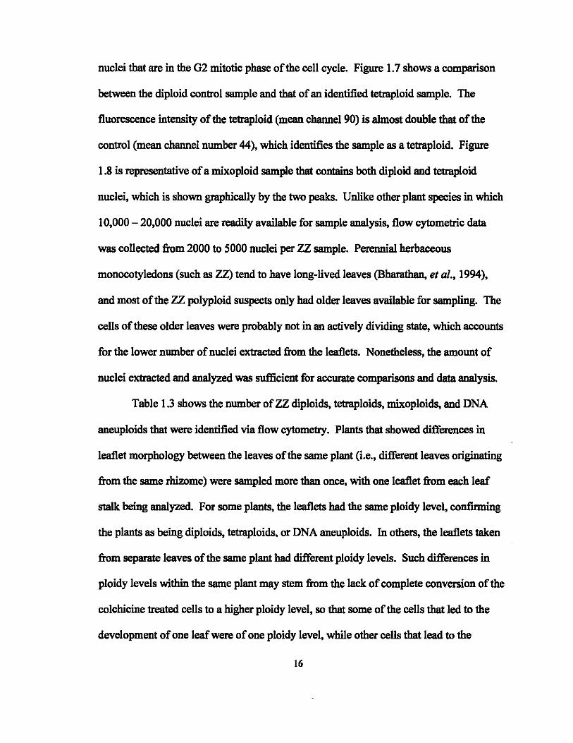
nuclei that are in the G2 mitotic phase of the cell cycle. Figure 1.7 shows a comparison
between the diploid control sample and that of an identified tetraploid sample. The
fluorescence intensity of the tetraploid (mean channel 90) is almost double that of the
control (mean channel number 44), which identifies the sample as a tetraploid. Figure
1.8 is representative of a mixoploid sample that contains both diploid and tetraploid
nuclei, which is shown graphically by the two peaks. Unlike other plant species in which
10,000 - 20,000 nuclei are readily available for sample analysis, flow cytometric data
was collected from 2000 to 5000 nuclei per ZZ sample. Perennial herbaceous
monocotyledons (such as ZZ) tend to have long-lived leaves (Bharathan, et aI., 1994),
and most of the ZZ polyploid suspects only had older leaves available for sampling. The
cells of these older leaves were probably not in an actively dividing state, which accounts
for the lower number of nuclei extracted from the leaflets. Nonetheless, the amount of
nuclei extracted and analyzed was sufficient for accurate comparisons and data analysis.
Table 1.3 shows the number of ZZ diploids, tetraploids, mixoploids, and DNA
aneuploids that were identified via flow cytometry. Plants that showed differences in
leaflet morphology between the leaves of the same plant (i.e., different leaves originating
from the same rhizome) were sampled more than once, with one leaflet from each leaf
stalk being analyzed. For some plants, the leaflets had the same ploidy level, confirming
the plants as being diploids. tetraploids. or DNA aneuploids. In others, the leaflets taken
from separate leaves of the same plant had different ploidy levels. Such differences in
ploidy levels within the same plant may stem from the lack of complete conversion of the
colchicine treated cells to a higher ploidy level, so that some of the cells that led to the
development of one leaf were of one ploidy level. while other cells that lead to the
16

development of an adjacent leafwere of another ploidy level. Leaflets with both diploid
and tetraploid cells within the same sample were also identified. The results obtained
indicate that this lack of complete conversion to tetraPloidy holds true for leaflets treated
before and after rhizome development, since mixoploids were identified in both cases.
The ZZ mixoploids identified in which one leaf possessed a certain ploidy level
while another leaf possessed another ploidy level may have been formed in one of two
ways: (i) before rhizome development: only some of the leaflet basaI cells that were
exposed to the colchicine were converted to tetraploids, so that a mixture of diploid and
tetraploid basal cells gave rise to the new rhizome. Consequently, some meristematic
regions within the rhizome were comprised of diploid cells, while other regions were
comprised of tetraploid cells, giving rise to adventitious buds that led to the development
of one plant with leaves of different ploidy levels; and (ii) qfter rhizome development:
not all of the treated rhizome cells developed 4x daughter cells; subsequently, the
meristematic regions that eventuaIly gave rise to new leaves were either of a diploid or
tetraploid nature. Though some researchers have reported that adventitious buds arise
from a single cell (Broertjes, Haccius, and Weidlich, 1968; Broertjes and Keen, 1980),
the mixoploids obtained from the colchicine treatment of leaflets do not support these
findings. The leaflet mixoploids indicate that more than one cell was involved in
adventitious bud formation. with both diploid and tetraploid cells giving rise to the de
novo buds that eventually developed into ZZ leaves with mixoploid leaflets.
The mixoploid ZZs that have been regenerated and shown to possess tetraploid
leaflets will not be discarded since they may prove to be an additional source for the
production of more tetraploid plants. The identified tetraploid leaves may be harvested
17

and used as propaguies from which new plants can be propagated If all the cells of the
leaflet were of tetraploid origin, the rhizome produced should give rise to solid tetraploid
plants.
Leaflet samples that produced a shift between 8% - 90% in the mean channel
nwnber of the Go + G I nuclei from that of the diploid control in the DNA histogram were
characterized as DNA aneuploids (Fig. 1.9). The term is synonymous with the
characterization of a leaf sample as having an abnormal DNA content, but it should not
be confused with the cytological term for "true" aneuploidy derived from karyotypic
evaluation (chromosome composition involving nwnber variations other than complete
genomes). Moreover, because flow cytometry analyzes nuclear DNA content and not the
nwnber of chromosomes, the Committee on nomenclature of the Society of Analytical
Cytology suggested that the flow cytometry results of ploidy analysis be considered
separately from those obtained from cytogenetic analysis (Hiddemann et al., 1984).
The degree of DNA aberration in the DNA aneuploids is expressed by the DNA
index (01), which is the ratio of the mode (or mean, as was used in this experiment) of
the relative DNA fluorescence of the GO/I cells of the sample (suspected polyploids)
divided by the mode (or mean) of the relative DNA fluorescence of the diploid GO/I
reference cells (Hiddemann, et aI., 1984). Cytometric analysis of the plants regenerated
from colchicine treated leaflets before and after rhizome production resulted in the
identification of 13 DNA aneuploids, with the DI index ranging from 1.2 - 3 (data not
shown). Though a review of literature has not provided a concrete explanation or
mechanism for the production of DNA aneuploids via colchicine treatment, further
microscopic investigation of the DNA aneuploids suggests that there are two contributing
18

factors for identification of DNA aneuploids in this project: (i) error in sample analysis
due to the production of cellular debris during sample preparation (tissue chopping)
which, in turn, affects the fluorescence intensity of the sample and (ii) error in sample
analysis due to unexpected sample browning, which may also affect light scatter after
sample illumination, and also affect the fluorescence intensity of that sample.
Due to the slow growing nature of z:z, only older. mature leaves were available
for sampling. These thick, older leaves exude various substances during chopping of the
tissue. and though samples are filtered through a 50J.UIl nylon mesh, inspection of some
of the prepared samples with an epifluorescence microscope showed debris in the
samples (Fig. 1.10). In addition, some samples showed slight browning after being
incubated on ice for 1 hour. The sample browning or the presence of sample debris may
produce slightly skewed results when the sample is analyzed, producing histograms that
may show DNA aneuploidy. Yanpaisan el aI. (1999) found that both cellular debris and
sample browning can contribute to higher peak CVs. Subsequently. sample resolution is
decreased, which may lead to the characterization of a sample as a DNA aneuploid
Brown el al .• (1991) provide various troubleshooting solutions for samples that
produce broad, unstable. and irreproducible histograms. She reports that the use of a
surfactant ([riton X-I 00) reduces adhesion of cellular debris, the addition of citrate
condenses DNA in the samples, and the routine addition of (3-mercaptoethanol minimizes
sample browning. Likewise. Ulrich and Ulrich (1991) were able to produce high
resolution histograms with coefficients of variation of 1-1.5% for various plant species by
pre-treatment with citric acid and Tween 20. These solutions must be considered for
further analysis of the DNA aneuploids identified in this study.
19
The identified tetraploids will continue to be monitored for changes in leaf shape
and overall growth morphology. Once the identified tetraploids have flowered, it will
also be necessary to confirm the ploidy status of the germinal cell line through
chromosome counts of the pollen produced. It has been shown that after colchicine
treatment. the ploidy level of leaves or other tissue may not necessarily be representative
of the ploidy level of the germinal cell line (Brown et al .• 1991). A complete
characterization of our regenerated tetraploids will contribute significantly to future
breeding studies utilizing these plants.
20

Tables and Figures
Table 1.1. Guard cell area measurements of ZZ leaflets treated with colchicine before rhizome development and visua\ly identified as possible polyploids. Area values shown were measured using a unitless graticule and are the averages obtained from the measurement of 15 guard cells, with one leaflet sampled per plant Values in column 4 represent the fiIctor of increase in area as compared to the control (treated pIant guard cell arealcontrol guard cell area). Factor values above 1.2 are classified as polyploids.
Colchicine PIant Identification Guard Cell Area Factor of incJdec. Concentration (length x width) compared to control
(lOA. I 127.80 0.2% 1 250.80 1.96 0.2% 2 257.47 2.01 0.2% 3 272.53 2.13 0.4% 1 237.87 1.86 0.4% 2 234.60 1.84 0.4% 3 248.27 1.94 0.4% 4 271.73 2.13 0.4% 5 243.87 1.91 0.4% 6 224.40 1.76 0.4% 7 212.07 1.66 0.4% 8 200.93 1.57
Table 1.2. Guard cell area measurements of ZZ leaflets treated with colchicine after rhizome development and v1sua\ly identified as possible polyploids. Area values shown were measured using a unitless graticule and are the averages obtained from 15 guard cells, with one leaflet sampled per plant Values in column 4 represent the fiIctor of increase or decrease in area as compared to the control (treated plant guard cell arealcontrol guard cell area). Factor values above 1.2 are classified as polyploids.
Colchicine PIant Identification Guard Cell Area Factor of incJdec. Concentration (length x width) compared to control
(lOA. 1 142.8 0.05% I 231.20 1.62 0.05% 2 227.93 1.60 0.05% 3 137.07 0.96 0.05% 4 216.93 1.52 0.05% 5 218.27 1.53 0.05% 6 262.73 1.84 0.05% 7 260.60 1.82 0.05% 8 127.07 0.89 0.05% 9 244.93 1.72 0.2% 1 236.07 1.65 0.2% 2 189.73 1.33 0.2% 3 238.73 1.67 0.2% 4 214.13 1.50 0.4% 1 118.93 0.83 0.4% 2 236.93 1.66 0.4% 3 140.27 0.98 0.4% 4 227.60 1.59 0.4% 5 181.40 1.27 0.4% 6 245.33 1.72
21
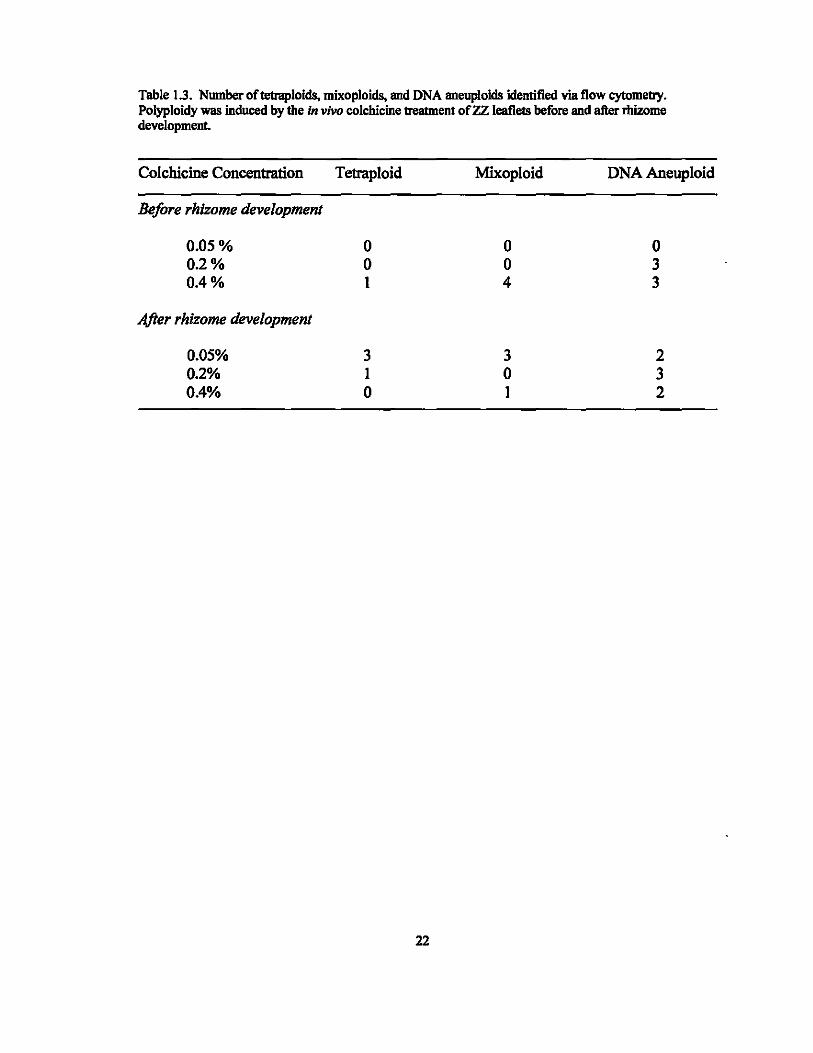
Table 1.3. Number oftetraploids, mixoploids, and DNA aneuploids identified via flow cytometry. Polyploidy was induced by the In vivo colchicine treatment of ZZ leaflets before and after rllizome development.
Colchicine Concentration Tetraploid Mixoploid DNA Aneuploid
Before rhizome development
0.05% 0 0 0 0.2% 0 0 3 0.4% 1 4 3
After rhizome development
0.05% 3 3 2 0.2% 1 0 3 0.4% 0 1 2
22
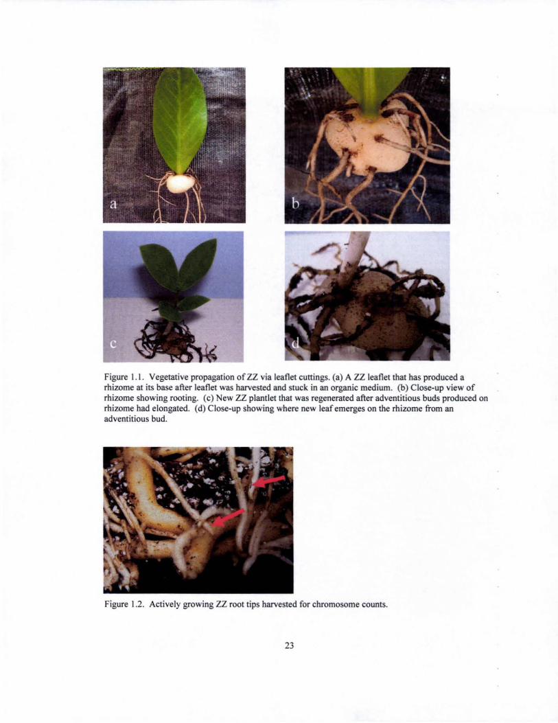
Figure 1.1. Vegetative propagation of ZZ via leaflet cuttings. (a) A ZZ leaflet that has produced a rhizome at its base after leaflet was harvested and stuck in an organic medium. (b) Close-up view of rhizome showing rooting. (c) New ZZ plant let that was regenerated after adventitious buds produced on rhizome had elongated. (d) Close-up showing where new leaf emerges on the rhizome from an adventitious bud.
Figure 1.2. Actively growing ZZ root tips harvested for chromosome counts.
23

Figure 1.3. Sample preparation for flow cytometric analysis. (a) Reagents and plant tissue kept on ice throughout the entire process. (b) Sample size varies from 0.5 - I em' . (c) and (d) Samples are chopped in the extraction buffer for 30-60 seconds with a double edged razor (e) suspension of nuclei is filtered, after which staining buffer containing RNAse and propidium iodide is added to the filtrate (I) samples are analyzed using a Beckman-Coulter (Miami, Florida) Altra flow cytometer.
24
Figure 1.4. (a) ZZ leaflet showing the typical diploid leaf morphology. (b) Leaflet of suspected ZZ tetraploid showing a much rounder shape. (c) Leaflet of another suspected ZZ tetraploid. Notice, the asymmetry of the leaflet: the left side is larger than that oftbe right, indicating the possibility that the leaflet may in fact be a chimera. (d) ZZ leaf structure
25
ZZ leaflet
ZZ leaf
Figure 1.5. Stained ZZ chromosomes isolated from single cells. (a) and (b) Chromosomes from two different cells that were stained using the modified Shi (2003) method. Cells were isolated from actively growing root tips of a regenerated control plant (0% colchicine) and show 2n = 34.
26

O.O.5Y. Colchicine: after rhizome production
2xDiploid
80 -
60 -
Number of Nuclei
2C
40 -
20 -
) LA~4C 04--~'~-'-1'--'-'1--~~~1r-~~-'-1~
o 30 60 90 120 DNA Fluorescence (Channel Number)
Figure 1.6. Distribution of DNA content of a suspected ZZ polyploid that resulted in being a diploid. Colchicine treatment of the parent leaflet after rhizome production did not result in the regeneration ofa tetraploid plant. At least 2000 nuclei were analyzed and both 2C (G I mitotic phase) and 4C (G2 mitotic phase) peaks have a CV<IO%. Cell nuclei were isolated from leaflet tissue and stained with propidium iodide prior to analysis.
27

80 -
60
40 -
20
Channel Number. 44 2x Diploid Control
Number of uclei 0 +--------------------1 Channel Number. 90
20 _ 4x Temploid
15 -
10 -
5 -
04--T~~~I ~~~I~~~L~I~~--r-I ~
o 30 60 90 120
DNA Fluorescence (Channel umber)
Figure 1.7. Distribution of D A conten! of diploid and tetraploid 12 samples. The DNA fluorescence of the tetraploid sample is almost double that of the diploid control. The tetraploid was produced by soaking a ZZ leaflet (with a small rhizome) in a 0.05% solution of colchicine. At least 2000 nuclei were analyzed and both the diploid and tetraploid peaks have a CV< IO%. Cell nuclei were isolated from leaflet tissue and stained with propidium iodide prior to analysis.
28

100 -
80 -
60 -
Number of Nuclei
40 -
20 -
o
0.05 ~. Colchicine: after rhizome production
30 60 90
2x + 4x Milcoploid
.. 4x 813
.. 2x 44.8
120
DNA Fluorescence (Channel umber)
Figure 1.8. Distribution of DNA content of a ZZ mixoploid sample showing both 2x (diploid) and 4x (tetraploid) nuclei. The mixoploid was produced by soaking a ZZ leaflet (with a small rhizome) in a 0.05% solution of colchicine. At least 2000 nuclei were analyzed and both the diploid and tetraploid peaks have a CV< IO%. Cell nuclei were isolated from leaflet tissue and stained with propidium iodide prior to analysis.
29
100
80
60 -
umber of Nuclei
40
20
o
DI - 1.4
30 60 90
DNA Fluorescence (Channel Number)
120
"4x " 2x .. DNA Aneuploid
Figure 1.9. Distribution of DNA content of a DNA aneuploid. The diploid peak has been assigned a DI of I, the tetraploid peak a DI of2, and the DNA aneuploid was calculated as having a DI of 1.4. At least 2000 nuclei were analyzed and all 3 peaks have a CV< IO%. Cell nuclei were isolated from leaflet tissue and stained with propidium iodide prior to analysis.
Figure 1.10. Photographs (a) and (b) were taken using an Olympus BX5 1 Epifluorescence Microscope with 480nm excitation frequency in order to view propidium iodide-stained nuclei (red dots) and detect cellular debris. Yellow arrows indicate debris.
30

CHAPTER 2: POL YPLOIDIZATION OF ZZ VIA ORYZALIN TREATMENT OF
CALLUS
Background Information
Though a simplc method was employed for the production of ZZ tetraploids in
Chapted, tissue culture has also been shown to be an effective tool in polyploidy
induction. Tissue culture increases the efficiency of mutagenic treatments for variation
induction, handling oflarge populations, use of ready selection methods, and mpid
cloning of selected variants (Predieri, 2001). The use of tissue culture in the successful
production oftetraploids has been shown in various plants including: Spathiphyllum
(Eeckhaut et. al, 2004), Alocasia (Thao et. al2003), Coco yam (Tambong, Sapra, and
Garton, 1998), Miscanthus (petersen, 2003) and Rhododendrons (Vainola and Repo,
2001; Vainola, 2000). The objective of this experiment was to produce a ZZ tetraploid
by the oryza!in treatment of ZZ callus.
In the design of a protocol for the in vitro production of ZZ tetraploids, callus
tissue was treated with oryzalin. The advantage of treating callus is twofold: (i) callus
tissue is typically in a high state of cell division, which has been shown to be most
responsive to chemical mutagenesis and (ii) plantlets regenerated from callus tissue may
express some genetic variability, which would contribute to the potential for obtaining a
new ZZ variety. To date, HePing and Peng (2003 and 2005) are the only researchers to
report an in vitro micropropagation protocol for ZZ. The 2003 publication, however,
describes a protocol for direct shoot organogenesis from ZZ leaf explants with no
31

intervening callus stage. Similarly, the 2005 publication describes a protocol for tuber
induction on leaflet explants. In this research project, a tissue culture protocol was
developed for the callus induction and plantlet regeneration of ZZ for the oryzalin
treatment of ZZ.
Materials and Methods
Callus Induction and Plantlet Regeneration
A newly emerging ZZ leaf (petiole staIk with leaflets) was harvested from a
juvenile-like stock plant for callus induction. The plant material was prepared for
disinfestation by soaking it in running water overnight and scrubbing it with liquid
antibiotic soap the following day. After rinsing with distilled water, explants (ZZ leaflets
and petioles) were harvested and wiped down with 95% ethanol then trimmed at the
edges. Explants were soaked in 0.65% sodium hypochlorite solution (10% v/v Clorox
Regular Bleach; The Clorox Company; Oakland, CA) for 10 minutes while shaking on a
rotator at 14Orpm, transferred to 0.13% sodium hypochlorite solution (2% v/v Clorox) for
more trimming, and then soaked for an additional 15 minutes in a 0.33% sodium
hypochlorite solution (5% v/v Clorox). Leaflet explants were trimmed down to a 1 x 1
em square (containing the major vein) while petioles were trimmed to about 2.5em in
length., then rinsed in sterilized distilled water before being transferred to 25mm test tubes
containing 10 ml solid callus inducing medium. Leaflet explants were stuck vertically in
the medium so that half of the explant was submerged into the medium, while the other
half was exposed. Similarly, the basal portion of the petiole was submerged in the
32
medium while the distal portion remained exposed. The callus inducing media is
composed of half strength MS macro- and micronutrients (Murasbige and Skoog, 1962),
half strength MS vitamins, 100mg r1 myo-inositol, 0.2mg r1 BA, 4mg r1 2,4-0, 20g r1
sucrose, and 3g r1 gellan gum (Miguel, 2004, see pg iii). All cultures were stored in the
dark at about 7S-S0"F. Once callus production had been initiated, cultures were
transferred to fresh callus inducing medium approximately every 2-4 weeks for callus
multiplication.
For shoot regeneration, callus cultures were removed from the dark, transferred to
shoot induction media, and placed on metal racks in the culture room under Gro-Lux
lights with a timer set for IS hour days. The shoot inducing medium used is composed of
half strength MS macro- and micronutrients, half strength MS vitamins, 100mg r1 myo
inositol, 1 mg r1 BA, 40g r1 sucrose, and 3g r1 gellan gum. Once the adventitious buds
had elongated and the leaf sheath was about 2.Scm, they were transferred to y. MS with
no plant growth regulators for further development. Rooted plantlets, about Scm in
height, where then transferred to community pots containing moist Pro-Mix 'BX'
medium (Premier Horticulture Ltd.; Dorval, Canada) in the greenhouse at Pope
Laboratory. Pots were covered with plastic wrap for about 4 weeks to allow for
acclimatization. Figure 2.1 shows the developmental stages observed, and provides an
overall view of the protocol that was developed for callus induction and plantlet
regeneration of ZZ.
33

Oryzalin Treatment ofZZ Callus Derivedfrom Leqf1et and Petiole Tissue
Callus cultures derived from leaflets were kept separately from those derived
from petiole tissue, and the experimental set up described below applies to both leaflet
and petiole callus. A completely randomized experimental design with a 2 x 3 factorial
treatment design with three oryzalin concentrations (0%, 0.005%, and om %) and two
durations (24 and 48 hours) was used for the oryzalln treatment of ZZ callus. Oryzalin
(phytoTechnology Laboratories; Shawnee Mission, KS) was dissolved in DMSO, and
added to liquid callus inducing medium to give a final concentration of 1 % DMSO.
Each treatment consisted of two reps with 10 experimental units (1 cm2) per rep for a total
of20 explants per treatment Due to limited explant material, however, the controls were
comprised of one rep with 5 experimental units per rep. A total of 90 callus pieces were
used.
Seven month old callus was cut into lcm2 units and transferred to a 125m1 flask
containing 50m! of the filter sterilized oryzalln solution, with 10 callus pieces per flask.
A 1 % DMSO solution of callus inducing medium was used as the control. Flasks were
placed on a rotary shaker at 75 rpm for 24 or 48 hours. After treatment with oryzalin, the
calli were rinsed in sterile distilled water, and each callus piece was transferred to a
25mm test tube containing 10m! solid callus inducing medium, with the callus piece
submerged in the medium. Cultures were kept in the dark and transferred to fresh
medium every two weeks, for a total of 6 transfers.
After 12 weeks on callus inducing medium, the cultures were transferred to Petri
dishes containing 20m! shoot inducing medium and cultures were placed under Oro-Lux
lights with 18 hour days. Cultures were transferred to fresh medium every 2 weeks.
34
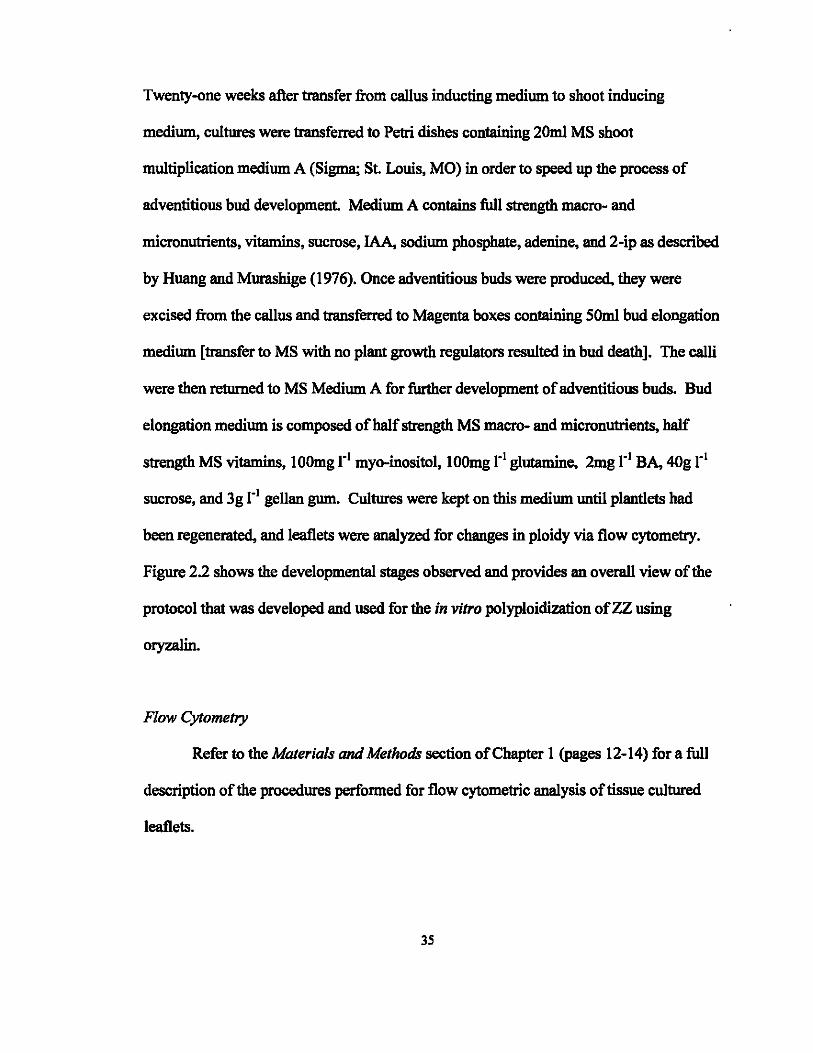
Twenty-one weeks after transfer from callus inducting medium to shoot inducing
medium, cultures were transferred to Petri dishes containing 20m! MS shoot
multiplication medium A (Sigma; St. Louis, MO) in order to speed up the process of
adventitious bud development. Medium A contains full strength macro- and
micronutrients, vitamins, sucrose, IAA, sodium phosphate, adenine, and 2-ip as described
by Huang and Murasbige (1976). Once adventitious buds were produced. they were
excised from the callus and transferred to Magenta boxes containing 50m! bud elongation
medium [transfer to MS with no plant growth regulators resulted in bud death]. The ca1li
were then returned to MS Medium A for further development of adventitious buds. Bud
elongation medium is composed of half strength MS macro- and micronutrients, half
strength MS vitamins, IOOmg rl myo-inositol, 100mg rl glutamine, 2mg rl BA, 40g rl
sucrose, and 3g rl gellan gum. Cultures were kept on this medium until pIantiets had
been regenerated, and leaflets were analyzed for changes in ploidy via flow cytometry.
Figure 2.2 shows the developmental stages observed and provides an overaI1 view of the
protocol that was developed and used for the in vitro polyploidization of ZZ using
oryzalin.
Flow Cytometry
Refer to the Materials and Methods section of Chapter 1 (pages 12-14) for a full
description of the procedures performed for flow cytometric analysis of tissue cultured
leaflets.
35
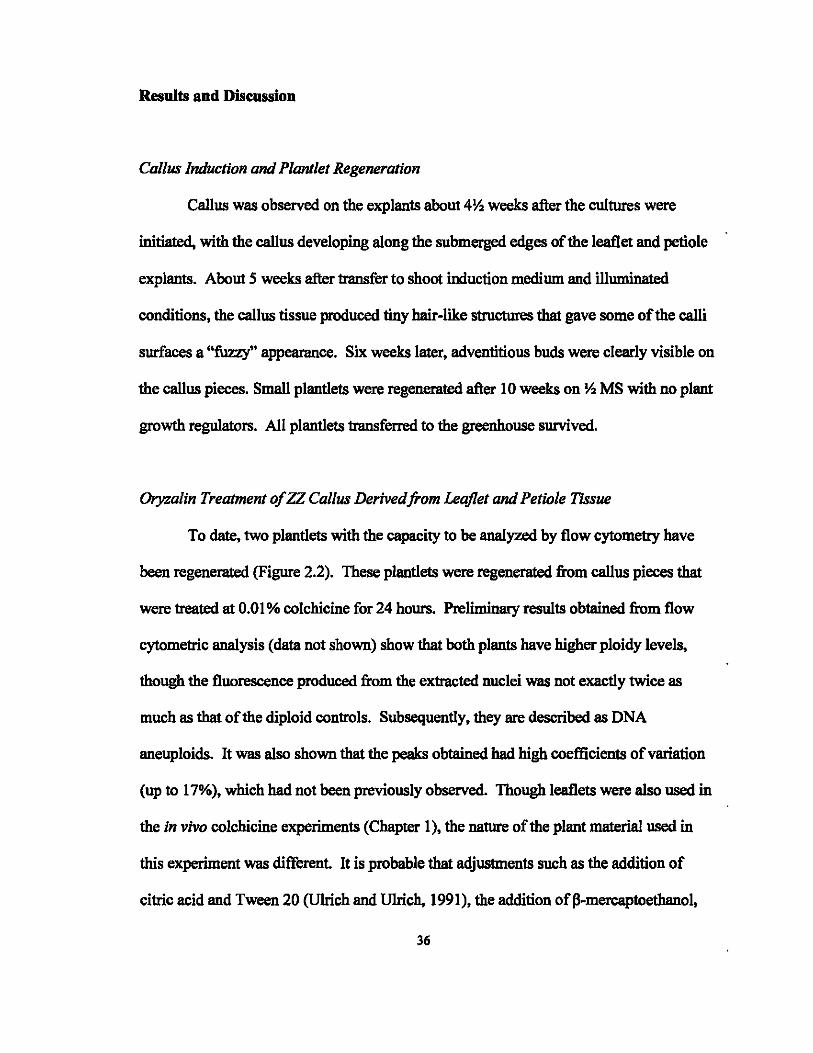
Results and Discussion
Callus Induction and Plantlet Regeneration
Callus was observed on the explants about 4Yo weeks after the cultures were
initiated. with the callus developing along the submerged edges of the leaflet and petiole
explants. About 5 weeks after transfer to shoot induction medium and illllminawl
conditions, the callus tissue produced tiny hair-like structures that gave some of the calli
surfaces a ''fuzzy'' appearance. Six weeks later, adventitious buds were clearly visible on
the callus pieces. Small plantlets were regenerated after 10 weeks on Yo MS with no plant
growth regulators. All plantlets transferred to the greenhouse survived.
Oryzalin Treatment olZZ Callus Derivedfrom Leqflet and Petiole Tissue
To date, two plantlets with the capacity to be analyzed by flow cytometry have
been regenerated (Figure 2.2). These plantlets were regenerated from callus pieces that
were treateA at 0.01 % colchicine for 24 hours. Preliminary results obtained from flow
cytometric analysis (data not shown) show that both plants have higher ploidy levels,
though the fluorescence produced from the extracted nuclei was not exactly twice as
much as that of the diploid controls. Subsequently, they are described as DNA
aneuploids. It was also shown that the peaks obtained had high coefficients of variation
(up to 17%), which had not been previously observed. Though leaflets were also used in
the in vivo colchicine experiments (Chapter 1), the nature of the plant material used in
this experiment was different It is probable that adjustments such as the addition of
citric acid and Tween 20 (Ulrich and Ulrich, 1991), the addition ofp-mercaptoethanol,
36

citrate, and the use of a surfactant (Brown et at., 1991) may need to be applied to the
procedure used for flow cytometric analysis of tissue cultured ZZ leaflets. More
adventitious buds will need to develop into plantlets in order for further tests to be run.
The length of time required for adventitious bud development to occur on the
callus tissue once the cultures had been transferred to shoot induction media varied
between the original tissue culture protocol developed for callus in induction in ZZ and in
the second procedure in which the calli were treated with oryzalin. In the callus
induction protocol, it took 11 weeks for adventitious buds to begin to form, while it took
over twice that long and a modification of the medium for all the petiole and leaflet callus
cultures (including controls) to develop adventitious buds after oryzalin trea1ment (26
weeks). It is suspected that DMSO, and not oryzaIin, may have affected the regeneration
capacity of the callus tissue. Unfortunately, it cannot be confirmed because a control
without DMSO was not included in the experiment.
The inability of the callus tissue to produce adventitious buds, however, was
overcome with the use ofMS Multiplication Medium A. The medium contains key
components that helped activate the callus tissues: adenine sulfate promotes cell division,
while sodium phosphate provides the cells with energy. Also, the high cytokinin (2-ip at
30 mg rl): low auxin (IAA at 0.3 mg rl) ratio helped induce adventitious bud
development Within 5 weeks of callus transfer to Medium A, adventitious buds had
developed and callus tissue continued to multiply. Bud elongation to form a new plantlet,
however, did not occur upon transfer of the buds to MS medium with no PGRs (MS.).
Within one to two weeks of transfer to MS .. browning occurred that led to eventual bud
death. It was suspected that the buds were affected by the high MS salt concentration in
37

medium, hence, a bud elongation media was devised that contained Y. the salt
concentration and BA was used as the cytokinin to promote elongation (modeled after the
original plantlet regeneration protocol devised). The use of this medium proved
successful and most buds are now elongating and developing into pIantiets.
Though a tetraploid ZZ plantlet has not been identified, there are several
emerging buds that must still be analyzed. More importantly, however, is that this
project has led to the development of a novel tissue culture protocol for callus induction
and pIantiet regeneration in ZamiocuJcas zamii/olia (Fig. 2.1). Leaflets or petioles are
harvested from a mature mother stock pIant, disinfested, and trimmed to an appropriate
size. The explant is then stuck into callus inducing medium and cultures are kept in the
dark. After sufficient callus has been produced, the callus tissue is transferred to shoot
induction medium and cultures are exposed to illuminated conditions. Callus pieces with
adventitious buds are then transferred to a medium with no pIant growth regulators to
allow for rooting and plantlet development The tissue culture protocol developed may
be used for further in vitro mutation induction studies in ZZ such as the irradiation of
callus tissue or adventitious buds. The protocol developed may also be adopted for the
commercial production of zz, which may allow for more uniform ZZ propagation.
38
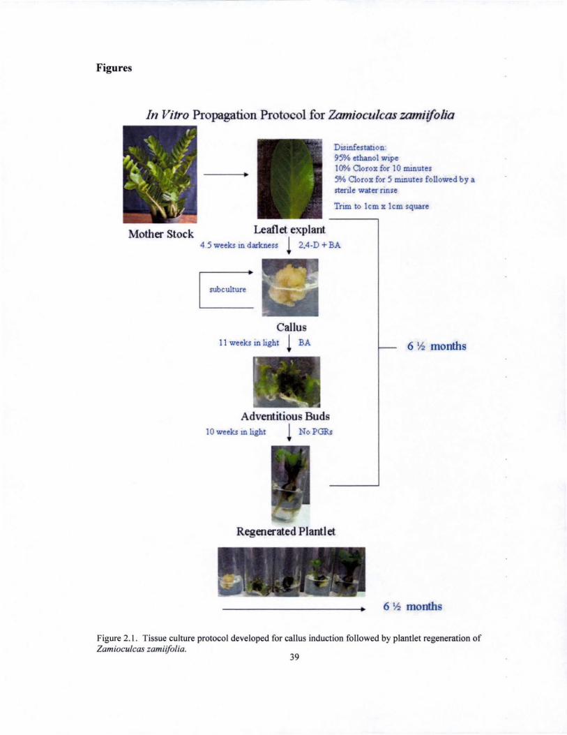
Figures
In Vitro Propagation Protocol for Zamioculcas zamiijolia
Mother Stock
•
Leaflet explant
DlSUlfestahon 95% ethanol wIpe 10% C1orox for 10 ll11Dute, 5% C1orox for 5 minutes followed by a rtenle water nnse
Tnm to lcm x lcm square
4 5 weeks III darkness 1 2,4-D + BA
I subculture '
Callus 11 weeks In light 1 BA
Adventitious Buds 10 weeks III hght NoPGRs
Regenerated Plantlet
I ...... , - ~ l - ;' ~ ....... ~ ....
6 Y.z months
6 Y.z months
Figure 2. 1. Tissue culture protocol developed for callus induction followed by plantlet regeneration of Zamioculcas zamiijolia.
39
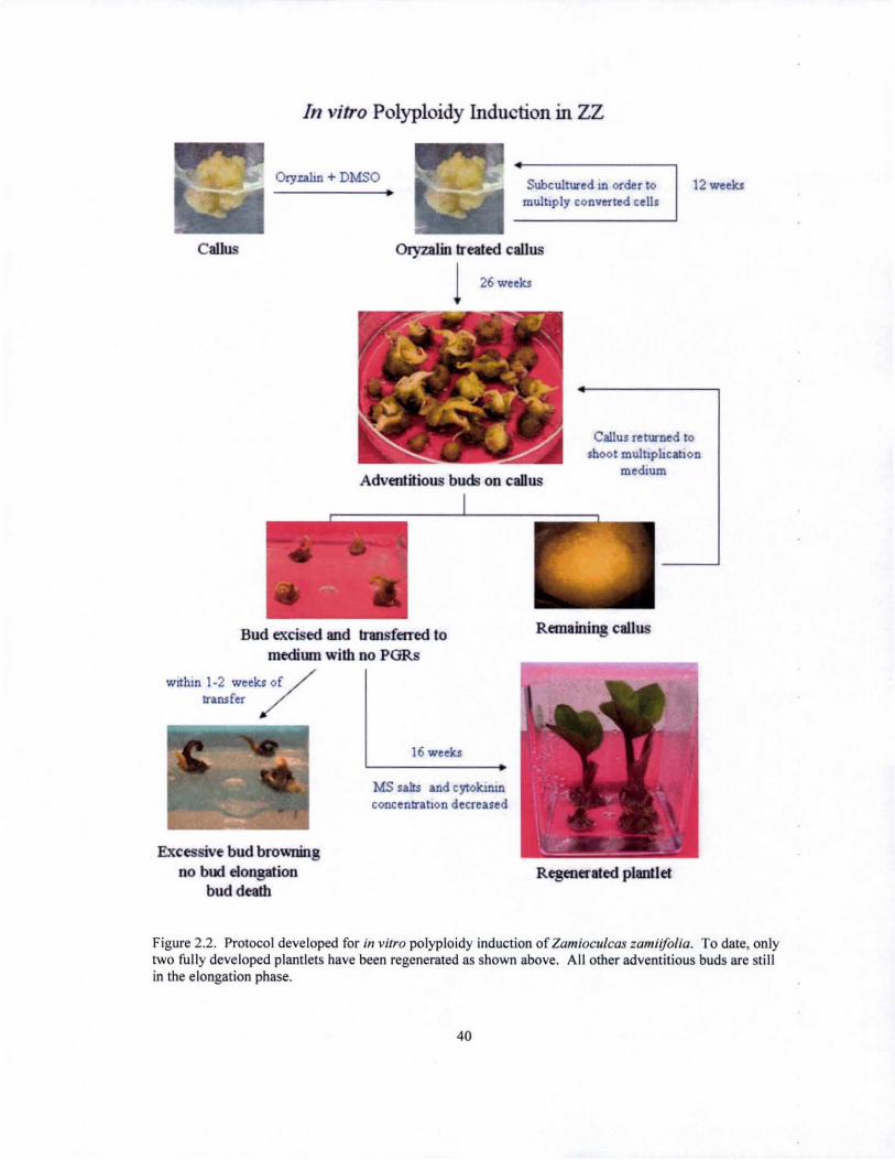
Callus
In vitro Polyploidy Induction in ZZ
Oryuhn + DMSO Subcultured Ul order to multiply converted cells
Oryzalin treated callus
26 weekJ
12 weekJ
Adventitious buds on callus
Callus returned 10
shoo t muihpi.tcatlon medium
Bud excised and transferred to medi\Ull with no PGRs
Wllhln 1-2 weeks of /
transfer /
Excessive bud browning no bud elongation
bud death
16 weeks
MS salls and cyloklllln concentration decreased
Remaining callus
Regenerated plantlet
Figure 2.2. Protocol developed for in vitro polyploidy induction of Zamioculcas zamiifolia . To date, only two fully developed plantlets have been regenerated as shown above. All other adventitious buds are still in the elongation phase.
40

CHAPTER 3: IDENTIFICATION OF A SUITABLE IRRADIATION DOSAGE
FORZAMIOCULCAS ZAMHFOUA
Ba~kground Information
Mutation breeding aims to improve an otherwise good cultivar by the
modification of one easily recognizable character. leaving the rest of the genotype
untouched (Broerges and Van Harten, 1988; Micke eta! .• 1987; Lapins. 1983). It is also
used to generate more genetic variability for selection and cross breeding (Micke et at .•
1987). making it a useful breeding tool for plant species that are to be incorporated into a
breeding program. It is for both of these reasons that ZZ is an ideal candidate for
mutation breeding.
One of the first steps in the establishment of a mutation breeding program is the
determination of a suitable irradiation dosage. typically identified as the lethal dosage or
LOx. The LOx is calculated as the exposure required to produce x percent lethality of the
irradiated material. expressed as a percentage of the control (Sparrow et at .• 1968). Many
breeders use LOso as the desirable irradiation dosage to induce mutations, unless the
observed results show otherwise (Pred:ieri, 2001; Cruz T. and Rubi A. 1995; Bbandari,
1993; Yamaguchi. 1988; Lapins. 1983). The objective of this work was to determine the
LOso for the irradiation of ZZ leaflets using x-rays.
41
Materials and Methods
Leaflets used for irradiation were harvested from mature (capable of flowering),
ZZ stock plants with fully expanded leaves. Harvested leaflets were graded according to
size and distributed evenly throughout all treatments, after which leaflets were stacked in
sets of (i) 10 leaflets (10 or 30 leaflets irradiated per treatment) or (ti) 11 leaflets (33
leaflets irradiated per treatment). Each set of leaflets was wrapped in a moist paper towel
and placed in the irradiator with the leaflets centered within the irradiation zone with the
petioles facing inward. After the desired irradiation dosage had been achieved, a stack
was randomly removed from the irradiator and placed in a labeled plastic bag until all
leaflets had received the appropriate irradiation dosage. Leaflets were then stuck in a
40cm x 55cm metal flat containing moistened Pro-Mix 'BX' (Premier Horticulture Ltd.;
Dorval, Canada) and placed under 70% shade in a greenhouse at Pope Laboratory (Figure
3.1). SAS statistical package version 9.1 (SAS Institute Inc., Cary, N . C.) and Microsoft
Excel were used to perform probit analysis and regression analyses on the data collected.
Irradiation of ZZ leaflets was performed on three separate occasions using a
Hewlett Packard 43804N X-Ray System Faxitron Series. Leaflets were irradiated at a
rate of 1.21 Grays per minute. Due to the limited availability of plant materials, only a
small number ofleaflets were irradiated during the first irradiation. The second and third
irradiations were performed based on the preliminary results obtained from the preceding
irradiation (Table 3.1). The first irradiation was performed in March 2005, the second in
June 2005, and the third in August 2005. Final leaflet survival data was collected in
August 2006, and leaflet survival was scored as a ZZ leaflet that regenerated a new plant.
42
Results and Discussion
Irradiation 1
LOuJO, the lowest exposure to produce 100% leaflet mortality, was found to be
40Gy, and all leaflets irradiated above this dosage died from exposure to the X-rays
(Table 3.2). Within 2 - 3 months, leaflet yellowing was observed, and within 4 months
the leaflets had developed a brown, "bumt" appearance (Fig. 3.2). Probit approximation
was the model used to determine the LOso from the results shown in Table 3.2. Pearson
Chi-Square and L. R. Chi-Square were used as goodness of fit tests, and both models
showed that there is no significant difference (p= 0.2142 and p=O.0686, respectively)
between the observed data and the expected data, hence the data were normally
distributed. Type III Analysis of Effects showed that irradiation dosage had a highly
significant (p = 0.0026) effect on leaflet death, and the LOso was identified as 19Gy. The
complete SAS output is provided in the Appendix (pages 77 - 80).
Irradiation 2
The second set of irradiations was designed after preliminary observations from
the first irradiation showed LOlOo = 40Gy (Table 3.3). It is inappropriate, however, to
apply probit analysis to the second data set in order to determine LOso because the data
obtained did not fit the normal distribution; hence, regression analysis was used to
estimate LOso. Irradiation dosage vs. percent death was plotted and a linear regression
equation was calculated as follows: Y = 0.0261X - 0.0545, where Y = LOso. and X =
equivalent dosage, r = -0.545, and R2 = 0.9348. Using the calculated regression line,
LOso was determined to be 2lGy (Fig. 3.3).
43

It should also be noted that of the 33 leaflets irradiated at 20Gy, one leaflet
regenerated a plant that shows a possible mutation (Fig. 3.4). One of the leaves produced
by the regenerated plant shows differences in leaflet size, with half of the leaflets
showing a reduction in size as compared to a typical 'ZZ leaflet. The distance between
the successive leaflets also appears to be shorter than is typicaJly observed. Because the
plant belongs to the MI generation (mutagen-treated plant), further observations of the
M2 and M3 generations (offspring of the mutagen treated plants) are necessary in order to
confinn the differences observed as a mutation. Nonetheless, the results obtained are
promising.
The data collected from the third irradiation could not be used to calculate the
LOso. since the highest percentage of death recorded was 36%, which was obtained at
25Gy, the highest irradiation dosage used in the third irradiation (Table 3.4). The
discrepancy between the results for LOso in irradiation 3 and those of irradiations 1 and 2
may have resulted from various reasons. According to Sparrow et aI., (1968), several
biological, radiological, and environmental factors may contribute to variations in the
radiobiological responses of plants. For example, the effectiveness of the prescribed
dosage varies according to the moisture content of the irradiated plant material.
Similarly, the stage of the plant growth cycle affects plant sensitivity to irradiation, where
actively growing plants are more sensitive to irradiation than plants in their dormant
stages. In this experiment, the first two irradiations were performed in March and June,
while the third irradiation was performed in August. It is plausible that the mother stock
44
plants used in the third experiment were in a physiologically different stage (growth cycle
and vegetative or floral stage of differentiation) than those that were used in the first two
irradiations, hence the variation in the calculated lethal dosages. In conclusion, the
results obtained have provided useful information that contributes significantly to the
establishment of a mutation breeding program for ZZ. Large number of leaflets may now
be irradiated at the calculated LDso of20:!: lOy in an attempt to produce ZZ mutants.
45
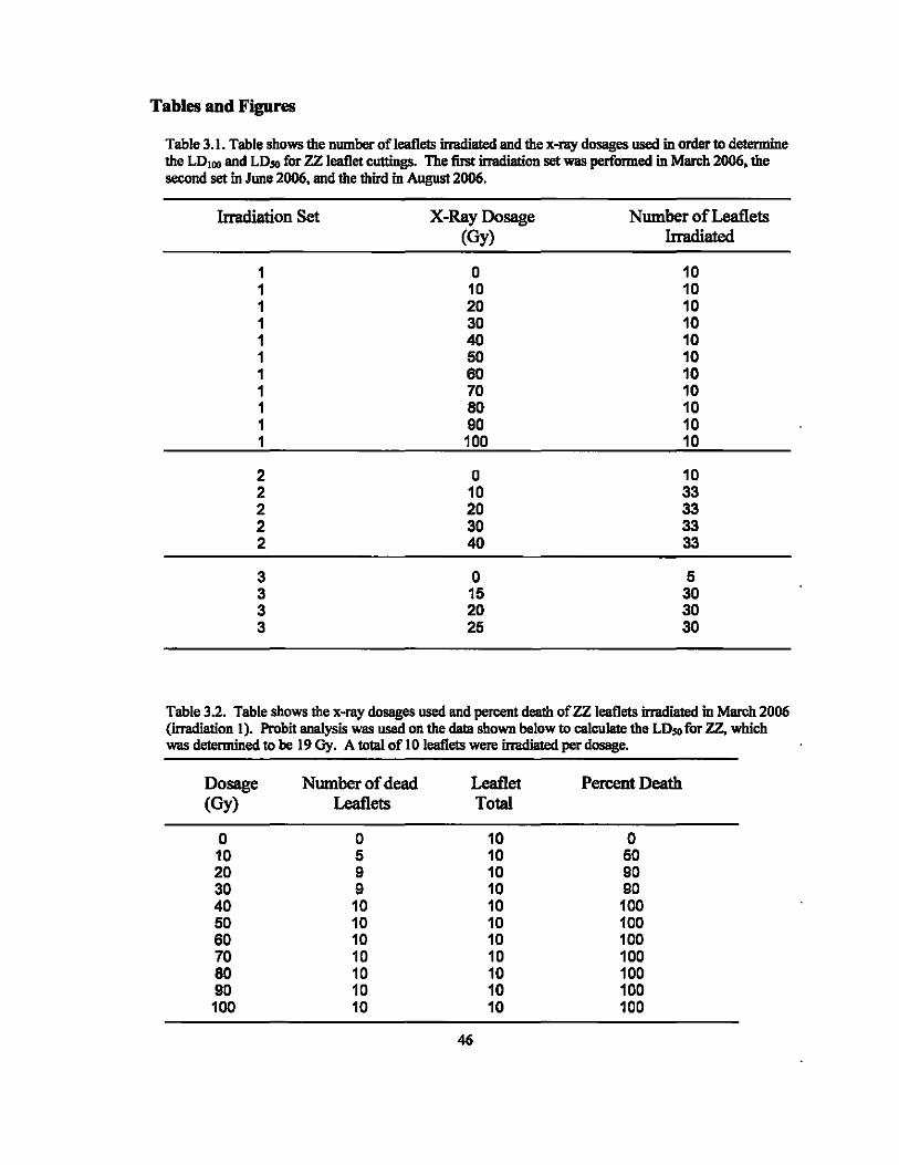
Tables and Figures
Table 3.1. Table shows the number of leaflets irradiated and the x-ray dosages used in order to determine the LDu .. and LD50 for ZZ leaflet cuttings. The fust imldiation set was performed in March 2006, the second set in JWle 2006, and the third in August 2006.
Irradiation Set X-Ray Dosage Number of Leaflets (Oy) Irradiated
1 0 10 1 10 10 1 20 10 1 30 10 1 40 10 1 50 10 1 60 10 1 70 10 1 80 10 1 90 10 1 100 10
2 0 10 2 10 33 2 20 33 2 30 33 2 40 33
3 0 5 3 15 30 3 20 30 3 25 30
Table 3.2. Table shows the x-my dosages used and percent death ofZZ leaflets irradiated in March 2006 (imldiation 1). Probit analysis was used on the data shown below to calculate the LD$O for zz. which was determined to be 19 Gy. A to1al of 10 leaflets were irradiated per dosage.
Dosage Number of dead Leaflet Percent Death (Oy) Leaflets Total
0 0 10 0 10 5 10 50 20 9 10 90 30 9 10 90 40 10 10 100 50 10 10 100 60 10 10 100 70 10 10 100 80 10 10 100 90 10 10 100 100 10 10 100
46

Table 3.3. Table shows the x-ray dosages used and the percent death of ZZ leaflets irradiated in June 2006 (irradiation 2). Regression analysis was used to calculate the LD50 for ZZ from the data shown below, and was detennined to be 21Gy. A total of 10 leaflets were irradiated fur the controls and 33 leaflets for each of the other treatments.
Dosage Number of dead Leaflet Percent Death Leaflets Total
0 0 10 0 10 2 33 6 20 20 33 60 30 22 33 66 40 33 33 100
Table 3.4. Table shows the results obtained from irradiation 3, which was performed in August 2006. The data collected could not be used to calculate the LD50 ofzz, since none of the x-ray dosages used produced 50"10 letbality.
Dosage Number of dead Leaflet Percent Death Leaflets Total
0 0 5 0 15 6 30 20 20 11 30 36 25 11 30 36
47

Figure 3, I, (a) Sets of leaflets wrapped in moist paper towels and ready to be irradiated, (b) Leaflets are kept within the irradiation zone on the shelf(c) irrad iated leaflets stuck in moist Pro-mix ' BX ' and kept under 70% shade,
Figure 3,2, (a) Leaflet on the right was irradiated at 100Gy, and exhibits heavy browning and has a "burnt" look when compared to the control on the left, The picture was taken approximately 4 months after exposure to the x-rays, (b) From left to right: leaflet irradiated at 80Gy, 60Gy, and OGy, The picture was taken approximately 3 months after exposure to the x-rays,
48

1.2
1.0
0.8
-5 '" Q 0.6
'5 t> 0.4 b
P..
0.2
0.0
-0.2
y=0 .0261x- 0.0545
R squared = 0.9348
•
o 10
•
20 30 40 50
Dosage (Gy)
Figure 3.3. Relationship between dosage and percent death of x-ray irradiated ZZ leaflets. Using the calculated regression equations. LD", is estimated at 21Gy.
Figure 3.4. ZZ plant regenerated from leaflet irradiated at 20Gy. Red arrows point to the leaflets that are smaller in comparison to those that are typical in size.
49

CHAPTER 4: GERMPLASM COLLECTION OF ZAMIOCULCAS ZAMIIFOLIA
Our Zamioculcas zamiifolia germplasm collection originally began withjust a few
plants, and has now grown to include plants from six different sources. Though several
of the inventoried plants seem to be of the same clone, we have kept records on the date
of receipt and source of collection for each of the different plants. Plants from these six
different sources have been propagated by division or leaf cuttings to increase our stock
numbers. A brief description of the collected plants is given below, followed by
photographs of several plants in the collection.
Hawaiian Slmshine Nursery. Oahu
Cultivated variety obtained from Hawaiian Slmshine Nursery in Wajmanalo, Oahu, on
several occasions. These are the mother stock plants used in my thesis research and are
also the variety of ZZ that is commercially grown.
National Botanic Garden of Belgium
Two accessions were purchased from the National Botanic Garden of Belgium:
(i) Zamioculcas zamiifolia (Lodd.) Engl. Accession: 19074359 cultivated material
(ii) Zamioculcas zamiifolia (Lodd.) Engl. 'Variegata' Accession: 19726043 cultivated material
Both accessions were received in February 2005.
Contact person: Frieda Billiet: Botanist, Living Collections, in charge of greenhouses National Botanic Garden of Belgium 8-1860 MEISE
50
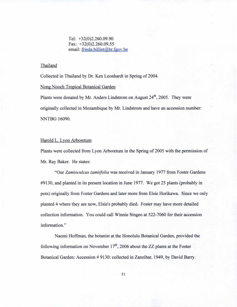
Tel: +32(0)2.260.09.90 Fax: +32(0)2.260.09.55 email : [email protected]
Thailand
Collected in Thailand by Dr. Ken Leonhardt in Spring of2004.
Nong ooch Tropical Botanical Garden
Plants were donated by Mr. Anders Lindstrom on August 24th, 2005. They were
originally collected in Mozambique by Mr. Lindstrom and have an accession number:
NNTBG 16090.
Harold 1. Lyon Arboretum
Plants were collected from Lyon Arboretum in the Spring of2005 with the pemUssion of
Mr. Ray Baker. He states:
"Our Zamioculcas zamiijolia was received in January 1977 from Foster Gardens
#9130, and planted in its present location in June 1977. We got 25 plants (probably in
pots) originally from Foster Gardens and later more from Elsie Horikawa. Since we only
planted 4 where they are now, Elsie's probably died. Foster may have more detailed
collection information. You could call Winnie Singeo at 522-7060 for their accession
information. "
Naomi Hoffman, the botanist at the Honolulu Botanical Garden, provided the
following information on ovember 17th, 2006 about the ZZ plants at the Foster
Botanical Garden: Accession # 9130: collected in Zanzibar, 1949, by David Barry.
51

Contact Person: Naomi Hoffinan [email protected] 808-S22-7066
Waimea Valley Audubon Center
Plants were donated on June 30th, 200S by Mr. David Orr.
(i) Accession: 7Sp2lS4 received from Tagami & Hart, Kahaluu Oahu.
(ii) Accession: 79p331 received from Conrad Flemming, St. Croix, Virgin Islands; possibly a plant at Fairchild Tropical Gardens Florida
Important Note: Both of these plants are planted in the exact same area at Waimea
Valley Audubon Center, so that the plant material we received can be either of the above.
Contact Person: Mr. David Orr [email protected] 808-638-9199
52

Figures
Figure 4.1 . Zamioculcas zamiiJolia (Lodd.) EngJ. acquired from Hawaiian Sunshine Nursery on Oahu. Commercial variety with thick, solid gray/green petioles, and green leaflets.
53

Figure 4.2. Zamioculcas zamiifolia (Lodd.) EngJ. acquired from the ational Botanic Garden of Belgium. Solid green leaflets are sl ightly thinner, but broader and larger than those of the commercial variety.
54
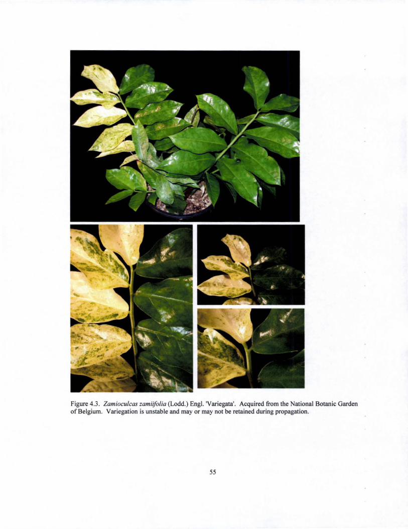
Figure 4.3. Zamioculcas zamiiJolia (Lodd.) Eng!. 'Variegata'. Acquired from the National Botanic Garden of Belgium. Variegation is unstable and mayor may not be retained during propagation.
55
Figure 4.4 Zamioculcas zamiiJolia (Lodd.) Engl. 'Variegata' acquired from the National Botanic Garden of Belgium. Different forms of ZZ variegation.
56

Figure 4.5. Zamioculcas zamiiJolia (Lodd.) Engl. 'Variegata' acquired ITom the National Botanic Garden of Belgium. Different forms of ZZ variegation.
57

Figure 4.6. Zamioculcas zamiifolia (Lodd.) Eng!. 'Variegata' acquired from the National Botanic Garden of Belgium. DitTerent forms of ZZ variegation.
58
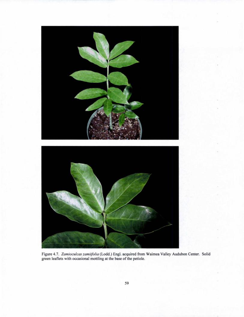
Figure 4.7. Zamioculcas zamiifolia (Lodd.) Eng!. acquired from Waimea Valley Audubon Center. Solid green leaflets with occasional monling at the base of the petiole.
59

Figure 4.8. Zamioculcas zamii[olia (Lodd.) Eng\. acquired from Harold L. Lyon Arboretum. Closely resembles ZZ from Waimea Valley Audubon Center.
60
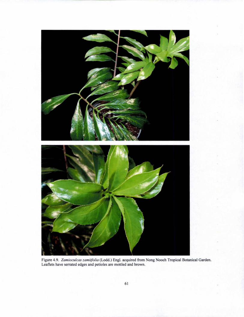
Figure 4.9. Zamioculcas zamiifolia (Lodd.) Eng!. acquired from ong Nooch Tropical Botanical Garden. Leaflets have serrated edges and petioles are mottled and brown.
61
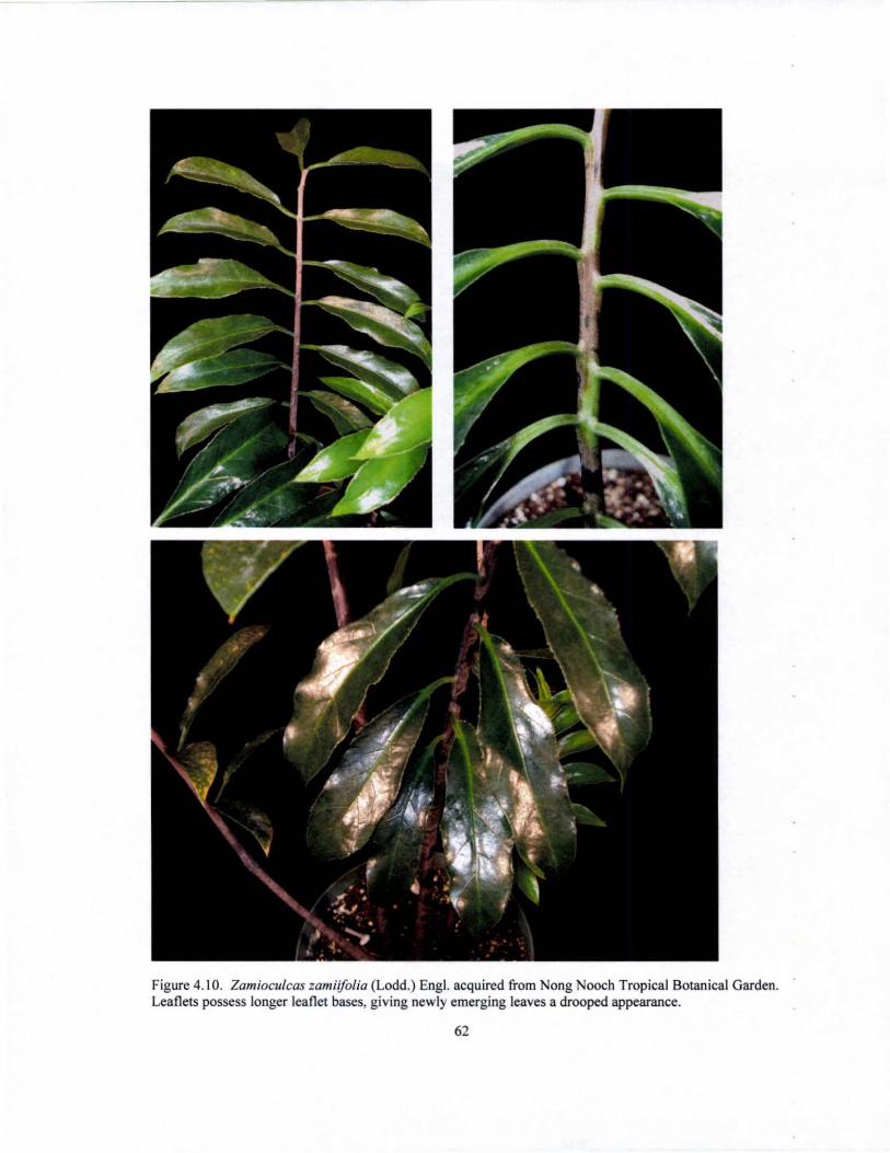
Figure 4.10. Zamioculcas zamii/olia (Lodd.) Eng!. acquired from Nong Nooch Tropical Botanical Garden. Leaflets possess longer leaflet bases, giving newly emerging leaves a drooped appearance.
62
SECTION 2
MARSDENIA FLORIBUNDA
CHAPTER 5: POL VPLOIDIZATION OF MARSDENlA FLORIBUNDA
Background Information
The botanist Paul Forster (1990) merged the well-known genus Stephanotis with
the larger genus Marsdenia, resulting in the name change of the commonly known
stephanotis plant to Marsdeniajlorlbunda (Brongn.). Of the 125 species that comprise
the genus Marsdenia, M. jloribunda is the only one of horticultural value. It is an
evergreen climber native to Madagascar that is cultivated in tropical and subtropical
regions (including Hawaii) for its white fragrant flowers. In Hawaii, M. jloribunda
flowers are either strung as lei lengthwise, requiring about 25 flowers, or they are pierced
through the side of the corolla, utilizing about 700 flowers (Staples and Herbst, 2005). It
has been suggested that winter production of M. jlorlbunda flowers under artificial light
(Kofranek and Kubota, 1982; Wikesjo,1982; Kofranek and Criley, 1983) would be an
economically feasible commercial venture for local growers, since there is a year-round
market for the flowers on the mainland (Criley, 1992).
The production of a cultivar possessing larger flowers with thicker, sturdier petals
would be beneficial to the lei industry, since fewer flowers would be required to produce
the lei. Criley (1992) reported the shelf life of M. jlorlbunda flowers as 10 - 14 days
when held in a plastic bag. The development of a cultivar with sturdier petals could
possibly extend the shelf life of the flower, allowing for even longer storage periods.
Such a cultivar could be produced by doubling the chromosome number of M. jloribunda
(2n = 22); hence, the objective of this experiment was to create a tetraploid M. jloribunda
64
by treating seeds with colchicine at various concentrations and durations. The resulting
tetraploid should theoretically possess larger flowers (Vainola, 2000; Kamemoto, 1985)
and could eventually be introduced into the Hawaii floriculture industry as an improved
variety.
Colchicine has been used for chromosome doubling in various plants (Kamemoto
et aJ., 1997; Kamemoto, 1985; Vainola, 2000; Tambong and Garton 1998). The flowers
produced by polyploid plants have been shown to be larger and the overall plant growth
more compact than their diploid forms. New leaves and stems growing from treated
sectors are also usually wrinkled, thicker, darker green, and have a coarser texture when
compared to untreated plants (Eigsti and Dustin, 1955). Such characteristics were used in
the preliminary screening for identification of tetraploid M.floribunda.
Materlals and Methods
Treatments
Colchicine (phytoTechnology Laboratories; Shawnee Mission, KS) was dissolved
in distilled water to make 0.05, 0.1, 0.2, and 0.4% colchicine solutions. Seeds were
treated in 25ml of each solution for 24 and 36 hours, with 44 seeds per treatment for a
total of 10 treatments. Distilled water was used in the 24 and 36 hour controls. Fifty
milliliter flasks containing the seed treatments were placed on a rotator at 100rpm for the
duration of each treatment, after which the seeds were rinsed with water and sown in
moist Pro-Mix 'BX' media (Premier Horticulture Ltd.; Dorval, Canada).
65
Guard Cell Measurements
After visual inspection of the regenerated seedlings, suspected tetraploids were
selected for further screening using stomatal guard cell measurements. All guard cell
measurements were taken at the same time of day in order to decrease any variation in
guard cell size due to natural daytime effects on stomatal opening. Clear nail polish
(Markwins Beauty Products Inc.; City of Industry. California) was thinly applied to the
abaxial side of the leaf. After the polish had dried (approximately 60 seconds). a strip of
Scotch 3M Transparent Tape with Gloss Finish (3M Products; St. Paul, MN) was
mounted on top of the dried polish. The tape. which now held the abaxial imprint, was
removed from the leaflet and mounted onto a coverslip for viewing at 400X
magnification under a Leitz Wetzlar light microscope. Fifteen guard cells were randomly
selected from each leaf sample and measured (length x width) using a unitless graticule.
Chromosome Counts
The method employed for chromosome counting of M. floribUTllkl was a modified
version of the technique used by Sharma and Mookerjea (1955). Actively growing root
tips were excised, rinsed in distilled water. and pre-treated in a saturated solution of
paradichiorobenzene for 4 hours at 10-12·C. The saturated solution was prepared by
dissolving 109 of the solid in 500ml distilled water and holding the solution at 60·C
overnight. The solution was used at room temperature. After pretreatment, the root was
transferred to an acid-dye fixation solution (2% aceto-orcein and IN HCl at a 9: 1 ratio)
and the mixture heated over a flame for 3-4 seconds. The contents were then poured onto
a watch glass and allowed to cool for at least 5 minutes. A drop of 1 % aceto-orcein
66
solution was placed on a dry slide and the root was transferred to the drop of stain. The
intensely colored portion of the tip was retained, while the remainder of the root was
discarded. The root tip was then squashed using a small scalpel and a cover slip placed
on top of the root. The root tip was further squashed by applying even pressure on the
cover slip. A Leitz Wetz1ar light microscope was used to observe the prepared slides at
1000X magnification and pictures were taken using a Nikon Coolpix 4S00 digital
camera.
Flow Cytometry
CyStain PI Absolute P DNA Staining Kit for Plant Genome Size (partee;
Munster, Germany) was used for nuclei extraction and DNA staining of nuclear DNA
from M floribunda leaves used for flow cytometric measurement. Approximately
0.Scm2 of each leaf was chopped for 30 - 60 seconds in SOOl1l ice cold nuclei extraction
buffer with a sharp doubled edged razor blade in a SSmm plastic Petri dish. The slurry
was then filtered through a SO~ Cell Trics filter (partee; Munster, Germany) and the
suspension of released nuclei was stained in a solution composed of staining buffer,
RNAse, and Hoechst 33342 (1:40 dye: staining solution volume), which was substituted
for the PI provided by nuclei extraction kit. Leaf samples and all reagents used were
kept on ice throughout the entire process from leaflet harvest to sample preparation and
all samples were prepared for flow cytometric measurement within 1-2 hours of harvest.
Figure 1.3 shows the process from sample preparation to sample analysis.
The relative fluorescence oftota! DNA of single nuclei was analyzed using a
Beckman-Coulter (Miami, Florida) AItra flow cytometer (www.soest.hawaii.edulsfct)
67
using the UV excitation of a Coherent 190C argon ion laser set at 200m W. Control
diploid plants were used as external standards, and these standards were intercalated
between samples. The linear, log and peak fluorescence signals of the Hoechst-stained
nuclei were collected (610 BP filter). along with forward and side scatter signals. Plots of
peak vs. linear Hoechst fluorescence were used to eliminate doublets (two nuclei stuck
together as they pass the laser/particle sensing point). The resulting data was analyzed
using Flow Jo (v. 6.3.4. Treestar Inc., www.flowjo.com). Means and coefficients of
variance percentages of the resulting peaks were calculated and histograms of linear
DNA fluorescence, which allowed for visual analyses of the data, were produced.
Results and DiscussioD
Five weeks after treatment with colchicine, the number of seedlings that had
emerged from each treatment was noted (Fig. 5.1). Though a statistical analysis of the
effect of increasing colchicine concentration and duration on seedling emergence of M
jloribunda could not be performed (due to the low Dumber of reps - one rep per
treatment), Figure 5.2 shows an apparent trend: as colchicine concentration increases,
percent seedling emergence decreases, with the fewest number of seed!ings emerging in
the 36 hour 0.4% colchicine treatment. Approximately 4 months after treatment, the
number of seedlings per treatment and seedling height (stem base to apical meristem) was
recorded (Table 5.1). Those seedlings that did not display polyploid-like qualities
(thicker, rounder, greener leaves) were discarded.
Guard cell measurements were performed on the remaining plants approximately
11 months after seed treatment. Of the 33 plants screened by guard cell measurements, 4
68

were identified as possible polyploids (Figure 5.3 and Table 5.2). Factors of increase in
cell area (as compared to the diploid controls) ranged from 1.5 to 1.95. Further analysis
of ploidy status via chromosome counts of root tips was unsuccessful. The nucleus of the
root tip cells is so small that individual chromosomes could not be identified.
Consequently, flow cytometric analysis was used to confirm the ploidy status of
the 4 suspects and of those plants that were identified as non converted diploids. The two
plants that showed guard cell areas of 1.59 and 1.66 were identified as mixoploids. the
plant with a guard cell area of 1.95 was identified as a diploid, and the plant with a guard
cell area of 1.50 was identified as the only tetraploid (Figures 5.4, 5.5, and 5.6,). Also,
the ploidy level of the plants that had been shown to be non converted diploids by guard
cell measurements was confirmed. The results obtained show that though guard cell
measurements may not allow one to distinguish between mixoploids and full tetraploids,
it is a useful tool for the screening of suspected polyploids. It cannot be used, however,
to confirm ploidy status; flow cytometry or chromosome counts are the most reliable
methods available for ploidy confirmation. If financial constraints are not a concern,
however, it is advisable to screen for ploidy changes via flow cytometry, since the
samples may be analyzed within a few hours as opposed to several days or weeks when
compared to chromosome counts.
The identified tetraploid and mixoploids will continue to be monitored, with
closer attention given upon flowering (no plants have flowered to date). Further
comparisons between the controls and the tetraploid can be made concerning number of
flowers produced, floral vase life, peta1 thickness, fragrance, and flower size.
Verification of the ploidy status of the germinal cell line will also be performed through
69

chromosome counts of the pollen produced, as researchers have found that after
colchicine treatment, the ploidy level of leaves or other tissue may not necessarily be
representative of the ploidy level of the germinal cell line (Brown et al., 1991). The
introduction of the M. j10ribunda tetraploid into the market as an improved variety may
be a significant new plant for lei flower producers.
70
Tables and Figures
Table 5.1. Number of remaining seedlings and average seedling height 4 months after seed treatment of M. floribunda with increasing colchicine concentrations and durations, with 44 seeds per treatment. The results show a decrease in the number germinated seedlings as the colchicine concentration and treatment durations increased. Increasing concentration also affected seedling height, with a decrease in height as colchicine concentration and duration increased. Treatment differences could not be statistically confirmed, however, due to the lack of treatment reps.
Treatment Duration (Hours) Concentration (%)
24 24 24 24 24 36 36 36 36 36
o 0.05 0.1 0.2 0.4 o
0.05 0.1 0.2 0.4
Number of Seedlings RemalDlDg
71
42 39 16 7 15 42 32 23 5 1
Average Height (em)
10.36 10.3 8.81 6.64 5.83 9.44 9.59 7.11 6.3 4.5
Table 5.2. Table shows guard cell area measurements of those Mfloribunda seedlings that were visually identified as possible polyploids after seed treatment with colchicine. Guard cell area values shown were measured with a unitless graticule and are the averages obtained from 15 guard cells measured per leaf, with one leaf sampled per plant. Values in column 5 represent the factor of increase or decrease in area as compared to the control (treated plant guard cell area/control guard cell area). Factor values above 1.2 are classified as polyploids and are highlighted in blue.
Colchicine Length of Seed Plant Identification Guard Cell Area Factor of inc.ldec. Concentration Treatment (days) (length x width) compared to control
0% I 1, 2, 3 (mean) 85 .34 0% 3 1,2,3 (mean) 87.4
0.05% I I 100.20 1.17 0.05% I 2 86.80 1.02 0.05% I 3 80.91 0.95 0.05% 3 I 71.04 0.81 0.05% 3 2 97.67 1.12 0.05% 3 3 86.76 0.99 0.05% 3 4 93.87 1.07 0.05% 3 5 90.16 1.03 0.05% 3 6 87.63 1.00 0.05% 3 7 93 .30 1.07 0.05% 3 8 96.65 1.11 0.1% 1 I 93.45 1.10 0. 1% I 2 101.67 1.19 0.1% I 3 141.33 1.66 0.1% 3 I 93 .87 1.07 0.1% 3 2 85.51 0.98 0.1% 3 3 98.40 1.13 0.1 % 3 4 84.43 0.97 0.1 % 3 5 74. 11 0.85 0.2% 1 1 81.98 0.96 0.2% I 2 99.76 1.17 0.2% I 3 89.28 1.05 0.2% I 4 72.00 0.84 0.2% I 5 87.63 1.03 0.2% 3 1 73.47 0.84 0.2% 3 2 84.43 0.97 0.2% I 3 138.92 1.59 0.4% I I 127.60 1.50 0.4% I 2 94.26 1.10 0.4% I 3 85.87 1.01 0.4% I 4 86.92 1.02 0.4% I 5 83.91 0.98 0.4% 3 I 170.52 1.95
72

24 Hours 36 Hours
Figure 5.1. Emerging 5 week old seedlings of M. jloribunda after seed treatment with increasing concentrations and durations of colchicine.
100 - .... - 24 hour seed soak --<)- 36 hour seed soak
80 ~ ~ \\
.g 60 \\ .... '" ~ \'\ . ~
40 \\ 0 \\ '5 \"-u ~ 20
b... "-Po. ',~
'""0-.=- - - - - ~ 0 ------0
0.0 0.1 0.2 0.3 0.4
Colchicine Concentration (%)
0.5
Figure 5.2. Percentage of seedling emergence of M. jloribunda seeds treated at various concentrations of 73

colchicine for 24 and 36 hours, with 44 seeds sown per treannent.
24 Hour Control 0.4% Colchicine for 24 hours
'.* 11 x8
Figure 5.3. Guard cell measurements of M.f1oribunda: 24 hour control measuring II x 8 (left) and suspected polyploid measuring 14 x 10 (right). The line visible in both photographs is 1.1 em and was superimposed on the photographs to allow for a visual comparison in size between both guard cells. The remaining three samples identified as possible polyploids by guard cell measurements are not shown .
74

100
80
Number of Nuclei 60 -
40
20
0
0 I I
50 100
2x + 4x ht ixoplold
.. 4x 133
.. 2x 36.7 I
150 I
200 DNA Fluorescence (Channel Number)
Figure 5.4. Distribution of D A content of a mixoploid sample showing both 2x (diploid) and 4x (tetraploid) nuclei. The mixoploid was produced by soaking M. f1oribunda seeds in a 0.1% solution of colchicine for I day. At least 5000 nuclei were analyzed and both the diploid and tetraploid peaks have a CV< IO%. Cell nuclei were isolated from leaf tissue and stained with Hoechst 33342 prior to analysis.
250 - 2xDlploid
200
150 2C D~
-I Number of Nuclei
100
50
j 4C DNA
\.,.~" 0 , I I
0 50 1 00 150 200
DNA Fluorescence (Channel Number)
Figure 5.5. Distribution of DNA content ofa suspected M. f10ribunda polyploid that showed a guard cell area of 1.95. Flow cytometric analysis showed that the plant that emerged from the colchicine treated seed was in fact a diploid, and had not been polyploidized. At least 5000 nuclei were analyzed and both 2C (G I mitotic phase) and 4C (G2 mitotic phase) peaks have a CV< IO%. Cell nuclei were isolated from leaf tissue and stained with Hoechs! 33342 prior to analysis.
75
200 -Diploid Control Channel Number 66
150 -
100 -
50 -
Number of uclei 0
Tetraploid
N Channel Number 132 80
60 -
40 -
20
o I I I I
o 50 100 150 200
DNA Fluorescence (Channel Number)
Figure 5.6. Distribution of DNA content of diploid and tetraploid M. jloribunda samples. The DNA fluorescence of the tetraploid sample is double that of the diploid control. The tetraploid was produced by soaking M jloribunda seeds in 0.4% colchicine solution for I day. At least 5000 nuclei were analyzed and both the diploid and tetraploid peaks have a CV< IO%. Cell nuclei were isolated from leaf tissue and stained with Hoechst 33342 prior to analysis.
76

APPENDIX
SASS data output for probit analysis of ZZ leaflets irradiated in March 2006. Inadiation dosages ranged from 0 -IOOGy. LD",for ZZ was determined to be 19 Gy. A total of 10 leaflets were irradiated per dosage. Leaflet survival was scored as ZZ leaflet that was capable of regenerating a new plant.
OUTPUT FROM IRRADIATION PROBIT 16:49 Friday, October 20,2006
Probit Procedure
Iteration History for Parameter Estimates
Iter Ridge Loglikelihood Intercept Dose
0 0 -50.321437 0 0 1 0 -23.563754 -0.985865807 0.0635619952 2 0 -21.74554 -1.693320317 0.0929560018 3 0 -21.421257 -2.11965983 0.112605381 4 0 -21.392417 -2.301251828 0.1203470989 5 0 -21.391893 -2.330811856 0.1215073582 6 0 -21.391892 -2.33151289 0.1215337199 7 0 -21.391892 -2.33151289 0.1215337199
Model Infornudion DataSet Events Variable Trials Variable Number of Observations Number of Events Number of Trials Name of Distribution Log Likelihood
Number of Observations Read Number of Observations Used Number of Events Number ofTrials
Parameter Infornudion
Parameter Effect Intercept Intercept
77
WORK.DEAD dead total
11 93
110 Normal
-21.39189244
11 11 93
110
dose dose
Last Evaluation of the Negative of the Gradient
Intercept
4.666559IE-7
Dose (Gy)
8.6813482E-7
Last Evaluation of the Negative of the Hessian
Intercept Dose
Intercept
5.079013505 I 104.82034014
Dose (Gy)
104.82034014 2776.1596451
Algorithm converged.
Goodness-of-Fit Tests for data collected on ZZ leaflets irradiation in March 2006. There is no significant difference between the observed and expected data; hence the data is normally distributed.
Statistic Value DF Pr> ChiSq
Pearson Chi-Square L.R. Chi-Square
Response-Covariate Profile
11.9843 9 15.9175 9
Response Levels 2 Number of Covariate Values 11
0.2142 0.0686
Since the chi-square is small (p> 0.1000). fiducial limits were calculated using a t value of 1.96.
Type m Analysis of Effects. Irradiation dosage is shown to have a highly significant effect on leaflet death.
Wald Effect OF Chi-Square Pr> ChiSq
dose I 9.0526 0.0026
78
Analysis of Parameter Estimates
Standard 95% Confidence Chi-Parameter DF Estimate Error Limits Square Pr > ChiSq
Intercept 1 dose 1 C 0
-2.3315 0.9444 -4.1824 -0.4806 6.10 0.0136 0.1215 0.0404 0.0424 0.2007 9.05 0.0026 0.5000 0.0000 0.5000 0.5000
Probit Model in Terms ofTolerance Distribution. LDso is calculated as 19Gy.
MU SIGMA
19.1840823 8.22816911
Estimated Covariance Matrix for Tolerance Parameters
MU SIGMA
MU 13.563394 SIGMA -1.321450
-1.321450 7.478839
79
Probit Analysis on Dose. Table shows the calculated lethal dosages and their fiducial limits base on the lethal dosage probabilities. LDso is calculated as 19.184 Gy. LD99 is shown to be 38Gy, which is close to the value observed for LDloo (40Gy) in Table 3.2 (page 46).
Probability (LD) O.ot 0.02 0.03 0.04 0.05 0.06 0.07 0.08 0.09 0.10 0.15 0.20 0.25 0.30 0.35 0.40 0.45 0.50 0.55 0.60 0.65 0.70 0.75 0.80 0.85 0.90 0.91 0.92 0.93 0.94 0.95 0.96 0.97 0.98 0.99
Dose (Gy)
0.042 2.285 3.709 4.779 5.650 6.391 7.041 7.623 8.152 8.639 10.656 12.259 13.634 14.869 16.014 17.099 18.150 19.184 20.218 21.269 22.355 23.499 24.734 26.109 27.712 29.729 30.216 30.745 31.327 31.977 32.718 33.589 34.660 36.083 38.326
95% Fiducial Limits
-40.282 -34.053 -30.125 -27.185 -24.805 -22.789 -21.029 -19.461 -18.040 -16.738 -11.418 -7.291 -3.848 -0.854 1.819 4.245 6.473 8.538 10.465 12.275 13.990 15.636 17.244 18.860 20.556 22.474 22.909 23.371 23.868 24.411 25.015 25.708 26.537 27.608 29.238
10.003 11.566 12.582 13.362 14.008 14.567 15.065 15.518 15.936 16.327 18.013 19.456 20.791 22.087 23.391 24.737 26.159 27.686 29.352 31.192 33.250 35.580 38.262 41.424 45.298 50.387 51.645 53.021 54.545 56.261 58.232 60.564 63.455 67.328 73.491
NOTE: The above quantiles and fiducial limits refer to effects due to the independent variable (x-ray dosage) and do not include any effect due to the natural threshold
80

LITERATURE CITED
Barker, R. E., J. A. Kilgore, R. L. Cook and A. E. Garay. Validity oftlow cytometry for
testing ploidy level in ryegrasses. Assessed on September 24, 2006. Available at:
http://cropandsoil.oregonstate.eduiseed-ext/Pub/1998/SR9919.pdf.
Bhandari, Mahesh. 1993. Influence of irradiated medium on radiosensitivity of
DaIbergia sissoo Roxb. Indian Journal of Forestry 16(3):232-235.
Bharathan, G., G. Lambert, D. W. Galbraith. 1994. Nuclear DNA Content of
Monocotyledons and Related Taxa. American Journal of Botany. 81 (3):381:386.
Blakesley, D., A. Allen, T. Pellny, A. Roberts. 2002. Natural and Induced Polyploidy in
Acacia dealbata Link. and Acacia mangium Willd. Annals of Botany 90:391-398.
Bown, D. 2000. Aroids: Plants of the Arum Family. 2nd Ed. Timber Press, Oregon.
Broerties, C. and A. M. van Hanen. 1988. Applied mutation breeding for vegetatively
propagated crops. Elsevier Science Publishers, Amsterdam.
Broerties, C. 1972. Mutation Breeding of Achimenes. Euphytica. 21 :48-62.
Broerties, C. 1969. Mutation Breeding ofStreptocarpus. Euphytica. 18:333-339.
Broerties, C. and A. Keen. 1980. Adventitious shoots: Do they develop from one cell?
Euphytica 29:73-87.
Broertjes, C., B. Haccius and S. Weidlich. 1968. Adventitious bud formation on isolated
leaves and its significance for mutation breeding. Euphytica 17:321-344.
Brown, S. C., C. Bergounioux, S. Tallet, D. Marie. 1991. Flow cytometry of nuclei for
ploidy and cell cycle analysis. From: A laboratory guide for cellular and molecular
plant biology. Basel: Birkhauser Verlag, Switzerland. pp. 326 - 345.
81
Chen, J. and R. J. Henny. 2003. ZZ: A Unique Tropical Ornamental Foliage Plant.
HortTecbnology 13(3):458-462.
Chen, J., R. J. Henny, D. B. McConnell. 2002. Development of New Foliage Cultivars.
In "Trends in new crops and new uses", pp. 466-472. J. Janick and A. Whipkey
(eels.). ASHS Press. Alexandria, VA.
Criley, R. 2000. Tropical rhododendrons as potted plants. Acta HorticuJturae 513:123-
127.
Criley, R. 1992. Stephanotis and Pakalana - Lei Flowers With Added Potential.
Horticulture Digest (Hawaii Cooperative Extension Service) 98:12-15.
De la Cruz T. and M. Rubi A. 1995. Advances on the radioinduced mutation breeding
programme on avocado at CICTAMEX. Proceedings of The World Avocado
Congress ill - Israel. Assessed on November 291h, 2004. Available at:
http://www.avocadosource.comlWAC3IWAC3pI24.htm.
Dolezel, J. 1991. Flow Cytometric Analysis of Nuclear DNA Content in Higher Plants.
Phytochemical Analysis. 2:143-154.
Eeckhaut T., S. Werbrouck, L. Leus, E. Van Bockstaele and P. Debergh. 2004.
Chemically induced polyploidization in Spathiphyl/um waflisil Regel through somatic
embryogenesis. Plant Cell, Tissue and Organ Culture 4669:1-6.
Eigsti, o. J. and P. Dustin, Jr. 1955. Colchicine in Agriculture Medicine Biology and
Chemistry. pp. 383. The Iowa State College Press. Iowa USA.
Forster, P. I. 1990. Proposal to conserve Marsdenia R. Br. against Stephanotis Thovars
(Asclepiadaceae). Taxon. 39(2):364-367.
Feng, P. 2004. Dwarfing effects ofpaclobutrazol and chlorocholine chloride on potted
82

Zantedeschia antedeschia. Journal of Plant Resources and Environment 13 (4):32-34.
HePing, S. and L. Pengo 2005. Tuber induction of 7Amioculcas zamiifolia Engl. leaf
explants and its plant regeneration. Acta Horticulturae Sinica. 32(1):29.
HePing, S. and L. Pengo 2003. Plantlet regeneration from leaf explants of Zamioculcas
zamiifolia. Acta Horticulturae Sinica. 30(5):621-622.
Hiddemann, W., J. Schumann, M. Andreeff, B. Barlogie, C. J. Herman. R. C. leU; B. H.
Mayall, R. F. Murphy, A. A. Sandberg. 1984. Convention on Nomenclature for DNA
Cytometry. Cytometry 5:445-446.
Huang, L. and T. Murashige. 1977. Plant tissue culture media: major constituents, their
preparation and some applications·. TCA Manual. 3(1):539-548.
Jones, G. E. 1957. Chromosome numbers and phylogenetic relationships in the Araceae.
PhD. Diss., Univ. Va., Charlottesville.
Kaicker, U. S. and D. Dhyani.1986. Induced colour mutations in roses through gamma
irradiation. Indian Journal of Horticulture. 43 (3/4):287-290.
Kamemoto, H., T. Amore, N. Kuanprasert, A. Kuehnle, N. Sugii. 1997. Evaluation of
Dendrobium crosses involving four new amphidiploid parents. University of Hawaii
Research Extension Series 166.
Kamemoto, H. 1985. Seed-propagated amphidiploid Dendrobium cultivars. HortScience
20(1):2, 163.
Kofranek, A. M., and J. Kubota. 1982. The influence of pruning and of extending the
photoperiod in the winter flowering of Stephanotisf/oribunda. Acta Horticulturae
128:69-78.
Kofranek, A. M., and R. A. Criley. 1983. Photoperiod and temperature effects on
83
Stephanotis flowering. Acta Horticulturae 147:211-218.
Lapins, K. O. 1983. Mutation Breeding. In "Methods in Fruit Breeding", pp. 75-99. J.
Moore and J. Janick (eds.). Purdue University Press. West Lafayettena, USA.
Manriquez, D. 2005. Prohexadione-ca1cium, a gibberellin biosynthesis inhibitor, can
reduce vegetative growth in 'Bing' sweet cherry trees. Acta Horticulturae 667 (2):447-
451.
Matsubara, H. 1982. Mutation breedings in ornamental plants -Technique used for
radiation induced mutant in Begonia, Chrysanthemum, Aberia, and Winter Daphne.
Gamma Field Symposia (21): 55- 66.
Meng, R. and Chad Finn. 1999. Using flow cytometry to determine ploidy level in
Rubus. Acta Horticulturae. 505:223-225.
Micke, A., B. Donini, and M. Maluszynski. 1987. Induced mutations for crop
improvement - a review. Tropical Agriculture (Trinidad). 64(4):259-278.
Miguel, Tilden. 2004. Personal Communication. Tilden is currently a student at the
University of Hawaii, and suggested the callus inducing media recipe utilized in
Chapter 2.
Mitrofanova, I. V., I. R. Zilbervarg, A. I. Yemets, O. V. Mitrofanova and Va. B. Blume.
2003. The effect of dinitroaniline and phosphorothioamidate herbicides on
polyploidization in vitro of Nepeta plants. Cell Biology International. 27 (3):229-
231.
Morejohn, L. C., T. E. Bureau, J. Mole-Bajer, A. S. Bajer and D. E. Fosket. 1987.
Oryzalin, a dinitroaniline herbicide, binds to plant tubulin and inhibits microtubule
polymerization in vitro. Planta 172 (2):252-264.
84
Murashige, T. and F. Skoog. 1962. A revised medium for rapid growth and bioassays
with tobacco tissue cultures. Physiologia Plantarum. 15:473-497.
Petersen, K., P. Hagberg and K. Kristiansen. 2003. Colchicine and oryzalin mediated
chromosome doubling in different genotypes of Miscanthus sinensis. Plant Cell,
Tissue and Organ Culture 73:137-146.
Predieri, S. 2001. Mutation induction and tissue culture in improving fruits. Plant Cell,
Tissue and Organ Culture. 64:185-210.
Ramulu, K., H. A. Verhoeven and P. Dijkhula. 1991. Mitotic blocking, micronucleation,
and chromosome doubling by oryzalin, amiprophos-methyl, and colchicine in potato.
Protoplasma 160:65-71.
Russell, Greig. 2004. Stomatal guard cell measurements using leaf imprints. CSA
Joumal4(3):137-139.
Shi, Xuebo. 2003. Genetic Improvement of Leucaena Spp. And Acacia koa Gray as
High-Value Hardwoods. Unpublished Ph.D. dissertation. University of Hawaii,
Manoa.
Sparrow, A. H., A. Rogers, S. Schwemmer. 1968. Radiosensitivity studies with woody
plants - I. Acute gamma irradiation survival data for 28 species and predictions for
190 species. Radiation Botany 8: 149-186.
Staples. G. and D. Herbst. 2005. Tropical Garden Flora. Plants cultivated in the
Hawaiian islands and other tropical places. pp. 143-144 Bishop Museum Press,
Honolulu Hawaii.
St. Marseille, P. and W. Grant. 1997. Segregation by morphological analyses of trisomy
types in Lotus tenuis (Fabaceae). Canadian Journal of Botany. 75:1209-1214
85
strachan, S. D. and D. A. Hess. 1983. The biochemical mechanism of action of the
dinitroaniline herbicide oryzalin. Pesticide Biochemist:ty and Physiology. 20(2):141-
150.
Takamura. T., K. B. Lim and J. M. Van Tuyl. 2002. Effect of a new compound on the
mitotic polyploidization of Lilium longiflorum and oriental hybrid lilies. Acta
Horticulturae 572:37-42.
Tambong, J., V. Sapra, S. Garton. 1998. In vitro induction oftetraploids in colchicine
treated cocoyam plantlets. Euphytica 104:191-197.
Thao, N., K. Ureshino, I. Miyajima, Y. Ozaki and H. Okubo. 2003. Induction of
tetraploids in ornamental Alocasio through colchicine and oryzalin treatments. Plant
Cell, Tissue and Organ Culture. 72:19-25.
Tosca, A., R. Pandolfi, S. Cittero, A. Fasoli and S. Sgorbati. 1995. Determination by
flow cytometry of the chromosome doubling capacity of colchicine and oryzalin in
gynogenetic haploids of Gerbera. Plant Cell Reports 14 (7):455-458.
Tuyl, J. M. van, B. Meijer and M. P. van Dien. 1990. The use oforyzalinas an
alternative for colchicine in in-vitro chromosome doubling ofLilium. Lily Yearbook
of the North American Lily Society, Inc. 43:19-22.
Ulrich, 1. and W. Ulrich. 1991. High-resolution flow cytometry of nuclear DNA in
higher plants. Rapid communication. Protoplasma. 165:212-215
Upadhyaya, M. K. and L. D. Nooden. 1976. Colchicine-like effects of dinitroaniline
herbicides. Plant Physiology 57 (5, Supplement):63.
USDA - National Agricultural Statistics Service. 2006. Floriculture and Nursery Crops
Yearbook. Assessed on September 2rJi', 2006. Available at:
86

http://www.ers.usdagovlPublicationsIFlo/2006/06JunlFL02006.pdfand
http://www.nass.usdagovlCharts and Maps/Floriculture CropslfleadstO.asp.
Vainola, Anu. 2000. Polyploidization and early screening of Rhododendron hybrids.
Euphytica. 112:239-244.
Vainola, A. and T. Repo. 2001. Polyploidization of Rhododendron cu1tivars in vitro and
how it affects cold hardiness. Acta Horticulturae 560: 319-322.
Wikesjo, K. 1982. Cultivation of Stephanotis floribunda. Minnesota State Florists
Bulletin. 31(2):5-7.
Yamaguchi, T. 1988. Mutation Breeding of Ornamental Plants. Acta radiobotanica et
genetica. Bulletin of the Institute of Radiation Breeding (7): 49-67.
Zalewska, M. 2001. In vitro formation of adventitious meristem and its significance for
mutation breeding of Dendranthema grandiflora Tzvelev. Acta Horticulturae
560:225-227.
87