mutational analysis of terminal ... · mutational analysis of terminal...
TRANSCRIPT

of June 26, 2018.This information is current as
RecombinationN-Nucleotide Addition in V(D)JDeoxynucleotidyltransferase- Mediated Mutational Analysis of Terminal
and David G. SchatzJamie A. E. Repasky, Elizabeth Corbett, Cristian Boboila
http://www.jimmunol.org/content/172/9/5478doi: 10.4049/jimmunol.172.9.5478
2004; 172:5478-5488; ;J Immunol
Referenceshttp://www.jimmunol.org/content/172/9/5478.full#ref-list-1
, 17 of which you can access for free at: cites 45 articlesThis article
average*
4 weeks from acceptance to publicationFast Publication! •
Every submission reviewed by practicing scientistsNo Triage! •
from submission to initial decisionRapid Reviews! 30 days* •
Submit online. ?The JIWhy
Subscriptionhttp://jimmunol.org/subscription
is online at: The Journal of ImmunologyInformation about subscribing to
Permissionshttp://www.aai.org/About/Publications/JI/copyright.htmlSubmit copyright permission requests at:
Email Alertshttp://jimmunol.org/alertsReceive free email-alerts when new articles cite this article. Sign up at:
Print ISSN: 0022-1767 Online ISSN: 1550-6606. Immunologists All rights reserved.Copyright © 2004 by The American Association of1451 Rockville Pike, Suite 650, Rockville, MD 20852The American Association of Immunologists, Inc.,
is published twice each month byThe Journal of Immunology
by guest on June 26, 2018http://w
ww
.jimm
unol.org/D
ownloaded from
by guest on June 26, 2018
http://ww
w.jim
munol.org/
Dow
nloaded from

Mutational Analysis of Terminal Deoxynucleotidyltransferase-Mediated N-Nucleotide Addition in V(D)J Recombination1
Jamie A. E. Repasky, Elizabeth Corbett, Cristian Boboila, and David G. Schatz2
The addition of nontemplated (N) nucleotides to coding ends in V(D)J recombination is the result of the action of a unique DNApolymerase, TdT. Although N-nucleotide addition by TdT plays a critical role in the generation of a diverse repertoire of Agreceptor genes, the mechanism by which TdT acts remains unclear. We conducted a structure-function analysis of the murine TdTprotein to determine the roles of individual structural motifs that have been implicated in protein-protein and protein-DNAinteractions important for TdT function in vivo. This analysis demonstrates that the N-terminal portion of TdT, including theBRCA-1 C-terminal (BRCT) domain, is not required for TdT activity, although the BRCT domain clearly contributes quanti-tatively to N-nucleotide addition activity. The second helix-hairpin-helix domain of TdT, but not the first, is required for activity.Deletional analysis also suggested that the entire C-terminal region of TdT is necessary for N-nucleotide addition in vivo. The longisoform of TdT was found to reduce N-nucleotide addition by the short form of TdT, but did not increase nucleotide deletion fromcoding ends in either human or rodent nonlymphoid cells. We consider these results in light of the recently reported structure ofthe catalytic region of TdT. The Journal of Immunology, 2004, 172: 5478–5488.
T he enormous diversity in Ig and TCR Ag binding sites iscreated during assembly of their genes from individual V,sometimes D, and J gene segments in a process called
V(D)J recombination (1). This diversity is generated at multiplelevels. Combinatorial diversity arises from the use of differentcombinations of V, D, and J segments. Superimposed on this isjunctional diversity, created by the imprecise joining of the genesegments during V(D)J recombination. Finally, Igs and TCRs areheterodimers of rearranged receptor genes, with both subunits con-tributing to Ag recognition, thus creating an additional layer ofcombinatorial diversity.
The first phase of V(D)J recombination begins with binding bythe recombination-activating gene 1 (RAG1)3/RAG2 proteins to apair of recombination signal sequences, followed by double-strandcleavage of the DNA to generate two hairpin sealed coding endsand two blunt signal ends. The second phase (end processing andjoining) is performed primarily by the ubiquitously expressed non-homologous ending joining machinery (DNA ligase IV, XRCC4,Ku70, Ku80, DNAPKcs, and Artemis) and results in the formationof a coding joint and a signal joint. Coding joints are typicallyimprecise, exhibiting both nucleotide deletion and the addition of
templated and nontemplated nucleotides. Signal joints are oftenprecise fusions of the two recombination signal sequence, althoughfrequent addition of nontemplated nucleotides to signal joints hasbeen observed for several Ag receptor loci (2–4) and with extra-chromosomal recombination substrates (5).
Nontemplated nucleotide (N-nucleotide) addition to coding andsignal joints in V(D)J recombination is due almost entirely to theaction of the enzyme TdT. TdT is a template-independent DNApolymerase that adds random nucleotides (with a GC bias) to the3�-hydroxyl of single- or dsDNA (reviewed in Ref. 6). TdT-defi-cient mice are viable and perform V(D)J recombination normally,although virtually no N-nucleotides are found in the assembled Igand TCR genes (7, 8). In addition, in the absence of TdT, codingjoints show a significant increase in the use of short stretches ofmicrohomology near the ends of the coding segments to mediatejoining, suggesting that TdT also influences the joining process(9). How TdT interferes with microhomology-mediated joining isuncertain (reviewed in Ref. 10). TdT is not expressed before birthin the mouse; in the adult it is expressed at most stages of thymo-cyte development and in pro-B, but not pre-B, cells (reviewed inRef. 11). Hence, adult TCR and IgH genes, but not Ig L chaingenes, routinely contain N-nucleotides.
TdT belongs to the pol X family of polymerases (12), a sub-group of an ancient nucleotidyltransferase superfamily defined byhomologies within nucleotide binding domains and active site mo-tifs (13). Although DNA polymerases � (pol �), � (pol �), and �
(pol �), preferentially add nucleotides complementary to those ona template strand, TdT is unique in that it adds nucleotides in anuntemplated fashion. Homology among members of the pol Xfamily is not limited to their catalytic center. Additional domainsshared by several of these polymerases include BRCA-1 C-termi-nal (BRCT) domains as well as helix-hairpin-helix (HhH) domains(see Fig. 1A for schematic of the TdT protein). The BRCT domainis a phosphopeptide binding motif (14, 15) that mediates protein-protein interactions and is commonly found in proteins involved inDNA recombination and repair, such as BRCA-1, XRCC4, and
Howard Hughes Medical Institute, Section of Immunobiology, Yale UniversitySchool of Medicine, New Haven, CT 06510
Received for publication June 26, 2003. Accepted for publication February 10, 2004.
The costs of publication of this article were defrayed in part by the payment of pagecharges. This article must therefore be hereby marked advertisement in accordancewith 18 U.S.C. Section 1734 solely to indicate this fact.1 This work was supported by Grant AI32524 from the National Institutes of Health(to D.G.S.). D.G.S. is an investigator with the Howard Hughes Medical Institute.2 Address correspondence and reprint requests to Dr. David G. Schatz, Howard HughesMedical Institute, Section of Immunobiology, Yale University School of Medicine, Box208011, New Haven, CT 06520-8011. E-mail address: [email protected] Abbreviations used in this paper: RAG, recombination-activating gene; BRCT,BRCA-1 C-terminal; Cam, chloramphenicol; CHO, Chinese hamster ovary;DNAPKcs, catalytic subunit of the DNA-dependent protein kinase; H2, secondHhH domain; HhH, helix-hairpin-helix; Kan, kanamycin; N, nontemplated; NLS,nuclear localization signal; pol, polymerase; TdT-FL, full-length TdT; TdTL, longisoform of TdT.
The Journal of Immunology
Copyright © 2004 by The American Association of Immunologists, Inc. 0022-1767/04/$02.00
by guest on June 26, 2018http://w
ww
.jimm
unol.org/D
ownloaded from
DNA ligase IV (16–18). One such BRCT domain was also iden-tified in the N-terminal regions of TdT, pol � and pol � (19).
Support is growing for the possibility that TdT is recruited tocoding ends during V(D)J recombination by components of thenonhomologous end-joining machinery. The lack of N-nucleotidesin the rare CJs found in Ku80�/� and scid mice led to the sug-gestion that the Ku heterodimer and/or DNAPKcs plays this role(5, 20, 21). Furthermore, an in vitro study demonstrated that theKu heterodimer interacts with the N-terminal portion of TdT incoimmunoprecipitation experiments (22). In contrast, Mickelsen etal. (23) reported that in gel shift assays, TdT was able to associatewith DNAPKcs on DNA ends, but not with the Ku heterodimer.DNAPKcs could modulate the activity of TdT in vitro with respectto the length and composition of nucleotide addition (23), suggest-ing that DNAPKcs directs and controls TdT activity during V(D)Jrecombination. Perhaps most intriguing is a recent biochemical studydemonstrating that TdT and pol � can be stably recruited to DNA byvirtue of interactions with Ku-XRCC4-DNA ligase IV, but that Kualone or XRCC4-ligase IV alone is not sufficient (24). If activerecruitment of TdT to coding ends does occur during V(D)J recom-bination, the domain(s) of TdT involved has not been defined, al-though the BRCT domain remains an attractive candidate.
HhH domains are nonsequence-specific DNA binding motifsthat contact DNA by interactions of peptide backbone nitrogenatoms with phosphate groups of the DNA (25). The two putativeHhH domains identified in TdT, pol �, pol �, and pol �, are thusexpected to bind to the substrate DNA and to position it properlyfor nucleotide addition. Although both HhH domains in pol �match the consensus motif (G�G, with � being a hydrophobicresidue, typically isoleucine or valine), in TdT, HhH1 differs fromthe consensus sequence (C�G instead of G�G). Because the twoglycines are critical for the sharp hairpin characteristic of HhHdomains, it is not clear whether this region of TdT can adopt thisstructure. Interestingly a similar variation of the HhH motif is alsopresent in DNA pol � and � (19).
TdT from a number of species has been reported or predicted toundergo alternative splicing (26). In mice, alternative splicing re-sults in the production of a long form of TdT (TdTL) containing a20-aa insertion near the C terminus of the protein. TdTL was ini-tially reported to display a thermolabile terminal transferase activ-ity in vitro, but to be unable to add N-nucleotides to V(D)J codingjoints (27, 28). More recently, however, TdTL was reported topossess a 3� to 5� exonuclease activity in vitro and to lack terminaltransferase activity in vitro and in vivo (26). The finding of anexonuclease activity associated with TdTL provided an explana-tion for the increased nucleotide deletion from coding ends ob-served in transient V(D)J recombination assays (in Chinese ham-ster ovary (CHO) cells) containing TdTL compared with assayscontaining no TdT or containing the short form of TdT (which wewill refer to in this report as full-length TdT (TdT-FL)) (26). It alsoexplained the observation that TdTL was able to down-modulatethe activity of the short isoform of TdT in transgenic mice (29) andin transient recombination assays (26). Because there remain someuncertainties concerning the catalytic activities associated withTdTL, we decided to re-examine its activities in vivo.
Recently, a crystal structure of TdT was reported that confirmsmany of the predictions presented above (30). The portion of TdTcrystallized (aa 130–510) excludes the putative nuclear localiza-tion signal (NLS) and BRCT domain, but retains catalytic activity(31). The structure of TdT closely resembles that of other DNApolymerases (30), with a shape similar to a right hand, consistingof an N-terminal finger domain, a central palm containing the ac-tive site, and a C-terminal thumb domain. TdT exhibits a partic-ularly strong structural similarity to pol �. Although the primary
amino acid sequence identity between the two proteins is only22%, almost all secondary structure elements have been con-served. Pol � can adopt two different conformations, open andclosed (32). The catalytic core of TdT adopts a closed conforma-tion, mediated by an interaction of the N-terminal portion of thecore protein with the C terminus. The active site of TdT containsthree conserved aspartate residues (D343, D345, and D434), inkeeping with a previous mutagenesis study (33), and mirrors thatof pol �, suggesting an identical two-divalent metal ion mecha-nism for catalysis (34).
Although TdT is well characterized structurally, virtually noth-ing is known about the contributions of its defined or proposedstructural domains to function in vivo. Therefore, we generatedmutant forms of murine TdT to assess the relevance of proposeddomains and motifs for the only known biological activity of TdT,the addition of N-nucleotides to coding joints in V(D)J recombi-nation. The results raise questions about the relevance of the pro-posed N-terminal NLS, reveal a nonessential role for the BRCTdomain, and define essential roles for the second HhH motif andthe entire C terminus of TdT. We were unable to obtain evidencefor an interaction between TdT and Ku by coimmunoprecipitationfrom cell extracts or for a nuclease activity associated with TdTLin human or rodent nonlymphoid cell lines.
Materials and MethodsCloning of TdT mutants
The untagged TdT mutants were PCR-amplified using Pfu polymerase(Stratagene, La Jolla, CA) from the pDTS TdT cDNA (27) and cloned intothe pEBB-RAG1 vector after digestion with EcoRI to remove the RAG1cDNA. Constructs generated included TdT-FL (primers MUT11 andMUT3prim), TdT-�N (MUT10 and MUT3prim), TdT-�B (MUT11 andMUT3), NLS alone, MUT5 and MUT3prim (digested with XbaI and ligatedbefore EcoRI cloning), TdT-�N�B (MUT19 and MUT3prim), TdT-232(MUT22 and MUT3prim), TdT-280 (MUT23 and MUT3prim), TdT-421 (MUT11 and MUT18), TdT-453 (MUT11 and MUT21), andTdT-481 (MUT11 and MUT20). TdTL sequences were PCR-amplifiedfrom the mTdTL cDNA (provided by T.-H. Thai and J. Kearney,University of Alabama, Birmingham, AL). Mutations were confirmedby DNA sequencing.
Site-directed mutagenesis
The TdT cDNA was subcloned into the pBluescript II KS� vector andmutagenized using the QuikChange kit and Pfu polymerase (Stratagene)according to the manufacturer’s instructions. The mutants included TdT-ASM (primers D343/5E-F and D343/5E-B, template TdT-FL), TdT-�H1(HHH1TOP and HHH1BOT, template TdT-FL), TdT-�H2 (HHH2TOPand HHH2BOT, template TdT-FL), and TdT-�H1/2 (HHH2TOP andHHH2BOT, template TdT-�H1). Mutations were confirmed by DNA se-quencing. The mutants were PCR-amplified and cloned into pEBB.
Transient transfection V(D)J recombination assay in 293T andCHO-K1 cells
293T cells were transfected by a calcium phosphate method as describedpreviously (35), using 5 �g of each plasmid: recombination substratepSF290 or pSF200 (35), pEBB-RAG1, and pEBB-RAG2, with or withoutpEBB-TdT mutants. After 48 h at 37°C, extrachromosomal DNA was har-vested by rapid alkaline lysis as described previously (35). DNA was thentransformed into MC1061 bacteria and plated on agar containing kanamy-cin (Kan) and chloramphenicol (Cam). When transformed into bacteria,pSF290 and pSF200 confer resistance to Kan in both the unrearranged andthe rearranged form, and after successful V(D)J recombination they conferresistance to Cam. To analyze the structure of the joints, individualKanR�CamR colonies were grown, and the portion of the plasmid con-taining the relevant junction was sequenced.
CHO-K1 cells were transfected using FuGene 6 (Roche, Indianapolis,IN) according to the manufacturer’s instructions. Briefly, FuGene 6 (3�l/�g DNA) was mixed with serum-free medium to a final volume of 100�l, and DNA was then added (2.5 �g of the recombination substratepJH290 (36) and 2 �g each of pEBB-RAG1, pEBB-RAG2, and, as appro-priate, pEBB-TdT-FL and/or pEBB-TdTL). The mixture was added to thecells, and extrachromosomal DNA was harvested 2 days after transfection
5479The Journal of Immunology
by guest on June 26, 2018http://w
ww
.jimm
unol.org/D
ownloaded from
and transformed into bacteria as described above. Recombined substrateswere isolated from AmpR�CamR bacterial colonies, and coding jointswere analyzed by sequencing.
Immunoprecipitation
Cells were washed with PBS, and the cell pellet was resuspended with0.5–1 ml of lysis buffer (1 mM PMSF, 20 �M leupeptin, 8 �M pepstatin,1 mM benzamidine, and 1% Nonidet P-40 in TBS, pH 7.5). The cells weresonicated three times for 10 s each time on ice. The solution was rotated for30 min at 4°C and spun at 14,000 rpm for 15 min at 4°C. Five microgramsof the Abs (TdT�, Ku70 (clone N3H10; Kamiya Biomedical, ThousandOaks, CA), Ku80 (clone 111; Kamiya Biomedical), DNA-PKcs (clone18-2; Neomarker), or IgG (Sigma-Aldrich, St. Louis, MO)) were added toeach sample and rotated overnight at 4°C. The samples were spun again at14,000 rpm for 10 min, and 30 �l of a washed protein G slurry was addedto the supernatant and rotated for 2 h at 4°C. Each sample was washed fivetimes with 1 ml of lysis buffer. The pellet was resuspended with 30 �l of2� loading buffer, boiled, and loaded on an SDS-polyacrylamide gel.
Immunofluorescence
Forty-eight hours after transfection, cells were split onto coverslips in a24-well plate (1 � 105 cells/200 �l) and incubated overnight at 37°C. Cellswere washed with PBS, fixed in methanol for 5 min at 4°C, washed withPBS four times, and permeabilized with PS� buffer (RPMI 1640, 10%newborn calf serum, 0.05% saponin, 10 mM HEPES, and 10 mM glycine)for 5 min at room temperature. The anti-TdT Ab (Supertech) was diluted1/10 in 30 �l of PS� buffer, and the cells were incubated upside down for30 min at room temperature in a moisture chamber. The coverslip wasreturned to the 24-well plate, and the cells were washed four times in PS�.The secondary Abs (1/300 goat anti-rabbit, 1/5000 SYTOX) were dilutedin 30 �l of PS� and incubated on the coverslips for 30 min at roomtemperature in the moisture chamber. Coverslips were washed twice withPS� PBS, dipped briefly in water, vacuum-dried, and mounted in 5 �l ofmounting solution.
Chromosomal V(D)J recombination in 3TGR cells
3TGR cells were seeded in a 24-well plate in 0.5 ml of medium and incubatedovernight at 37°C. Lipofectamine 2000 (3 �l; Invitrogen, Carlsbad, CA) wasdiluted in 50 �l of Opti-MEM medium (Life Technologies, Gaithersburg,MD) and incubated for 5 min at room temperature. A solution of pEBB-RAG1and pEBB-RAG2, with or without pEBB-TdT mutants (3 �g of DNA total),was added to the Lipofectamine/Opti-MEM solution, incubated at roomtemperature for 20 min, added to the cells, and incubated for 48 h at 37°C.Cells were harvested, washed with PBS, resuspended in 500 �l of lysis buffer(50 mM Tris (pH 8), 20 mM EDTA, 1% sodium lauryl sulfate, and proteinaseK), and incubated at 55°C overnight. The genomic DNA was phenol/chloroform-extracted twice, precipitated in ethanol, and resuspended in 50 �lof TE (10 mM Tris, pH 8.0, 1 mM EDTA). CJs were amplified using nestedPCR and Taq polymerase (Life Technologies). First-round amplification(25-�l reaction) was performed for 15 cycles using primers V2 (5�-CTCCTCATCTATCGTGCATCCAACC-3�) and BR-1 (5�-GGAAGCGAGAAGAATCATAATGGG-3�). One microliter of the first-round reactionwas added to a fresh mix containing primers BR-3 clone (5�-GCGGAATTCGGAAGGCCATCCAGCCTCGCGTCG-3�) and V5 clone (5�-GCGGAATTCCAACCTAGAATCTGGGATCCC-3�) and amplified for an addi-tional 25 cycles.
Statistical analysis
Statistical analysis of N-nucleotide addition, coding end deletion, and mi-crohomology use was conducted using a two-sample t test with DataDeskversion 5.0 (Data Description, Ithaca, NY).
ResultsTdT mutants
The first set of mutants was designed to inactivate individual pre-dicted functional domains (all mutants are depicted schematicallyin Fig. 1B). The respective regions were either deleted entirely, orcritical amino acids within these domains were mutated to abolishtheir function: TdT-FL (TdT-FL cDNA, short form; Fig. 1A), TdT-ASM (pol X active site motif mutant, D343E, D345E, identicalwith the catalytic mutant used by Yang et al. (33)), TdT-�N (de-letion of putative NLS, aa 1–27 (37)), TdT-�B (deletion of theBRCT domain, aa 26–143), TdT-�H1 (mutated first HhH domain,
GIP213–215 AAA), TdT-�H2 (mutated second HhH domain,GVG257–259 AAA), and TdT-�H1/2 (both HhH domains mutated).
The second panel of mutants comprised a series of C- and N-terminal truncations: TdT-�N�B (aa 1–143 deleted), TdT-232 (N-terminal truncation after the first HhH, lacking aa 1–232), TdT-280(N-terminal truncation after the second HhH, lacking aa 1–280),TdT-421 (C-terminal truncation, deletion of aa 421–501), TdT-453(C-terminal truncation, deletion of aa 453–501), and TdT-481 (C-terminal truncation, deletion of aa 481–501). All TdT mutantswere expressed without an epitope tag, because in preliminary ex-periments the presence of even a short C-terminal Myc-epitope tagwas found to dramatically reduce activity (data not shown). Fi-nally, we also analyzed the long isoform of murine TdT, TdTL,which contains 20 additional aa near its C terminus as a result ofalternative splicing (TdTL cDNA provided by T.-H. Thai and J.Kearney).
All TdT mutants were tested for their ability to add N-nucleo-tides to coding and signal joints. For selected TdT mutants, theassays were a performed with either full-length or truncated coreRAG proteins to determine whether there exists a functional in-teraction between TdT and the noncore regions of the RAG pro-teins (which are dispensable for V(D)J recombination in such as-says). For a subset of TdT mutants, including TdTL, activitieswere measured in both extrachromosomal and chromosomalV(D)J recombination assays. In all cases duplicate sequences ob-tained from a single transfection were counted only once. Histo-grams showing the distribution of coding end nucleotides deletedor N-nucleotides added for each mutant are available upon request.
N-nucleotide addition to coding joints using episomalrecombination substrates
The activity of all TdT mutants was first assessed using a V(D)Jrecombination assay involving episomal substrates transfected intohuman 293T cells. Cells were transfected with expression vectorsfor RAG1, RAG2, and TdT and with artificial recombination sub-strate pSF290 or pSF200 to test for CJ or SJ formation, respec-tively. After 48 h the plasmids were harvested from the cells andtransformed into bacteria, individual KanR�CamR colonies weregrown, and the portion of the plasmid containing the relevant junc-tion was sequenced. Coding junctions were analyzed for the fre-quency of addition of N-nucleotides, the number of N-nucleotidesadded, the number of nucleotides deleted from the coding ends,and whether microhomologies were present. Signal joints wereanalyzed for the frequency of addition of N-nucleotides. The re-sults obtained in assays using the full-length RAG proteins areshown in Fig. 1B, whereas those obtained with the core RAG pro-teins are shown in Fig. 2. Western blotting demonstrated that allmutants of TdT were expressed at levels comparable to that ofTdT-FL, with the exception of TdT-�N�B, which accumulated tolower levels (data not shown).
With full-length RAG1/RAG2, wild-type TdT (TdT-FL) addedN-nucleotides to 59% of CJs and 44% of SJs (Fig. 1B, line 2),whereas in the absence of TdT, only a single, 1-bp addition wasobserved in the 36 junctions examined (line 1). For TdT-FL, theaverage number of N-nucleotides found in all CJs sequences was2.0, whereas this value rose to 3.3 when only those CJs with N-nucleotides added were considered. The nucleotides added by TdTin the in vivo assay showed a preference for the addition of G andC nucleotides (60%), as observed in endogenous Ag receptorgenes (6).
To confirm that the pol X catalytic core motif is required forenzymatic activity, an active site mutant, TdT-ASM, was exam-ined. This TdT variant (DD343,345EE) is identical with the cat-alytic mutant analyzed in a previous biochemical study (33). These
5480 MUTATIONAL ANALYSIS OF TdT
by guest on June 26, 2018http://w
ww
.jimm
unol.org/D
ownloaded from

mutations maintain the charge and chemical nature of the sidechains and thus are not expected to have a significant effect on theglobal structure of the protein. As expected, this TdT mutant wasfound to be inactive in vivo; no N-nucleotides were detected in the22 joints obtained from four independent transfections (Fig. 1B,line 3). Thus, the active site identified within the pol X familycatalytic core of TdT is essential for polymerase activity in vivoand in vitro.
Of the other 12 TdT mutants examined, four were found to beactive in vivo: TdT-�N, TdT-�B, TdT-�N�B, and TdT-�H1(Table IB, lines 4 –7). Although three of these mutants, TdT-�N(80%), TdT-�B (55%), and TdT-�H1 (63%), demonstratedfrequencies of N-nucleotide addition to CJ comparable toTdT-FL (59%), TdT-�N�B added N-nucleotide to only 34% of
joints. As noted above, TdT-�N�B was not expressed as wellas the other TdT proteins, and this could explain its reducedactivity. The average length of the N regions added to CJs was,however, reduced in all four cases. Although the averagenumber of nucleotides added by TdT-�N was 80% of thewild-type level (2.7 nt compared with 3.3 nt), for TdT-�B,TdT-�N�B, and TdT-�H1, N regions were, on the average, atleast 1 bp shorter, containing only 1.9 –2.2 N-nucleotides (Fig.1B). This difference is statistically significant for TdT-�N�B( p � 0.05) and borderline significant for TdT-�B ( p � 0.057).Whether this reduction is due to partial misfolding of the mutantproteins, reduced expression (in the case of TdT-�N�B), or adirect contribution of these deleted domains to the catalytic activ-ity remains to be elucidated. In summary, neither the NLS, the
FIGURE 1. Activity of TdT mutants in 293Tcells with full-length RAG proteins. A, Structureof TdT-FL. The domains of TdT are depicted asrectangles and labeled as follows: NLS, BRCA-1C-terminal domain (BRCT), helix-hairpin-helix(HhH1 and HhH2), and pol X active site. Num-bers refer to the murine TdT-FL sequence. The20-aa insertion found in TdTL occurs after aa 482.B, 293T cells were transfected with vectors ex-pressing the full-length RAG proteins, a recombi-nation substrate (pSF290 or pSF200), and full-length (FL) or mutant TdT, as indicated. CJs andSJs were amplified and sequenced. Mutation, de-leted or mutated amino acids; # Rx, number ofindependent recombination reactions; CJ N-nt ad-dition, percentage with N-nt added (number ofsequences with N-nucleotide added/total numberof sequences); Avg Length N-nt addition, averagenumber of N-nucleotides added to CJs based onlythose sequences with N-nucleotides (based on allsequences); statistical analysis of the N-nucleotideaddition in comparison with N-nucleotide additionby TdT-FL is indicated as follows: �, p � 0.05;��, p � 0.001); microhomology use, number ofsequences with at least one nucleotide of homol-ogy at the CJ (percentage; statistical analysis incomparison with microhomology use in presenceof TdT-FL is indicated as above); SJ N-nt addi-tion, percentage of SJs with at least one N-nucleotide added (number of sequences); AvgNt-deletion, average total number of nucleotidesdeleted from the two coding ends (std dev); ND,not determined.
5481The Journal of Immunology
by guest on June 26, 2018http://w
ww
.jimm
unol.org/D
ownloaded from
BRCT domain, nor the first HhH motif is essential for the additionof N-nucleotide to CJs by TdT. In particular, the first 27 aa,spanning the putative NLS, are largely dispensable for activity inthis assay.
In contrast, all seven remaining mutants, TdT-�H2, TdT-�H1/2, TdT-232, TdT-280, and TdT-421, TdT-453, and TdT-481,show no appreciable activity in vivo. This indicates that the secondHhH domain is critical for TdT activity, and that TdT cannot un-dergo even a 20-aa deletion at its C terminus without a completeloss of activity.
N-nt addition to signal joints
A somewhat different picture was seen when nucleotide addition toSJs was analyzed. The wild type protein, TdT-FL, was the onlyform that added N-nt to a significant number of SJs (44%). Threemutants with partial activity at CJs exhibited a much greater deficitin N-nt addition to SJs (Fig. 1B, lines 4, 5, and 7). Deletion of theputative NLS, which has almost no effect on activity at CJs, re-duces activity at SJs by �60%, and deletion of the BRCT domainor mutation of HhH1 essentially eliminates N-nt addition to SJs.The other mutant forms of TdT analyzed for N-nt addition to SJswere also defective in this activity (lines 8, 9, and 12). As observedfor CJs, the N-nucleotides added to SJs were G/C rich (57% withfull length RAG proteins; 67% with core RAG proteins) (data notshown). Altogether, TdT is less active at signal ends as comparedwith coding ends, and the activity at signal ends is particularlysensitive to mutations in TdT. This is consistent with the previousobservation that higher levels of TdT are required for addition ofN-nucleotides to SJs than to CJs (38), and is in keeping with the
relatively poor activity of TdT on blunt ends (39), such assignal ends.
N-nucleotide addition in the context of the core RAG proteins
The core RAG proteins appear to perform DNA cleavage in vivoefficiently, as measured by steady state levels of signal ends andcoding ends, but they are significantly compromised for SJ and CJformation relative to the full-length proteins (40–42). To deter-mine whether the noncore regions of the RAG proteins are re-quired for efficient recruitment of TdT to coding ends, TdT-FL,TdT-ASM, TdT-�B, and TdT-421 were analyzed using the coreRAG proteins by transient transfection of 293T cells. As expected,TdT-ASM and TdT-421 were inactive (Fig. 2, lines 3 and 5).TdT-FL and TdT-�B both mediated N-nucleotide addition at CJs,but deletion of the BRCT domain resulted in a reduction in thefrequency and average length of N-nucleotide addition (39%, 2.6nt for TdT-�B; 68%, 3.9 nt for TdT-FL; p � 0.046; Fig. 2, lines4 and 2). These results further support the conclusion that theBRCT domain of TdT is not essential for function, but contributesto the overall level of activity.
Interestingly, N-nt addition to SJs was reduced using the coreRAG proteins compared with their full-length counterparts (com-pare Fig. 2 with Fig. 1). Previous results led to the suggestion thatthe nonessential portions of the RAG proteins, missing from thecore proteins, facilitate remodeling of postcleavage complexes andthereby enhance signal joint formation (42). It may be thatpostcleavage complexes containing the core RAG proteins onlyinefficiently assume a conformation conducive to TdT activity
FIGURE 2. Activity of TdT mutants in 293T cellswith core RAG proteins. 293T cells were transfectedwith vectors expressing the core RAG proteins, arecombination substrate (pSF290 or pSF200), and full-length (FL) or mutant TdT, as indicated. CJs and SJswere amplified and sequenced. Other symbols andabbreviations are explained in Fig. 1.
5482 MUTATIONAL ANALYSIS OF TdT
by guest on June 26, 2018http://w
ww
.jimm
unol.org/D
ownloaded from
at signal ends, perhaps because the 3�-hydroxyl groups are notmade accessible.
TdT- and microhomology-mediated coding joint formation
Previous results indicated that TdT reduces the use of micro-homologies to mediate CJ formation (see introduction). Our resultsconfirm this. The presence of TdT-FL significantly reduces the useof microhomologies in CJs relative to the situation where only theRAG proteins were expressed (16 vs 63%; Fig. 1B, lines 2 and 1).To separate possible effects of TdT on microhomology joining dueto its enzymatic activity as opposed to the mere presence of TdTin the end-joining machinery, the CJs formed in the presence of thecatalytic mutant, TdT-ASM, were analyzed. As noted above, thesubtle mutations in this protein maintain the charge and chemicalnature of the side chains and are not expected to have an effect onthe global structure of the protein. Thus, putative interactions withthe DNA substrate and other proteins should remain normal. In-terestingly, TdT-ASM does not reduce microhomology-basedjoining compared with TdT-FL. Sixty-four percent of the CJs inthe presence of TdT-ASM were created by the microhomologypathway, nearly identical with the 63% observed in the absence ofTdT (Fig. 1B, lines 1 and 3). This suggests that the enzymaticactivity of TdT, i.e., the addition of N-nucleotides, and not recruit-ment of the enzyme to the DNA ends, plays an important role inthe choice of the joining pathway. Two additional findings providefurther support for this model. First, all TdT mutants that lackpolymerase activity show frequencies of microhomology-mediatedCJ formation comparable to those found in the absence of TdT(Fig. 1B, compare line 1 to lines 3 and 8–14). Second, all activemutants, TdT-�N, TdT-�B, TdT-�N�B, and TdT-�H1, just likethe wild-type form TdT-FL, reduce microhomology use in CJs toless than half the value observed when expressing the RAG pro-teins alone (compare line 1 to lines 2 and 4–7).
TdT and nucleotide deletion from coding ends
TdT is only one factor in a group of enzymes involved in process-ing of coding ends before joining. We were interested in deter-mining whether TdT or mutants thereof have any effect on otherend-processing activities. We thus analyzed all of our sequences,from reactions with full-length or core RAG proteins, for the num-ber of nucleotides deleted from the coding ends. Even though theuse of microhomology for joining is reduced by active forms ofTdT, the average number of nucleotides deleted was not statisti-cally affected by the expression of full-length or any mutant formof TdT (all p � 0.1 compared with the nucleotide deletion in theabsence of TdT; Figs. 1B and 2). The most common junctionssustained a net loss of four, six, seven, or eight nucleotides, whichcorresponds to junctions generated by microhomology-mediatedjoining. Although coding ends with different sequences have notbeen analyzed, this indicates that end-processing activities that re-sult in nucleotide deletion act independently of TdT. We also notethat full-length and core RAG proteins gave rise to essentiallyindistinguishable patterns of nucleotide deletion from coding ends,indicating that nucleotide removal from coding ends is not signif-icantly affected by the N-terminal region of RAG1 or the C-ter-minal region of RAG2.
Activities of TdTL
Several observations emerged from the recombination assays weperformed with TdTL. First, TdTL is capable of adding N-nucle-otides to coding joints with both the full-length and core RAGproteins and exhibits some activity at signal joints as well (Fig. 1B,line 15, and Fig. 2, line 6). Second, the frequency and length ofN-nucleotide additions to CJs are much reduced compared with
those of TdT-FL. Third, TdTL appears to be less efficient thanTdT-FL in blocking microhomology use, but still has some activityin this regard.
Benedict et al. (29) proposed that TdTL is involved in the reg-ulation of TdT-FL (also referred to as the short isoform of TdT)activity in vivo. Specifically, TdTL was observed to reduce theaverage length of N-nucleotide additions when coexpressed withTdT-FL in transgenic mice (29) A plausible explanation for thiswas provided by the observation that TdTL is a 3� to 5� exonu-clease, and hence is presumably able to delete nucleotides fromcoding ends (26). Evidence for this was provided by Thai et al.(26), who reported that the expression of TdTL resulted in in-creased nucleotide deletion from coding ends in a transient trans-fection assay in CHO cells.
Our data in 293T cells are consistent with a subset of theseobservations. Coexpression of TdTL with TdT-FL reduced the av-erage length of N-nucleotide additions compared with TdT-FLalone: from 3.3 to 1.6 with full-length RAG proteins (Fig. 1B, line16) and from 3.9 to 2.4 with core RAG proteins (Fig. 2, line 7).The frequency of CJs containing N-nucleotides was not reduced(full-length RAG proteins; Fig. 1) or was modestly reduced (coreRAG proteins; Fig. 2). These results are consistent with those pre-viously reported (26, 29) and support the model that TdTL acts todown-modulate the activity of TdT-FL without substantially af-fecting its recruitment to coding ends. Interpretation of these datais complicated by our observation that coexpression of TdT-ASMwith TdT-FL resulted in a comparable reduction in N-nucleotidelength, but only in reactions using the full-length RAG proteins(Fig. 1B, line 17), not with the core RAG proteins (Fig. 2, line 8).We conclude that TdTL is indeed capable of reducing the averagelength of N regions added in the presence of TdT-FL (discussedbelow).
In contrast with previously published results in CHO cells (26),we did not observe any increase in nucleotide deletion from codingends when TdTL was expressed in 293T cells. This was true whenTdTL was expressed by itself or when it was coexpressed withTdT-FL, and was true in reactions using either the full-length orcore RAG protein (Fig. 1B, lines 15 and 16, and Fig. 2, lines 6 and7). Hence, TdTL does not enhance nucleotide deletion from codingends using an extrachromosomal substrate in 293T cells.
The difference between our results and those reported by Thai etal. (26) might be due to the use of different cell lines derived fromdifferent species. We therefore repeated a subset of these experi-ments in the CHO-K1 cell line using the coding joint recombina-tion substrate pJH290 (36). The results were, for the most part,very similar to those we obtained in 293T cells. The expression ofTdTL in CHO-K1 cells (confirmed in all experiments by Westernblot) resulted in detectable N-nucleotide addition and did not causean increase in deletion of nucleotides from coding ends (Table I).In contrast to results in 293T cells, we did not detect a decrease inN-nucleotide addition in transfections containing TdT-FL andTdTL compared with those containing only TdT-FL (Table I).Hence, using a hamster cell line similar to that used by Thai et al.(26), we did not detect evidence for an exonuclease activity asso-ciated with TdTL capable of removing nucleotides from codingends. As noted in the next section, TdTL also did not enhancenucleotide deletion from coding ends using a chromosomal recom-bination substrate in mouse 3T3 fibroblasts. Hence, in both humanand rodent nonlymphoid cells, with both extrachromosomal andchromosomal substrates, TdTL was not able to increase nucleotidedeletion from coding ends. Possible explanations for the differencebetween our results and those obtained by Thai et al. (26) arediscussed below.
5483The Journal of Immunology
by guest on June 26, 2018http://w
ww
.jimm
unol.org/D
ownloaded from
TdT activities on a chromosomal substrate
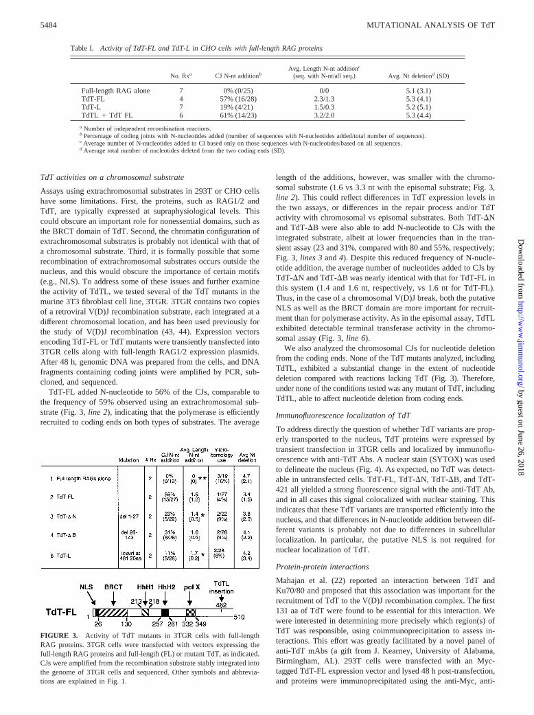
Assays using extrachromosomal substrates in 293T or CHO cellshave some limitations. First, the proteins, such as RAG1/2 andTdT, are typically expressed at supraphysiological levels. Thiscould obscure an important role for nonessential domains, such asthe BRCT domain of TdT. Second, the chromatin configuration ofextrachromosomal substrates is probably not identical with that ofa chromosomal substrate. Third, it is formally possible that somerecombination of extrachromosomal substrates occurs outside thenucleus, and this would obscure the importance of certain motifs(e.g., NLS). To address some of these issues and further examinethe activity of TdTL, we tested several of the TdT mutants in themurine 3T3 fibroblast cell line, 3TGR. 3TGR contains two copiesof a retroviral V(D)J recombination substrate, each integrated at adifferent chromosomal location, and has been used previously forthe study of V(D)J recombination (43, 44). Expression vectorsencoding TdT-FL or TdT mutants were transiently transfected into3TGR cells along with full-length RAG1/2 expression plasmids.After 48 h, genomic DNA was prepared from the cells, and DNAfragments containing coding joints were amplified by PCR, sub-cloned, and sequenced.
TdT-FL added N-nucleotide to 56% of the CJs, comparable tothe frequency of 59% observed using an extrachromosomal sub-strate (Fig. 3, line 2), indicating that the polymerase is efficientlyrecruited to coding ends on both types of substrates. The average
length of the additions, however, was smaller with the chromo-somal substrate (1.6 vs 3.3 nt with the episomal substrate; Fig. 3,line 2). This could reflect differences in TdT expression levels inthe two assays, or differences in the repair process and/or TdTactivity with chromosomal vs episomal substrates. Both TdT-�Nand TdT-�B were also able to add N-nucleotide to CJs with theintegrated substrate, albeit at lower frequencies than in the tran-sient assay (23 and 31%, compared with 80 and 55%, respectively;Fig. 3, lines 3 and 4). Despite this reduced frequency of N-nucle-otide addition, the average number of nucleotides added to CJs byTdT-�N and TdT-�B was nearly identical with that for TdT-FL inthis system (1.4 and 1.6 nt, respectively, vs 1.6 nt for TdT-FL).Thus, in the case of a chromosomal V(D)J break, both the putativeNLS as well as the BRCT domain are more important for recruit-ment than for polymerase activity. As in the episomal assay, TdTLexhibited detectable terminal transferase activity in the chromo-somal assay (Fig. 3, line 6).
We also analyzed the chromosomal CJs for nucleotide deletionfrom the coding ends. None of the TdT mutants analyzed, includingTdTL, exhibited a substantial change in the extent of nucleotidedeletion compared with reactions lacking TdT (Fig. 3). Therefore,under none of the conditions tested was any mutant of TdT, includingTdTL, able to affect nucleotide deletion from coding ends.
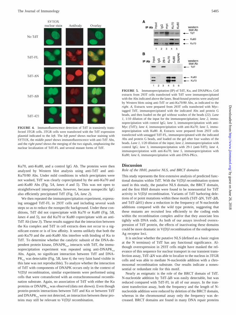
Immunofluorescence localization of TdT
To address directly the question of whether TdT variants are prop-erly transported to the nucleus, TdT proteins were expressed bytransient transfection in 3TGR cells and localized by immunoflu-orescence with anti-TdT Abs. A nuclear stain (SYTOX) was usedto delineate the nucleus (Fig. 4). As expected, no TdT was detect-able in untransfected cells. TdT-FL, TdT-�N, TdT-�B, and TdT-421 all yielded a strong fluorescence signal with the anti-TdT Ab,and in all cases this signal colocalized with nuclear staining. Thisindicates that these TdT variants are transported efficiently into thenucleus, and that differences in N-nucleotide addition between dif-ferent variants is probably not due to differences in subcellularlocalization. In particular, the putative NLS is not required fornuclear localization of TdT.
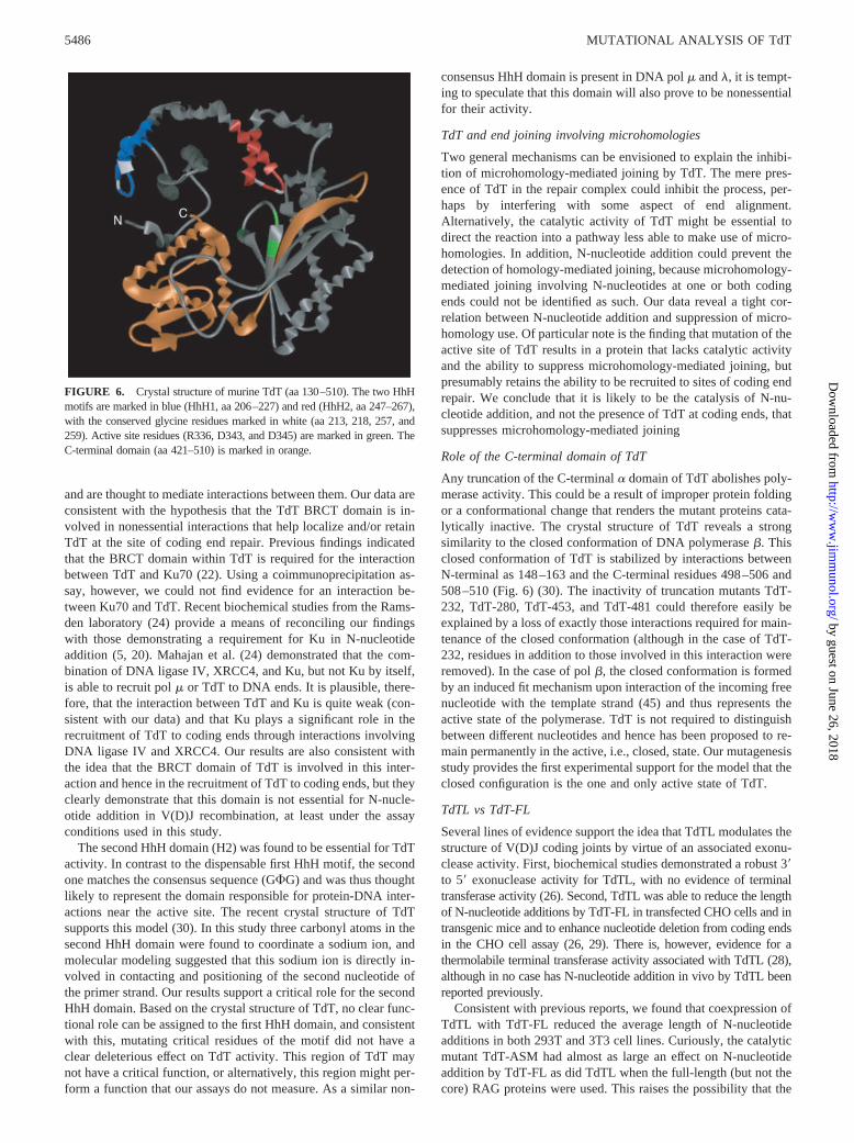
Protein-protein interactions
Mahajan et al. (22) reported an interaction between TdT andKu70/80 and proposed that this association was important for therecruitment of TdT to the V(D)J recombination complex. The first131 aa of TdT were found to be essential for this interaction. Wewere interested in determining more precisely which region(s) ofTdT was responsible, using coimmunoprecipitation to assess in-teractions. This effort was greatly facilitated by a novel panel ofanti-TdT mAbs (a gift from J. Kearney, University of Alabama,Birmingham, AL). 293T cells were transfected with an Myc-tagged TdT-FL expression vector and lysed 48 h post-transfection,and proteins were immunoprecipitated using the anti-Myc, anti-
Table I. Activity of TdT-FL and TdT-L in CHO cells with full-length RAG proteins
No. Rxa CJ N-nt additionbAvg. Length N-nt additionc
(seq. with N-nt/all seq.) Avg. Nt deletiond (SD)
Full-length RAG alone 7 0% (0/25) 0/0 5.1 (3.1)TdT-FL 4 57% (16/28) 2.3/1.3 5.3 (4.1)TdT-L 7 19% (4/21) 1.5/0.3 5.2 (5.1)TdTL � TdT FL 6 61% (14/23) 3.2/2.0 5.3 (4.4)
a Number of independent recombination reactions.b Percentage of coding joints with N-nucleotides added (number of sequences with N-nucleotides added/total number of sequences).c Average number of N-nucleotides added to CI based only on those sequences with N-nucleotides/based on all sequences.d Average total number of nucleotides deleted from the two coding ends (SD).
FIGURE 3. Activity of TdT mutants in 3TGR cells with full-lengthRAG proteins. 3TGR cells were transfected with vectors expressing thefull-length RAG proteins and full-length (FL) or mutant TdT, as indicated.CJs were amplified from the recombination substrate stably integrated intothe genome of 3TGR cells and sequenced. Other symbols and abbrevia-tions are explained in Fig. 1.
5484 MUTATIONAL ANALYSIS OF TdT
by guest on June 26, 2018http://w
ww
.jimm
unol.org/D
ownloaded from
Ku70, anti-Ku80, and a control IgG Ab. The proteins were thenanalyzed by Western blot analysis using anti-TdT and anti-Ku70/80 Abs. Under mild conditions in which precipitates werenot washed, TdT was clearly coprecipitated by the anti-Ku70 andanti-Ku80 Abs (Fig. 5A, lanes 4 and 5). This was not open tostraightforward interpretation, however, because nonspecific IgGalso efficiently precipitated TdT (Fig. 5A, lane 2).
We then repeated the immunoprecipitation experiment, express-ing untagged TdT-FL in 293T cells and including several washsteps so as to reduce the nonspecific background. Under these con-ditions, TdT did not coprecipitate with Ku70 or Ku80 (Fig. 5B,lanes 4 and 5), nor did Ku70 or Ku80 coprecipitate with an anti-TdT Ab (lane 3). These results suggest that the interaction betweenthe Ku complex and TdT in cell extracts does not occur to a sig-nificant extent or is of low affinity. It seems unlikely that both theanti-Ku70 and the anti-Ku80 Abs interfere with binding of Ku toTdT. To determine whether the catalytic subunit of the DNA-de-pendent protein kinase, DNAPKcs, interacts with TdT, the immu-noprecipitation experiment was repeated using anti-DNAPKcs
Abs. Again, no significant interaction between TdT and DNA-PKcs was detectable (Fig. 5B, lane 6; the very faint band visible inthis lane was not reproducible). To analyze whether the interactionof TdT with components of DNAPK occurs only in the context ofV(D)J recombination, similar experiments were performed usingcells that were cotransfected with an extrachromosomal recombi-nation substrate. Again, no association of TdT with either the Kuproteins or DNAPKcs was observed (data not shown). Even thoughprotein-protein interactions between TdT and Ku or between TdTand DNAPKcs were not detected, an interaction between these pro-teins may still be relevant to V(D)J recombination.
DiscussionRole of the HhH, putative NLS, and BRCT domains
This study represents the first extensive analysis of predicted func-tional domains within TdT. With the V(D)J recombination systemused in this study, the putative NLS domain, the BRCT domain,and the first HhH domain were found to be nonessential for TdTactivity in V(D)J recombination. Variants of TdT harboring dele-tions of or point mutations within these motifs (TdT-�N, TdT-�B,and TdT-�H1) show a reduction in the frequency of N-nucleotideadditions compared with the wild type protein, suggesting thatthese mutants are recruited less efficiently to the coding endswithin the recombination complex and/or that they associate lesstightly with DNA ends. As both of our assays involved overex-pression of TdT protein, the effects of inactivating these domainscould be more dramatic in V(D)J recombination of the endogenousAg receptor loci.
It is unclear whether the putative NLS (defined as a basic regionat the N terminus) of TdT has any functional significance. Al-though overexpression in 293T cells might have masked the rel-evance of this sequence for nuclear transport in our transient trans-fection assay, TdT-�N was able to localize to the nucleus in 3TGRcells and was able to mediate N-nucleotide addition with a chro-mosomal recombination substrate. Our results indicate a nones-sential or redundant role for this motif.
Nearly as enigmatic is the role of the BRCT domain of TdT.N-nucleotide addition by TdT-�B was easily detectable, but wasreduced compared with TdT-FL in all of our assays. In the tran-sient transfection assay, both the frequency and the length of N-nucleotide addition were reduced by deletion of the BRCT domain,whereas in the chromosomal assay only the frequency was de-creased. BRCT domains are found in many DNA repair proteins
FIGURE 4. Immunofluorescence detection of TdT in transiently trans-fected 3TGR cells. 3TGR cells were transfected with the TdT expressionplasmid indicated to the left. The left panel shows nuclear staining withSYTOX, the middle panel shows immunofluorescence with anti-TdT Abs,and the right panel shows the merging of the two signals, emphasizing thenuclear localization of TdT-FL and several mutant forms of TdT.
FIGURE 5. Immunoprecipitation (IP) of TdT, Ku, and DNAPKcs. Cellextracts from 293T cells transfected with TdT were immunoprecipitatedwith the Abs indicated above the lanes. Bead-bound proteins were analyzedby Western blots using anti-TdT or anti-Ku70/80 Abs, as indicated to theright. A, Extracts were prepared from 293T cells transfected with Myc-tagged TdT, immunoprecipitated with the indicated Abs and protein Gbeads, and then loaded on the gel without washes of the beads (22). Lane1, 1/10 dilution of the input for the immunoprecipitation; lane 2, immu-noprecipitation with control IgG; lane 3, immunoprecipitation with anti-Myc (TdT); lane 4, immunoprecipitation with anti-Ku70; lane 5, immu-noprecipitation with Ku80. B, Extracts were prepared from 293T cellstransfected with untagged TdT-FL, immunoprecipitated with the indicatedAbs and protein G beads, and loaded on the gel after four washes of thebeads. Lane 1, 1/20 dilution of the input; lane 2, immunoprecipitation withcontrol IgG; lane 3, immunoprecipitation with 29-1 (anti-TdT); lane 4,immunoprecipitation with anti-Ku70; lane 5, immunoprecipitation withKu80; lane 6, immunoprecipitation with anti-DNA-PKcs.
5485The Journal of Immunology
by guest on June 26, 2018http://w
ww
.jimm
unol.org/D
ownloaded from
and are thought to mediate interactions between them. Our data areconsistent with the hypothesis that the TdT BRCT domain is in-volved in nonessential interactions that help localize and/or retainTdT at the site of coding end repair. Previous findings indicatedthat the BRCT domain within TdT is required for the interactionbetween TdT and Ku70 (22). Using a coimmunoprecipitation as-say, however, we could not find evidence for an interaction be-tween Ku70 and TdT. Recent biochemical studies from the Rams-den laboratory (24) provide a means of reconciling our findingswith those demonstrating a requirement for Ku in N-nucleotideaddition (5, 20). Mahajan et al. (24) demonstrated that the com-bination of DNA ligase IV, XRCC4, and Ku, but not Ku by itself,is able to recruit pol � or TdT to DNA ends. It is plausible, there-fore, that the interaction between TdT and Ku is quite weak (con-sistent with our data) and that Ku plays a significant role in therecruitment of TdT to coding ends through interactions involvingDNA ligase IV and XRCC4. Our results are also consistent withthe idea that the BRCT domain of TdT is involved in this inter-action and hence in the recruitment of TdT to coding ends, but theyclearly demonstrate that this domain is not essential for N-nucle-otide addition in V(D)J recombination, at least under the assayconditions used in this study.
The second HhH domain (H2) was found to be essential for TdTactivity. In contrast to the dispensable first HhH motif, the secondone matches the consensus sequence (GG) and was thus thoughtlikely to represent the domain responsible for protein-DNA inter-actions near the active site. The recent crystal structure of TdTsupports this model (30). In this study three carbonyl atoms in thesecond HhH domain were found to coordinate a sodium ion, andmolecular modeling suggested that this sodium ion is directly in-volved in contacting and positioning of the second nucleotide ofthe primer strand. Our results support a critical role for the secondHhH domain. Based on the crystal structure of TdT, no clear func-tional role can be assigned to the first HhH domain, and consistentwith this, mutating critical residues of the motif did not have aclear deleterious effect on TdT activity. This region of TdT maynot have a critical function, or alternatively, this region might per-form a function that our assays do not measure. As a similar non-
consensus HhH domain is present in DNA pol � and �, it is tempt-ing to speculate that this domain will also prove to be nonessentialfor their activity.
TdT and end joining involving microhomologies
Two general mechanisms can be envisioned to explain the inhibi-tion of microhomology-mediated joining by TdT. The mere pres-ence of TdT in the repair complex could inhibit the process, per-haps by interfering with some aspect of end alignment.Alternatively, the catalytic activity of TdT might be essential todirect the reaction into a pathway less able to make use of micro-homologies. In addition, N-nucleotide addition could prevent thedetection of homology-mediated joining, because microhomology-mediated joining involving N-nucleotides at one or both codingends could not be identified as such. Our data reveal a tight cor-relation between N-nucleotide addition and suppression of micro-homology use. Of particular note is the finding that mutation of theactive site of TdT results in a protein that lacks catalytic activityand the ability to suppress microhomology-mediated joining, butpresumably retains the ability to be recruited to sites of coding endrepair. We conclude that it is likely to be the catalysis of N-nu-cleotide addition, and not the presence of TdT at coding ends, thatsuppresses microhomology-mediated joining
Role of the C-terminal domain of TdT
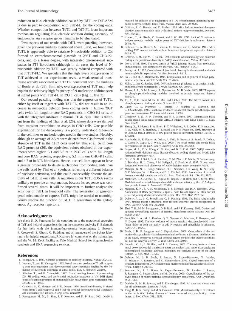
Any truncation of the C-terminal � domain of TdT abolishes poly-merase activity. This could be a result of improper protein foldingor a conformational change that renders the mutant proteins cata-lytically inactive. The crystal structure of TdT reveals a strongsimilarity to the closed conformation of DNA polymerase �. Thisclosed conformation of TdT is stabilized by interactions betweenN-terminal as 148–163 and the C-terminal residues 498–506 and508–510 (Fig. 6) (30). The inactivity of truncation mutants TdT-232, TdT-280, TdT-453, and TdT-481 could therefore easily beexplained by a loss of exactly those interactions required for main-tenance of the closed conformation (although in the case of TdT-232, residues in addition to those involved in this interaction wereremoved). In the case of pol �, the closed conformation is formedby an induced fit mechanism upon interaction of the incoming freenucleotide with the template strand (45) and thus represents theactive state of the polymerase. TdT is not required to distinguishbetween different nucleotides and hence has been proposed to re-main permanently in the active, i.e., closed, state. Our mutagenesisstudy provides the first experimental support for the model that theclosed configuration is the one and only active state of TdT.
TdTL vs TdT-FL
Several lines of evidence support the idea that TdTL modulates thestructure of V(D)J coding joints by virtue of an associated exonu-clease activity. First, biochemical studies demonstrated a robust 3�to 5� exonuclease activity for TdTL, with no evidence of terminaltransferase activity (26). Second, TdTL was able to reduce the lengthof N-nucleotide additions by TdT-FL in transfected CHO cells and intransgenic mice and to enhance nucleotide deletion from coding endsin the CHO cell assay (26, 29). There is, however, evidence for athermolabile terminal transferase activity associated with TdTL (28),although in no case has N-nucleotide addition in vivo by TdTL beenreported previously.
Consistent with previous reports, we found that coexpression ofTdTL with TdT-FL reduced the average length of N-nucleotideadditions in both 293T and 3T3 cell lines. Curiously, the catalyticmutant TdT-ASM had almost as large an effect on N-nucleotideaddition by TdT-FL as did TdTL when the full-length (but not thecore) RAG proteins were used. This raises the possibility that the
FIGURE 6. Crystal structure of murine TdT (aa 130–510). The two HhHmotifs are marked in blue (HhH1, aa 206–227) and red (HhH2, aa 247–267),with the conserved glycine residues marked in white (aa 213, 218, 257, and259). Active site residues (R336, D343, and D345) are marked in green. TheC-terminal domain (aa 421–510) is marked in orange.
5486 MUTATIONAL ANALYSIS OF TdT
by guest on June 26, 2018http://w
ww
.jimm
unol.org/D
ownloaded from
reduction in N-nucleotide addition caused by TdTL or TdT-ASMis due in part to competition with TdT-FL for the coding ends.Whether competition between TdTL and TdT-FL is an importantmechanism regulating N-nucleotide addition during assembly ofendogenous Ag receptor genes remains to be elucidated.
Two aspects of our results with TdTL were puzzling, however,given the previous findings mentioned above. First, we found thatTdTL is apparently able to catalyze N-nucleotide addition to CJsformed on extrachromosomal plasmids in 293T and CHO-K1cells, and, to a lesser degree, with integrated chromosomal sub-strates in 3T3 fibroblasts (although in all cases the level of N-nucleotide addition by TdTL was greatly reduced compared withthat of TdT-FL). We speculate that the high levels of expression ofTdT achieved in our experiments reveal a weak terminal trans-ferase activity associated with TdTL, consistent with the findingsof Boule et al. (28). Similarly, overexpression of TdT may helpexplain the relatively high frequency of N-nucleotide addition seenat signal joints with TdT-FL in 293 T cells (Fig. 1, line 2).
Our second puzzling finding was that the expression of TdTL,either by itself or together with TdT-FL, did not result in an in-crease in nucleotide deletion from coding ends in human 293Tcells (with full-length or core RAG proteins), in CHO-K1 cells, orwith the integrated substrate in murine 3TGR cells. This is differ-ent from the findings of Thai et al. (26), whose data were derivedfrom transient recombination assays in CHO cells. One possibleexplanation for the discrepancy is a poorly understood differencein the cell lines or methodologies used in the two studies. Notably,although an average of 2.5 nt were deleted from CJs formed in theabsence of TdT in the CHO cells used by Thai et al. (with coreRAG proteins) (26), the equivalent values obtained in our exper-iments were higher: 6.1 and 5.8 nt in 293T cells with full-lengthand core RAG proteins, respectively; 5.1 nt in our CHO-K1 cells;and 4.7 nt in 3T3 fibroblasts. Hence, our cell lines appear to havea greater propensity to delete nucleotides from coding ends thanthe cells used by Thai et al. (perhaps reflecting a different balanceof nuclease activities), and this could conceivably obscure the ac-tivity of TdTL in our cells. A mutation in our TdTL cDNA seemsunlikely to provide an explanation, because its sequence was con-firmed several times. It will be important to further analyze theactivities of TdTL in lymphoid cells. The generation of gene-tar-geted mice unable to express TdTL might be needed to unambig-uously resolve the function of TdTL in generation of the endog-enous Ag receptor repertoire.
AcknowledgmentsWe thank S. D. Fugmann for his contribution to the mutational strategiesof TdT and helpful suggestions during the sequence analysis; F. Balamuthfor her help with the immunofluorescence experiments; J. Sweasy,P. Cresswell, S. Ghosh, C. Radding, and all members of the Schatz labo-ratory for helpful suggestions; J. Kearney for comments on the manuscript;and the W. M. Keck Facility at Yale Medical School for oligonucleotidesynthesis and DNA sequencing services.
References1. Tonegawa, S. 1983. Somatic generation of antibody diversity. Nature 302:575.2. Iwasato, T., and H. Yamagishi. 1992. Novel excision products of T cell receptor
� gene rearrangements and developmental stage specificity implied by the fre-quency of nucleotide insertions at signal joints. Eur. J. Immunol. 22:101.
3. Shimizu, T., and H. Yamagishi. 1992. Biased reading frames of pre-existingDH–JH coding joints and preferential nucleotide insertions at VH–DJH signaljoints of excision products of immunoglobulin heavy chain gene rearrangements.EMBO J. 11:4869.
4. Candeias, S., K. Muegge, and S. K. Durum. 1996. Junctional diversity in signaljoints from T cell receptor � and � loci via terminal deoxynucleotidyl transferaseand exonucleolytic activity. J. Exp. Med. 184:1919.
5. Purugganan, M. M., S. Shah, J. F. Kearney, and D. B. Roth. 2001. Ku80 is
required for addition of N nucleotides to V(D)J recombination junctions by ter-minal deoxynucleotidyl transferase. Nucleic Acids Res. 29:1638.
6. Gilfillan, S., C. Benoist, and D. Mathis. 1995. Mice lacking terminal deoxynu-cleotidyl transferase: adult mice with a fetal antigen receptor repertoire. Immunol.Rev. 148:201.
7. Komori, T., A. Okada, V. Stewart, and F. W. Alt. 1993. Lack of N regions inantigen receptor variable region genes of TdT-deficient lymphocytes. Science261:1171.
8. Gilfillan, S., A. Dierich, M. Lemeur, C. Benoist, and D. Mathis. 1993. Micelacking TdT: mature animals with an immature lymphocyte repertoire. Science261:1175.
9. Gerstein, R. M., and M. R. Lieber. 1993. Extent to which homology can constraincoding exon junctional diversity in V(D)J recombination. Nature 363:625.
10. Lewis, S. M. 1994. The mechanism of V(D)J joining: lessons from molecular,immunological, and comparative analyses. Adv. Immunol. 56:27.
11. Feeney, A. J. 1992. Comparison of junctional diversity in the neonatal and adultimmunoglobulin repertoires. Int. Rev. Immunol. 8:113.
12. Ito, J., and D. K. Braithwaite. 1991. Compilation and alignment of DNA poly-merase sequences. Nucleic Acids Res. 19:4045.
13. Holm, L., and C. Sander. 1995. DNA polymerase � belongs to an ancient nucle-otidyltransferase superfamily. Trends Biochem. Sci. 20:345.
14. Manke, I. A., D. M. Lowery, A. Nguyen, and M. B. Yaffe. 2003. BRCT repeatsas phosphopeptide-binding modules involved in protein targeting. Science302:636.
15. Yu, X., C. C. Chini, M. He, G. Mer, and J. Chen. 2003. The BRCT domain is aphospho-protein binding domain. Science 302:639.
16. Casey, G., S. Plummer, G. Hoeltge, D. Scanlon, C. Fasching, andE. J. Stanbridge. 1993. Functional evidence for a breast cancer growth suppressorgene on chromosome 17. Hum. Mol. Genet. 2:1921.
17. Critchlow, S. E., R. P. Bowater, and S. P. Jackson. 1997. Mammalian DNAdouble-strand break repair protein XRCC4 interacts with DNA ligase IV. Curr.Biol. 7:588.
18. Zhang, X., S. Morera, P. A. Bates, P. C. Whitehead, A. I. Coffer, K. Hainbucher,R. A. Nash, M. J. Sternberg, T. Lindahl, and P. S. Freemont. 1998. Structure ofan XRCC1 BRCT domain: a new protein-protein interaction module. EMBO J.17:6404.
19. Aoufouchi, S., E. Flatter, A. Dahan, A. Faili, B. Bertocci, S. Storck, F. Delbos,L. Cocea, N. Gupta, J. C. Weill, et al. 2000. Two novel human and mouse DNApolymerases of the polX family. Nucleic Acids Res. 28:3684.
20. Bogue, M. A., C. Y. Wang, C. M. Zhu, and D. B. Roth. 1997. V(D)J recombi-nation in Ku86-deficient mice: distinct effects on coding, signal, and hybrid jointformation. Immunity 7:37.
21. Gu, Y. S., K. J. Seidl, G. A. Rathbun, C. M. Zhu, J. P. Manis, N. Vanderstoep,L. Davidson, H. L. Cheng, J. M. Sekiguchi, K. Frank, et al. 1997. Growth retar-dation and leaky scid phenotype of Ku70-deficient mice. Immunity 7:653.
22. Mahajan, K. N., L. Gangi-Peterson, D. H. Sorscher, J. S. Wang, K. N. Gathy,N. P. Mahajan, W. H. Reeves, and B. S. Mitchell. 1999. Association of terminaldeoxynucleotidyl transferase with Ku. Proc. Natl. Acad. Sci. USA 96:13926.
23. Mickelsen, S., C. Snyder, K. Trujillo, M. Bogue, D. B. Roth, and K. Meek. 1999.Modulation of terminal deoxynucleotidyltransferase activity by the DNA-depen-dent protein kinase. J. Immunol. 163:834.
24. Mahajan, K. N., S. A. N. McElhinny, B. S. Mitchell, and D. A. Ramsden. 2002.Association of DNA polymerase � (pol �) with Ku and ligase IV: Role for pol� in end-joining double-strand break repair. Mol. Cell. Biol. 22:5194.
25. Doherty, A. J., L. C. Serpell, and C. P. Ponting. 1996. The helix-hairpin-helixDNA-binding motif: a structural basis for non-sequence-specific recognition ofDNA. Nucleic Acids Res. 24:2488.
26. Thai, T. H., M. M. Purugganan, D. B. Roth, and J. F. Kearney. 2002. Distinct andopposite diversifying activities of terminal transferase splice variants. Nat. Im-munol. 3:457.
27. Bentolila, L. A., M. F. Dandon, Q. T. Nguyen, O. Martinez, F. Rougeon, andN. Doyen. 1995. The two isoforms of mouse terminal deoxynucleotidyl trans-ferase differ in both the ability to add N regions and subcellular localization.EMBO J. 14:4221.
28. Boule, J. B., F. Rougeon, and C. Papanicolaou. 2000. Comparison of the twomurine deoxynucleotidyltransferase terminal isoforms: a 20-amino acid insertionin the highly conserved carboxyl-terminal region modifies the thermosensitivitybut not the catalytic activity. J. Biol. Chem. 275:28984.
29. Benedict, C. L., S. Gilfillan, and J. F. Kearney. 2001. The long isoform of ter-minal deoxynucleotidyl transferase enters the nucleus and, rather than catalyzingnontemplated nucleotide addition, modulates the catalytic activity of the shortisoform. J. Exp. Med. 193:89.
30. Delarue, M., J. B. Boule, J. Lescar, N. Expert-Bezancon, N. Jourdan,N. Sukumar, F. Rougeon, and C. Papanicolaou. 2002. Crystal structures of atemplate-independent DNA polymerase: murine terminal deoxynucleotidyltrans-ferase. EMBO J. 21:427.
31. Sukumar, N., J. B. Boule, N. Expert-Bezancon, N. Jourdan, J. Lescar,F. Rougeon, C. Papanicolaou, and M. Delarue. 2000. Crystallization of the cat-alytic domain of murine terminal deoxynucleotidyl transferase. Acta Crystallogr.D 56:1662.
32. Doublie, S., M. R. Sawaya, and T. Ellenberger. 1999. An open and closed casefor all polymerases. Structure 7:R31.
33. Yang, B., K. N. Gathy, and M. S. Coleman. 1994. Mutational analysis of residuesin the nucleotide binding domain of human terminal deoxynucleotidyl trans-ferase. J. Biol. Chem. 269:11859.
5487The Journal of Immunology
by guest on June 26, 2018http://w
ww
.jimm
unol.org/D
ownloaded from

34. Steitz, T. A., and J. A. Steitz. 1993. A general two-metal-ion mechanism forcatalytic RNA. Proc. Natl. Acad. Sci. USA 90:6498.
35. Fugmann, S. D., and D. G. Schatz. 2001. Identification of basic residues in RAG2critical for DNA binding by the RAG1-RAG2 complex. Mol. Cell. 8:899.
36. Lieber, M. R., J. E. Hesse, S. Lewis, G. C. Bosma, N. Rosenberg, K. Mizuuchi,M. J. Bosma, and M. Gellert. 1988. The defect in murine severe combined im-mune deficiency: joining of signal sequences but not coding segments in V(D)Jrecombination. Cell 55:7.
37. Koiwai, O., T. Yokota, T. Kageyama, T. Hirose, S. Yoshida, and K. Arai. 1986.Isolation and characterization of bovine and mouse terminal deoxynucleotidyl-transferase cDNAs expressible in mammalian cells. Nucleic Acids Res. 14:5777.
38. Lieber, M. R., J. E. Hesse, K. Mizuuchi, and M. Gellert. 1988. Lymphoid V(D)Jrecombination: nucleotide insertion at signal joints as well as coding joints. Proc.Natl. Acad. Sci. USA 85:8588.
39. Chang, L. M., and F. J. Bollum. 1986. Molecular biology of terminal transferase.CRC Crit. Rev. Biochem. 21:27.
40. McMahan, C. J., M. J. Difilippantonio, N. Rao, E. S. Spanopoulou, andD. G. Schatz. 1997. A basic motif in the N-terminal region of RAG1 enhancesrecombination activity. Mol. Cell. Biol. 17:4544.
41. Roman, C. A. J., S. R. Cherry, and D. Baltimore. 1997. Complementation ofV(D)J recombination deficiency in RAG-1�/� B cells reveals a requirement fornovel elements in the N-terminus of RAG-1. Immunity 7:13.
42. Steen, S. B., J. O. Han, C. Mundy, M. A. Oettinger, and D. B. Roth. 1999. Rolesof the “dispensable” portions of RAG-1 and RAG-2 in V(D)J recombination.Mol. Cell. Biol. 19:3010.
43. Schatz, D. G., and D. Baltimore. 1988. Stable expression of immunoglobulin geneV(D)J recombinase activity by gene transfer into 3T3 fibroblasts. Cell 53:107.
44. Schatz, D. G., M. A. Oettinger, and D. Baltimore. 1989. The V(D)J recombina-tion activating gene (RAG-1). Cell 59:1035.
45. Sawaya, M. R., R. Prasad, S. H. Wilson, J. Kraut, and H. Pelletier. 1997. Crystalstructures of human DNA polymerase � complexed with gapped and nickedDNA: evidence for an induced fit mechanism. Biochemistry 36:11205.
5488 MUTATIONAL ANALYSIS OF TdT
by guest on June 26, 2018http://w
ww
.jimm
unol.org/D
ownloaded from