mutational analysis of the central domain of adenovirus virus
TRANSCRIPT

JOURNAL OF VIROLOGY, June 1993, p. 3534-35430022-538X/93/063534-10$02.00/0Copyright © 1993, American Society for Microbiology
Mutational Analysis of the Central Domain of AdenovirusVirus-Associated RNA Mandates a Revision of
the Proposed Secondary StructureTSAFRIRA PE'ERY,lt KENNETH H. MELLITS,"12t AND MICHAEL B. MATHEWS"*Cold Spring Harbor Laboratory, P.O. Box 100, Cold Spring Harbor, New York 11724,1 and
Genetics Program, State University ofNew York, Stony Brook; New York 117902Received 23 November 1992/Accepted 4 March 1993
Protein synthesis in adenovirus-infected cells is regulated during the late phase of infection. The rate ofinitiation is maintained by a small viral RNA, virus-associated (VA) RNA,, which prevents the phosphorylationof eukaryotic initiation factor eIF-2 by a double-stranded RNA-activated protein kinase, DAI. On the basis ofnuclease sensitivity analysis, a secondary-structure model was proposed for VA RNA. The model predicts acomplex stem-loop structure in the central part of the molecule, the central domain, joining two duplexedstems. The central domain is required for the inhibition ofDAI activation and participates in the binding ofVARNA to DAI. To assess the significance of the postulated stem-loop structure in the central domain, we
generated compensating, deletion, and substitution mutations. A substitution mutation which disrupts thestructure in the central domain abolishes VA RNA function in vitro and in vivo. Base-compensating mutationsfailed to restore the function or structure of the mutant, implying that the stem-loop structure may not exist.To confirm this observation, we tested mutants with alterations in the hypothetical loop and short stem thatconstitute the main features of the wild-type model structure. The upper part of the hypothetical loop could bedeleted without abolishing the ability of the RNA to block DAI activation in vitro, whereas other loop mutationswere deleterious for function and caused major rearrangements in the molecule. Base-compensating mutationsin the stem did not restore the expected base pairing, even though the mutant RNAs were still functional invitro. Surprisingly, a mutant with a noncompensating substitution mutation in the stem was more effective thanwild-type VA RNA, in DAI inhibition assays but was ineffective in vivo. The structural and functionalconsequences of these mutations do not support the proposed model structure for the central domain, and wetherefore suggest an alternative structure in which tertiary interactions may play a significant role in shapingthe specificity ofVA RNA function in the infected cell. Discrepancies between the functionality of mutant formsof VA RNA in vivo and in vitro are consistent with the existence of additional roles for VA RNA in the cell.
The virus-associated (VA) RNA, of adenovirus type 2(Ad2) is a key viral product which confers resistance to an
interferon-induced cellular antiviral defense mechanism (fora review, see reference 23). VA RNA, is a highly structuredRNA of 160 nucleotides (nt), which is synthesized by RNApolymerase III (pol III) and accumulates to high concentra-tion in the cytoplasm during the late phase of infection. Itcompetes with double-stranded RNA (dsRNA) for binding tothe protein kinase DAI, the dsRNA-activated inhibitor ofprotein synthesis. In the absence of VA RNA, activated DAIphosphorylates eukaryotic initiation factor 2, thereby indi-rectly impeding translational initiation (for a review, see
reference 25). VA RNA can also stimulate the expression ofgenes introduced into uninfected cells by transfection (16,36). No viral sequences are required in the reporter geneconstruct for this effect to be manifested. More recently, VARNA has also been reported to increase the accumulation ofmRNA from cotransfected plasmids: in one case VA RNApreferentially enhanced the accumulation of longer mRNAs(37), and in another it increased the stability of the plasmid-generated mRNA (35).On the basis of nuclease sensitivity analysis (27), a sec-
* Corresponding author.t Present address: Department of Cell Biology, Weizmann Insti-
tute, Rehovot, Israel.t Present address: Imperial Cancer Research Fund, Lincoln's Inn
Field, London WC2A 3PX, England.
ondary-structure model was proposed for VA RNA consist-ing of three main regions: the apical stem-loop, the centraldomain, and the terminal stem (Fig. 1A). In this model, thecentral domain, which resides between the apical stem andthe terminal stem, contains a number of features typical ofRNA secondary structure. The most prominent feature inthe central domain is stem-loop 6, and there is also a
bifurcation loop and an interior loop. Several aspects of thismodel structure are supported by mutational analyses inwhich foreign sequences or deletions were introduced intothe VA RNA molecule. Examination of the secondarystructure, biological function, and binding to DAI of thesediverse mutants indicated that the central domain is requiredfor the inhibition of DAI activation by dsRNA and impli-cated the apical stem in the binding of the VA RNA to DAI(6, 26, 27, 29). Subsequent studies suggested that the centraldomain also participates in the binding of VA RNA to DAI,but probably to a lesser extent than the apical stem (1, 7).
It is well established that secondary structural elements inRNA, such as duplexes, hairpins, internal loops, bulgedbases, and pseudoknots, are intimately connected with bio-logical functions (30, 31). Despite its critical role in VA RNAfunction, the complex stem-loop structure of the centraldomain has not been subjected to a detailed examination. Todefine the features of this stem-loop structure, we introducedforeign sequences into a 20-nt region within the centraldomain and made deletions in the proposed loop structure.The structural and functional consequences of these muta-
3534
Vol. 67, No. 6

CENTRAL DOMAIN OF VA RNA 3535
Loop 2o
GUGG C-
\ C-GU -A
G-C 4oG AU-AG-CC-GC-G
o U *GStem lo- UC-G
C-C
U-AC-G soA C
C-GC-CG-CUCU5p-_G-C C Un
~AC'a, A C>r-Loop 5 G C-
VC C'0
U g'6° G A
G UG-C
C-GAPICAL A cSTEM G-C 80
Stem 4b C-GA-Uso,,G-CG-CC-GG-CG-C
CENTRAL 42 A-U(~~~~~U-C-G-sD C CG Cc CA-U-A100 G~-CA 1o C C
CGGyUU _ Stem- r.G-CAUGCGlUUCU,&
\ DOMAIN U lo c~~~~~~~~~UCC AA Loop C-G UpCC AAGC
UGU G-CI30 G U G
GC 9.4°
,AAA~U-A
DOMAIN C
TERMINAL~~~~--U G-U*G
Stem3b 4G6U GG, C4
Loop2b UC-C
IsC-G
U-AC-C '40
C A
U-A
C-C
TERMINAL C-G
C-GU -A
C-G so0A C
C-C
C-C
A. S.G B.5sp-G-C CUn
A Co,~AhA 0lA.
G C,VC COOU GV
As'°o G Ao
G *UC.G-CDG-CC-GC-GA C
G-C 80C-GA-U
so G-C'6- C
C-GG-CG-CU * G
42 A-UcU-C-Gs:'A-U-A
G * UCUCG-C
U "C -00 A
-WA AUGG CC120G G lAC C
16 C-GW CU~~~A~~~ C~U-A C GG-C A
G A 4CGUGU~
U-A 130
G-CC-GC-GU *GU GC-GU-AC-GA C
C-C
G-CGCU C.
5 p-G-C C Un
FIG. 1. Secondary structure models for Ad2 VA RNA,. (A) Schematic diagram based on reference 27 showing the principal regions of themolecule. (B) As in panel A, with the cleavage sites marked according to the following convention: large arrowheads with shaft, very strong
cuts; large arrowheads, strong cuts; small arrowheads, weaker cuts; solid symbols, single-strand-specific sites; open symbols, nuclease V1sites. (C) Alternative model with cleavage sites marked as in panel B.
tions were investigated. Secondary-structure analysis showedthat in all cases the structural changes were confined to thecentral domain, including loop 2a, stem 3, and stem-loop 6(Fig. 1A), and did not affect the apical stem-loop or theterminal stem, implying that the central domain is a rela-tively autonomous structural element with considerable in-trinsic tertiary interactions. Surprisingly, the stem-loop,which is the principal feature of the central domain in theoriginal model, is not supported by structural analysis or bystudy of the function of the mutant VA RNAs in vivo and invitro. These findings suggest that an alternative conforma-tion of the central domain is important for VA RNA func-tion.
MATERUILS AND METHODS
Construction of mutants. All deletion and substitutionmutants constructed here were generated within the VARNA gene of plasmid pT7VA (28) by oligonucleotide-di-rected mutagenesis (18, 41) with Escherichia coli CJ 236. Forexperiments conducted in vivo, the DraI restriction endonu-clease site ('l'lT'AAA) was modified to TT'T'GG by usingoligonucleotide-directed mutagenesis to allow termination ofVA RNA transcription by pol III in vivo. The fidelity oftermination was verified in pol III transcription experimentsin vitro (data not shown). Two RNA bands were generated,
one the size of wild-type VA RNA, and the other a fewnucleotides shorter, possibly because of 5'-end changes (4,40). The efficiency of site-specific mutation was approxi-mately 50%. Oligonucleotides were synthesized by using a
380-A DNA synthesizer (Applied Biosystems), and mutantswere analyzed by dideoxy sequencing (33) with a kit (UnitedStates Biochemical Corporation).
Kinase assay. Wild-type and mutant VA RNAs were
purified by sequential electrophoresis through denaturingand nondenaturing 8% polyacrylamide-7 M urea gels, fol-lowed by cellulose CF-11 chromatography (28). The VARNA was then incubated with DAI (purified to the hydrox-ylapatite stage [41]) and [y-32P]ATP (ICN Biochemicals,Inc.) in either the presence or absence of reovirus dsRNA asdescribed previously (28).RNA secondary structure. The secondary structures of
wild-type and mutant VA RNAs were determined by theRNase sensitivity technique (27). Each mutant RNA was
labeled at its 5' or 3' end and subjected to partial digestionunder conditions such that most RNA molecules were notdigested and the remainder were cleaved only once. Weemployed the single-strand-specific RNase T1, Bacilluscereus RNase, RNase U2, and RNase T2 (specific for Gresidues, pyrimidines, A residues, and any exposed base,respectively), as well as nuclease V1 from cobra venom
(specific for helical and stacked nucleotides). Digestion
VOL. 67, 1993

3536 PE'ERY ET AL.
TABLE 1. Sequence and function of central-domain mutants
Sequence' DAI inhibition Reticulocyte CATVA RNA ( L/l sa tmltoStem 6 Loop 6 Stem 6 ( ./l sa tmlto
100 110 120
Wild type GCGGUU ACCGCCCGCGUGUCG AACCCA ++ ++ +1s3 ----CA GAUCUGx-----ls3a GAUCUGx-----ls3b ----CA GAUCUGx----- UG---- -de1103-109 ------x----x----++de1110-117 ----Y-X]xx-xx---x +sub11O-117 ----UACACAUC ----+sub101,102 ----CA ----------------------- -- - --isub108,119 ---------------- UG----sublO2,118,119 -----*A ..--------------- UG----
subl0l,102,118,119 ----CA ..-------- UG---- +
-, same as wild type; x, deleted nucleotide. The sequence shown includes two nucleotides flanking stem 6 at both the 5' and 3' ends.
products were analyzed in denaturing gels run for variouslengths of time to resolve all regions of the RNA molecules,and the positions and intensities of nuclease cleavages werenoted. RNA secondary-structure models were derived bymanual folding and the use of a computer program whichincorporated both empirical data and estimates of energiesinherent in base pairing (15, 42).Enhancement ofCAT expression. Human 293 cells (8) were
cotransfected with 3 ,ug of ,3-globin-CAT12 (37) and 10 ,ug ofthe appropriate VA RNA-encoding plasmids as describedpreviously (12). Transfections were performed in duplicate.The cells were -60% confluent in 6-cm dishes, and the totalquantity of DNA was adjusted to 15 p,g with pUC119 DNA.Following a glycerol shock at 6 h posttransfection, the cellswere harvested at 48 h for chloramphenicol acetyltransferase(CAT) assay (12). Negative controls contained the vectorpUC119 or pEM-7, which carries the T7 RNA polymerasepromoter and human immunodeficiency virus type 1 TARsequence (11) in place of the VA RNA sequence. Dot blothybridization showed that comparable amounts of wild-typeand mutant VA RNA had accumulated in the cytoplasm by48 h posttransfection.
Cell-free translation. DAI activation was assayed by trans-lation in vitro in two stages. First, rabbit reticulocyte lysatewas preincubated for 15 min at 30°C with reovirus dsRNAand purified wild-type or mutant VA RNA (28). Then thereaction mixture was completed by addition of_lobin mRNA(4 Lg/ml; Bethesda Research Laboratories), [3 SJmethionine(5 mCi/ml; ICN Biomedicals), 72 tnM potassium acetate, 0.4mM magnesium acetate, 0.4 mM GTP, and other compo-nents specified previously (3). After incubation for a further30 min at 30°C, the translation products were resolved in a15% polyacrylamide-sodium dodecyl sulfate (SDS) gel andvisualized by fluorography. The globin band was quantifiedby using a Fuji Bio-image Analyzer.
RESULTS
The current model for the secondary structure of VARNA(Fig. 1A) contains two imperfectly duplexed stems joined bya complex stem-loop structure, the central domain. Thismodel was derived as a fit for the empirical data obtained byRNase sensitivity analysis, placing nucleotides that arecleaved by single-strand (ss)-specific nucleases in unpairedregions and assigning nucleotides that are cleaved by nucle-ase V1 in regions of secondary structure (Fig. 1B). However,
the data can be accommodated equally well by other models,such as that shown in Fig. 1C, with alternative structures inthe central domain. The apical stem is at least partiallyresponsible for the efficient binding of VA RNA to DAI (26,29), while the central domain is implicated in the inhibition ofDAI activation (6, 26, 27). In this study we wanted to explorethe role of stem-loop 6 in the central domain in determiningthe inhibition function of VA RNA. A previous study (26)had shown that large deletions or substitutions in the centraldomain abolished function and caused major rearrangementsin the structure of the VA RNA molecule. Here we haveintroduced small deletions and substitutions by site-directedmutagenesis to test the important features of stem-loop 6 andto challenge the existence of this predicted structure in thecentral domain.The mutations are listed in Table 1, and their sequences
are displayed schematically in Fig. 2 superimposed on themodel structure of Fig. 1A. All the constructs were intro-duced into a vector which allows efficient production of VARNA in vitro by using T7 RNA polymerase (28), and thesecondary structures of the mutant RNAs were studied bythe nuclease sensitivity technique (27). This analysis indi-cated that the structures are perturbed primarily in thecentral domain, while the rest of the structure remains thesame as in wild-type VA RNA. To assess the functionalconsequences of the mutations, we investigated their abilityto inhibit the activation of DAI in vitro, as measured byautophosphorylation. None of the RNAs synthesized invitro activated DAI when tested over a wide range ofconcentrations (5 nglml to 50 ,g/ml [data not shown]),verifying that they were free of dsRNA contaminants (28)and confirming that the interaction between VA RNA andDAI is fundamentally different from that between dsRNAand DAI.Attempts to restore the stem-loop structure in the central
domain. Base-compensating mutations are often designed tolend support to a predicted secondary structure. We focusedinitially on the mutant 1s3, in which part of the short stemand the top of the presumed loop of stem-loop 6 weresubstituted by the linker-derived sequence CAGAUCUG(Fig. 2). This substitution should not allow the pairingnecessary for the formation of stem-loop 6. Mutant 1s3 isnonfunctional both in vivo and in DAI autophosphorylationinhibition assays in vitro (26, 27, 29). To determine whetherfunction is restored by reestablishing the ability to formstem-loop 6, we first made the base-compensating mutants
J. VIROL.

CENTRAL DOMAIN OF VA RNA 3537
ccC 7oGGAucc Is3bG UCUC G A GCGo GG cIcsECoU 00 G GC CUGAUCGC Is3aC
G I3G A Ue10-1
GAU CG deIlO13-109C AUGCGGUU
o ,~~~~~~~~~~1
UG CCA CC SUUGAC de111-1717GA
C 4
A ICACCA
GUC subC I-117AGACa14A Is3A
uC YAUCU
GG GCCAGCG
C
G 15CGCUC C U0
CC G Cc sub 101, 102GCGGa CGlo
CCAA C'20 G G
CU G U
20r.g/mi ReodsRNA - +
VARNA ug/mI 0 0 50 20 5
Ad2 VA RNAI do
del 103-109 _
CCGCC sub 1 1 8, 119GCGGUU C
G oC CFG7 c120G GG GU
CC G C sub 102, 118,G C G G A C 119
GC jGq_ GU G U
CCGC sub 101, 102,G CC 118,119GCGGr Go
120 GCU G U
FIG. 2. Schematic of mutant sequences. The sequences aresuperimposed on the original secondary-structure model (27).
Is3 _
sub 110-I 17
Is3b go
SUb 101,102
sub 118,119 b
FIG. 3. Inhibition of DAI activation by VA RNA mutants.Kinase assays were conducted with or without reovirus dsRNA andthe indicated RNAs at the concentrations indicated. Shown aresections of autoradiograms illustrating DAI autophosphorylation.
1s3a and 1s3b (Fig. 2). Substitution 1s3a was obtained byreplacing nt 101 and 102 in 1s3 (CA) with the wild-typesequence UU to restore the proposed pairing with thewild-type sequence AA (nt 118 and 119). Conversely, sub-stitution 1s3b was made by replacing the sequence AA (nt118 and 119) in 1s3 with the sequence UG to allow the pairingof nt 118 to 102 and 119 to 101, thereby recreating a
presumptive short stem in stem-loop 6 but with a differentsequence (Fig. 2).These potentially base-compensating mutants did not fully
restore wild-type function. As illustrated in Fig. 3 and shownin Table 2, only at the highest concentration tested (50,ug/ml) did mutant 1s3a inhibit DAI autophosphorylation asefficiently as wild-type VA RNA did. It was less efficient atthe lower concentration of 20 ,ug/ml and completely lost itsinhibitory function at 5 ,ug/ml. Mutant ls3b, on the otherhand, completely failed to restore the lost inhibitory functionof mutant 1s3 (29) and was inactive even at the high concen-tration of 50 ,ug/ml (Fig. 3, Table 2).To seek an explanation for these results, we examined the
changes in the secondary structure of each of the compen-sating mutants. The nuclease sensitivity pattern of 1s3a RNAin stem-loop 6 (Fig. 4A) is more similar to that of wild-typeVA RNA than to that of the nonfunctional mutant 1s3 (Fig.5). However, the three A bases in loop 2a which are verysusceptible to attack by ss-specific nucleases in wild-typeVA RNA are resistant in mutant 1s3a. Moreover, mildcleavages in stem 3b suggest melting of the stem and newnuclease V1 cuts that appear in the substituted sequenceindicate that this sequence may be involved in a secondaryor tertiary structure. These changes are diagrammed in Fig.5, in which the cleavage patterns are superimposed on thewild-type central domain structure. Thus, the structure andpartial function of 1s3a RNA are compatible with pairing instem-loop 6, and alterations elsewhere in the central domain
could explain its lack of function at 5 ,ug/ml. However,structural analysis of mutant ls3b suggests that the substi-tuted bases CA (nt 101 and 102) and UG (nt 118 and 119) donot pair since there is an ss cut after the C at nt 101 and thereare no nuclease V1 cuts after nt 99 (Fig. 5). One interpreta-tion of these results is that nt 101, 102, 118, and 119 areimportant for function but do not form the stem structuredepicted in Fig. 1A.
Functional and structural analysis of mutants with changesin loop 6. The reduced function of mutant ls3a could be
TABLE 2. Inhibition of DAI activation in vitroby central-domain mutants
Inhibition when present at:VA RNA
50 p.g/ml 20 jxg/mI 5 Lg/ml
Wild-type VA RNA, +++ +++ + +
Loop mutantsdellO3-109 +++ +++ ++delll1-117 + + + + +subll1-117 + + + + +1s3a +++ ++
Stem mutantssubll8,119 +++ ++-sublOl,102 +++ +++ +++sublO2,118,119 +++ +++-sublOl,102,118,119 +++ +++ +
Stem-loop mutants1s3 + -1s3b - -
a + + +, complete inhibition; + +, partial inhibition; +, slight inhibition; -,no inhibition.
VOL. 67, 1993
AAGcuu
60 GGGGccAGcA
so GGcGGu
-2 Acu-CA-U
GGGAAcGc
AA AU UU 30
AG U G G
u,a C
10I
si-

3538 PE'ERY ET AL.
1s3aSTRUiCTU RESPROBIING SEC>.
z T: BCU2T2VI TIlo <G {j OH
am_ a
.L..
u~~
I
Ad2 Wild-type
STRUCTUREO PROBING SEQEr .- --_ i!
z Ti BCU2T`2VI T ,F-G I wslsS (0)
- 120
-110
- 100
- 60
- 50
40 stem4a
a L
_ !Q_ - 30 F,MD slem 3o
N~~~~~_~~~~~~~
20 loop2o
_94msiem ia__,
stem lb [ _
stem- _- -
stem 4b
loop5 _.f
I
S TR JCT RE
:7: PR0J.2'S ^
E77.2 2 ..... ......
-~~~~~~~~~~~ls
-120_ -
-- II1W)0 -140
100-
II6-3
- 50
Ad2 Wild-type
STRU~CTJRE, PROB NG SEQ= T BC U2 T2 VI TI;.% .G1' U IA) 7s) 2dS (131
aEEN
C
I.- licA
sro
-w - 3v
a -
. 20
U-_ _
- i0l
FIG. 4. Nuclease sensitivity analysis. Wild-type VA RNA, and ls3a RNA were labeled with 32P at their 5' ends and then partially digestedwith the enzymes marked. (The specificities of the enzymes are given in parentheses.) Shown are autoradiograms of polyacrylamide gels run
for short (A) and long (B) times. The regions of the molecule and nucleotide numbers are marked. Abbreviations: Ti, RNase T1; BC, B. cereusRNase; U2, RNase U2; T2, RNase T2; Vi, nuclease V1 from cobra venom.
attributed to the presence of the mutant sequence GAUCUGin the top part of the proposed loop. This sequence couldinterfere with the formation of wild-type structure (as indi-cated by the Is3a RNA digestion pattern shown in Fig. 5), orit could be that the wild-type primary sequence is essentialper se for function. To test these possibilities, we mademutant de1103-109, corresponding to ls3a, by deleting theloop sequence between nt 103 and 109. In addition, todetermine whether the lower part of the potential loop isimportant for function, we introduced two mutations intothis region. Nucleotides 110 to 117 were deleted to producemutant delllO-117, and the same bases were substituted bythe sequence CUACACAU to produce mutant subllO-117(Table 1; Fig. 2).Mutant dellO3-109, in which the 15-base loop was short-
ened to 8 nt, inhibited DAI autophosphorylation nearly asefficiently as wild-type VA RNA did (Fig. 3; Table 2),indicating that the sequence between nt 103 and 109 is notessential for function in vitro and implying that nt 103 to 109in Is3 are detrimental, perhaps because they interact withother sequences in the VA RNA molecule, thereby disturb-ing its structure. However, deleting nt 110 to 117 impairedVARNA function to some extent at all concentrations tested(Table 2). Even at the high concentration of 50 jg/ml, thismutant RNA did not completely inhibit activation of DAI bydsRNA. The loop substitution mutant subllO-117 at concen-trations of 20 and 5 p,g/ml was less efficient than wild-typeVA RNA (Fig. 3; Table 2). These results suggest that, unlikethe upper part of stem-loop 6, the wild-type VA RNA
sequence in the lower part of the loop may be important forfunction.The digestion pattern of the functional deletion mutant,
dellO3-109, is compatible with that of wild-type VA RNA,except that nuclease V1 cuts appear in stem-loop 6 and stem3a (Fig. 5). These cuts are compatible with the modelstructure suggested for wild-type VA RNA (Fig. 1A) andwith alternative structures such as that illustrated in Fig. 1C.In contrast, the deletion of nt 110 to 117 caused majorrearrangements in the central domain, as can be deducedfrom the structure analysis (Fig. 5). Strong ss-specific cleav-ages appeared in the sequence immediately adjacent to thedeletion, and weaker cuts appeared in stem 3b. New nucle-ase V1 cuts in stem 3a and between nt 95 and 99 suggest thatthese sequences are involved in base pairing. The cleavagepattern of subllO-117 depicted in Fig. 4B shows new strongss cuts between nt 118 and 122 and nt 129 and 136, while nt123 to 127, which are extensively cut by ss-specific nucleasesin wild-type VA RNA, are resistant in the mutant and newnuclease V1 cuts appear after nt 126 and 128 (Fig. 5). Thesechanges may correlate with the reduced function of themutant.Mutants with substitutions in the short stem of stem-loop 6:
functional and structural analysis. An essential feature of theproposed central domain structure is the formation of a stemcontaining a minimum of 3 bp. Analysis of the structure ofthe base-compensating mutant, ls3b, suggests that this pair-ing does not exist, possibly because of the new sequenceintroduced between nt 103 and 109. Strong nuclease V1 cuts
A.
stem lb E
stem-loop6
stem4b I
loop5
stem4|
stem 3o F
loop 2a !
F
stem 10
3O-
ii - 13
II~ E12 1-~
LI J
0
4v-
am-
_ '"'
-101
J. VIROL.

CENTRAL DOMAIN OF VA RNA 3539
G-C Is3aU *G0
:. A-U<u--C-Ggo
A- *U-A a V1 tP'G-C 9',G-C A U G C GG U9U;A -A AA
G^U:CGC
\_/.8C-GU -AG-C14|
J G-C del 103-109u 0G
,. A-U
UC-G go
'-A-U-A
I G-14
0~~~110
GU CCAA M_\cJG#Cio wCUCU0
G-CIo 0A' <C G
'8C-G
U -A
G-C 10
_j UGC sub 101,102*2A-Uu< - G
4 -A G.c
U_C-GgoA-U-A
G-CGa
-'U C A A
-G
U-AC-G
FIG. 5. Cleavage sites in the central domain. The results of analyses such as in Fig. 4 are summarized by using the convention defined inFig. 1. Wt, wild type.
in the substituted bases in the loop may indicate that they areinvolved in a secondary structure, perhaps involving otherparts of the molecule, that can interfere with the pairing atthe base of the loop (Fig. 5).To examine this possibility, we generated four "stem
mutants" based on the model of Fig. 1B, by substituting twoor more nucleotides in the proposed stem of stem-loop 6(Fig. 2). Mutant sublOl,102,118,119 is a base-compensatingmutant expected to pair in the base of the central-domainloop. Unlike mutant ls3b, in which the substituted bases in
VOL. 67, 1993
Ad2 Wt

3540 PE'ERY ET AL.
A80 1
B50 1
>%0
CI-
c)
a-CZa):I
EZZL
WT Is3 Is3a Is3b WT sub101, subl 18, sub101, del103-102 119 102,118, 109
119FIG. 6. Activity of mutant VA RNAs in vivo. Plasmids encoding the RNAs indicated were cotransfected with a CAT expression plasmid,
and the units of CAT resulting were measured. (A) CAT activity is expressed relative to a negative control transfected with pUC119 insteadof a VA RNA plasmid. The control marked (-) was transfected with pEM-7 instead of a VA RNA plasmid. (B) CAT activity is expressedrelative to a pEM-7 control. WT, wild type.
the loop may create spatial disturbances, sublOl,102,118,119has wild-type sequences in the loop. Indeed, sublO1,102,118,119 is effective in blocking DAI activation (although it isless effective at 5 ,ug/ml [Table 2]), but nuclease sensitivityanalysis suggests that the expected pairing of C at nt 101 withG at nt 119 does not occur. The nuclease digestion pattern(Fig. 5) shows a single-stranded cut after C at nt 101, andnew nuclease V1 cleavages appear in stem 3a and between nt95 and 99. Also notable is a strong V1 cut after C at nt 116(missing in wild-type VA RNA) and weak V1 cleavages atthe base of loop 2a. These minor changes in the digestionpattern, taken together, lead to the conclusion that in thismutant the central domain is composed of a different struc-ture from the one suggested for wild-type VA RNA. Anattractive possibility is that nt 96 to 99 (UGCG) pair with nt35 to 32 (ACGC), as indicated in the alternative model (Fig.1C).Mutant sublO2,118,119, which is another base-compensat-
ing mutant with a C--U transversion compared with sublOl,102,118,119 (Fig. 2), lends support to the conclusion that thecentral-domain structure in these mutants is incompatiblewith the original model structure for the central domain. Thedigestion pattern of sublO2,118,119 RNA is similar to that ofsublOl,102,118,119, except that there is a ss cut after G-100,again indicating that the short stem is not formed (Fig. 5).sublO2,118,119 RNA is effective in blocking DAI activationat the high concentration of 50 ,ug/ml and at 20 ,ug/ml butdoes not inhibit activation at 5 ,ug/ml (Table 2). This arguesstrongly in favor of the view that the stem-loop structure perse is not important for function in vitro but that changing thesequence either of nt 101 and 102 or of nt 118 and 119 isdeleterious.
Substitution mutants sublO1,102 and subll8,119 distin-guish between these two possibilities. The AA-to-GUchange in subll8,119 impaired the inhibition function at 20and 5 ,ug/ml but did not cause significant changes in thenuclease digestion pattern (Fig. 5). This result may suggestthat the bases AA in positions 118 and 119 are important forfunction and that replacing them affects the efficiency of theRNA in blocking DAI activation. Mutant sublOl,102 was
designed to test simultaneously the significance of the AAdinucleotide for function and the validity of stem-loop 6structure in the central domain by replacing UU with CA inthe presumed short stem (Fig. 2). When sublO1,102 RNAwas tested in in vitro kinase assays, it was found to blockDAI activation efficiently even at the low concentration of 5,ug/ml (Fig. 3; Table 2). Moreover, when the activity ofsublOl,102 RNA was compared with wild-type VA RNA bymaking serial dilution of the RNAs, it was estimated thatsublOl,102 is four times more efficient than wild-type VARNA in blocking DAI activation by dsRNA (data notshown). Hence the sequence UU in wild-type VA RNA isnot essential for function in vitro and can be replaced by thesequence CA. This is consistent with the conclusion that astem-loop structure in the central domain is not essential forfunction.Mutant function in transient-expression and cell-free trans-
lation assays. In previous studies, the ability of VA RNAmutants to rescue protein synthesis in vivo was tested incells infected with the VA RNA,-negative virus AdS d1331(27). To assess the functional consequences of some of thenew central domain mutants, we examined mutants s3, ls3a,and ls3b in this assay. For this purpose, the original 3'-endsequence was restored to allow pol III termination: theT7-based wild-type plasmid stimulated CAT expression asefficiently as did pMHVA (27), which contains the unmodi-fied VA RNA gene (data not shown). On the basis of thekinase assays described above, 1s3 and 1s3b RNAs wereexpected to be inactive while 1s3a might exhibit partialactivity compared with wild-type VA RNA. All three mu-tants proved to be inactive, however (data not shown).Because the range of the d1331 rescue assay is small (<six-fold) and the activity of 1s3a might therefore have beenmissed, we turned to an alternative assay in which VA RNAenhances the expression of a cotransfected CAT reportergene (37). As seen in Fig. 6A, in this assay wild-type VARNA, increased CAT expression by over 70-fold while themutants 1s3, 1s3a, and 1s3b were almost ineffective, givingincreases of only 2- to 3-fold over the results obtained withthe control plasmid. Thus, in the case of 1s3a there is a
J. VIROL.
CENTRAL DOMAIN OF VA RNA 3541
60]
50 A0
.-C
0
0
CA,
V)
(o4-
n
CD
a)C
0
0
40
303
20
10
o-
---
10 -----
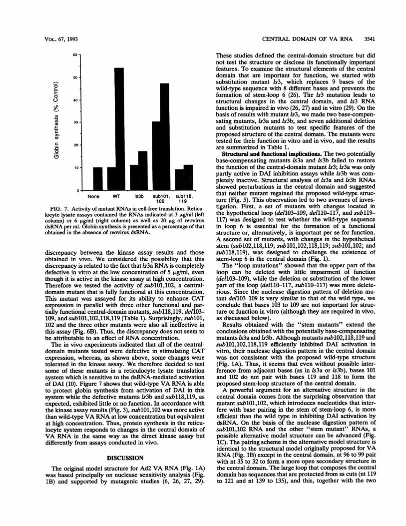
None WT Is3b sub101, subl 18,102 119
FIG. 7. Activity of mutant RNAs in cell-free translation. Reticu-locyte lysate assays contained the RNAs indicated at 3 ,ug/ml (leftcolumn) or 6 ,ug/ml (right column) as well as 20 pg of reovirusdsRNA per ml. Globin synthesis is presented as a percentage of thatobtained in the absence of reovirus dsRNA.
discrepancy between the kinase assay results and thoseobtained in vivo. We considered the possibility that thisdiscrepancy is related to the fact that ls3a RNA is completelydefective in vitro at the low concentration of 5 ,ug/ml, eventhough it is active in the kinase assay at high concentration.Therefore we tested the activity of sublO1,102, a central-domain mutant that is fully functional at this concentration.This mutant was assayed for its ability to enhance CATexpression in parallel with three other functional and par-tially functional central-domain mutants, subll8,119, dellO3-
109, and sublOl,102,118,119 (Table 1). Surprisingly, sublOl,102 and the three other mutants were also all ineffective inthis assay (Fig. 6B). Thus, the discrepancy does not seem tobe attributable to an effect of RNA concentration.The in vivo experiments indicated that all of the central-
domain mutants tested were defective in stimulating CATexpression, whereas, as shown above, some changes weretolerated in the kinase assay. We therefore decided to testsome of these mutants in a reticulocyte lysate translationsystem which is sensitive to the dsRNA-mediated activationof DAI (10). Figure 7 shows that wild-type VA RNA is ableto protect globin synthesis from activation of DAI in thissystem while the defective mutants ls3b and subll8,119, as
expected, exhibited little or no function. In accordance withthe kinase assay results (Fig. 3), sublOl,102 was more activethan wild-type VA RNA at low concentration but equivalentat high concentration. Thus, protein synthesis in the reticu-locyte system responds to changes in the central domain ofVA RNA in the same way as the direct kinase assay butdifferently from assays conducted in vivo.
DISCUSSION
The original model structure for Ad2 VA RNA (Fig. 1A)was based principally on nuclease sensitivity analysis (Fig.1B) and supported by mutagenic studies (6, 26, 27, 29).
These studies defined the central-domain structure but didnot test the structure or disclose its functionally importantfeatures. To examine the structural elements of the centraldomain that are important for function, we started withsubstitution mutant 1s3, which replaces 9 bases of thewild-type sequence with 8 different bases and prevents theformation of stem-loop 6 (26). The 1s3 mutation leads tostructural changes in the central domain, and 1s3 RNAfunction is impaired in vivo (26, 27) and in vitro (29). On thebasis of results with mutant 1s3, we made two base-compen-sating mutants, Is3a and Is3b, and seven additional deletionand substitution mutants to test specific features of theproposed structure of the central domain. The mutants weretested for their function in vitro and in vivo, and the resultsare summarized in Table 1.
Structural and fumctional implications. The two potentiallybase-compensating mutants Is3a and Is3b failed to restorethe function of the central-domain mutant 1s3; Is3a was onlypartly active in DAI inhibition assays while Is3b was com-pletely inactive. Structural analysis of Is3a and Is3b RNAsshowed perturbations in the central domain and suggestedthat neither mutant regained the proposed wild-type struc-ture (Fig. 5). This observation led to two avenues of inves-tigation. First, a set of mutants with changes located inthe hypothetical loop (de1103-109, delllO-117, and subll9-117) was designed to test whether the wild-type sequencein loop 6 is essential for the formation of a functionalstructure or, alternatively, is important per se for function.A second set of mutants, with changes in the hypotheticalstem (sublO2,118,119; sublOl,102,118,119; sublOl,102; andsubll8,119), was designed to challenge the existence ofstem-loop 6 in the central domain (Fig. 1).The "loop mutations" showed that the upper part of the
loop can be deleted with little impairment of function(de1103-109), while the deletion or substitution of the lowerpart of the loop (delllO-117, subllO-117) was more delete-rious. Since the nuclease digestion pattern of deletion mu-tant de1103-109 is very similar to that of the wild type, weconclude that bases 103 to 109 are not important for struc-ture or function in vitro (although they are required in vivo,as discussed below).
Results obtained with the "stem mutants" extend theconclusions obtained with the potentially base-compensatingmutants Is3a and ls3b. Although mutants sublO2,118,119 andsublOl,102,118,119 efficiently inhibited DAI activation invitro, their nuclease digestion pattern in the central domainwas not consistent with the proposed wild-type structure(Fig. 1A). Thus, it seems that even without possible inter-ference from adjacent bases (as in Is3a or Is3b), bases 101and 102 do not pair with bases 119 and 118 to form theproposed stem-loop structure of the central domain.A powerful argument for an alternative structure in the
central domain comes from the surprising observation thatmutant sublOl,102, which introduces nucleotides that inter-fere with base pairing in the stem of stem-loop 6, is moreefficient than the wild type in inhibiting DAI activation bydsRNA. On the basis of the nuclease digestion pattern ofsublO1,102 RNA and the other "stem mutant" RNAs, a
possible alternative model structure can be advanced (Fig.1C). The pairing scheme in the alternative model structure isidentical to the structural model originally proposed for VARNA (Fig. 1B) except in the central domain. nt 96 to 99 pairwith nt 35 to 32 to form a more open secondary structure inthe central domain. The large loop that composes the centraldomain has sequences that are protected from ss cuts (nt 119to 121 and nt 139 to 135), and this, together with the two
VOL. 67, 1993

3542 PE'ERY ET AL.
major nuclease V1 cuts after nt 115 and 131, suggests thattertiary interactions are involved in shaping the centraldomain. More support for a tertiary structure comes frommutant ls3a, in which substitutions in nt 103 to 109 affect thedigestion pattern in loop 2a on the opposite side of the model(Fig. 5). It should be emphasized, however, that othersecondary structures are also compatible with the dataavailable (20). Furthermore, Furtado et al. have suggestedthat a drastically altered VA RNA molecule can rearrange toform an active structure (6). Evidently the critical features ofthe central domain remain to be discovered.
Implications of the discrepancies between in vivo and invitro experiments. Some of the new central-domain mutantsbehaved differently in the in vivo assays used in this study,the d1331 rescue assay and the CAT cotransfection assay,than in the in vitro kinase and reticulocyte assays. Whilewild-type VA RNA increased CAT gene expression by over70-fold, even central-domain mutants that were functional invitro failed to show a similar effect. Mutant sublO1,102,which does not allow the formation of stem-loop 6 and isineffective in vivo, is fully functional in vitro and is evenmore active than the wild type at low concentrations. Thisobservation supports our suggestion that VA RNA adopts analternative structure in which base changes in the centraldomain can be functionally tolerated.What is the structure of the central domain, and why is
there a discrepancy between the in vivo and in vitro assays?Our data do not suggest any additional Watson-Crick basepairing in the central domain of the secondary structureproposed in Fig. 1C, forcing us to invoke other types ofinteraction that can contribute to the tertiary structure.Possibilities include base stacking and hydrogen bonds be-tween bases and phosphates or sugar 2' OH groups. Suchinteractions are seen in yeast phenylalanine-tRNA (14) andin RNA hairpins that contain the GNRA loops found in themost frequently occurring RNA hairpins in 16S and 23SrRNA (13). Tertiary structures are also crucial for functionin the tRNA-like structure at the 3' terminus of turnip yellowmosaic virus RNA and other plant viruses (2, 32). In theseviral RNAs, the formation of a specific three-dimensionalstructure based on an anomalous cloverleaf secondary struc-ture is the key to faithful recognition of the RNA terminus byaminoacyl-tRNA synthetases. It is altogether likely that aspecific three-dimensional structure is necessary for therecognition of VA RNA by DAI.
Several central-domain mutants are active in vitro, asshown here, but no central-domain mutant has been pro-duced so far which is functional in vivo (6, 27; see above).The discrepancy between the two kinds of assay could beexplained by assuming that VA RNA exerts two differentactions, one of which is observed only in vivo. In thereticulocyte lysate translation system, as in the in vitrokinase assay, VA RNA competes with dsRNA for binding tothe RNA-binding site on DAI (9, 17). Previous data showedthat the integrity of the apical stem is essential for thebinding ofVA RNA at low RNA concentrations (26, 29) andthat the central domain may stabilize the binding (1, 7). Thiseffect of VA RNA may be less dependent on an optimalstructure of the central domain, and therefore some basechanges can be accommodated. In the transient-expressionassay, VA RNA increases the expression of a reporter geneat least in part by preferentially enhancing the accumulationof certain mRNAs, probably by regulating their stability onpolysomes (35, 37). This second effect of VA RNA may bedependent on an optimal structure of the central domain,which can be achieved only with the wild-type primary
sequence. It is also possible that some changes in the centraldomain can be tolerated for this function as well but that nosuch changes were included in the central-domain mutantstested. Resolution of these issues must await further analysisof the interactions between RNA and DAI (9, 22), as well asrefinement of the VA RNA structure by new methods (5)including three-dimensional modeling (21) and phylogeneticanalysis (20). An alternative resolution of the discrepancyrecognizes that assays conducted in vitro are influenced bythe assay conditions selected. For instance, deviations in theionic conditions can lead to changes in the RNA structure(1). Moreover, cells contain many potential ligands and otherfactors which may affect the interaction between VA RNAand DAI. Both of these entities are to some extent ribosomeassociated in the cell (17, 34), and a fraction of the VA RNAis also associated with the La antigen (19, 24); furthermore,the related Epstein-Barr virus RNAs (EBERs) are associ-ated with a ribosomal protein (38, 39). Interactions of thissort may place restrictions on the structure of VA RNA suchthat only optimal central-domain structures can be toleratedin vivo. The significance of these additional interactions forVA RNA structure and function is not yet understood.
ACKNOWLEDGMENTS
We thank Aaron Shatkin for reovirus RNA; Lisa Manche fortechnical help; Ronnie Packer for cell culture; and Paul Clarke,Simon Green, and Yuliang Ma for valuable discussions.This work was supported by grant CA13106 from the National
Cancer Institute.
REFERENCES1. Clarke, P. A., T. Pe'ery, Y. Ma, and M. B. Mathews. Unpub-
lished data.2. Dreher, T. W., and T. C. Hall. 1988. Mutational analysis of thetRNA mimicry of brome mosaic virus RNA. J. Mol. Biol.201:41-55.
3. Dunn, A. R., M. B. Mathews, L. T. Chow, and J. SambrooL1978. A supplementary adenoviral leader sequence and its rolein messenger translation. Cell 15:511-526.
4. Fowlkes, D. M., and T. Shenk 1980. Transcriptional controlregions of the adenovirus VAI RNA gene. Cell 22:405-413.
5. Frankel, A. D., I. W. Mattaj, and D. C. Rio. 1991. RNA-proteininteractions. Cell 67:1041-1046.
6. Furtado, M. R., S. Subramanian, R. A. Bhat, D. M. Fowlkes, B.Safer, and B. Thimmappaya. 1989. Functional dissection ofadenovirus VAI RNA. J. Virol. 63:3423-3434.
7. Ghadge, G. D., S. Swaminathan, M. G. Katze, and B. Thimma-paya. 1991. Binding of the adenovirus VAI RNA to the inter-feron-induced 68 kDA protein kinase correlates with function.Proc. Natl. Acad. Sci. USA 88:7140-7144.
8. Graham, F. L., J. Smiley, W. C. Russell, and R. Nairn. 1977.Characteristics of a human cell line transformed by DNA fromhuman adenovirus type 5. J. Gen. Virol. 36:59-72.
9. Green, S. R., and M. B. Mathews. 1992. Two RNA bindingmotifs in the double-stranded RNA activated protein kinase,DAI. Genes Dev. 6:2478-2490.
10. Gunnery, S., S. R. Green, and M. B. Mathews. 1992. HIV-1TAR RNA stimulates protein synthesis in vivo and in vitro:relationship between structure and function. Proc. Natl. Acad.Sci. USA 89:11557-11561.
11. Gunnery, S., A. P. Rice, H. D. Robertson, and M. B. Mathews.1990. Tat-responsive region RNA of human immunodeficiencyvirus 1 can prevent activation of the double-stranded-RNA-activated protein kinase. Proc. Natl. Acad. Sci. USA 87:8687-8691.
12. HerLmann, C., C. V. D6ry, and M. B. Mathews. 1987. Transac-tivation of host and viral genes by the adenovirus E1B 19Ktumor antigen. Oncogene 2:25-35.
J. VIROL.
CENTRAL DOMAIN OF VA RNA 3543
13. Heus, H. A., and A. Pardi. 1991. Structural features that giverise to the unusual stability of RNA hairpins containing GNRAloops. Science 253:191-194.
14. Holbrook, S. H., J. L. Suyssman, R. Wade Warrant, and K.Sung-Hou. 1978. Crystal structure of yeast phenylalanine trans-fer RNA. J. Mol. Biol. 123:631-660.
15. Jacobson, A., L. Good, J. Simonetti, and M. Zuker. 1984. Somesimple computational methods to improve the folding of largeRNAs. Nucleic Acids Res. 12:45-66.
16. Kaufman, R. J., and P. Murtha. 1987. Translational controlmediated by eukaryotic initiation factor-2 is restricted to spe-cific mRNAs in transfected cells. Mol. Cell. Biol. 7:1568-1571.
17. Kostura, M., and M. B. Mathews. 1989. Purification and activa-tion of the double-stranded RNA-dependent eIF-2 kinase DAI.Mol. Cell. Biol. 9:1576-1586.
18. Kunkel, T. A. 1985. Rapid and efficient site-specific mutagenesiswithout phenotypic selection. Proc. Natl. Acad. Sci. USA82:488-492.
19. Lerner, M. R., J. A. Boyle, J. A. Hardin, and J. A. Steitz. 1981.Two novel classes of small ribonucleoproteins detected byantibodies associated with lupus erythematosus. Science 211:400-402.
20. Ma, Y., and M. B. Mathews. Unpublished data.21. Major, F., M. Turcotte, D. Gantheret, G. Lapolme, E. Fillion,
and R. Cedergren. 1991. The combination of symbolic andnumerical computation for three-dimensional modeling of RNA.Science 253:1255-1260.
22. Manche, L., S. R. Green, C. Schmedt, and M. B. Mathews. 1992.Interactions between double-stranded RNA regulators and theprotein kinase DAI. Mol. Cell. Biol. 12:5238-5248.
23. Mathews, M. B. 1993. Viral evasion of cellular defense mecha-nisms: regulation of the protein kinase DAI by RNA effectors.Semin. Virol. 4:247-258.
24. Mathews, M. B., and A. M. Francoeur. 1984. La antigenrecognizes and binds to the 3'-oligouridylate tail of a smallRNA. Mol. Cell. Biol. 4:1134-1140.
25. Mathews, M. B., and T. Shenk. 1991. Adenovirus virus-associ-ated RNA and translational control. J. Virol. 65:5657-5662.
26. Mellits, K. H., M. Kostura, and M. B. Mathews. 1990. Interac-tion of adenovirus VA RNA, with the protein kinase DAI:non-equivalence of binding and function. Cell 61:843-852.
27. Mellits, K. H., and M. B. Mathews. 1988. Effects of mutations instem and loop regions on the structure and function of adeno-virus VA RNA,. EMBO J. 7:2849-2859.
28. Mellits, K. H., T. Pe'ery, L. Manche, H. D. Robertson, and
M. B. Mathews. 1990. Removal of double-stranded contami-nants from RNA transcripts: synthesis of adenovirus VA RNA,from a T7 vector. Nucleic Acids Res. 18:5401-5406.
29. Mellits, K. H., T. Pe'ery, and M. B. Mathews. 1992. Structureand function of adenovirus VA RNA: Role of the apical stem. J.Virol. 66:2369-2377.
30. PleU, C. W. A., K. Reitveld, and L. Bosch. 1985. A new principleof RNA folding based on pseudoknotting. Nucleic Acids Res.13:1717-1731.
31. Puglisi, J. D., J. R. Wyatt, and I. Tinoco, Jr. 1988. Apseudoknotted RNA oligonucleotide. Nature (London) 321:283-286.
32. Rietveld, K., R. VanPelgeest, C. W. A. PleU, and L. Bosch. 1982.The tRNA-like structure at the 3' terminus of turnip yellowmosaic virus RNA. Differences and similarities with canonicaltRNA. Nucleic Acids Res. 10:1929-1946.
33. Sanger, F., S. Nicklen, and A. R. Coulson. 1977. DNA sequenc-ing with chain-terminating inhibitors. Proc. Natl. Acad. Sci.USA 74:5463-5467.
34. Schneider, R. J., C. Weinberger, and T. Shenk 1984. Adenovi-rus VAI RNA facilitates the initiation of translation in virus-infected cells. Cell 37:291-298.
35. Strijker, R., D. T. Fritz, and A. D. Levinson. 1989. AdenovirusVAI-RNA regulates gene expression by controlling stability ofribosome-bound RNAs. EMBO J. 8:2669-2675.
36. Svensson, C., and G. Akusjarvi. 1985. Adenovirus VA RNA,mediates a translational stimulation which is not restricted tothe viral mRNAs. EMBO J. 4:957-964.
37. Svensson, C., and G. Akusjirvi. 1990. A novel effect of adeno-virus VA RNA, on cytoplasmic mRNA abundance. Virology174:613-617.
38. Toczysk, D. P. W., and J. Steitz. 1991. EAP, a highly conservedcellular protein associated with Epstein-Barr virus small RNAs(EBERs). EMBO J. 10:459-466.
39. Toczysk, D. P. W., J. Steitz, and I. Wool. 1993. Personalcommunication.
40. Wu, G.-J., J. F. Railey, and R. E. Cannon. 1987. Defining thefunctional domains in the control region of the adenovirus type2 specific VA RNA1 gene. J. Mol. Biol. 194:423-442.
41. Zoller, M. J., and M. Smith. 1984. Oligonucleotide-directedmutagenesis: a simple method using two oligonucleotide prim-ers and a single-stranded DNA template. DNA 3:479-488.
42. Zuker, M., and P. Stiegler. 1981. Optimal computer folding oflarge RNA sequences using thermodynamics and auxiliaryinformation. Nucleic Acids Res. 9:133-148.
VOL. 67, 1993