mutations. some definitions 1. mutation any change in the dna which if occurs in germ cells is...
TRANSCRIPT

Mutations
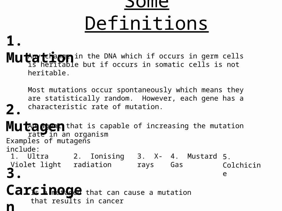
Some Definitions1. Mutation
Any change in the DNA which if occurs in germ cells is heritable but if occurs in somatic cells is not heritable.
Most mutations occur spontaneously which means they are statistically random. However, each gene has a characteristic rate of mutation.
2. MutagenAn agent that is capable of increasing the mutation rate in an organism
Examples of mutagens include:
1. Ultra Violet light 2. Ionising radiation 3. X-rays 4. Mustard Gas 5. Colchicine
3. CarcinogenIs a mutagen that can cause a mutation that results in cancer
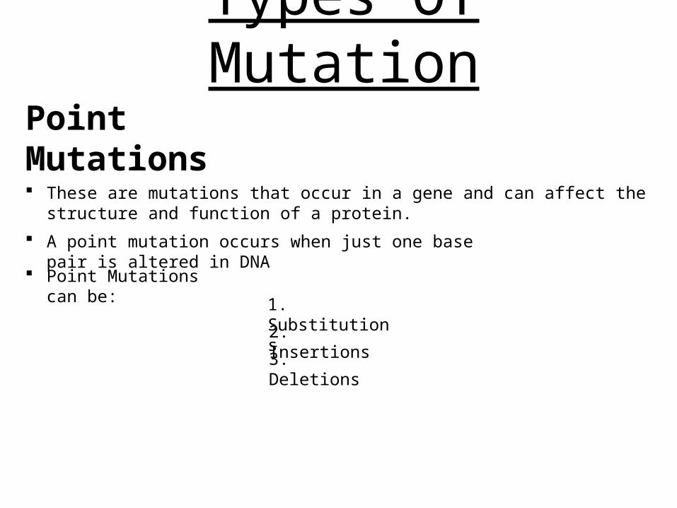
Types Of MutationPoint Mutations
These are mutations that occur in a gene and can affect the structure and function of a protein.
A point mutation occurs when just one base pair is altered in DNA
Point Mutations can be:
1. Substitutions
2. Insertions
3. Deletions
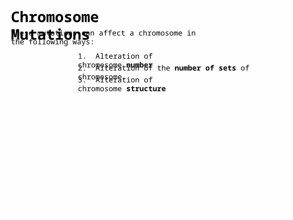
Chromosome MutationsThese mutations can affect a chromosome in the following ways:
1. Alteration of chromosome number
3. Alteration of chromosome structure
2. Alteration of the number of sets of chromosome.
Point Mutations
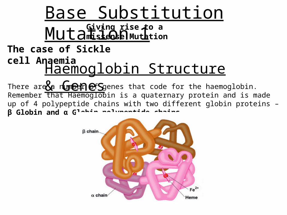
Base Substitution Mutation 1
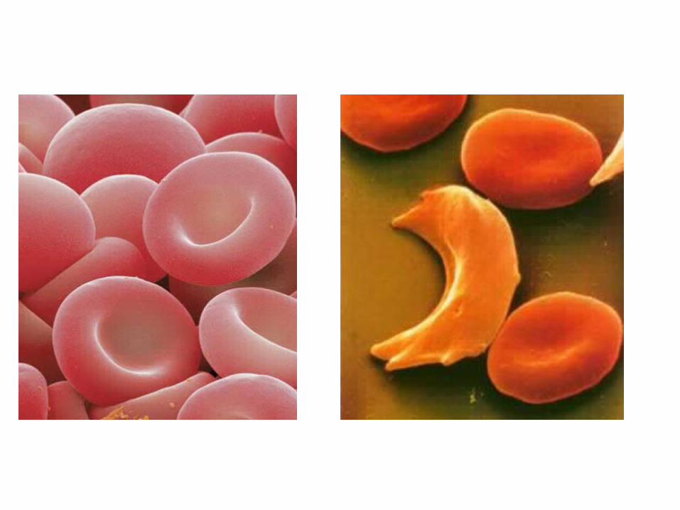
The case of Sickle cell Anaemia
Haemoglobin Structure & GenesThere are a number of genes that code for the haemoglobin. Remember that Haemoglobin is a quaternary protein and is made up of 4 polypeptide chains with two different globin proteins – β Globin and α Globin polypeptide chains.
Giving rise to a missense Mutation
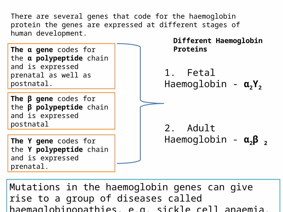
There are several genes that code for the haemoglobin protein the genes are expressed at different stages of human development.
The α gene codes for the α polypeptide chain and is expressed prenatal as well as postnatal.
The β gene codes for the β polypeptide chain and is expressed postnatal
The ϒ gene codes for the ϒ polypeptide chain and is expressed prenatal.
Different Haemoglobin Proteins
1. Fetal Haemoglobin - α2ϒ2
2. Adult Haemoglobin - α2β 2
Mutations in the haemoglobin genes can give rise to a group of diseases called haemaglobinopathies, e.g. sickle cell anaemia.
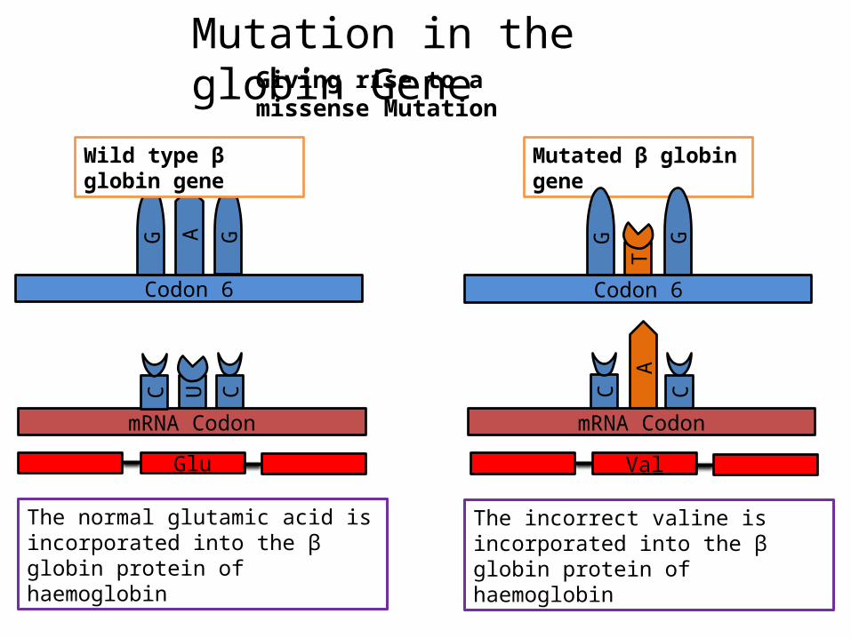
Codon 6
AG G
Wild type β globin gene
Glu
The normal glutamic acid is incorporated into the β globin protein of haemoglobin
Mutated β globin gene
Codon 6
G G
T
CU
mRNA Codon
C C
mRNA Codon
C
A
Val
The incorrect valine is incorporated into the β globin protein of haemoglobin
Mutation in the globin GeneGiving rise to a missense Mutation
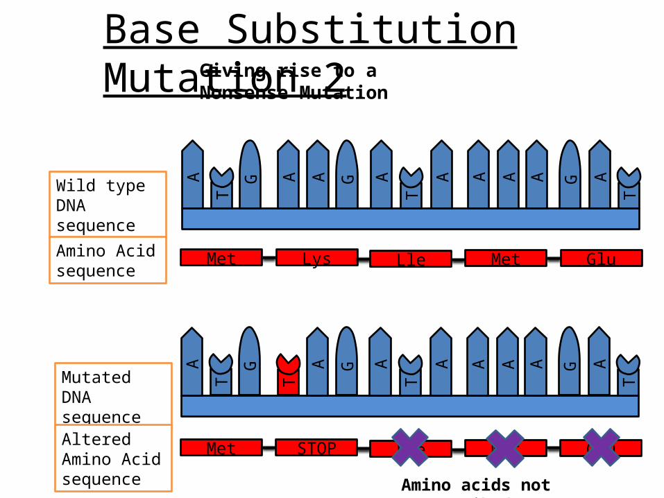
Base Substitution Mutation 2Giving rise to a Nonsense Mutation
Met Lys Lle Met Glu
T
A GG
T
GA A
T
A A A A A
T
Wild type DNA sequence
Amino Acid sequence
Mutated DNA sequence
A GG
T
GA A
T
A A A A A
T
A
Met STOP Lle Met GluAltered Amino Acid sequence
Amino acids not transcribed

Base Substitution Mutation 3Giving rise to a Silent Mutation
Met Lys Lle Met Glu
Wild type DNA sequence
Amino Acid sequence
A GG
T
GA A
T
A A A A A
T
A
A GG
T
GA A
T
A A A A AA G
Mutated DNA sequence
Amino Acid sequence
Met Lys Lle Met Glu
Amino Acid still transcribed
Insertion and deletion mutations
Insertion and deletion mutations have a disastrous effect on the protein more often than substitution mutations.
Insertion and deletion mutations result when nucleotide pairs are either inserted or deleted from a gene.
As the genetic code is read as codons, if the insertion or deletion of nucleotide pairs is not in multiples of three a frameshift mutation can occur.
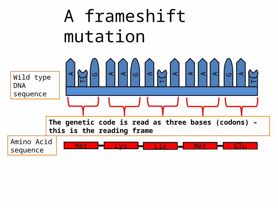
Met Lys Lle Met Glu
Wild type DNA sequence
Amino Acid sequence
A GG
T
GA A
T
A A A A A
T
A
A frameshift mutation
The genetic code is read as three bases (codons) – this is the reading frame
Wild type DNA sequence
A GG
T
GA A
T
A A A A A
T
A
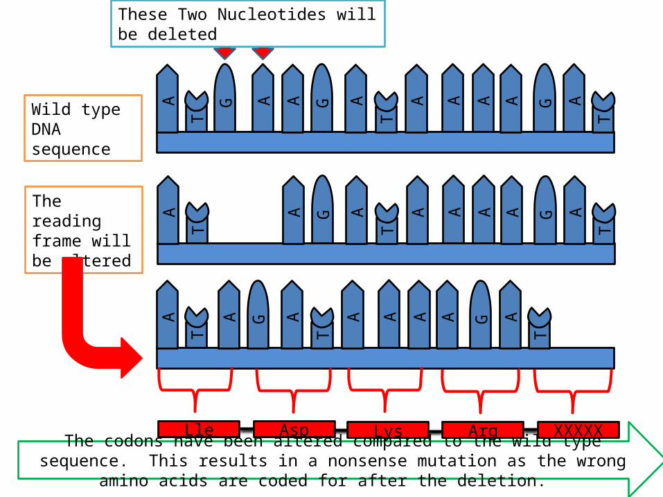
These Two Nucleotides will be deleted
GG
T
A A
T
A A A A A
T
A
GG
T
A A
T
A A A A A
T
A
The reading frame will be altered
The codons have been altered compared to the wild type sequence. This results in a nonsense mutation as the wrong amino acids are coded for after the deletion.
Lle Asp Lys Arg XXXXX
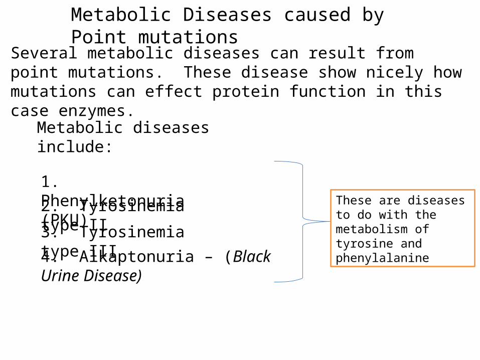
Metabolic Diseases caused by Point mutations
Several metabolic diseases can result from point mutations. These disease show nicely how mutations can effect protein function in this case enzymes.
Metabolic diseases include:
1. Phenylketonuria (PKU)2. Tyrosinemia type II
3. Tyrosinemia type III
4. Alkaptonuria – (Black Urine Disease)
These are diseases to do with the metabolism of tyrosine and phenylalanine
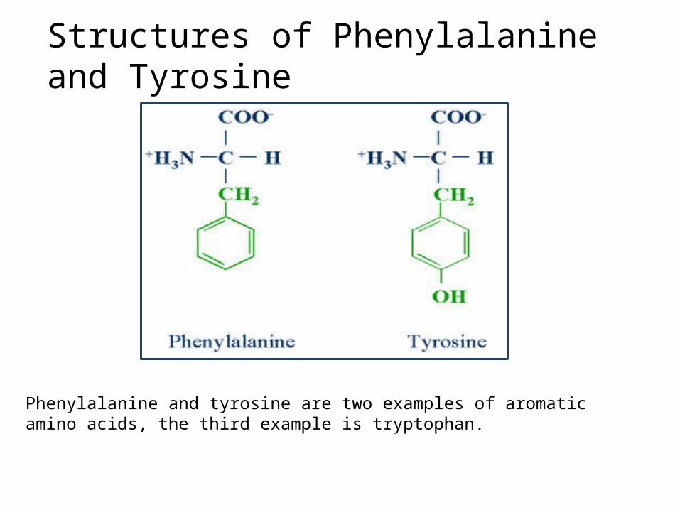
Structures of Phenylalanine and Tyrosine
Phenylalanine and tyrosine are two examples of aromatic amino acids, the third example is tryptophan.
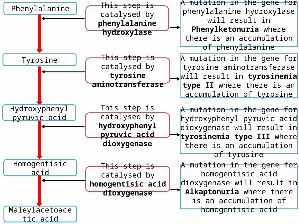
Phenylalanine
Tyrosine
This step is catalysed by phenylalanine
hydroxylase
A mutation in the gene for phenylalanine hydroxylase will result in Phenylketonuria where there is an
accumulation of phenylalanine
Hydroxyphenyl pyruvic acid
This step is catalysed by tyrosine aminotransferase
Homogentisic acid
Maleylacetoacetic acid
A mutation in the gene for tyrosine aminotransferase will result in
tyrosinemia type II where there is an accumulation of tyrosine
This step is catalysed by hydroxyphenyl pyruvic
acid dioxygenase
A mutation in the gene for hydroxyphenyl pyruvic acid
dioxygenase will result in tyrosinemia type III where there is an accumulation of tyrosine
This step is catalysed by homogentisic acid
dioxygenase
A mutation in the gene for homogentisic acid dioxygenase will
result in Alkaptonuria where there is an accumulation of homogentisic acid
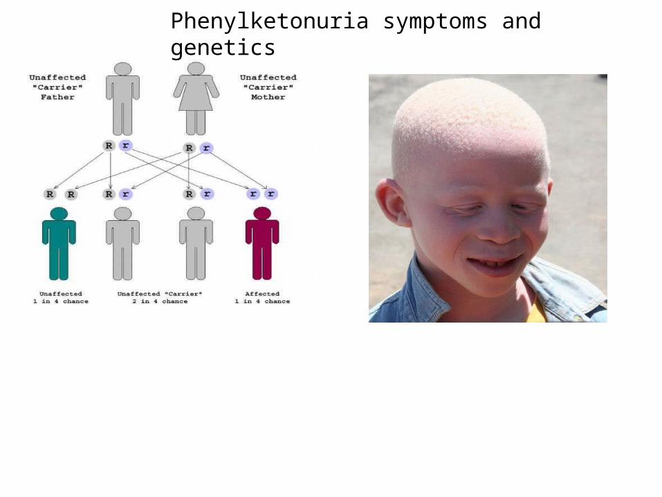
Phenylketonuria symptoms and genetics
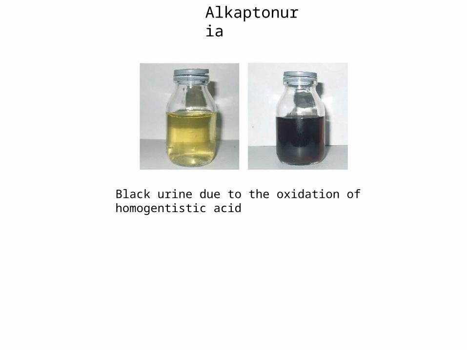
Alkaptonuria
Black urine due to the oxidation of homogentistic acid
Cystic fibrosis
An unusual frameshift mutation – the case of muscular dystrophy
There are two main forms of muscular dystrophy:
1. Duchenne muscular dystrophy (DMD).
2. Becker muscular dystrophy (BMD).
Both are mainly X linked recessive disorders in which skeletal muscle and to some extent cardiac and smooth muscles waste away.
DMD onset occurs in early child hood and is very severe. Suffers are in wheelchairs before the age of 11.
BMD onset is later and the condition is much milder then with DMD and has a slower progression of muscle weakness.
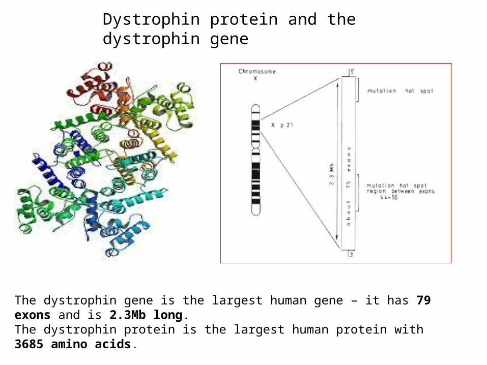
Dystrophin protein and the dystrophin gene
The dystrophin gene is the largest human gene – it has 79 exons and is 2.3Mb long.The dystrophin protein is the largest human protein with 3685 amino acids.
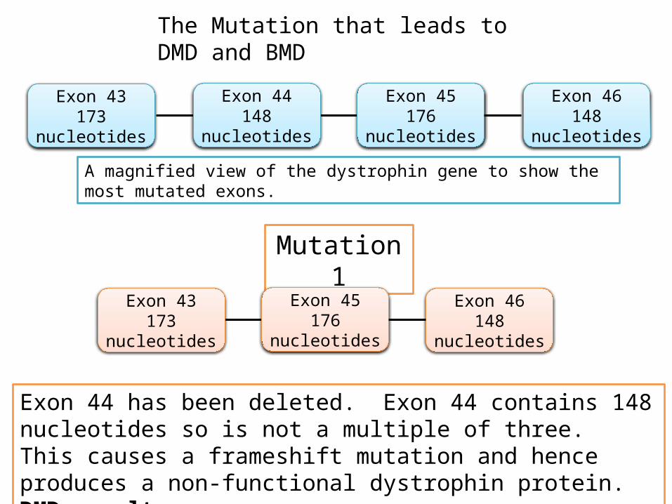
The Mutation that leads to DMD and BMD
Exon 43173 nucleotides
Exon 44148 nucleotides
Exon 45176 nucleotides
Exon 46148 nucleotides
A magnified view of the dystrophin gene to show the most mutated exons.
Mutation 1
Exon 43173 nucleotides
Exon 45176 nucleotides
Exon 46148 nucleotides
Exon 44 has been deleted. Exon 44 contains 148 nucleotides so is not a multiple of three. This causes a frameshift mutation and hence produces a non-functional dystrophin protein. DMD results.
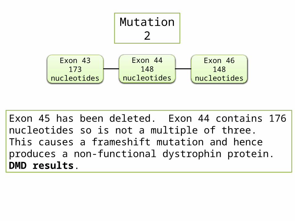
Mutation 2
Exon 43173 nucleotides
Exon 44148 nucleotides
Exon 46148 nucleotides
Exon 45 has been deleted. Exon 44 contains 176 nucleotides so is not a multiple of three. This causes a frameshift mutation and hence produces a non-functional dystrophin protein. DMD results.
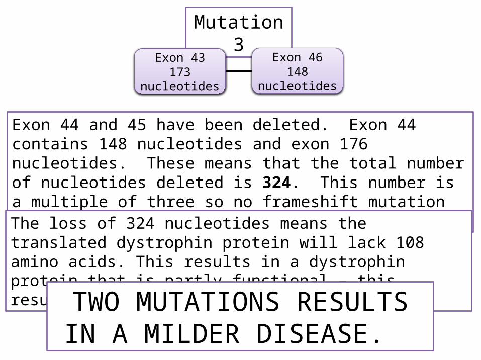
Mutation 3
Exon 43173 nucleotides
Exon 46148 nucleotides
Exon 44 and 45 have been deleted. Exon 44 contains 148 nucleotides and exon 176 nucleotides. These means that the total number of nucleotides deleted is 324. This number is a multiple of three so no frameshift mutation occurs.
The loss of 324 nucleotides means the translated dystrophin protein will lack 108 amino acids. This results in a dystrophin protein that is partly functional – this results in BMD.
TWO MUTATIONS RESULTS IN A MILDER DISEASE.
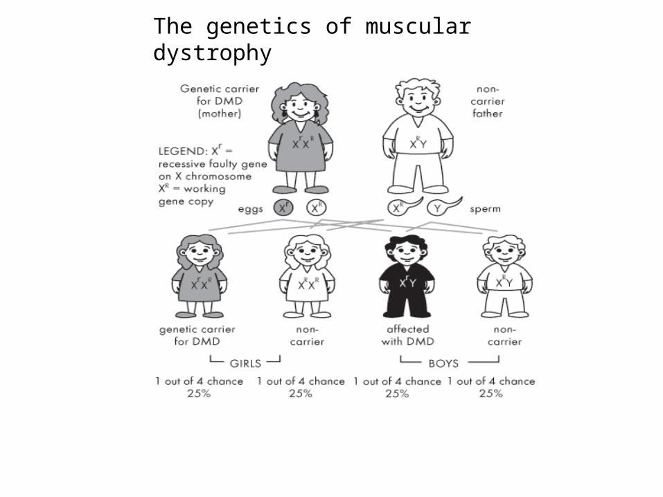
The genetics of muscular dystrophy
Chromosome Mutations
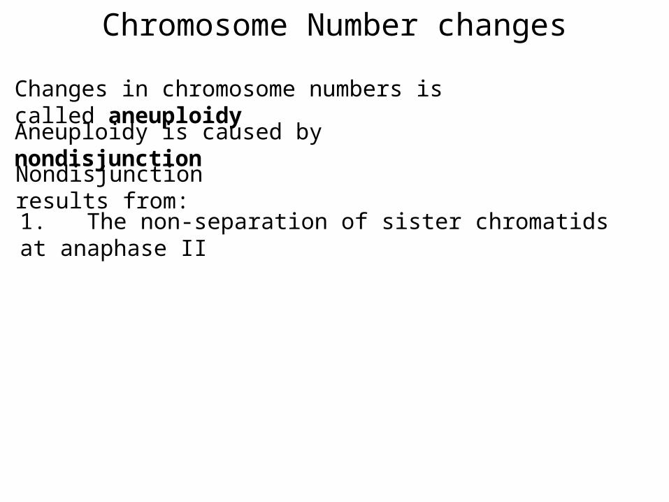
Chromosome Number changes
Changes in chromosome numbers is called aneuploidy
Aneuploidy is caused by nondisjunction
Nondisjunction results from:
1. The non-separation of sister chromatids at anaphase II
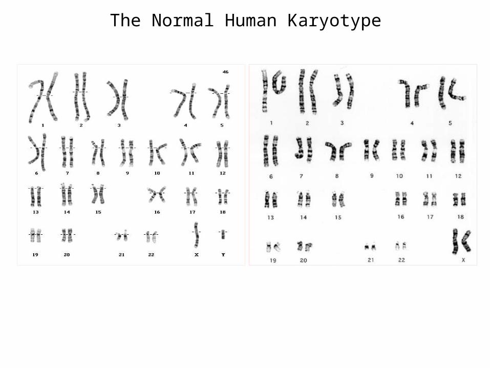
The Normal Human Karyotype
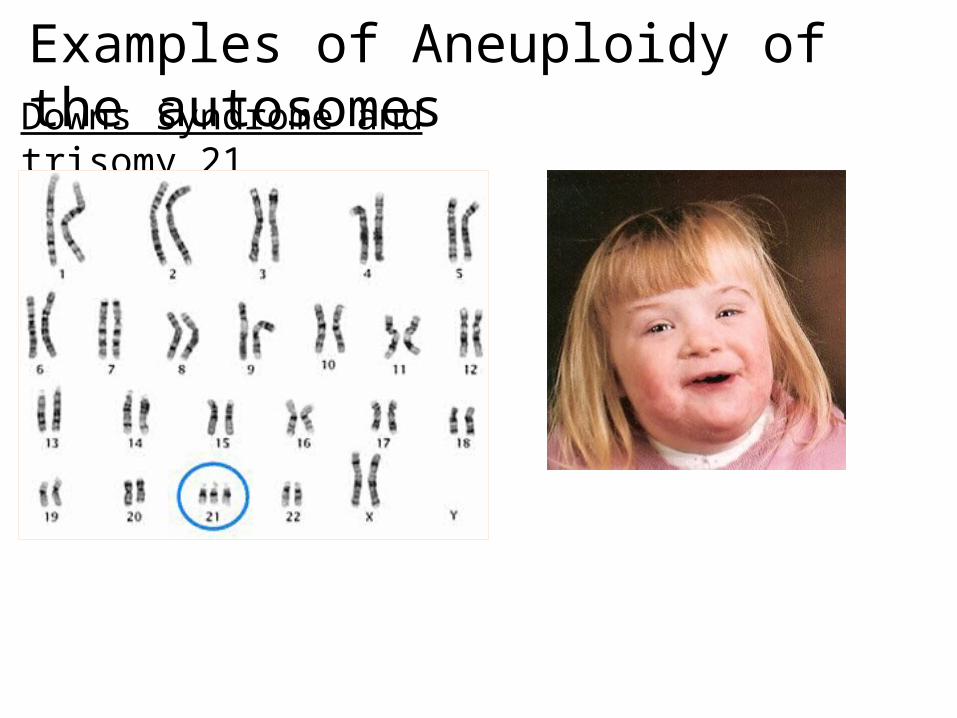
Downs Syndrome and trisomy 21
Examples of Aneuploidy of the autosomes
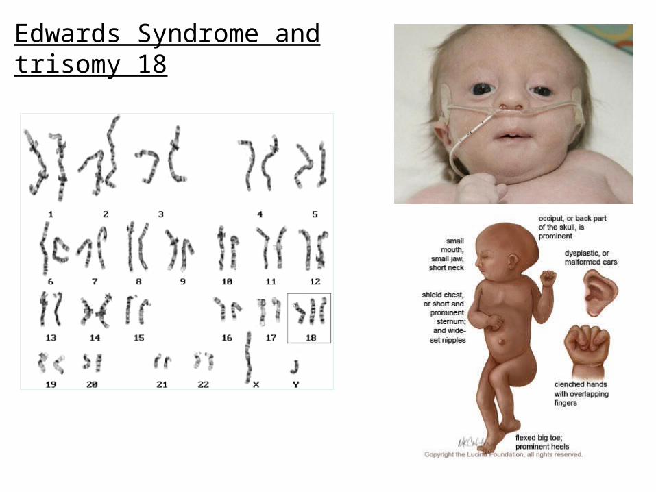
Edwards Syndrome and trisomy 18
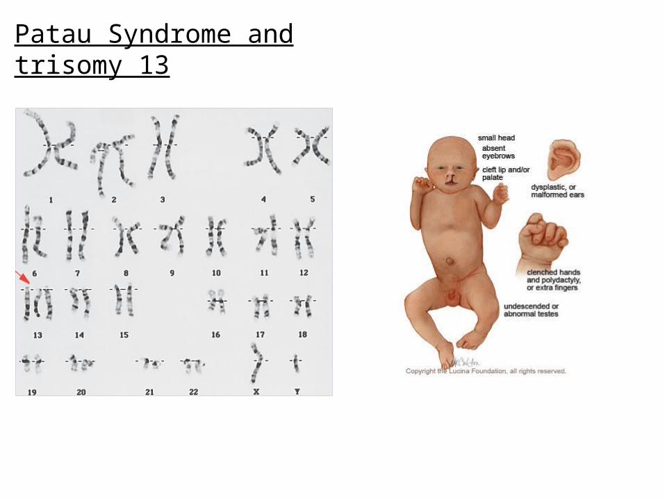
Patau Syndrome and trisomy 13
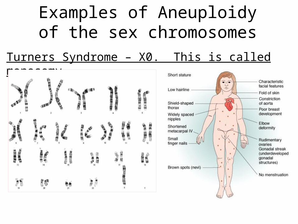
Examples of Aneuploidy of the sex chromosomes
Turners Syndrome – X0. This is called monosomy
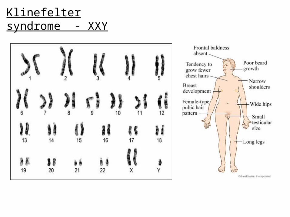
Klinefelter syndrome - XXY
Changes in whole sets of chromosomes
Changes in whole sets of chromosomes is called polyploidy and occurs because of non- disjunction in anaphase I.
The non-separation of homologous chromosomes at anaphase I
Summary video of non-disjunction
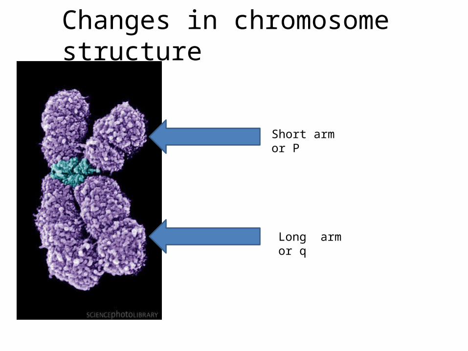
Short arm or P
Long arm or q
Changes in chromosome structure
A B DC E F G H
A DC E F G H
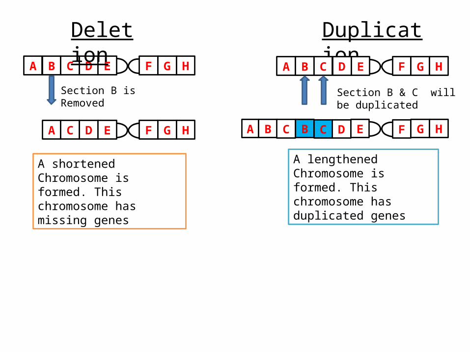
Deletion
Section B is Removed
A shortened Chromosome is formed. This chromosome has missing genes
Duplication
A B DC E F G H
Section B & C will be duplicated
A B DC E F G HB C
A lengthened Chromosome is formed. This chromosome has duplicated genes
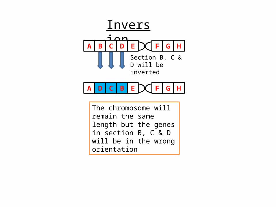
Inversion
A B DC E F G H
Section B, C & D will be inverted
A D BC E F G H
The chromosome will remain the same length but the genes in section B, C & D will be in the wrong orientation
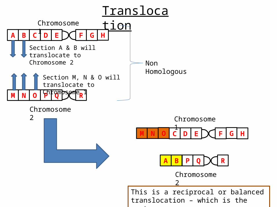
Translocation
A B DC E F G H
M N PO Q R
Non Homologous
Section A & B will translocate to Chromosome 2
Chromosome 1
Chromosome 2
Section M, N & O will translocate to Chromosome 1
DC E F G HM N O
A B P Q R
Chromosome 1
Chromosome 2
This is a reciprocal or balanced translocation – which is the most common.
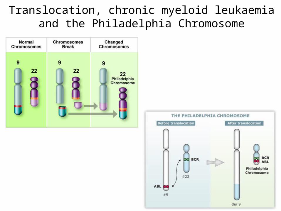
Translocation, chronic myeloid leukaemia and the Philadelphia Chromosome
How the Philadelphia Chromosome causes cancer
The ABL Gene
Summary Video of changes in chromosome structure
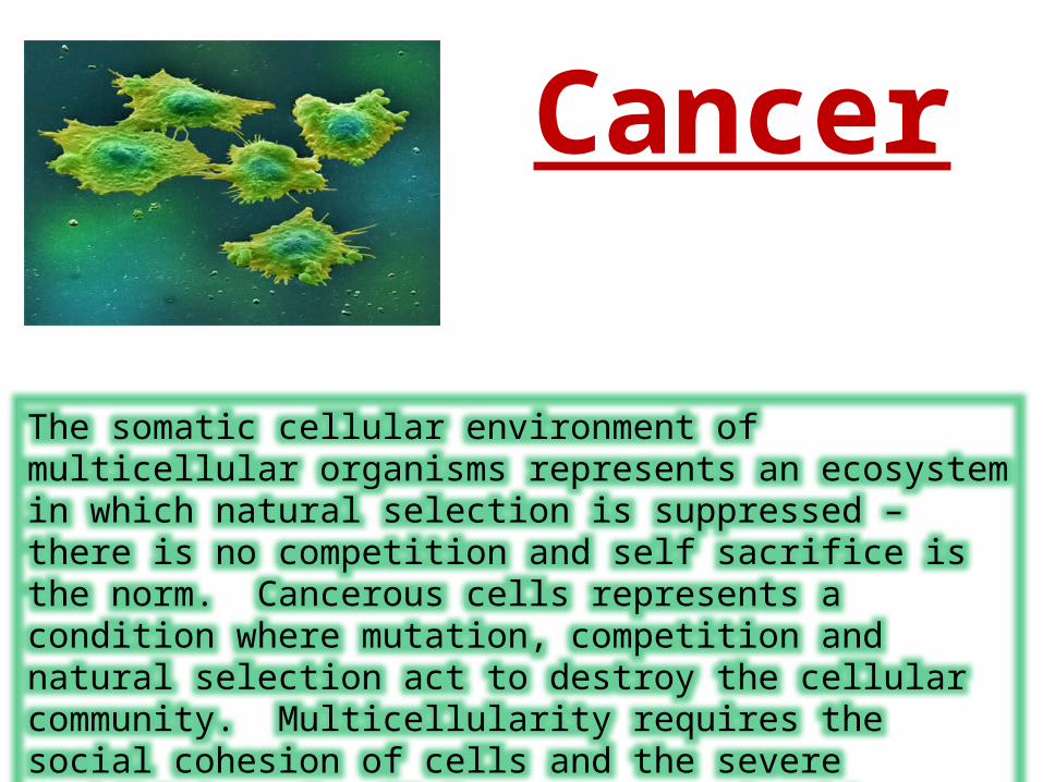
The somatic cellular environment of multicellular organisms represents an ecosystem in which natural selection is suppressed – there is no competition and self sacrifice is the norm. Cancerous cells represents a condition where mutation, competition and natural selection act to destroy the cellular community. Multicellularity requires the social cohesion of cells and the severe prohibition of cellular autonomy; in this respect cancer seems to be a reversion to a unicellular selfishness.
Cancer
A Definition Of Cancer
There is no one “all encompassing” definition of cancer
•The reason for this is that cancer is not one disease – there are approximately 200 different types of cancer.
•Each individual Cancer, therefore, has its own unique properties and characteristics.
•Despite this, however, all cancers share some common features that we can use to construct a definition.
Cancer is a disorder of cell division and regulation that results in uncontrolled growth and proliferation of cells resulting in the formation of a tumour that has the capacity leave their organ of origin and travel in the blood to form secondary tumours in distant organs
The development of cancer (carcinogenesis)
•A cell does not become cancerous due to a single event. NO one mutation will cause cancer.
•Carcinogenesis is, therefore, described as a multi-stage process which occurs over many years and the number of mutations accumulate.
Oncogenes and tumour suppressor genes
An oncogene is a gene that, if mutated, becomes active and causes the cell to become cancerous.
This is called a gain of function mutation
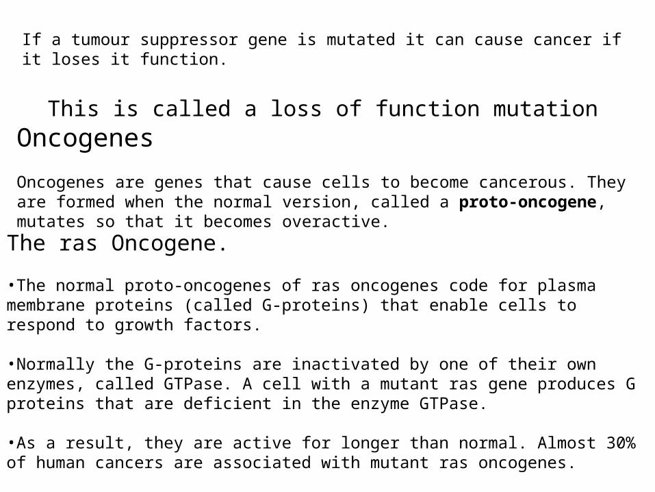
If a tumour suppressor gene is mutated it can cause cancer if it loses it function.
This is called a loss of function mutation
Oncogenes
Oncogenes are genes that cause cells to become cancerous. They are formed when the normal version, called a proto-oncogene, mutates so that it becomes overactive.
The ras Oncogene.
•The normal proto-oncogenes of ras oncogenes code for plasma membrane proteins (called G-proteins) that enable cells to respond to growth factors.
•Normally the G-proteins are inactivated by one of their own enzymes, called GTPase. A cell with a mutant ras gene produces G proteins that are deficient in the enzyme GTPase. •As a result, they are active for longer than normal. Almost 30% of human cancers are associated with mutant ras oncogenes.
The myc oncogene
•The myc proto-oncogene is located on human chromosome 8.
•The protein encoded by the myc proto-oncogene stimulates transcription of genes required for cell division.
•In a common mutation, this proto-oncogene switches from chromosome 8 to a site on chromosome 14 – this is called translocation.
•In its new position, the gene acts as an oncogene because it is over expressed and over-stimulates cell division.
Tumour Suppressor genes
•Tumour suppressor genes are also associated with cell division. They act in a different way to oncogenes.
•Whereas proto-oncogenes are converted to oncogenes by mutations that increase the gene’s activity, tumour suppressor genes are converted to oncogenes by mutations that reduce their normal activity.
•A normal, unmutated tumour suppressor gene inhibits cell division.
The Case of Retinoblastoma
Retinoblastoma is a childhood tumour of the eye and is an example of a cancer that is caused by the loss of a tumour suppressor gene. In this case the tumour suppressor gene, called RB1, is located on human chromosome 13: it inhibits the transcription of proto-oncogenes such as myc.
With a mutated RB1, the myc gene becomes overactive and a tumour results.
P53
The guardian of the genome
The Cellular Caretaker
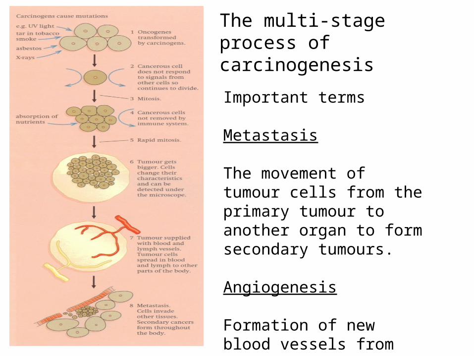
The multi-stage process of carcinogenesis
Important terms
Metastasis
The movement of tumour cells from the primary tumour to another organ to form secondary tumours.
Angiogenesis
Formation of new blood vessels from pre-existing ones.
Tumour Suppressor genes and Oncogenes
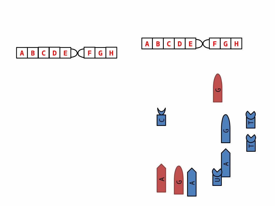
TT
GA
A G
G
A U
C
A B DC E F G HA B DC E F G H