mycoplasma diseases of ruminants
TRANSCRIPT


MYCOPLASMA DISEASES OF RUMINANTS

This page intentionally left blank

CABI is a trading name of CAB International
CABI Head Offi ce CABI North American Offi ceNosworthy Way 875 Massachusetts AvenueWallingford 7th FloorOxfordshire OX10 8DE Cambridge, MA 02139UK USA
Tel: +44 (0)1491 832111 Tel: +1 617 395 4056Fax: +44 (0)1491 833508 Fax: +1 617 354 6875E-mail: [email protected] E-mail: [email protected]: www.cabi.org
© CAB International 2008. All rights reserved. No part of this publication may be reproduced in any form or by any means, electronically, mechanically, by photocopying, recording or otherwise, without the prior permission of the copyright owners.
A catalogue record for this book is available from the British Library, London, UK.
Library of Congress Cataloging-in-Publication DataNicholas, Robin, 1951- Mycoplasma diseases of ruminants / Robin Nicholas, Roger Ayling, and Laura McAuliffe. p.; cm. Includes bibliographical references and index. ISBN 978-0-85199-012-5 (alk. paper) 1. Ruminants–Diseases. 2. Mycoplasma diseases in animals. I. Ayling, Roger. II. McAuliffe, Laura. III. C.A.B. International. IV. Title. [DNLM: 1. Mycoplasma Infections–veterinary. 2. Ruminants.3. Mycoplasma–physiology. 4. Mycoplasma Infections--diagnosis.SF 997.5.R86 N599m 2008]
SF809.M9N53 2008 636.2089'692–dc22 2008018903
ISBN-13: 978 0 85199 012 5
Typeset by AMA DataSet Ltd, UK.Printed and bound in the UK by Biddles Ltd, Kings Lynn, Norfolk.

v
Contents
Contributors vii
Preface viii
Acknowledgements x
PART I: METHODS 1
Chapter 1: Isolation and Growth of Mycoplasmas from Ruminants 3
Chapter 2: Detection and Differentiation of Mycoplasma Species Using PCR/Denaturing Gradient Gel Electrophoresis 15
Chapter 3: Detection of Mycoplasma Species Using Polymerase Chain Reaction (PCR) 28
Chapter 4: Molecular Typing of Mycoplasma Species 35
Chapter 5: Antigenic Analysis of Mycoplasmas 53
Chapter 6: Antimicrobial Sensitivity Testing 58
PART II: DISEASES 67
Chapter 7: Contagious Bovine Pleuropneumonia 69
Chapter 8: Contagious Agalactia 98
Chapter 9: Contagious Caprine Pleuropneumonia 114

Contentsvi
Chapter 10: Bovine Respiratory Disease 132 Overview 132 Diseases caused by Mycoplasma bovis 133 Other bovine mycoplasmoses 154
Chapter 11: Respiratory Diseases of Small Ruminants 169 Overview 169 Atypical pneumonia of sheep and goats 171 Diseases caused by Mycoplasma mycoides subsp. capri and Mycoplasma mycoides subsp. mycoides LC 179
In vitro susceptibility of small ruminant mycoplasmas to antimicrobials 190
Chapter 12: Eye Infections of Ruminants 199 Mycoplasma bovoculi 200 Mycoplasma conjunctivae 202
Chapter 13: Reproductive Diseases of Cattle 208 Mycoplasma bovigenitalium 208 Ureaplasmas 215
Chapter 14: New, Emerging and Unculturable Mycoplasmas in Ruminants 225
Index 233
The colour plate section can be found following p.118.

vii
Contributors
The following made a major contribution to parts of this book:
The Mycoplasma Group at the Veterinary Laboratories Agency, in particular Joanna Lawes, Katie Parham and Colin Churchward (identifi cation and molec-ular typing)
Guido Ruggero Loria, Istituto Zooprofi lattico Sperimentale, Palermo, Italy (conta-gious agalactia, eye infections)
Rosário Gonçalves, Laboratório Nacional Investigaçao Veterinaria, Estrada di Benfi ca, Lisbon, Portugal (immunoblotting, contagious bovine pleuropneumonia and atypical pneumonia)
Otto J.B. Hubschle, Chief Veterinary Offi cer, Windhoek, Namibia (contagious bovine pleuropneumonia)
Umit Ozdemir, Pendik Veterinary Control and Research Institute, Pendik, Istanbul, Turkey (contagious caprine pleuropneumonia)
Massimo Scacchia, Istituto Zooprofi lattico Sperimentale, Teramo, Italy (contagious bovine pleuropneumonia and caprine respiratory diseases)
José Regalla, formerly of the Laboratório Nacional di Investigaçao Veterimaria, Estrada di Benfi ca, Lisbon, Portugal (contagious bovine pleuropneumonia)
Mark Fielder, Kingston University, Penrhyn Road, Kingston, Surrey, UK (atypical pneumonia)
Former PhD students: Bilal Houshaymi, Waleed Al-Momani, L. Ali Khan, Masoud Shahram, Paola de Santis and Yi Ching Lin of King’s College London; and Hiren Patel of Kingston University (many aspects)
And the late Roger Miles of King’s College London (many aspects)

viii
Preface
Mycoplasma diseases of livestock such as contagious bovine pleuropneumonia (CBPP), contagious caprine pleuropneumonia (CCPP) and contagious agalactia still pose major problems for animal health authorities worldwide. CBPP remains one of the biggest impediments to cattle farming in many countries in sub- Saharan Africa. New regions and new animal species have been infected by CCPP since the turn of the century and little effective control exists for conta-gious agalactia, which is endemic in sheep and goat populations in countries surrounding the Mediterranean and in the Middle East, this despite a major research effort stimulated by the European Union following the unexpected reappearance of CBPP in Italy in the early 1990s. Sadly, and probably as a result of the eradication of CBPP from Europe at the end of the 20th century, many groups active in mycoplasma research a decade ago have had to develop research interests elsewhere because of fi nancial constraints. Consequently, the develop-ment of improved vaccines and better treatments for mycoplasma diseases in developing countries now move more slowly than ever. However, many signifi -cant improvements have been seen as a result of research into these degenerate bacteria, mainly in the area of diagnosis, with the availability of exquisite new molecular tools to detect minute quantities of these pathogens, some of them highly fastidious or unculturable and often in mixed bacterial culture, directly from clinical samples. Epidemiological fi ngerprinting tools are now available to identify the likely origins of outbreaks as well as providing fascinating insights into the evolution of these organisms. Fundamental research on biofi lm produc-tion has also shown for the fi rst time how these fragile organisms can withstand hostile environments and antimicrobials. The last decade has also shed light on other mycoplasmas, most importantly Mycoplasma bovis, which causes a whole range of clinical conditions, including calf pneumonia, mastitis, arthritis, eye diseases and, more recently, brain diseases. The increasing interest shown by the major pharmaceutical companies, evident with the production of new anti-microbials and the funding of vaccine research, is an indication of the fi nancial

Preface ix
rewards that can be made for developing control measures against these endemic and often untreatable diseases of livestock.
Despite the introduction of 16S rDNA sequence data to help describe new and unclassifi ed species, mycoplasma taxonomy moves agonisingly slowly. It seems likely that the unclassifi ed M. ovine/caprine serogroup 11 will soon be merged into the species M. bovigenitalium. Attempts to reclassify the mycoplasmas of the mycoides cluster will, no doubt, grind on for many more years to come. A great deal of genetic, biochemical and immunological evidence has accumulated to enable the merging of the two caprine pathogens, M. mycoides subsp. capri and M. mycoides subsp. mycoides large colony (LC) into a single subspecies or even species. However, offi cial acceptance of this recommendation is unlikely to take place before publication of this book, although several authors have begun using the name M. mycoides subsp. capri to describe both mycoplasmas. Consequently, readers will come across both names, often used synonymously and we apologise for any confusion this may cause.
This book aims to encapsulate research and development carried out on mycoplasma diseases of sheep, goats and cattle over the last decade by laborato-ries worldwide but focusing on that performed by our group at the Veterinary Laboratories Agency (Weybridge, UK) and its many international collaborators, in particular those at the Istituti Zooprofi lattico Sperimentale in Sicily and Abru-zzo e Molise (Teramo) and the University of Milan in Italy; the Central Veterinary Laboratory in Namibia; the Pendik Veterinary Control and Research Institute near Istanbul, Turkey; the Laboratorio Nacional de Investigacao Veterinaria, Lisbon, Portugal; the University of Missouri, Columbia, USA; the Department of Primary Industries, New South Wales, Australia; and, closer to home, King’s College and Imperial College London.
x
Acknowledgements
We are very grateful for continued funding by the Department for the Environ-ment, Food and Rural Affairs, UK, which has supported much of our research, and to the management of the Veterinary Laboratories Agency for permission to write this book. We are very appreciative of the enthusiastic staff at the VLA’s regional laboratories for bringing mycoplasma diseases to our attention, which has greatly helped our work. We also wish to thank Pfi zer and Novartis Animal Health for fi nancial support for specifi c projects on CBPP and calf pneumonia, respectively. We wish to acknowledge international agencies such as the FAO and the World Organisation for Animal Health (OIE) for providing forums for our research and the EU for funding early work which stimulated research in the fi eld of ruminant mycoplasmas.
Personal thanks go to: Chris Nicholas for proofreading the manuscript and providing a lay view of the work, Ashley Nicholas for translating old German texts and to Mrs Kathy Holley for her encouragement. Final thanks to Federigo Santini for showing us our fi rst case of CBPP.

I Methods

This page intentionally left blank

© CAB International 2008. Mycoplasma Diseases of Ruminants(R. Nicholas et al.) 3
1 Isolation and Growth of Mycoplasmas from Ruminants
Mycoplasmas, or more correctly mollicutes, the bacterial class which incorporates all the degenerate, wall-less bacteria, including mycoplasmas, acholeplasmas, ureaplasmas, spiroplasmas, entomoplasmas, mesoplasmas, phytoplasmas and the recently reclassifi ed eperythrozoans and haemobartonellas, are characterized by their small genome size (0.58–1.38 Mbp), a low G+C content (23–40 mol%) of the genome and a permanent lack of a cell wall. Over 200 species have so far been described. According to the recent taxonomy of prokaryotes, the mollicutes belong to the phylum Firmicutes, which contains the Gram-positive bacteria and also comprises the Bacilli and Clostridia, from which the mollicutes have derived by a process of degenerative evolution. The terms mycoplasmas and molllicutes are used synonymously throughout this book.
Growth and Nutritional Requirements of Mycoplasmas
The diffi culty of culturing mycoplasmas in vitro is a major obstacle to research and laboratory diagnosis of these fastidious organisms, and it is highly likely that many more mycoplasmas exist in nature but have not yet been isolated, despite great efforts over many years (Razin et al., 1998) including the introduction of PCR; in addition, many isolated mycoplasmas still grow very poorly even on the best mycoplasma medium (Razin, 1994).
The limited capability of mycoplasmas to synthesize macromolecules essen-tial for growth refl ects their evolutionary development, which has resulted in the small size of the mycoplasma genome. To overcome these defi ciencies, complex media are used for their cultivation. The medium is usually based on beef heart infusion, peptone, yeast extract and serum with various supplements (Razin, 1991). Mycoplasmas are completely dependent on the host for exogenous fatty acids and require amino acids, nucleic acid precursors, lipid precursor molecules and vitamins. The medium must contain sterol such as cholesterol, which may be

Chapter 14
replaced by other sterols such as cholestanol or ergosteroll (Rodwell and Mitchell, 1979). Glycerol oxidation is very important for the synthesis of glycophospholipid and glycerides, which is consequently important for lipid synthesis.
Glucose is the main source of energy in fermentative mycoplasmas, as well as a source of carbon for the synthesis of other sugars and polysaccharides. The fer-mentative mycoplasmas can also use maltose, trehalose, starch and glycogen (Razin and Freundt, 1984); pyruvate can replace glucose in non-fermentative mycoplasmas such as Mycoplasma bovis and M. agalactiae for energy production (Miles et al., 1988).
Peptones provide the media with different polypeptides, di-peptides and amino acids (Miles, 1992). A novel medium, called TSB-1, that is devoid of ruminant peptone and which may improve isolation of animal mycoplasmas from tissues and increase growth yields for antigen and vaccine production, has been reported (Khan et al., 2005; Patel et al., 2008); the use of vegetable peptones also reduces the risk of contamination of vaccines with agents causing the transmissible spongiform encephalopathies. Different types of animal sera (calf, horse, porcine) are used at 5–20% as a source of essential lipids. Other nutrients are provided by the sera, including sugars, urea and inorganic ions. There is a considerable differ-ence in the nutritional properties of different animal sera, which depends on their lipid concentration. Animal sera are usually inactivated by heating in a water bath at 56°C for 30 min to reduce the complement component of the serum, which can cause cell lysis. Continual efforts have been made to replace the serum com-ponent with albumin, fatty acids and cholesterol supplemented with serum albu-min to neutralize free fatty acid toxicity (Razin, 1978), but few of these efforts have been successful.
Beef heart infusion and yeast extract provide a variety of nutrients, including nucleotides, vitamins and mineral salts. Fresh yeast extracts are superior to com-mercial dehydrated extracts because they contain labile components which are destroyed during commercial processing. The addition of organic components including DNA and NADH (a coenzyme present in animal tissue and yeast extracts) may enhance the growth of different types of mycoplasmas by lowering the oxida-tion–reduction potential of the media and making them more suitable for the growth of anaerobic or microaerophilic organisms (Miles, 1992). Energy sources are provided through the inclusion of glucose, pyruvate, arginine or urea.
Mycoplasmas lack a cell wall and therefore they are more susceptible to cell lysis in hypo-osmotic media than other cell-walled bacteria, so they need sodium chloride to increase medium tonicity. They also require an osmotic pressure of 7–14 atmospheres for optimal growth. For most mycoplasmas growth is best at pH 7–8 (Rodwell and Mitchell, 1979) and typical media have a pH of 7.6. The growth of mycoplasmas is sensitive to any change in pH; a decrease in pH to less than 6.5 due to sugar fermentation causes a limit to growth and consequently leads to cell death; an increase in pH above 8.0 may also lead to cell death. Myco-plasma media should be well buffered because of the narrow range of pH values for mycoplasma growth. The buffers mostly used are phosphate buffer and N-2 hydroxyethylpiperazine-N-2-ethanesulfonic acid (HEPES) (Miles, 1992).
Oxygen plays an important role in mycoplasma growth. The need for oxygen depends on the strain; some mycoplasmas prefer to grow in anaerobic conditions
Isolation and Growth of Mycoplasmas 5
while others prefer microaerophilic conditions. Gentle aeration may increase growth rates, which may be due to the fact that this increases the rate of oxidation and thus the production of ATP during the metabolism of glucose or other carbo-hydrate (Miles and Agbanyim, 1998), while excessive aeration may reduce the culture viability.
Although the numerous nutritional requirements of mollicutes dictate the need for complex growth media, the notion that the richer the medium the better may be wrong. It appears that the lack of growth of mycoplasmas in a rich medium is, in some cases, not the result of the lack of specifi c nutrients but rather is due to the presence of a component toxic to mycoplasma. The growth inhibitors found in the complex media are mostly components of the peptone and yeast extract.
While DNA amplifi cation techniques are being used with ever-increasing fre-quency for the detection and identifi cation of mycoplasmas, the isolation of a mycoplasma by conventional techniques is still required by most national and international authorities, particularly where diseases of great importance are concerned, such as contagious bovine pleuropneumonia (CBPP) and contagious caprine pleuropneumonia (CCPP). Furthermore, live mycoplasmas are required in antibiotic sensitivity tests, molecular typing, vaccines and for use as antigens in diagnostic procedures. Consequently, the use of a high-yielding, and ideally selec-tive, medium is still essential.
Mycoplasma Culture
To date over 30 mollicute species have been isolated from small and large ruminants; however, this chapter will concentrate on methods for sampling, transporting and isolating the small number of mycoplasmas which have been shown to be pathogenic, in particular those that cause respiratory disease such as M. mycoides subsp. mycoides SC, M. capricolum subsp. capripneumoniae and M. bovis, the causes of CBPP, CCPP and calf pneumonia, respectively, and those that cause contagious agalactia, in particular M. agalactiae and M. mycoides subsp. capri.
With the exception of M. c. capripneumoniae and M. dispar, a cause of respira-tory disease in calves, the majority of pathogenic mycoplasmas are not intrinsi-cally diffi cult to grow and most general-purpose mycoplasma media (Eaton’s, Friis modifi ed, Chanock’s or Hayfl ick’s) will suffi ce. A highly productive medium, called PRM, was developed specifi cally for M. m. mycoides SC by Rice and Miles at King’s College London (Table 1.1) and is equally useful for other fermenting mycoplas-mas (Nicholas et al., 2000). In response to the need to remove ruminant proteins from media because of the risk of TSE (transmissible spongiform encephalopa-thies), Khan et al. (2005) developed a medium containing vegetable proteins for M. bovis, and Patel et al. (2008) developed a similar medium for M. ovipneumoniae. Both media showed a similar, if not increased, production of antigens compared with media containing ruminant proteins. Despite this, most laboratories use two media for primary isolation, in order to maximize isolation. What may complicate isolation, however, is bacterial contamination, the heavy presence of antibiotics

Chapter 16
in the clinical samples and/or where there is overgrowth by less important but rapidly growing mollicutes, notably acholeplasmas. Various strategies are avail-able to offset these problems, such as the use of antibacterial agents like the toxic thallium acetate in addition to the usual range of non-mycoplasmastatic antibiot-ics. The use of selective inhibitors such as the mycoplasma-resistant nisin (Abu-Amero et al., 1996) promises important advances in the development of selective media. Not only is the compound capable of suppressing acholeplasmas but it would also contribute to controlling cell-walled bacteria.
Since the development of a medium which would support the growth of M. c. capripneumoniae for the fi rst time in 1976 by MacOwan and Minette (1976), several others (Bölske, 1988; Thiaucourt et al., 1996) have been reported, includ-ing a commercially available one (Mycoplasma Experience, UK), which appear suitable for most strains. As with all media the quality of the components is crucial.
M. dispar is a particularly fastidious and slow-growing mycoplasma, espe-cially on solid media. It is easily overgrown by M. bovirhinis, a commonly occur-ring mycoplasma of little signifi cance which is often present in the same samples. A selective medium has been reported which suppresses M. bovirhinis while promoting the growth of M. dispar (Friis, 1979).
Sample Collection and Transport
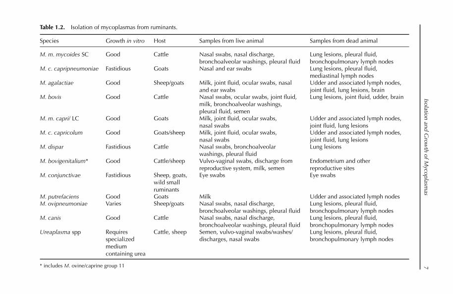
Table 1.2 shows the range of samples that can be taken for the isolation of the pathogenic mycoplasmas. The normal bacteriological procedures apply to sample taking. To ensure optimal recovery, fresh samples of milk and synovial fl uid must be taken. Lung lavage techniques have been advocated for detecting invading mycoplasmas in the lower respiratory tract (Thomas et al., 2002).
Nose, eye or ear swabs
A sterile cotton swab is pre-wetted in transport medium then inserted deep into the nasal passage or ear canal; the surface of the conjunctiva may also be gently
Table 1.1. Composition of PRM medium (pH 7.6).
Component Concentration (l–1)
Peptone (g) 20Yeast extract (g) 5Fresh yeast extract (ml) (Freundt, 1983) 100Glucose (g) 5Sodium pyruvate (g) 2Glycerol (g) 5Sodium chloride (g) 5HEPES (mmol) 30–120Heat-inactivated porcine serum (ml) 100
Isolation and Grow
th of Mycoplasm
as7
Table 1.2. Isolation of mycoplasmas from ruminants.
Species Growth in vitro Host Samples from live animal Samples from dead animal
M. m. mycoides SC Good Cattle Nasal swabs, nasal discharge, bronchoalveolar washings, pleural fl uid
Lung lesions, pleural fl uid, bronchopulmonary lymph nodes
M. c. capripneumoniae Fastidious Goats Nasal and ear swabs Lung lesions, pleural fl uid, mediastinal lymph nodes
M. agalactiae Good Sheep/goats Milk, joint fl uid, ocular swabs, nasal and ear swabs
Udder and associated lymph nodes, joint fl uid, lung lesions, brain
M. bovis Good Cattle Nasal swabs, ocular swabs, joint fl uid, milk, bronchoalveolar washings, pleural fl uid, semen
Lung lesions, joint fl uid, udder, brain
M. m. capri/ LC Good Goats Milk, joint fl uid, ocular swabs, nasal swabs
Udder and associated lymph nodes, joint fl uid, lung lesions
M. c. capricolum Good Goats/sheep Milk, joint fl uid, ocular swabs, nasal swabs
Udder and associated lymph nodes, joint fl uid, lung lesions
M. dispar Fastidious Cattle Nasal swabs, bronchoalveolar washings, pleural fl uid
Lung lesions
M. bovigenitalium* Good Cattle/sheep Vulvo-vaginal swabs, discharge from reproductive system, milk, semen
Endometrium and other reproductive sites
M. conjunctivae Fastidious Sheep, goats, wild small ruminants
Eye swabs Eye swabs
M. putrefaciens Good Goats Milk Udder and associated lymph nodesM. ovipneumoniae Varies Sheep/goats Nasal swabs, nasal discharge,
bronchoalveolar washings, pleural fl uidLung lesions, pleural fl uid, bronchopulmonary lymph nodes
M. canis Good Cattle Nasal swabs, nasal discharge, bronchoalveolar washings, pleural fl uid
Lung lesions, pleural fl uid, bronchopulmonary lymph nodes
Ureaplasma spp Requires specialized medium containing urea
Cattle, sheep Semen, vulvo-vaginal swabs/washes/discharges, nasal swabs
Lung lesions, pleural fl uid, bronchopulmonary lymph nodes
* includes M. ovine/caprine group 11
Chapter 18
swabbed. The swab is then placed into the transport medium. The swab is agitated briskly in the medium and then discarded. Some workers advocate leaving the swab in the transport medium, which enables the swab to be used to streak the solid medium, which can lead to the direct isolation of mycoplasma colonies. However, in our experience gross contamination often results.
Lung samples
Lesions are located and the exterior of the organ is sterilized by searing with a hot instrument, fl aming or boiling if possible. Small pieces of tissue from the interface between diseased and healthy tissue are aseptically removed. Where possible a fresh set of sterile instruments should be used for each tissue. Each piece of 1–3 cm3 tissue is placed in a separate sterile screw-capped jar containing trans-port medium. Where lesions are encapsulated, samples from the internal surface should be taken or scraped using a scalpel blade and placed in transport medium.
Milk samples
The tip of the animal’s teat should be cleansed. The initial stream of milk should be discarded. A sterile tube should be fi lled with the next stream of milk and the milk allowed to stand. Two drops of the milk layer should be placed into a broth using a Pasteur pipette. If there is clotting, a portion of the clot should be used in preference to the supernatant liquid. The whole milk should be used if no layer develops. Bulk or individual milk which appears normal (and is therefore likely to contain only small numbers of mycoplasmas) can be incubated whole after the addition of ampicillin to between 1 and 10 mg/ml, and subcultured after 1 or 2 days. This procedure increases the levels of contamination with ampicillin- resistant organisms but will occasionally result in isolation from milk which proves negative by the standard procedure. Smears may be made for polychrome methylene blue or fl uorescent antibody staining.
Pleural fl uid
Pleural fl uid is the sample of choice for the diagnosis of CBPP and CCPP but is only present in animals in the acute phase of the disease (Provost et al., 1987; Nicholas and Baker, 1998). Mycoplasmas can be isolated from this sample in pure culture and in high numbers. Animals in the acute phase of disease are identifi ed by clinical signs and euthanazed by humane means; the carcass should be posi-tioned carefully for post-mortem examination (raising the carcass vertically should be avoided). Alternatively, it is possible for a veterinary surgeon to take a few ml of pleural fl uid from the acutely affected live animal by puncturing the thoracic cavity in its sloping part between the 7th and 8th ribs. The chest should be opened and at least 10 ml of straw-coloured pleural fl uid removed aseptically.

Isolation and Growth of Mycoplasmas 9
Transport
Samples should be sent to the laboratory as quickly as possible, preferably the same day, keeping the samples cool (about 4°C). If microbiological examination cannot be performed immediately, samples and whole, or parts of, organs should be stored deep frozen, where mycoplasmas will remain viable for up to several months. For international transport, where freezing during transport is not pos-sible, samples should be lyophilized. Freeze-drying lung homogenates from CCPP-affected goats before transport overseas has proved highly successful with the recovery of M. c. capripneumoniae (Houshaymi et al., 2000). Many countries require a special import licence to be obtained in advance for any biological material, especially for tissues which contain animal pathogens, including mycoplasmas.
Isolation of Mycoplasmas from Samples
Isolation on solid and liquid media
Tenfold dilutions (10–1 to 10–6) are made of liquid sample (pleural fl uid, nasal exudate, synovial fl uid, etc.) or lung homogenate in appropriate medium. Tissue samples are best chopped with scissors then shaken vigorously or pulverized in medium (10% w/v). Dilution of the samples has a number of benefi ts: fi rst, it reduces the effects of mycoplasmacidal substances, including antibiotics released by the tissues; and second, it reduces bacterial contamination and the overgrowth by less important but more exuberant mollicutes. A few drops of each sample are deposited and spread on the solid medium and a 10% (v/v) inoculum dispensed into liquid medium. In addition, a direct impression should be made on the solid medium with the cut surface of a lung lesion or lymph node without spreading it. Swabs (if available) are streaked directly on to solid medium. The broths ( optimally with gentle shaking) and plates are incubated at 37°C in a humidifi ed atmosphere with 5% CO2.
The broths are examined daily for signs of growth or changes of pH, indicated by a colour change in the medium. Broths should be examined against a good, even light. Bacterial contamination will be seen as gross turbidity evident within 24 h. Mycoplasma growth will appear between 3 and 5 days and is usually seen as a very fi ne cloudiness, usually described as ‘opalescence’; it may be necessary to compare with an uninoculated broth to see the growth, particularly in the case of M. c. capripneumoniae. M. m. mycoides grows well and usually produces ‘whirls’ from the bottom of the tube when shaken. If fi lm appears on the surface of the liquid medium which also has an orange colour, M. bovis should be suspected if the isolate is from cattle and M. agalactiae if from small ruminants. The plates are inspected after 2–3 days under 35× magnifi cation, or 100× if colonies are small, for the typical ‘fried egg’ appearance. Mycoplasmas grow into the agar, which makes the use of a loop for subculturing unsatisfactory. All members of the M. mycoides cluster except M. c. capripneumoniae grow within 3 days, producing colonies of between 1 and 3 mm. M. ovipneumoniae can be suspected when iso-lated from small ruminant lung when the colonies are centreless and do not stick
Chapter 110
to the agar surface. In general, however, colonies may vary greatly in size in a single culture. The form may also vary in different cultures of the same strain, for example in having small or large centres or a granular or smooth appearance. Occasionally colonies or a pure culture on the same plate will differ in appearance, perhaps because of age. It follows then that colonial morphology is relatively valueless as a typing aid.
Subculturing
For most serological identifi cation tests, it is necessary to grow the mycoplasma on solid medium. Broths should be subcultured immediately growth is apparent or pH change is seen. Some mycoplasmas, notably M. m. mycoides, will die rapidly at a pH much below 7, and in the case of M. m. mycoides, which produces acid rap-idly in the medium, subculture at 3-day intervals is likely to result in loss of the strain. Suspected samples are subcultured three times before rejecting the mate-rial as negative. Previous subcultures should be held so that at the end the pri-mary broth will have been incubated for 3 weeks. Plates are not normally incubated for more than a week. For broth to broth subculturing, 10% (v/v) inoc-ulum should be placed into the new broth using a Pasteur pipette. For broth to plate, the plate is put on a level surface and a single drop (about 25 µl) of incu-bated broth is carefully streaked across the agar. The drop is allowed to soak in before the plate is moved. For plate to broth subculturing, a block with colonies on is cut out with a sterile spatula and dropped into the broth. It is possible to pick up
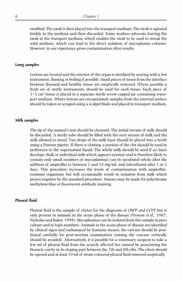
Fig. 1.1. Colonies of Mycoplasma bovirhinis (10).
Isolation and Growth of Mycoplasmas 11
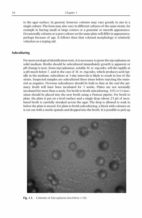
Fig. 1.2. Colonies of Mycoplasma agalactiae (15).
Fig. 1.3. Colonies of Mycoplasma mycoides subsp. capri (25).
a single colony by taking out a plug with a Pasteur pipette or a syringe and hypo-dermic needle, or just a needle. For plate to plate subculturing, a block with colo-nies is cut out and placed face down on the new plate, and is then carefully slid about on the surface.
Chapter 112
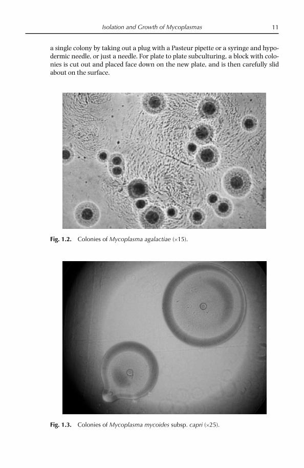
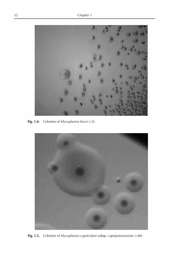
Fig. 1.4. Colonies of Mycoplasma bovis (5).
Fig. 1.5. Colonies of Mycoplasma capricolum subsp. capripneumoniae (40).

Isolation and Growth of Mycoplasmas 13
Once isolated the mycoplasmas can be identifi ed by conventional biochemical and immunological methods (Poveda and Nicholas, 1998) or increasingly by molecular methods such as polymerase chain reaction (PCR) assays, which can be found in Chapters 2 and 3 of this book.
Figures 1.1–1.6 show colony morphology of several important mycoplasmas of ruminants.
References
Abu-Amero, K.K., Halablab, M.A. and Miles, R.J. (1996) Nisin resistance distinguishes mycoplasma spp. and provides a basis for selective growth media. Applied and Envi-ronmental Microbiology 62, 3107–3111.
Bölske, G. (1988) Survey of mycoplasma infections in cell cultures and a comparison of detection methods. Zentralblat. fur Bakteriologie Hygiene A 269, 331–340.
Freundt, E.A. (1983) Culture media for classic mycoplasmas. In: Razin, S. and Tully, J.G. (eds) Methods in Mycoplasmology, Vol 1. Academic Press, New York, pp. 127–135.
Friis, N.F. (1979) Selective isolation of slow growing acidifying mycoplasmas from swine and cattle. Acta Veterinaria Scandinavia 20, 607–609.
Houshaymi, B.M., Miles, R.J. and Nicholas, R.A.J. (2000) Studies on strains of Mycoplasma capricolum subsp. capripneumoniae isolated from outbreaks of contagious caprine pleu-ropneumonia. Small Ruminant Research 45, 139–143.
Khan, A., Loria, G., Ramirez, S., Fielder, M., Miles, R.J. and Nicholas, R.A.J. (2005) Biochemi-cal characterisation of some non fermenting, non arginine hydrolysing mycoplasmas of ruminants. Veterinary Microbiology 109, 129–134.
Fig. 1.6. Colonies of Mycoplasma ovipneumoniae (50).

Chapter 114
MacOwan, K.J. and Minette, J.E. (1976) A mycoplasma from acute contagious caprine pleuropneumonia in Kenya. Tropical Animal Health and Production 8, 91–95.
Miles, R.J. (1992) Catabolism in mollicutes. Journal of General Microbiology 138, 1773–1783.
Miles, R.J. and Agbanyim, C.D. (1998) Determination of substrate utilization rates by my-coplasmas. In: Miles, R.J. and. Nicholas, R.A.J (eds) Mycoplasma Protocols. Humana Press, Totowa, New Jersey, pp. 95–104.
Miles, R.J., Wadher, B.J., Henderson, C.L. and Mohan, K. (1988) Increased growth yields of Mycoplasma species in the presence of pyruvate. Letters in Applied Microbiology 7, 149–151.
Nicholas, R.A.J and Baker, S.E. (1998) Recovery of mycoplasmas from animals. In: Miles, R.J and Nicholas, R.A.J (eds) Mycoplasma Protocols. Humana Press, Totowa, New Jersey, pp. 37–44.
Nicholas, R.A.J., Bashiruddin, J.B., Ayling, R.D. and Miles, R.J. (2000) Contagious bovine pleuropneumonia: a review of recent development. Veterinary Bulletin 70, 827–838.
Patel, H., Mackintosh, D., Ayling, R.D., Nicholas, R.A.J. and Fielder, M.D. (2008) A novel medium devoid of ruminant peptone for high yield growth of Mycoplasma ovipneumoniae. Veterinary Microbiology 127, 309–314.
Poveda, J.B. and Nicholas, R.A.J. (1998) Serological identifi cation of mycoplasmas by growth and metabolism inhibition tests. In: Miles, R.J. and Nicholas, R.A.J. (eds) Mycoplasma Protocols. Humana Press, Totowa, New Jersey, pp. 105–112.
Provost, A., Perreau, P., Breard, A., le Goff, C., Martel, J.L. and Cottew, G.S. (1987) Conta-gious bovine pleuropneumonia. Review Scientifi que et Technique. Offi ce International Epizooties 6, 625–679.
Razin, S. (1978) The mycoplasmas. Microbiological Review 42, 414–470.Razin, S. (1991) The genera Mycoplasma, Ureaplasma, Acholeplasma, Anaeroplasma, and
Asteroplasma. In: Balows, A., Truper, H.G., Dworkin, M., Harder, W. and Schleifer, K.H. (eds) The Prokaryotes, Vol. 2, 2nd edn. Springer-Verlag, New York, pp. 1937–1959.
Razin, S. (1994) DNA probes and PCR in diagnosis of mycoplasma infections. Molecular and Cellular Probes 8, 497–511.
Razin, S. and Freundt, E.A. (1984) The mycoplasmas. In: Krieg, N.R. and Holt, J.G. (eds) Bergey’s Manual of Systematic Bacteriology, Vol. 1. Williams & Wilkins, Baltimore, Maryland, pp. 740–770.
Razin, S., Yogev, D. and Naot, Y. (1998) Molecular biology and pathogenicity of mycoplas-mas. Microbiology and Molecular Biology Reviews 62, 1094–1156.
Rodwell, A.W. and Mitchell, A. (1979) Nutrition, growth and reproduction. In: Barile, M.F. and Razin, S. (eds) The Mycoplasmas, Vol. 1. Academic Press, New York, pp. 103–113.
Thiaucourt, F., Bölske, G., Leneguersh, B., Smith, D. and Wesonga, H. (1996) Diagnosis and control of contagious caprine pleuropneumonia. Review Scientifi que et Technique. Offi ce International Epizooties 15, 1415–1429.
Thomas, A., Dizier, I., Trolin, A., Mainil, J. and Linden, A. (2002) Comparison of sampling procedures for isolating pulmonary mycoplasmas in cattle. Veterinary Research Communications 26, 333–339.

© CAB International 2008. Mycoplasma Diseases of Ruminants(R. Nicholas et al.) 15
2 Detection and Differentiation of Mycoplasma Species using PCR/Denaturing Gradient Gel Electrophoresis
Introduction
Mycoplasmas cause a wide range of disease in both humans and animals and are commonly associated with pneumonia, arthritis, conjunctivitis, infertility and abortion. Specifi c diagnosis of mycoplasma infections is often diffi cult due to the limitations of current diagnostic tests together with the similarity in the diseases that they cause. Mycoplasmas are highly fastidious, typically taking weeks to cul-ture, and many serological tests are non-specifi c and insensitive. More recently PCR has been used to detect a number of Mycoplasma species. However, with over 125 mycoplasmas currently recognized it is not feasible to develop PCR tests for each species and there is a pressing need for a single, generic test that can both detect and differentiate mycoplasmas. Denaturing gradient gel electrophoresis (DGGE) can theoretically detect single base mutations in DNA (Fisher and Lerman, 1983; Lerman and Beldjord, 1999). The method is based on the prevention of migration of DNA fragments following strand separation caused by chemical denaturants. DGGE has been used extensively for diversity analysis in microbial ecology (Muyzer, 1999) but has not been widely used for the identifi cation and differentiation of pathogenic bacteria except for the detection and identifi cation of Listeria species (Schmolker et al., 1998) and for the molecular typing of Staphy-lococcus aureus (Gürtler et al., 2001) and Campylobacter species (Nielsen et al., 2000). Previously we demonstrated the ability of DGGE using universal primers for the V3 region of 16S rDNA to detect and differentiate 27 mycoplasmas of vet-erinary importance (McAuliffe et al., 2003). The development of mycoplasma-specifi c primers has enabled the application of this method directly to clinical material such as swabs and tissue samples. In addition, we have also extended the scope of the DGGE method to include human, equine, sea mammal, canine and feline Mycoplasma species and a variety of fi eld isolates (McAuliffe et al., 2005). The generic nature of the test may lead to the detection of mycoplasma infections

Chapter 216
that would be diffi cult to identify using traditional culture techniques. The appli-cability of this method to mixed infections is also described.
Design of Mycoplasma-specifi c Primers
A specifi c reverse primer for mollicutes was designed using PRIMROSE (Ashelford et al., 2002). A reverse primer, R543 5′-ACC TAT GTA TTA CCG CG, for Myco-plasma species was designed by aligning 102 Mycoplasma species. The forward primer of Muyzer, GC341, was used as described below (Muyzer et al., 1993). A 340 bp PCR product was generated with all 72 mycoplasmas tested. The mollicute-specifi c reverse primer was tested against a range of other bacterial pathogens to ensure specifi city. A gradient thermocycler (Biorad, iCycler) was used to test a range of annealing temperatures to ensure specifi city. For all future experiments an annealing temperature of 56°C was used.
DNA Extraction and 16S PCR
Mycoplasma DNA was extracted from a 1ml aliquot of stationary-phase culture using the Genelute genomic DNA kit according to the manufacturer’s instruc-tions (Sigma, Poole, UK). DNA was extracted from swabs by swirling the swab in 1 ml of PBS, removing the swab and then using the Genelute kit as described above. DNA was extracted from tissue samples by removing a 1 cm2 portion of tissue using sterile instruments, placing it in 1ml of PBS, homogenizing to produce a suspension and extracting DNA using a Sigma tissue kit according to the manu-facturer’s instructions. Amplifi cation of the V3 region of the 16S RNA gene was performed according to the method of Muyzer with minor modifi cations (Muyzer et al., 1993) using the universal bacterial primer GC-341F 5′-CGC CCG CCG CGC GCG GCG GGC GGG GCG GGG GCA CGG GGG GCC TAC GGG AGG CAG CAG and the mollicute-specifi c primer R543. For the PCR, 1 µl of lysate was added as a template to 49 µl of a reaction mixture containing 10 mM Tris-HCl (pH 9.0), 1.5 mM MgCl2, 50 mM KCl, 0.1% Triton X-100, 0.2 mM of each deoxynucleoside triphosphate and 0.5 U of Taqgold (Applied Biosystems). The cycling conditions were: denaturation at 94°C for 5 min, followed by 30 cycles of 95°C for 1 min, 56°C for 45 s and 72°C for 1 min, a fi nal extension step of 72°C for 10 min and samples were kept at 4°C until analysis. Aliquots were checked for correct ampli-fi cation by electrophoresis on a 2% agarose gel followed by visualization with ethidium bromide under UV illumination.
Denaturing Gradient Gel Electrophoresis
DGGE was performed using the Ingeny PhorU 2 × 2 apparatus (GRI Molecular Biology, Essex, UK). Samples (20 µl) were loaded on to 10% polyacyrlamide/bis (37.5:1) gels with denaturing gradients from 30 to 60% (where 100% is 7 M urea and 40% (v/v) deionized formamide) in 1 × TAE electrophoresis buffer (Severn

Detection and Differentiation by PCR/DGGE 17
Biotech Ltd, Worcestershire, UK). Electrophoresis was performed at 100 V at a temperature of 60°C for 18 h. Gels were then stained with SBYR Gold (Cambridge BioScience, Cambridgeshire, UK) in 1 × TAE for 30 min at room temperature and visualized under UV illumination.
Mycoplasma-specifi c Primers
Members of mycoplasma, acholeplasma and ureaplasma groups could be ampli-fi ed using the mycoplasma-specifi c primer R543 and the universal primer GC341; however, members of the related haemoplasma group could not. All 72 Myco-plasma species tested produced a PCR product of approximately 340 bp. These products were subjected to DGGE in groups according to host animal. In the majority of cases only one band was seen, indicating that there was no interspe-cifi c variation in the amplifi ed sequence. The presence of multiple bands indicated that more than one 16S rDNA operon was present and that there were some sequence differences between the copies. The migration of the bands in the gels is a function of the melting behaviour of the amplicons in the chemical gradient used. A faint background band was sometimes seen on the DGGE gels, which is probably due to a degree of primer–dimer formation and should as such be con-sidered an artefact. The background band was easily differentiated from bands generated from 16S operons as it was faint in intensity, had an irregular shape and was not straight.
Applicability of DGGE Directly to Clinical Samples
In order to test the practicality of DGGE in the clinical laboratory DNA extraction was performed directly on swabs and tissue samples received for veterinary diag-nostic investigations. In total 202 clinical samples were analysed, of which 89 were found to be positive for mycoplasma infection. Mycoplasma DNA was suc-cessfully amplifi ed for DGGE from a wide variety of diagnostic samples, including nasal, eye, ear and foot swabs, lung tissue, milk, brain tissue, synovial joint fl uid and tissue from an aborted bovine fetus. In order to test the robustness of the pro-cedure on samples that had undergone long-term storage, DGGE was used on bovine lung samples obtained from outbreaks of contagious bovine pleuropnemo-nia in Botswana and Tanzania that had been frozen at –80°C for approximately 9 years. DGGE identifi ed M. mycoides SC in eight out of nine samples; culture of the lung samples also yielded M. mycoides SC in eight out of nine samples.
Use of DGGE to Detect Mixed Cultures
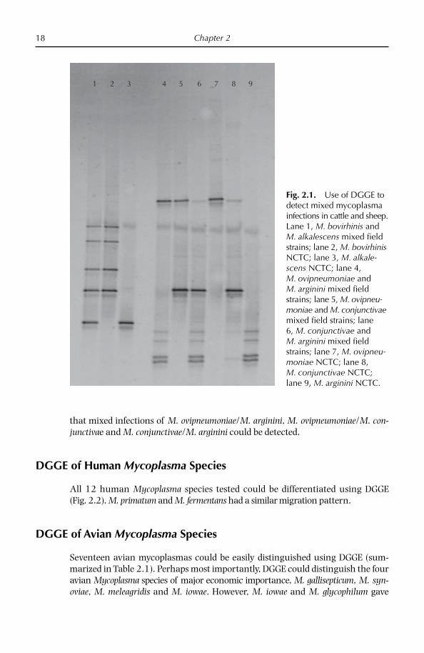
DGGE using mycoplasma-specifi c primers was particularly useful for detecting mixed cultures. As shown in Fig. 2.1, analysis of a number of bovine diagnostic samples demonstrated that a mixed infection of M. bovirhinis/M. alkalescens could be detected easily. In addition, analysis of small ruminant clinical samples showed
Chapter 218
that mixed infections of M. ovipneumoniae/M. arginini, M. ovipneumoniae/M. con-junctivae and M. conjunctivae/M. arginini could be detected.
DGGE of Human Mycoplasma Species
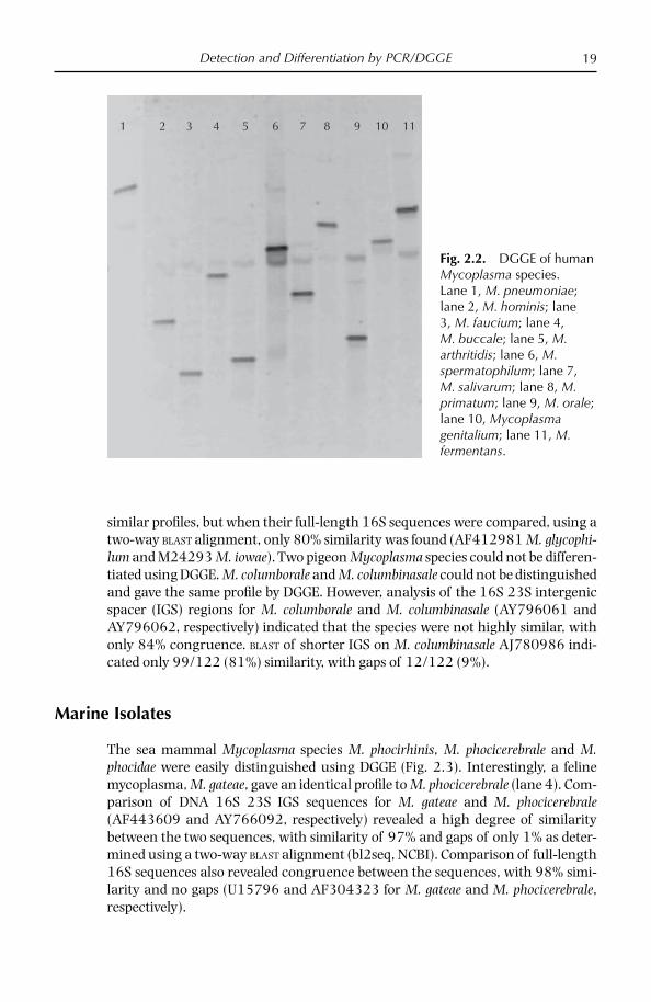
All 12 human Mycoplasma species tested could be differentiated using DGGE (Fig. 2.2). M. primatum and M. fermentans had a similar migration pattern.
DGGE of Avian Mycoplasma Species
Seventeen avian mycoplasmas could be easily distinguished using DGGE (sum-marized in Table 2.1). Perhaps most importantly, DGGE could distinguish the four avian Mycoplasma species of major economic importance, M. gallisepticum, M. syn-oviae, M. meleagridis and M. iowae. However, M. iowae and M. glycophilum gave
1 2 3 4 5 6 7 8 9
Fig. 2.1. Use of DGGE to detect mixed mycoplasma infections in cattle and sheep. Lane 1, M. bovirhinis and M. alkalescens mixed fi eld strains; lane 2, M. bovirhinis NCTC; lane 3, M. alkale-scens NCTC; lane 4, M. ovipneumoniae and M. arginini mixed fi eld strains; lane 5, M. ovipneu-moniae and M. conjunctivae mixed fi eld strains; lane 6, M. conjunctivae and M. arginini mixed fi eld strains; lane 7, M. ovipneu-moniae NCTC; lane 8, M. conjunctivae NCTC; lane 9, M. arginini NCTC.
Detection and Differentiation by PCR/DGGE 19
similar profi les, but when their full-length 16S sequences were compared, using a two-way BLAST alignment, only 80% similarity was found (AF412981 M. glycophi-lum and M24293 M. iowae). Two pigeon Mycoplasma species could not be differen-tiated using DGGE. M. columborale and M. columbinasale could not be distinguished and gave the same profi le by DGGE. However, analysis of the 16S 23S intergenic spacer (IGS) regions for M. columborale and M. columbinasale (AY796061 and AY796062, respectively) indicated that the species were not highly similar, with only 84% congruence. BLAST of shorter IGS on M. columbinasale AJ780986 indi-cated only 99/122 (81%) similarity, with gaps of 12/122 (9%).
Marine Isolates
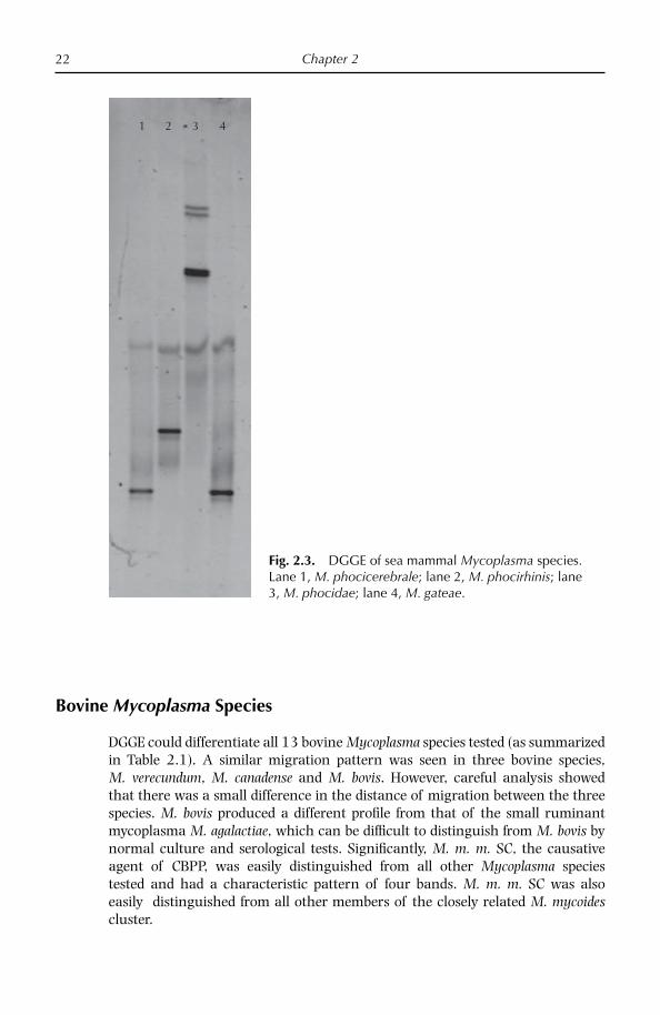
The sea mammal Mycoplasma species M. phocirhinis, M. phocicerebrale and M. phocidae were easily distinguished using DGGE (Fig. 2.3). Interestingly, a feline mycoplasma, M. gateae, gave an identical profi le to M. phocicerebrale (lane 4). Com-parison of DNA 16S 23S IGS sequences for M. gateae and M. phocicerebrale (AF443609 and AY766092, respectively) revealed a high degree of similarity between the two sequences, with similarity of 97% and gaps of only 1% as deter-mined using a two-way BLAST alignment (bl2seq, NCBI). Comparison of full-length 16S sequences also revealed congruence between the sequences, with 98% simi-larity and no gaps (U15796 and AF304323 for M. gateae and M. phocicerebrale, respectively).
1 2 3 4 5 6 7 8 9 10 11
Fig. 2.2. DGGE of human Mycoplasma species. Lane 1, M. pneumoniae; lane 2, M. hominis; lane 3, M. faucium; lane 4, M. buccale; lane 5, M. arthritidis; lane 6, M. spermatophilum; lane 7, M. salivarum; lane 8, M. primatum; lane 9, M. orale; lane 10, Mycoplasma genitalium; lane 11, M. fermentans.
Chapter 220
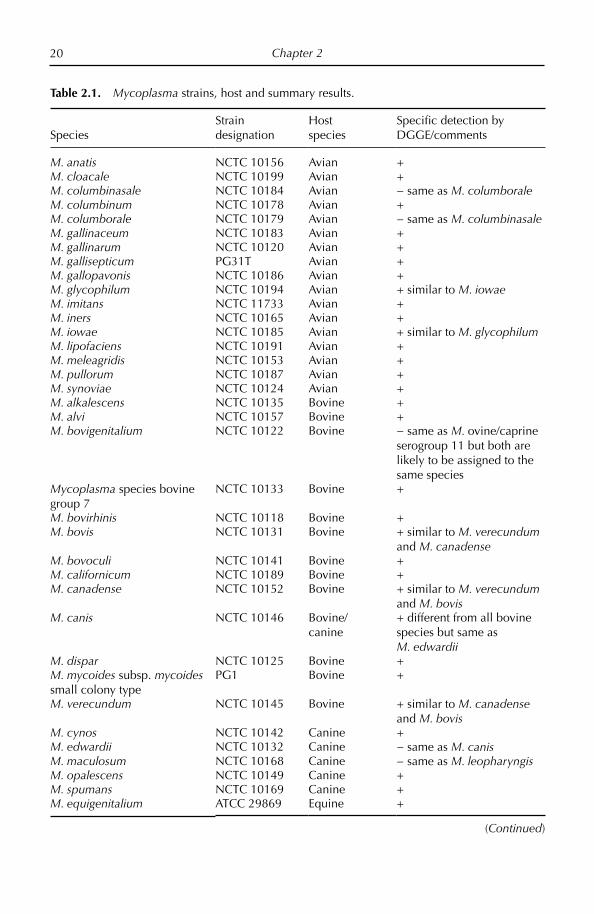
Table 2.1. Mycoplasma strains, host and summary results.
SpeciesStrain designation
Host species
Specifi c detection by DGGE/comments
M. anatis NCTC 10156 Avian +M. cloacale NCTC 10199 Avian +M. columbinasale NCTC 10184 Avian − same as M. columboraleM. columbinum NCTC 10178 Avian +M. columborale NCTC 10179 Avian − same as M. columbinasaleM. gallinaceum NCTC 10183 Avian +M. gallinarum NCTC 10120 Avian +M. gallisepticum PG31T Avian +M. gallopavonis NCTC 10186 Avian +M. glycophilum NCTC 10194 Avian + similar to M. iowaeM. imitans NCTC 11733 Avian +M. iners NCTC 10165 Avian +M. iowae NCTC 10185 Avian + similar to M. glycophilumM. lipofaciens NCTC 10191 Avian +M. meleagridis NCTC 10153 Avian +M. pullorum NCTC 10187 Avian +M. synoviae NCTC 10124 Avian +M. alkalescens NCTC 10135 Bovine +M. alvi NCTC 10157 Bovine +M. bovigenitalium NCTC 10122 Bovine − same as M. ovine/caprine
serogroup 11 but both are likely to be assigned to the same species
Mycoplasma species bovine group 7
NCTC 10133 Bovine +
M. bovirhinis NCTC 10118 Bovine +M. bovis NCTC 10131 Bovine + similar to M. verecundum
and M. canadenseM. bovoculi NCTC 10141 Bovine +M. californicum NCTC 10189 Bovine +M. canadense NCTC 10152 Bovine + similar to M. verecundum
and M. bovisM. canis NCTC 10146 Bovine/
canine+ different from all bovine species but same as M. edwardii
M. dispar NCTC 10125 Bovine +M. mycoides subsp. mycoides small colony type
PG1 Bovine +
M. verecundum NCTC 10145 Bovine + similar to M. canadense and M. bovis
M. cynos NCTC 10142 Canine +M. edwardii NCTC 10132 Canine − same as M. canisM. maculosum NCTC 10168 Canine − same as M. leopharyngisM. opalescens NCTC 10149 Canine +M. spumans NCTC 10169 Canine +M. equigenitalium ATCC 29869 Equine +
(Continued)

Detection and Differentiation by PCR/DGGE 21
Table 2.1. Continued.
SpeciesStrain designation
Host species
Specifi c detection by DGGE/comments
M. equirhinis NCTC 10148 Equine +M. fastidiosum NCTC 10180 Equine +M. felis NCTC 10160 Equine + M. subdolum NCTC 10175 Equine +M. arthriditis NCTC 10162 Human +M. buccale NCTC 10136 Human +M. faucium NCTC 10174 Human +M. fermentans NCTC 10117 Human + similar to M. primatumM. genitalium NCTC 10195 Human +M. hominis NCTC 10111 Human +M. lipophilum NCTC 10173 Human +M. orale NCTC 10112 Human +M. pneumoniae NCTC 10119 Human +M. primatum NCTC 10163 Human + similar to M. fermentansM. salivarum NCTC 10113 Human +M. spermatophilum NCTC 11720 Human +M. fl occulare NCTC 10143 Porcine + M. hyopneumoniae NCTC 10110 Porcine +M. hyorhinis NCTC 10130 Porcine +M. hyosynoviae NCTC 10167 Porcine +M. gateae NCTC 10161 Sea mammal/
feline− same as M. phocicerebrale
M. phocicerebrale NCTC 11721 Sea mammal − same as M. gateaeM. phocidae Strain 105 Sea mammal +M. phocirhinis NCTC 11722 Sea mammal +M. agalactiae NCTC 10123 Small ruminant +M. arginini NCTC 10129 Small ruminant +M. capricolum subsp. capricolum
NCTC 10154 Small ruminant +
M. capricolum subsp. capripneumoniae
NCTC 10192 Small ruminant +
M. conjunctivae NCTC 10147 Small ruminant +M. cottewii NCTC 11732 Small ruminant − same as M. yeatsiiM. mycoides subsp. capri NCTC 10137 Small ruminant − indistinguishable from M.
m. m. LC but both are likely to be the same species
M. mycoides subsp. mycoides LC
F30 Small ruminant − indistinguishable from M. m. capri but both are likely to be the same species
M. ovipneumoniae NCTC 10151 Small ruminant +Mycoplasma ovine/caprine serogroup 11
Strain 2D Small ruminant − same as M. bovigenitalium but both are likely to be assigned to the same species
M. putrefaciens NCTC 10155 Small ruminant +M. yeatsii NCTC 11730 Small ruminant − same as M. cottewii
Chapter 222
Bovine Mycoplasma Species
DGGE could differentiate all 13 bovine Mycoplasma species tested (as summarized in Table 2.1). A similar migration pattern was seen in three bovine species, M. verecundum, M. canadense and M. bovis. However, careful analysis showed that there was a small difference in the distance of migration between the three species. M. bovis produced a different profi le from that of the small ruminant mycoplasma M. agalactiae, which can be diffi cult to distinguish from M. bovis by normal culture and serological tests. Signifi cantly, M. m. m. SC, the causative agent of CBPP, was easily distinguished from all other Mycoplasma species tested and had a characteristic pattern of four bands. M. m. m. SC was also easily distinguished from all other members of the closely related M. mycoides cluster.
1 2 3 4
Fig. 2.3. DGGE of sea mammal Mycoplasma species. Lane 1, M. phocicerebrale; lane 2, M. phocirhinis; lane 3, M. phocidae; lane 4, M. gateae.
Detection and Differentiation by PCR/DGGE 23
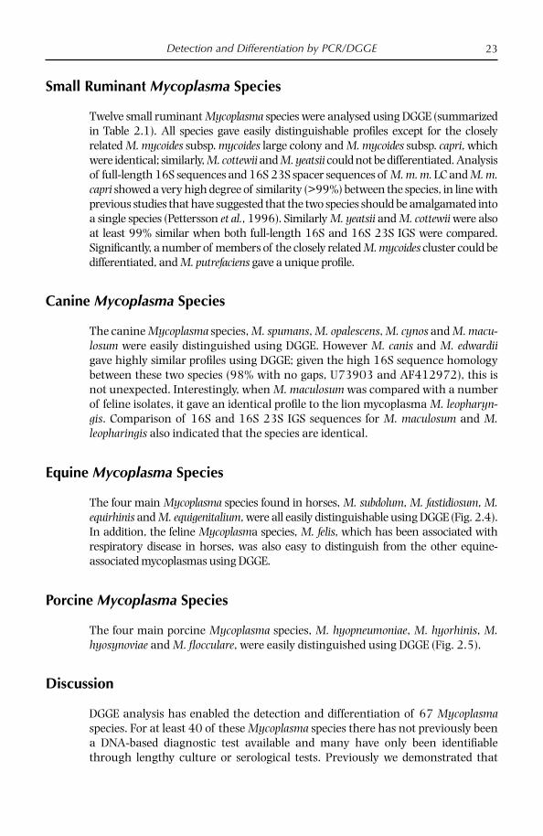
Small Ruminant Mycoplasma Species
Twelve small ruminant Mycoplasma species were analysed using DGGE (summarized in Table 2.1). All species gave easily distinguishable profi les except for the closely related M. mycoides subsp. mycoides large colony and M. mycoides subsp. capri, which were identical; similarly, M. cottewii and M. yeatsii could not be differentiated. Analysis of full-length 16S sequences and 16S 23S spacer sequences of M. m. m. LC and M. m. capri showed a very high degree of similarity (>99%) between the species, in line with previous studies that have suggested that the two species should be amalgamated into a single species (Pettersson et al., 1996). Similarly M. yeatsii and M. cottewii were also at least 99% similar when both full-length 16S and 16S 23S IGS were compared. Signifi cantly, a number of members of the closely related M. mycoides cluster could be differentiated, and M. putrefaciens gave a unique profi le.
Canine Mycoplasma Species
The canine Mycoplasma species, M. spumans, M. opalescens, M. cynos and M. macu-losum were easily distinguished using DGGE. However M. canis and M. edwardii gave highly similar profi les using DGGE; given the high 16S sequence homology between these two species (98% with no gaps, U73903 and AF412972), this is not unexpected. Interestingly, when M. maculosum was compared with a number of feline isolates, it gave an identical profi le to the lion mycoplasma M. leopharyn-gis. Comparison of 16S and 16S 23S IGS sequences for M. maculosum and M. leopharingis also indicated that the species are identical.
Equine Mycoplasma Species
The four main Mycoplasma species found in horses, M. subdolum, M. fastidiosum, M. equirhinis and M. equigenitalium, were all easily distinguishable using DGGE (Fig. 2.4). In addition, the feline Mycoplasma species, M. felis, which has been associated with respiratory disease in horses, was also easy to distinguish from the other equine- associated mycoplasmas using DGGE.
Porcine Mycoplasma Species
The four main porcine Mycoplasma species, M. hyopneumoniae, M. hyorhinis, M. hyosynoviae and M. fl occulare, were easily distinguished using DGGE (Fig. 2.5).
Discussion
DGGE analysis has enabled the detection and differentiation of 67 Mycoplasma species. For at least 40 of these Mycoplasma species there has not previously been a DNA-based diagnostic test available and many have only been identifi able through lengthy culture or serological tests. Previously we demonstrated that

Chapter 224
DGGE could be used to differentiate 27 Mycoplasma species of veterinary impor-tance (McAuliffe et al., 2003). This has now been extended to include 67 Myco-plasma species and presents signifi cant improvements to the technique, including the use of mycoplasma-specifi c primers. Whereas DGGE using universal primers required a media-enrichment step to ensure that only mollicute DNA was ampli-fi ed (McAuliffe et al., 2003), with the advent of mollicute-specifi c primers, DGGE can be applied directly to clinical material. As a result of this, mycoplasma infections can now be diagnosed in less than 24 h compared with 1 to 2 weeks for traditional culture. The use of mycoplasma-specifi c primers has also enabled the detection of mixed cultures, which would have been diffi cult to detect by conventional methods, as less fastidious species would be outgrown.
DGGE may prove to be particularly useful for human mycoplasmas and is the fi rst generic test capable of differentiating 12 species. Previously a multiplex PCR has been used to differentiate genital Mycoplasma species (Stellrecht et al., 2004) and a reverse line blotting procedure has been used to differentiate fi ve human mollicute pathogens (Wang et al., 2004), but there has not been a single generic
1 2 3 4 5
Fig. 2.4. DGGE of equine Mycoplasma species. Lane 1, M. fastidiosum; lane 2, M. subdolum; lane 3, M. felis; lane 4, M. equirhinis; lane 5, M. equigenitalium.

Detection and Differentiation by PCR/DGGE 25
test for other human Mycoplasma species. Signifi cantly M. genitalium and M. pneu-moniae can be differentiated easily by DGGE, thus demonstrating the specifi city of the technique as there is 98% similarity between the two species based on 16S homology (Jensen et al., 2003).
A number of mycoplasmas could not be differentiated using DGGE and gave identical profi les. For example, M. mycoides subsp. capri and M. mycoides LC were indistinguishable, indicating that there was no variation in the 16S rDNA sequence over the V3 region amplifi ed. This may provide further support for the notion that M. mycoides subsp. mycoides LC and M. mycoides subsp. capri are in fact the same species (Pettersson et al., 1996). Some unexpected isolates also gave identical profi les by DGGE, for example the feline mycoplasma M. gateae and the sea mammal species M. phocicerebrale. These results were also supported by com-parison of full-length 16S and 16S 23S IGS sequences for the isolates, which also indicated a very high degree of similarity between the species. If these species are closely related it is diffi cult to explain how they could have been transmitted between two very different hosts, cats and seals, which seem unlikely to have come
1 2 3 4
Fig. 2.5. DGGE of porcine mycoplasmas. Lane: 1, M. fl occulare; lane 2, M. hyopneumoniae; lane 3, M. hyosynoviae; lane 4, M. hyorhinis.
Chapter 226
into close contact with one another. Similarly, the canine mycoplasma M. maculo-sum showed a high degree of similarity to the lion mycoplasma M. leopharingis by 16S and 16S 23S IGS analysis and gave identical DGGE profi les. Previous studies have also highlighted the high degree of similarity in the 16S sequence and identical biochemical characteristics of these species (Pettersson et al., 2001).
Two canine Mycoplasma species, M. canis and M. edwardii, gave indistinguish-able DGGE profi les. This is not unexpected as previous analysis of full-length 16S sequences and 16S 23S IGS sequences found that the species are highly similar (Chalker and Brownlie, 2004). Interestingly, M. cynos could be differentiated from all other canine Mycoplasma species whereas previous studies based on sequence analysis have shown it grouped closely with M. canis and M. edwardii (Chalker and Brownlie, 2004).
Two species, M. columbinum and M. columbinasale, could not be distinguished, although previous studies have indicated that they are less than 97% similar by 16S sequence (Pettersson et al., 2001). Even when cultures were obtained from several different collections, the two isolates gave identical profi les. It is likely that the species were previously identifi ed using serological tests and emphasizes the need for DNA sequencing of historical isolates in collections to ensure that they are correctly identifi ed. Nevertheless, whether species should be designated based on serological or molecular methods is still a contentious issue within mollicute taxonomy. Recently, denaturing HPLC analysis has been used to detect and type bacterial pathogens (Domann et al., 2003; Hurtle et al., 2003) and could theoretically be used as an alternative to DGGE to target single nucleotide polymorphisms in the V3 region of 16S rDNA of Mycoplasma species. However, dHPLC would require expensive, specialized equipment and more laborious standardization and interpretation compared with DGGE.
In conclusion, DGGE enables the rapid detection and differentiation of Myco-plasma species and can be used to diagnose infections either directly from tissues or from cultured isolates. It is capable of detecting mixed cultures or even new mollicute species and is suitable for routine use in the diagnostic laboratory. In the event of the detection of a new or unusual profi le then the product can be identi-fi ed by 16S rDNA sequencing; however, if there is no matched sequence in the database then the isolate will need to be characterized by conventional methods.
References
Ashelford, K.E., Weightman, A.J. and Fry, J.C. (2002) PRIMROSE: a computer program for generating and estimating the phylogenetic range of 16S rRNA oligonucleotide probes and primers in conjunction with the RDP-II database. Nucleic Acids Research 30, 3481–3489.
Chalker, V.J. and Brownlie, J. (2004) Taxonomy of the canine mollicutes by 16S rRNA gene and 16S/23S rRNA intergenic spacer region sequence comparison. International Journal of Systematic Evolutionary Microbiology 54, 537–542.
Domann, E., Hong, G., Imirzalioglu, C., Turschner, S., Kuhle, J., Watzel, C., Hain, T., Hossain, H. and Chakraborty, T. (2003) Culture-independent identifi cation of pathogenic bacteria and polymicrobial infections in the genitourinary tract of renal transplant recipients. Journal of Clinical Microbiology 41, 5500–5510.
Detection and Differentiation by PCR/DGGE 27
Fisher, S.G. and Lerman, L.S. (1983) DNA fragments differing by single base-pair substitu-tions are separated in denaturing gradient gels: correspondence with theory. Proceedings of the National Academy of Sciences USA 80, 1579–1583.
Gürtler, V., Barrie, H.D. and Mayall, B.C. (2001) Use of denaturing gradient gel electrophore-sis to detect mutations in VS2 of the 16S-23S rDNA spacer amplifi ed from Staphylococ-cus aureus isolates. Electrophoresis 22, 1920–1924.
Hurtle, W., Bode, E., Kaplan, R.S., Garrison, J., Kearney, B., Shoemaker, D., Henchal, E. and Norwood D. (2003) Use of denaturing high-performance liquid chromatography to identify Bacillus anthracis by analysis of the 16S-23S rRNA interspacer region and gyrA gene. Journal of Clinical Microbiology 41, 4758–4766.
Jensen, J.S., Borre, M.B. and Dohn, B. (2003) Detection of Mycoplasma genitalium by PCR amplifi cation of the 16S rRNA gene. Journal of Clinical Microbiology 41, 261–266.
Lerman, L.S. and Beldjord, C. (1999) Comprehensive mutation detection with denaturing gradient gel electrophoresis. In: Cotton, R.G.H., Edkins, E. and Forrest, S. (eds) Mutation Detection. Oxford University Press, Inc., New York, pp. 35–61.
McAuliffe, L., Ellis, R.J., Ayling, R.D. and Nicholas, R.A.J. (2003) Differentiation of Mycoplas-ma species by 16S ribosomal DNA PCR and denaturing gradient gel electrophoresis fi ngerprinting. Journal of Clinical Microbiology 41, 4844–4847.
McAuliffe, L., Ellis, R., Lawes, J., Ayling, R.D. and Nicholas, R.A.J. (2005) 16S rDNA and DGGE: a single generic test for detecting and differentiating Mycoplasma species. Journal of Medical Microbiology 54, 1–9.
Muyzer, G. (1999) DGGE/TGGE: a method for identifying genes from natural ecosystems. Current Opinions in Microbiology 2, 317–322.
Muyzer, G., de Waal, E.C. and Uitterlinden, A.G. (1993) Profi ling of complex microbial populations by denaturing gradient gel electrophoresis analysis of polymerase chain reaction-amplifi ed genes coding for 16S rRNA. Applied and Environmental Microbiology 59, 695–700.
Nielsen, E.M., Engberg, J., Fussing, V., Petersen, L., Brogren, C.H. and On, S.L. (2000) Evalua-tion of phenotypic and genotypic methods for subtyping Campylobacter jejuni isolates from humans, poultry, and cattle. Journal of Clinical Microbiology 38, 3800–3810.
Pettersson, B., Leitner, T., Ronaghi, M., Bolske, G., Uhlen, M. and Johansson, K.E. (1996) Phylogeny of the Mycoplasma mycoides cluster as determined by sequence analysis of the 16S rRNA genes from the two rRNA operons. Journal of Bacteriology 178, 4131–4412.
Pettersson, B., Tully, J.G., Bolske, G. and Johansson, K.E. (2001) Re-evaluation of the clas-sical Mycoplasma lipophilum cluster and description of two new clusters in the hominis group based on 16S rDNA sequences. International Journal Systematic Evolutionary Microbiology 51, 633–643.
Schmolker, N., van Ommen Kloeke, F. and Geesey, G. (1998) Use of Dcode system to detect the food-borne bacterial pathogen Listeria monocytogenes. In: Bio-Rad Mutation Analysis, tech note 2403.
Stellrecht, K.A., Woron, A.M., Mishrik, N.G. and Venezia, R.A. (2004) Comparison of mul-tiplex PCR assay with culture for detection of genital mycoplasmas. Journal of Clinical Microbiology 42, 1528–1533.
Wang, H., Kong, F., Jelfs, P., James, G. and Gilbert, G.L. (2004) Simultaneous detection and identifi cation of common cell culture contaminant and pathogenic mollicutes strains by reverse line blot hybridization. Applied and Environmental Microbiology 70, 1483–1486.

© CAB International 2008. Mycoplasma Diseases of Ruminants28 (R. Nicholas et al.)
3 Detection of Mycoplasma Species Using Polymerase Chain Reaction (PCR)
Introduction
Although a number of novel generic techniques such as denaturing gradient gel electrophoresis (DGGE) (McAuliffe et al., 2003a, 2005), amplifi ed rDNA restric-tion analysis (ARDRA) (Stakenborg et al., 2005) and reverse line blot (Wang et al., 2004) have been developed which enable the identifi cation of Mycoplasma spe-cies, PCR remains the most useful, simple and rapid method of detecting specifi c mycoplasmas. With the advent of real-time PCR it is now possible not only to achieve results quickly but also to quantify the number of organisms present. New, small, portable PCR and real-time PCR systems have also been developed, which means that detection in the fi eld using these methods is now possible.
PCR is one of the most sensitive of the existing rapid methods to detect infec-tious pathogens in clinical specimens. PCR is particularly useful for pathogens such as mycoplasmas which are diffi cult to culture in vitro or require a long culti-vation period. However, the application of PCR to clinical specimens has many potential pitfalls due to the susceptibility of PCR to inhibitors, contamination and experimental conditions. Moreover, it is known that the sensitivity and specifi city of a PCR assay is dependent on many variables including target genes, primer sequences, PCR techniques, DNA extraction procedures and PCR product detec-tion methods.
Previously PCRs were frequently designed based solely on the 16S rDNA gene owing to a lack of sequenced mycoplasma genes. However, with the increased number of complete genome sequences, PCRs are now being developed on a num-ber of different gene targets. The specifi city and sensitivity of PCR can vary enor-mously depending on the chosen gene target and is a factor worthy of consideration when designing PCRs.
Many PCRs have been developed which enable the specifi c detection of a number of ruminant mycoplasmas including members of the closely related Mycoplasmas mycoides cluster: M. mycoides SC, M. mycoides LC, M. c. capricolum,

Detection Using PCR 29
M. c. capripneumoniae, M. species bovine group 7, M. putrefaciens, M. agalactiae, M. bovis, M. ovipneumoniae, M. conjunctivae, M. fermentans, M. dispar, M. canis and others (Table 3.1). PCRs are, however, still lacking for a number of important mycoplasma species, including M. canadense, M. verecundum and M. californicum. Real-time PCR has only been developed to date for a selected few species, including M. mycoides subsp. mycoides SC, M. agalactiae and M. conjunctivae (as summarized in Table 3.2).
PCR has been particularly useful for the detection and differentiation of mem-bers of the M. mycoides cluster as these species are diffi cult or impossible to differ-entiate using traditional culture and serological techniques. The causative agent of CBPP, M. mycoides SC, was among the fi rst species for which PCRs were devel-oped. Initial tests for M. mycoides SC were based on the Cap21 gene fragments, and although useful as a screening test to rule out the presence of exotic mycoplasmas
Table 3.1. PCRs available for ruminant Mycoplasma species.
Mycoplasma species Gene target Reference(s)
M. mycoides SC IS1296/8.8kb deletion regionCap21
Miles et al., 2006Bashiruddin, 1998
M. mycoides SC/LC/capri Cap21 Bashiruddin, 1998Mycoides cluster Cap21
GlkBashiruddin, 1998Woubit et al., 2007
M. sp. bovine group 7 gts Frey et al., 1998M. c. capricolum LppA Monnerat et al., 1999M. c. capripneumoniae Adi Woubit et al., 2004M. agalactiae urvC Subramaniam et al., 1998M. bovis uvrC Subramaniam et al., 1998M. ovipneumoniae 16S McAuliffe et al., 2003bM. bovigenitalium 16S Parham et al., unpublished resultsM. conjunctivae 16S, lppS Giacometti et al., 1999; Belloy
et al., 2003M. putrefaciens 16S Unpublished
Peyraud et al., 2003M. canis 16S UnpublishedM. dispar 16S Miles et al., 2004 M. fermentans IS1550 Afshar et al., 2007
Table 3.2. Real-time PCR for ruminant Mycoplasma species.
Mycoplasma species Gene target Reference
M. mycoides SC 16S rRNA and hypothetical lipoproteins
Gorton et al., 2005
M. agalactiae P81 Lorusso et al., 2007M. bovis 16S Cai et al., 2005M. conjunctivae lppS Vilei et al., 2007

Chapter 330
in areas free of CBPP, a second time-consuming enzyme-digestion step was neces-sary to differentiate M. mycoides SC from the closely related small ruminant patho-gen M. mycoides LC. More recently, PCRs based on the insertion sequence IS1296 have enabled not only the specifi c detection of M. mycoides SC but also the differ-entiation of African/Australian strains from the European strains (Miles et al., 2006). The development of Taqman real-time PCR for M. mycoides SC shows great promise and will also enable the quantifi cation of the number of organisms present (Gorton et al., 2005).
Specifi c PCRs have been developed for other members of the M. mycoides clus-ter, such as M. c. capricolum, M. c. capripneumoniae and M. species bovine group 7 (as summarized in Table 3.2).
PCRs have also been developed for the important ruminant pathogens M. bovis and M. agalactiae. These mycoplasmas are closely related and can be diffi cult to distinguish using 16S rDNA alone as they only differ by a few bases. Currently used PCRs for both M. bovis and M. agalactiae are based on the uvrC gene. Recently it has been shown that a number of unusual M. bovis strains, which have proved to be M. bovis using serology and 16S rDNA sequencing, are negative on the urvC PCR (McAuliffe et al., 2008, unpublished results). A real-time test has been devel-oped for M. bovis and shows improved sensitivity compared with conventional PCR (Lorusso et al., 2007). M. agalactiae was also positive using this method but could be distinguished as it had a different melt peak. Real-time PCR has also been developed as a diagnostic test for M. agalactiae based on the p81 lipoprotein gene; this PCR was highly sensitive, capable of detecting 101 copies in a 10 µl template from milk (Lorusso et al., 2007).
Limitations of PCR
PCR only enables the detection of a single Mycoplasma species and will not enable diagnosis of new or unusual Mycoplasma species or those species in unusual hosts. For this reason we fi nd that generic tests such as PCR/DGGE (see Chapter 2) are advantageous as they enable the detection of mixed cultures; it is not uncommon to fi nd between two and six different Mycoplasma species present in clinical sam-ples, particularly in birds, that would be missed by conventional single-species PCR.
Sensitivity and Specifi city
Mycoplasma PCRs vary in sensitivity: the M. mycoides SC cap21 PCR was shown to be relatively sensitive, detecting between 10 and 100 organisms (Bashiruddin et al., 1994), whereas other PCRs for non-ruminant mycoplasmas such as M. hyopneumoniae only detect 500 organisms (Baumeister et al., 1998).
The sensitivity of PCR can be increased in some instances by using a nested system. These PCRs are ideally suited to detecting very small numbers of patho-gens in clinical samples. The process utilizes two consecutive PCRs. The fi rst PCR contains an external pair of primers, while the second contains either two nested primers that are internal to the fi rst primer pair or one of the fi rst primers and a

Detection Using PCR 31
single nested primer. The larger fragment produced by the fi rst reaction is used as the template for the second PCR. The sensitivity and specifi city of DNA amplifi ca-tion can be considerably improved by using such nested PCR, sometimes with 1000 times more sensitivity than a standard PCR. Nested PCRs have been used for M. bovis (Pinnow et al., 2001), where it had a sensitivity of 51 cfu/ml, greater than culture of traditional PCR.
Both traditional and nested PCR were evaluated for the detection of M. con-junctivae and it was found that the detection level was estimated to be 20 cells per swab when the nested PCR procedure was used and 2 × 10⁵ by the single PCR method (Giacometti et al., 1999).
The sensitivity of PCR has also been enhanced by the use of real-time PCR technology. Real time, developed for M. bovis, gave a detection limit of 550 cfu/ml in milk and 650 cfu/25 mg in lung tissue. In validation testing of clinical samples, the relative sensitivity and specifi city were 100 and 99.3%, respectively for individ-ual milks and 96.6 and 100%, respectively for the lung tissue. M. agalactiae was also positive using this method but could be distinguished as it had a different melt peak. As mentioned above, real-time PCR has also been developed as a diagnostic test for M. agalactiae based on the p81 lipoprotein gene (Lorusso et al., 2007).
False Positive/Negative Results
As PCR is based on amplifi cation of DNA, false positive or false negative results may easily occur. In particular, as a single PCR cycle results in very large numbers of amplifi able molecules, these can potentially contaminate subsequent amplifi -cations of the same target sequence and generate false positive results. Carryover contamination of reagents, pipetting devices, laboratory surfaces or even the clothing and skin of workers can all lead to false positive results. To control car-ryover contamination, the physical transfer of DNA between amplifi ed samples and between positive and negative experimental controls must be prevented. It is recommended that samples are prepared for PCR in a room or biosafety hood sep-arate from that in which the reactions are performed. Using a pipette tip with an aerosol barrier is also essential for avoiding cross and carryover contamination. Using uracil N-glycosylase (UNG) to cleave the dUTP incorporated in PCR prod-ucts is considered a powerful protocol to prevent carryover amplicon contamina-tion enzymatically (Longo et al., 1990), particularly in a clinical laboratory that is performing PCR extensively. This is performed by substituting dUTP for dTTP and adding UNG to the master mixture. To protect the dUTP-containing product, UNG must be inactivated chemically or by heat before the PCR product can be analysed further. Therefore, the dUTP protocol requires only two changes in a standard PCR protocol: the substitution of dUTP for dTTP in all reactions and the incuba-tion of all PCR mixtures with UNG prior to temperature cycling. The exposure of laboratory surface, pipettes and racks to UV is also able to destroy contaminating amplicons but is effi cient only on amplicons greater than 300 bp size (Espy et al., 1993).
False negative results can also create problems in the diagnostic laboratory. The choice of suitable DNA extraction methods is crucial, as when PCR is applied
Chapter 332
directly to DNA extracted from clinical samples the effect of inhibitors must be considered as they may interfere with amplifi cation. Inhibitors include haemoglo-bin, lactoferrin, CSF, bone marrow aspirates, faeces and the blood anticoagulants heparin and SPS. The effect of inhibitors can be minimized by careful DNA extrac-tion methods, and the addition of surfactants may be particularly useful for extraction from milk samples as used in some M. bovis PCR protocols (Pinnow et al., 2001). The use of internal controls may be prudent to ensure that false neg-ative results are not obtained. Inhibitors can be detected either by spiking negative samples with the target DNA by diluting the specimen to minimize the effects of inhibitors or by adding a control template.
Previously Unpublished PCRs
Mycoplasma canis
This PCR is based on the 16S rDNA gene of M. canis and amplifi es a 400 bp frag-ment in both canine and bovine M. canis isolates. The primers McaF1 TGA TGA TTA GCT GAT AGT AGA ACT and McaR1 GAT TTG CTT GAC GTC GCC GTT are used at a concentration of 50 pmoles/µl. Total genomic DNA from M. canis (NCTC 10146) is used as a positive control. A MgCl2 concentration of 2.0 mM is used and the cycling conditions are as follows: 94°C for 5 min, followed by 33 cycles of 94°C for 30 s, 60°C for 30 s, 72°C for 30 s, followed by 72°C for 7 min and then 4°C until analysis. For the test to be valid, a band of 400 bp should be present for the positive control and no bands should be present in the negative controls upon agarose gel electrophoresis, staining with ethidium bromide and visualization under UV light.
Mycoplasma putrefaciens
This PCR is based on the 16S rDNA gene of M. putrefaciens and amplifi es a 340 bp fragment in all isolates. The primers SSF1 GCG GCA TGC CTA ATA CAT GC and SSR1 AGC TGC GGC GCT GAG TTC A are used at a concentration of 50 pmoles/µl. Total genomic DNA from M. putrefaciens (NCTC10155) is used as a positive con-trol. A MgCl2 concentration of 2.0 mM is used and the cycling conditions are as follows: 94°C for 5 min, followed by 25 cycles of 94°C for 30 s, 64°C for 30 s, 72°C for 30 s, followed by 72°C for 7 min and then 4°C until analysis. For the test to be valid, a band of 340 bp should be present for the positive control and no bands should be present in the negative controls upon agarose gel electrophoresis, stain-ing with ethidium bromide and visualization under UV light.
Mycoplasma bovigenitalium
The forward non-specifi c primer was MbgF (GGC TGT GTG CCT AAT ACA TGC), and the reverse specifi c primer was MbgR (CCT AGA GTG CTC AAT TG), designed

Detection Using PCR 33
to give a PCR amplicon size of 1061 bp. The PCR cycling conditions used were 94°C for 4 min followed by 30 cycles of 95°C for 30 s, 57°C for 30 s, 72°C for 48 s, followed by a fi nal extension at 72°C for 7 min.
References
Afshar, B., Pitcher, D., Nicholas, R.A.J. and Miles, R.J. (2007) An evaluation of PCR methods to detect strains of Mycoplasma fermentans. Biologicals 36, 117–121.
Bashiruddin, J.B. (1998) PCR and RFLP methods for the specifi c detection and identifi cation of Mycoplasma mycoides subsp. mycoides SC. Methods in Molecular Biology 104, 167–178.
Bashiruddin, J.B., Taylor, T.K. and Gould, A.R. (1994). A PCR-based test for the specifi c identifi cation of Mycoplasma mycoides subsp. mycoides SC. Journal of Veterinary Diagnostic Investigation 6, 428–434.
Baumeister, A.K., Runge, M., Ganter, M., Feenstra, A.A., Delbeck, F. and Kirchhoff, H. (1998) Detection of Mycoplasma hyopneumoniae in bronchoalveolar lavage fl uids of pigs by PCR. Journal of Clinical Microbiology 36, 1984–1988.
Belloy, L., Janovsky, M., Vilei, E.M., Pilo, P., Giacometti, M. and Frey, J. (2003) Molecular epidemiology of Mycoplasma conjunctivae in Caprinae: transmission across species in natural outbreaks. Applied and Environmental Microbiology 69, 1913–1919.
Cai, H.Y., Bell-Rogers, P., Parker, L. and Prescott, J.F. (2005) Development of a real-time PCR for detection of Mycoplasma bovis in bovine milk and lung samples. Journal of Veterinary Diagnostic Investigations 17, 537–545.
Espy, M.J., Smith, T.F. and Persing, D.H. (1993) Dependence of polymerase chain reaction product inactivation protocols on amplicon length and sequence composition. Journal of Clinical Microbiology 31, 2361–2365.
Frey, J., Cheng, X., Monnerat, M.P., Abdo, E.M., Krawinkler, M., Bolske, G. and Nicolet, J. (1998) Genetic and serological analysis of the immunogenic 67-kDa lipoprotein of Mycoplasma sp. bovine group 7. Research in Microbiology 149, 55–64.
Giacometti, M., Nicolet, J., Johansson, K-E., Naglic, T., Degiorgis, M.P. and Frey, J. (1999) Detection and identifi cation of Mycoplasma conjunctivae in infectious keratoconjunc-tivitis by PCR based on the 16S rRNA gene. Zentralblatt fur Veterinarmedizin B. 46, 173–180.
Gorton, T.S., Barnett, M.M., Gull, T., French, R.A., Lu, Z., Kutish, G.F., Adams, L.G. and Geary, S.J. (2005) Development of real-time diagnostic assays specifi c for Mycoplasma mycoides subsp. mycoides Small Colony. Veterinary Microbiology 30, 51–58.
Longo, M.C., Berninger, M.S. and Hartley, J.L. (1990) Use of uracil DNA glycosylase to control carry-over contamination in polymerase chain reactions. Gene 93, 125–128.
Lorusso, A., Decaro, N., Greco, G., Corrente, M., Fasanella, A. and Buonavoglia, D. (2007) A real-time PCR assay for detection and quantifi cation of Mycoplasma agalactiae DNA. Journal of Applied Microbiology 103, 918–923.
McAuliffe, L., Ellis, R.J., Ayling, R.D. and Nicholas, R.A.J. (2003a) Differentiation of Myco-plasma species by 16S ribosomal DNA PCR and denaturing gradient gel electrophoresis fi ngerprinting. Journal of Clinical Microbiology 41, 4844–4847.
McAuliffe, L., Hatchell, F.M., Ayling, R.D., King, A.I.M. and Nicholas, R.A.J. (2003b) Detec-tion of Mycoplasma ovipneumoniae in Pasteurella-vaccinated sheep fl ocks with respira-tory disease in England. Veterinary Record 153, 687–688.
McAuliffe, L., Ellis, R., Lawes, J., Ayling, R.D. and Nicholas, R.A.J. (2005) 16S rDNA and DGGE: a single generic test for detecting and differentiating Mycoplasma species. Journal of Medical Microbiology 54, 1–9.
Chapter 334
Miles, K., McAuliffe, L., Ayling, R.D. and Nicholas, R.A.J. (2004) Rapid detection of Myco-plasma dispar and M. bovirhinis using allele specifi c polymerase chain reaction protocols. FEMS Microbiology Letters 241, 103–107.
Miles, K., Churchward, C.P., McAuliffe, L., Ayling, R.D. and Nicholas, R.A.J. (2006) Identi-fi cation and differentiation of European and African/Australian strains of Mycoplasma mycoides subsp. mycoides small-colony type using polymerase chain reaction analysis. Journal of Veterinary Diagnostic Investigation 18, 168–171.
Monnerat, M.P., Thiaucourt, F., Poveda J.B., Nicolet, J. and Frey, J. (1999) Genetic and sero-logical analysis of lipoprotein lppA in Mycoplasma mycoides subsp. mycoides LC and Mycoplasma mycoides subsp. capri. Clinical and Diagnostic Laboratory Immunology 6, 224–230.
Peyraud, A., Woubit, S., Poveda, J.B., De La Fe, C., Mercier, P. and Thiaucourt, F. (2003) A specifi c PCR for the detection of Mycoplasma putrefaciens, one of the agents of the con-tagious agalactia syndrome of goats. Molecular and Cellular Probes 17, 289–294.
Pinnow, C.C., Butler, J.A., Sachse, K., Hotzel, H., Timms, L.L. and Rosenbusch, R.F. (2001) Detection of Mycoplasma bovis in preservative treated milk samples. Journal of Dairy Science 84, 1640–1645.
Stakenborg, T., Vicca, J., Butaye, P., Maes, D., De Baere, T., Verhelst, R., Peeters, J., de Kruif, A., Haesebrouck, F. and Vaneechoutte M. (2005) Evaluation of amplifi ed rDNA restric-tion analysis (ARDRA) for the identifi cation of Mycoplasma species. BMC Infectious Diseases 5, 46.
Subramaniam, S., Bergonier, D., Poumarat, F., Capaul, S., Schlatter, Y., Nicolet, J. and Frey, J. (1998) Species identifi cation of Mycoplasma bovis and Mycoplasma agalactiae based on the uvrC genes by PCR. Molecular and Cellular Probes 12, 161–169.
Vilei, E.M., Bonvin-Klotz, L., Zimmermann, L., Ryser-Degiorgis, M.P., Giacometti, M. and Frey, J. (2007) Validation and diagnostic effi cacy of a TaqMan real-time PCR for the detection of Mycoplasma conjunctivae in the eyes of infected Caprinae. Journal of Micro-biological Methods 70, 384–386.
Wang, H., Kong, F., Jelfs, P., James, G. and Gilbert, G.L. (2004) Simultaneous detection and identifi cation of common cell culture contaminant and pathogenic mollicutes strains by reverse line blot hybridization. Applied and Environmental Microbiology 70, 1483–1486.
Woubit, S., Lorenzon, S., Peyraud, A., Manso-Silvan, L. and Thiaucourt, F. (2004) A spe-cifi c PCR for the identifi cation of Mycoplasma capricolum subsp. capripneumoniae, the causative agent of contagious caprine pleuropneumonia (CCPP). Veterinary Microbiology 104, 125–132.
Woubit, S., Manso-Silvan, L., Lorenzon, S., Gaurivaud, P., Poumarat, F., Pellet, M.P., Singh, V.P. and Thiaucourt, F. (2007) A PCR for the detection of mycoplasmas belonging to the Mycoplasma mycoides cluster: application to the diagnosis of contagious agalactia. Molecular and Cellular Probes 21, 391–399.

© CAB International 2008. Mycoplasma Diseases of Ruminants(R. Nicholas et al.) 35
4 Molecular Typing of Mycoplasma Species
Introduction
Analysis of populations and assessment of strain variability provide important information regarding bacterial pathogens. Molecular epidemiological analysis enables the genotyping of strains and has uses in the tracing and control of dis-ease outbreaks. In many cases pathogenic strains of bacteria have been identifi ed by typing methods as belonging to distinct clonal lineages that demonstrate a unique combination of virulence alleles or genes (Musser, 1996). Therefore typ-ing may enable the detection of certain genotypes that are associated with more severe or unusual disease signs. These isolates, or particularly those genotypes associated with low virulence, may be useful in vaccine development. Recently developed molecular typing methods are capable of distinguishing whether genetic variation is due to recombination or point mutation; they may also have applications for fundamental research as they can enable the defi nition and further understanding of microbial population structures and dynamics.
Over the past decade, nucleic acid-mediated procedures have largely replaced serology and protein-based methods, and procedures that were once laborious, expensive and complex have now been adapted to the high-throughput working environment of the diagnostic laboratory. Many methods are now available for use in molecular typing studies and the use of PCR has enabled the development of a plethora of rapid typing methods for strain identifi cation. Traditional methods such as pulsed fi eld gel electrophoresis (PFGE) are still a valuable tool for geno-typing mycoplasmas, and more recently the increased number of sequenced mycoplasma genomes has meant that methods such as multilocus sequence tech-niques (MLST) and variable number tandem repeat (VNTR) analysis are gaining popularity and being applied to an increasing range of Mycoplasma species. The development of species-specifi c DNA-based methods means that typing can now, in theory, be directly applied to clinical samples without the need for culture, offering

Chapter 436
the possibility in the near future of rapid pen-side typing methods for use in out-break situations.
The choice of typing technique depends on available equipment, time con-siderations and cost but also on the need for discrimination power and repro-ducibility. Ideally, methods of strain differentiation must have high enough discriminatory power to clearly differentiate unrelated strains, as well as to dem-onstrate the relationship of isolates from individuals infected through the same source. Genotyping methods should also have a high degree of reproducibility, which is particularly important during construction of reliable databases. Fur-thermore, genotyping methods should be easy to interpret and rapid to perform. It is frequently necessary to have multiple typing methods available for use in geno-typing mycoplasmas as studies have shown that different techniques are suited to different Mycoplasma species; as some methods may detect little variation between strains, alternative methods may be more suitable and capable of differentiating strains.
Amplifi ed Fragment Length Polymorphism
Amplifi ed fragment length polymorphism (AFLP) analysis is based on the selective PCR amplifi cation of restriction fragments from a total digest of genomic DNA. The technique involves three steps: (i) restriction of the DNA and ligation of oli-gonucleotide adapters; (ii) selective amplifi cation of sets of restriction fragments; and (iii) gel analysis of the amplifi ed fragments. PCR amplifi cation of restric-tion fragments is achieved by using the adapter and restriction site sequence as target sites for primer annealing. The selective amplifi cation is achieved by the use of primers that extend into the restriction fragments, amplifying only those fragments in which the primer extensions match the nucleotides fl anking the restriction sites. Using this method, sets of restriction fragments may be visu-alized by PCR without knowledge of nucleotide sequence. The method allows the specifi c co-amplifi cation of high numbers of restriction fragments. AFLP has been applied to M. bovis (McAuliffe et al., 2004), M. gallisepticum (Boettger and Dohms, 2006), M. hyosynoviae (Kokotovic et al., 2002), M. mycoides SC (Kusiluka et al., 2001) and others. AFLP is a highly discriminatory technique but requires specialist equipment, including an automated DNA sequencer or separation of DNA fragments. The use of AFLP for molecular epidemiology studies of M. bovis is described below.
AFLP analysis of M. bovis
M. bovis is a primary cause of bovine pneumonia, arthritis and mastitis and has also been associated with keratoconjunctivitis, otitis, meninigitis, infertility and abortion (Pfützner and Sachse, 1996; Nicholas and Ayling, 2003). M. bovis was fi rst isolated in 1961 in the USA from a cow with severe mastitis (Hale et al., 1962). Subsequently, infection has been reported throughout the world, includ-ing most European countries (Nicholas and Ayling, 2003). As the prevalence of

Molecular Typing of Mycoplasma Species 37
M. bovis varies widely across the world, there are important trade implications and a pressing need for monitoring cattle for M. bovis.
AFLP was carried out on 60 M. bovis isolates from the UK. DNA restriction, ligation of AFLP adapters and amplifi cation of the modifi ed fragments was car-ried out as described previously (Kusiluka et al., 2000). Briefl y, the template DNA was simultaneously restricted with 5 U BglII and 5 U MfeI (New England Biolabs, Ipswich, MA) at 37°C for 2 h in a restriction buffer containing 10 mM Tris-acetate (pH 7.5), 10 mM Mg-acetate, 50 mM K-acetate, 5 mM dithiothreitol and 50 ng/µl bovine serum albumin. A 5 µl aliquot of the DNA digest was added to 15 µl of a ligation mixture containing 2 pmol of BglII adapter and 20 pmol MfeI adapter, 1 U of T4 DNA ligase (Amersham Pharmacia Biotech, Bucks, UK), 2 µl of 10 × ligase buffer (supplied with the enzyme) and 8 µl restriction buffer. Ligation reactions were carried out overnight at room temperature.
The modifi ed genomic fragments were amplifi ed by using a Bgl-2F-0 primer, which was labelled at the 5′ end with 6-carboxyfl uorescein (FAM), and an unla-belled MfeI-0 primer, as described previously (Kokotovic et al., 1999). The PCR reac-tions were performed in a 50 µl total volume containing 2 µl (tenfold diluted) ligation product, 0.2 mM of each dNTP, 10 mM Tris-HCl (pH 8.3), 50 mM KCl, 2.5 mM MgCl2, 65 ng of each primer and 1.5 U Taq polymerase (Gibco BRL, Delhi, India). Cycling conditions included an initial denaturation step of 94°C for 3 min and 25 cycles of denaturation (94°C for 60 s), annealing (56°C for 60 s) and exten-sion (72°C for 90 s). The last cycle also included a fi nal extension step at 72°C for 10 min. The PCR products were detected using an ABI 377 automated DNA sequencer (Perkin Elmer, Waltham, MA) as described previously (Kusiluka et al., 2000).
In total 54 isolates were typed using AFLP and 35 different profi les were obtained. By comparison of AFLP fragments in the size range 50–400 bp, the M. bovis isolates could be separated into two distinct groups (A and B) with about 79% similarity between groups (Fig. 4.1). Several bands were detected that were not shared between members of the two groups: fragments of 199, 269 and 337 bp were detected only among the members of group B, whilst fragments of 223, 230, 248 and 387 bp were present only among the members of group A. In addi-tion, all of the members of group A had a fragment of either 254 or 255 bp that was not detected among any of the group B strains.
Distinct differences in heterogeneity were seen between group A and group B. Group B exhibited the greatest genetic diversity, with 93% of isolates having unique profi les compared with 39% of group A isolates. Group B could also be further divided into two subgroups (Ba and Bb). Three fragments (157, 267 and 287 bp) were present exclusively among the strains of subgroup Ba. Interestingly, a single fragment (137 bp) was detected that was present in all subgroup Ba strains and all group A strains but none of the Bb strain.
Random Amplifi ed Polymorphic DNA Analysis
Random amplifi ed polymorphic DNA (RAPD) typing is a PCR technique which uses short primers with a low annealing temperature to randomly amplify target DNA. The primers are intended to bind multiple times within the genome, and

Chapter 438
187B02
80
Sj (%)
85
A
Ba
B
Bb
90 95 100
29B0215B01139B01166B01124B0217B01163B02125B01208B0286B96151B02233B02159B0040B97144B02162B0268B98145B0254B98149B00189B02217B02147B02124B962B01171B02
231B0240B01201B0221B01222B026B018B01202B0218B01164B029B99152B02150B02226B01148B02158B0130B0162B98232B027B9976B9814B0171B01235B02236B02133B00
PG45
A1A1A1A1A1A1A1A2A3A4A4A4A4A4A4A5A5A5A5A5A6A7A8A8A9A9A10
Ba1Ba2Ba3Ba4Ba5Ba6Ba7Ba8Ba9Ba10Ba11Ba12Ba13Ba14Ba15Bb1Bb2Bb3Bb4Bb5Bb6Bb6Bb7Bb8Bb8Bb9
A11
200220022001200120012002200120022001200219962002200220002997200220021998200219982000200220022002199620012002
20022001200220012002200120012002200120021999200220022001200220012001199820021999199820012001200220022000
1962
N. YorkshireNottinghamshireCornwallTyne and WearWarwickshireN. YorkshireCornwallShropshireLancashireDevonLancashireSomersetWarwickshireHampshireWarwickshireDumfriesCumbriaLancashireDumfriesN. YorkshireTyne and WearCumbriaN. YorkshireShropshireN. YorkshireTyne and WearLancashire
CumbriaN. YorkshireShropshireBordersN. YorkshireN. YorkshireN. YorkshireShropshireN. YorkshireN. YorkshireN. YorkshireCumbriaCumbriaLancashireCumbriaShropshireShropshireHampshireShropshireHampshireCumbriaShropshireLancashireN. YorkshireN. YorkshireBorders
USA
i tAFLP y o
Fig. 4.1. Genetic relationship between M. bovis strains based on comparison of AFLP profi les (50–400 bp fragment size range) produced by amplifi cation of BglII and MfeI DNA templates with non-selective primers. The dendrogram was produced with the UPGMA method with Jaccard similarity coeffi cient (SJ) matrix. i, strain designation; tAFLP, arbitrarily assigned AFLP type; y, year of isolation; o, origin of strains.
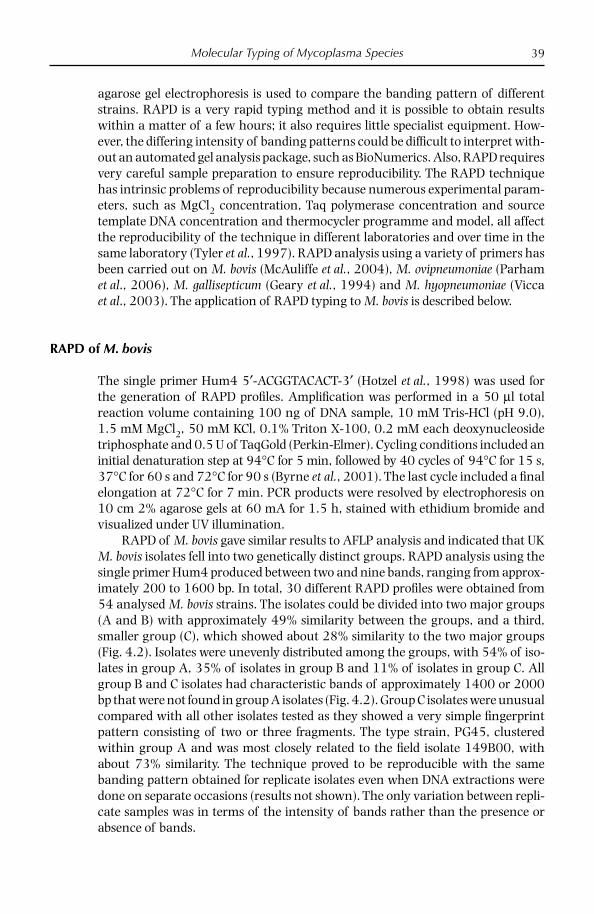
Molecular Typing of Mycoplasma Species 39
agarose gel electrophoresis is used to compare the banding pattern of different strains. RAPD is a very rapid typing method and it is possible to obtain results within a matter of a few hours; it also requires little specialist equipment. How-ever, the differing intensity of banding patterns could be diffi cult to interpret with-out an automated gel analysis package, such as BioNumerics. Also, RAPD requires very careful sample preparation to ensure reproducibility. The RAPD technique has intrinsic problems of reproducibility because numerous experimental param-eters, such as MgCl2 concentration, Taq polymerase concentration and source template DNA concentration and thermocycler programme and model, all affect the reproducibility of the technique in different laboratories and over time in the same laboratory (Tyler et al., 1997). RAPD analysis using a variety of primers has been carried out on M. bovis (McAuliffe et al., 2004), M. ovipneumoniae (Parham et al., 2006), M. gallisepticum (Geary et al., 1994) and M. hyopneumoniae (Vicca et al., 2003). The application of RAPD typing to M. bovis is described below.
RAPD of M. bovis
The single primer Hum4 5′-ACGGTACACT-3′ (Hotzel et al., 1998) was used for the generation of RAPD profi les. Amplifi cation was performed in a 50 µl total reaction volume containing 100 ng of DNA sample, 10 mM Tris-HCl (pH 9.0), 1.5 mM MgCl2, 50 mM KCl, 0.1% Triton X-100, 0.2 mM each deoxynucleoside triphosphate and 0.5 U of TaqGold (Perkin-Elmer). Cycling conditions included an initial denaturation step at 94°C for 5 min, followed by 40 cycles of 94°C for 15 s, 37°C for 60 s and 72°C for 90 s (Byrne et al., 2001). The last cycle included a fi nal elongation at 72°C for 7 min. PCR products were resolved by electrophoresis on 10 cm 2% agarose gels at 60 mA for 1.5 h, stained with ethidium bromide and visualized under UV illumination.
RAPD of M. bovis gave similar results to AFLP analysis and indicated that UK M. bovis isolates fell into two genetically distinct groups. RAPD analysis using the single primer Hum4 produced between two and nine bands, ranging from approx-imately 200 to 1600 bp. In total, 30 different RAPD profi les were obtained from 54 analysed M. bovis strains. The isolates could be divided into two major groups (A and B) with approximately 49% similarity between the groups, and a third, smaller group (C), which showed about 28% similarity to the two major groups (Fig. 4.2). Isolates were unevenly distributed among the groups, with 54% of iso-lates in group A, 35% of isolates in group B and 11% of isolates in group C. All group B and C isolates had characteristic bands of approximately 1400 or 2000 bp that were not found in group A isolates (Fig. 4.2). Group C isolates were unusual compared with all other isolates tested as they showed a very simple fi ngerprint pattern consisting of two or three fragments. The type strain, PG45, clustered within group A and was most closely related to the fi eld isolate 149B00, with about 73% similarity. The technique proved to be reproducible with the same banding pattern obtained for replicate isolates even when DNA extractions were done on separate occasions (results not shown). The only variation between repli-cate samples was in terms of the intensity of bands rather than the presence or absence of bands.
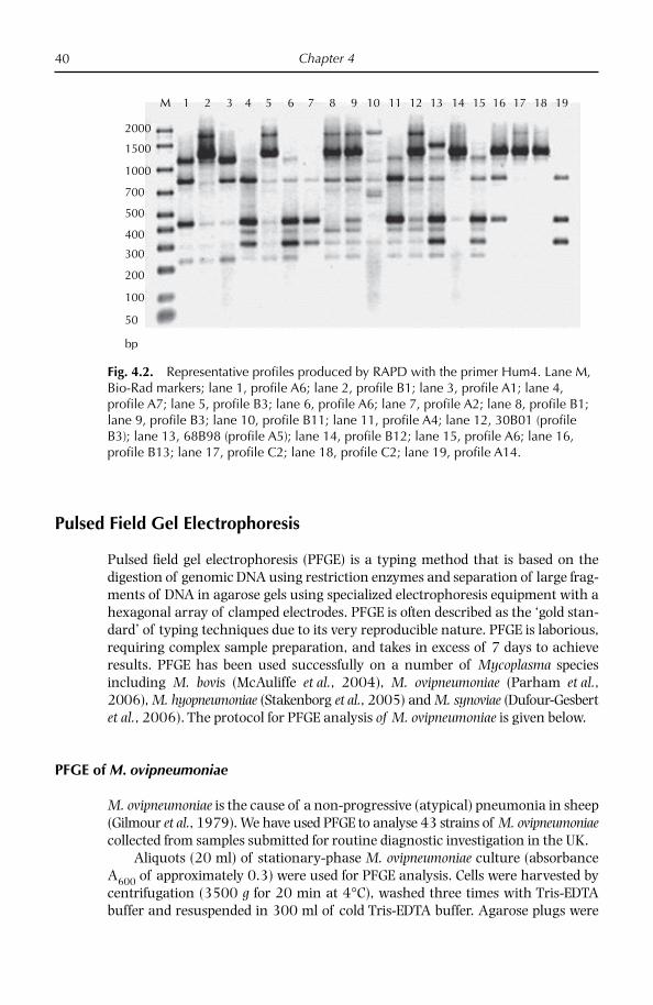
Chapter 440
Pulsed Field Gel Electrophoresis
Pulsed fi eld gel electrophoresis (PFGE) is a typing method that is based on the digestion of genomic DNA using restriction enzymes and separation of large frag-ments of DNA in agarose gels using specialized electrophoresis equipment with a hexagonal array of clamped electrodes. PFGE is often described as the ‘gold stan-dard’ of typing techniques due to its very reproducible nature. PFGE is laborious, requiring complex sample preparation, and takes in excess of 7 days to achieve results. PFGE has been used successfully on a number of Mycoplasma species including M. bovis (McAuliffe et al., 2004), M. ovipneumoniae (Parham et al., 2006), M. hyopneumoniae (Stakenborg et al., 2005) and M. synoviae (Dufour-Gesbert et al., 2006). The protocol for PFGE analysis of M. ovipneumoniae is given below.
PFGE of M. ovipneumoniae
M. ovipneumoniae is the cause of a non-progressive (atypical) pneumonia in sheep (Gilmour et al., 1979). We have used PFGE to analyse 43 strains of M. ovipneumoniae collected from samples submitted for routine diagnostic investigation in the UK.
Aliquots (20 ml) of stationary-phase M. ovipneumoniae culture (absorbance A600 of approximately 0.3) were used for PFGE analysis. Cells were harvested by centrifugation (3500 g for 20 min at 4°C), washed three times with Tris-EDTA buffer and resuspended in 300 ml of cold Tris-EDTA buffer. Agarose plugs were
2000
M 1 2 3 4 5 6 7 8 9 10 11 12 13 14 15 16 17 18 19
1500
1000
700
500
400
300
200
100
50
bp
Fig. 4.2. Representative profi les produced by RAPD with the primer Hum4. Lane M, Bio-Rad markers; lane 1, profi le A6; lane 2, profi le B1; lane 3, profi le A1; lane 4, profi le A7; lane 5, profi le B3; lane 6, profi le A6; lane 7, profi le A2; lane 8, profi le B1; lane 9, profi le B3; lane 10, profi le B11; lane 11, profi le A4; lane 12, 30B01 (profi le B3); lane 13, 68B98 (profi le A5); lane 14, profi le B12; lane 15, profi le A6; lane 16, profi le B13; lane 17, profi le C2; lane 18, profi le C2; lane 19, profi le A14.

Molecular Typing of Mycoplasma Species 41
made from a 1:1 mixture of 2% low-melting-point agarose (Bio-rad, Hercules, CA) and the cell suspension. Plugs were incubated in lysis buffer (10 mM Tris-HCl, 1 mM EDTA, 1% lauroyl sarcosine, 1 mg/ml proteinase K) for 48 h at 56°C. Plugs were washed four times with Tris-EDTA buffer for 30 min at 4°C. Slices (2 mm) were cut aseptically from plugs and equilibrated in restriction buffer (Promega, Madison, WI) for 1 h. Subsequently, restriction digestion was performed by using 30 U of SmaI (Promega) for 16 h according to the manufacturer’s instructions. The fragments were resolved on 1% pulsed fi eld-certifi ed agarose (Biorad) gels using a CHEF-DRIII system (Biorad) at 6 V/cm, with a running time of 20 h at 14°C, included angle of 120°, initial pulse time of 4 s and fi nal pulse time of 40 s. Gels were stained with ethidium bromide (0.5 mg/ml) for 15 min, destained in distilled water for 1 h and photographed under UV light. A lambda ladder PFGE marker (Sigma, Ronkonkoma, NY) was used for fragment size determination.
PFGE analysis of M. ovipneumoniae from the UK gave a high degree of genetic heterogeneity, with 40 different PFGE profi les generated from 43 isolates. These results are summarized in Fig. 4.3. There was no link between the genotype and geographical origin of isolates.
Variable Number Tandem Repeat Analysis
Variable number tandem repeats (VNTRs) are simple repeated DNA sequences that vary in copy number and have been described for a variety of organisms, including Staphylococcus aureus (Sabat et al., 2003), Mycobacterium tuberculosis (Frothingham and Meeker-O’Connell, 1998), Borrelia spp. (Farlow et al., 2002), Leptospira spp. (Slack et al., 2005), Brucella spp. (Whatmore et al., 2006), Francisella tularensis (Farlow et al., 2001) and Legionella pneumophila (Pourcel et al., 2003), but have not previously been used to differentiate Mycoplasma strains. VNTRs have been shown to give a high level of discriminatory power and can provide information regarding both evolutionary and functional bacterial diversity (van Belkum et al., 1998). With the increasing number of sequenced genomes and the development of VNTR databases, VNTR is an increasingly amenable technique for bacterial typing. VNTR requires little specialized equipment, only thermocy-clers and agarose gel electrophoresis, and does not necessitate sequencing, unlike some typing methods such as MLST analysis. Despite their small genome size, many Mycoplasma species contain a large proportion of repetitive DNA, therefore the use of typing tools which target repetitive DNA is likely to be most promising.
VNTR analysis has been successfully used for the molecular typing of MmmSC (McAuliffe et al., 2007) and is described in more detail below.
VNTR analysis of M. mycoides subsp. mycoides SC
Undoubtedly, the most important mycoplasma disease is contagious bovine pleuropneumonia (CBPP), an Offi ce International des Epizooties (OIE) notifi able disease, which is caused by M. mycoides subsp. mycoides small colony (MmmSC). Studies to date on the molecular epidemiology of MmmSC are consistent with there
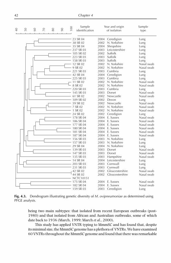
Chapter 442
being two main subtypes: that isolated from recent European outbreaks (post-1980) and that isolated from African and Australian outbreaks, some of which date back to 1936 (March, 1999; March et al., 2000).
This study has applied VNTR typing to MmmSC and has found that, despite its minimal size, the MmmSC genome has a plethora of VNTRs. We have examined 60 VNTRs throughout the MmmSC genome and found that there was remarkable
23 SR 04
Sampleidentification40 50 60 70 80 90 10
0 Year and originof isolation
Sampletype
38 SR 0235 SR 04237 SR 03105 SR 02223 SR 03158 SR 0312 SR 029 SR 02221 SR 0342 SR 04225 SR 0311 SR 028 SR 02220 SR 03145 SR 0361 SR 02109 SR 0259 SR 027 SR 021 SR 0224 SR 02178 SR 04186 SR 04177 SR 04180 SR 04181 SR 04187 SR 04156 SR 03157 SR 0329 SR 04139 SR 03147 SR 03135 SR 0314 SR 04203 SR 03231 SR 0342 SR 0244 SR 02NCTC10151175 SR 04182 SR 04159 SR 03
200420022004200320022003200320022002200320042003200220022003200320022002200220022002200220042004200420042004200420032003200420032003200320042003200320022002
200420042003
CeredigionN. YorkshireShropshireLeicestershireSuffolkSuffolkSuffolkN. YorkshireN. YorkshireCumbriaCeredigionCumbriaN. YorkshireN. YorkshireCumbriaDorsetNewcastleDevonNewcastleN. YorkshireN. YorkshireCeredigionE. SussexE. SussexE. SussexE. SussexE. SussexE. SussexN. YorkshireN. YorkshireN. YorkshireDorsetDorsetHampshireLeicestershireCornwallCornwallGloucestershireGloucestershire
E. SussexE. SussexCeredigion
LungLungLungLungLungLungLungNasal swabNasal swabLungLungLungNasal swabNasal swabLungNasal swabNasal swabLungNasal swabNasal swabNasal swabLungNasal swabNasal swabNasal swabNasal swabNasal swabNasal swabLungLungLungNasal swabNasal swabNasal swabLungLungLungNasal swabNasal swab
Nasal swabNasal swabLung
Fig. 4.3. Dendrogram illustrating genetic diversity of M. ovipneumoniae as determined using PFGE analysis.

Molecular Typing of Mycoplasma Species 43
genetic homogeneity between strains from different geographical origins, with only three VNTRs exhibiting variation between strains.
Three primer sets were used for VNTR analysis: VNTR 4F AATCAGCGTGATTCAGCTTT, 4R TTGCTTTGATTAACTTGTTGTTTT;5F TGAACAAAATAAATTACGTGAAATG, 5R AAGTTTAAGATGCAAAACAAACG,; and 42F ATTATGATGATGGAGAGCAA, 42R CATTCTGCTTTTCAAGATGTF.
For the PCR, 1 µl of lysate was added as a template to 49 µl of a reaction mix-ture containing 10 mM Tris-HCl (pH 9.0), 1.5 mM MgCl2, 50 mM KCl, 0.1% Tri-ton X-100, 0.2 mM each deoxynucleoside triphosphate and 0.5 U of Taqgold (Applied Biosystems, Foster City, CA). The cycling conditions were: denaturation at 94°C for 5 min, followed by 30 cycles of 95°C for 1 min, 56°C for 45 s and 72°C for 1 min, a fi nal extension step of 72°C for 10 min and samples were kept at 4°C until analysis. Aliquots were checked for correct amplifi cation by electrophoresis on 2.5% agarose gels followed by visualization with ethidium bromide under UV illumination. Selected VNTRs were subjected to sequence analysis.
VNTR 4 and 5 were located within a similar region of the genome and all were located in a region containing the gene natA, encoding the ATP-binding component of a Na+ ABC transporter, MSC_0398 and a hypothetical transmem-brane protein MSC_0399. VNTR 5 spans a region at the end of natA and an inter-genic non-coding region and ends within MSC_0399. VNTR 4 is contained within the gene natA. VNTR 42 was positioned within the hypothetical surface-located membrane protein MSC_0523.
VNTR 4 analysis showed that there was intraspecifi c variation between the strains, with eight different-sized PCR products found. Strains were assigned arbi-trary numbers, depending on the size of the PCR product obtained. VNTR 5 gave two different-sized PCR products, all isolates tested giving an identical-sized product with the exception of the African strain Afade, which gave a smaller-sized PCR product. There was no difference in size of PCR product found for VNTR 42. However, it enabled the differentiation of African/Australian and European iso-lates as it was present in African and Australian strains and absent in European isolates. This VNTR is in the previously identifi ed 8.8 kb region that is known to be deleted in European strains (Vilei et al., 2000). In total 12 different groups were identifi ed by combining the VNTR analysis to give a three-number profi le, for example 031. The European strains tested gave four profi les; African/Australian strains were more heterogeneous compared with European strains, with eight different profi les obtained. These results are summarized in Table 4.1.
This study has also enabled the fi rst subgrouping of European strains. Using VNTR analysis four profi les were obtained from the 19 European strains tested. Previous studies have found that European strains are largely homogeneous, although studies of whole cell proteins showed that many Italian strains lacked a 98 kDa band that was present in other European strains (Goncalves et al., 1998). MLST analysis only tested six European strains but found that they fell into three groups, with all European strains being identical except the single Portuguese strain tested and the unusual ancestral strain PO1967 (Lorenzon et al., 2003). Insertion sequence analysis using IS1634 of European strains was homogeneous (Vilei et al., 1999). Similarly, analysis of Vmm showed that European strains were

Chapter 444
homogeneous, with the exception of a single strain that demonstrated variation (Persson et al., 2002).
In conclusion, VNTR analysis is capable of subtyping MmmSC strains and it may prove to be an interesting complement to existing typing tools, and owing to its rapid nature and ease of use, it may make an ideal fi rst-line screening tool for isolates.
Insertion Sequence Analysis
Insertion sequences (IS) are mobile genetic elements of between 0.7 and 2.5 kb and encode only those proteins required for their transposition. IS elements are capable of moving within the genome and generating extensive DNA rearrange-ments. Mycoplasmas are thought to contain a high density of IS elements, and as such IS analysis is a promising typing technique. IS typing is based on the diges-tion of genomic DNA using restriction enzymes, the separation of digested DNA using agarose gel electrophoresis, the Southern blotting of DNA fragments to a membrane, probing of the membrane with a labelled insertion sequence probe and detection of probe binding using a colorimetric or chemiluminescent sub-strate. The different banding pattern exhibited between different strains can then be compared. IS analysis has previously been used for M. bovis (Miles et al., 2005), M. agalactiae (Pilo et al., 2003) and MmmSC (Vilei et al., 1999; Westberg et al., 2002). The application of IS analysis to M. bovis is described below.
IS analysis of M. bovis
IS analysis of M. bovis was carried out using the insertion sequences ISMbov2 and ISMbov3. DIG-labelled probes for IsMbov2 and IsMbov3 were amplifi ed using the primers 5im_ismmy1 and 3im_ismmy1 (Westberg et al., 2002) and IS1634F CAGGAAATGTTGCTGATCCA and IS1634R TTGTTTGCTTCCAGCTTT in the
Table 4.1. Summary of VNTR results for representative strains.
Strain Origin VNTR 42 profi le VNTR 4 profi le VNTR 5 profi le
M545/91 Portugal 1991 0 3 1138/5 Italy 1992 0 4 1197 Italy 1992 0 5 1Astercous Spain 1987 0 6 1Tan8 Tanzania 1998 1 1 1SF0177 Namibia 2004 1 2 1Mandingwan Namibia 2001 1 4 1Gladysdale Australia pre-1964 1 5 1Afade Chad 1968 1 5 2IS31 Tanzania 1998 1 6 1M375 Botswana 1996 1 7 1KH3J Sudan 1940 1 8 1
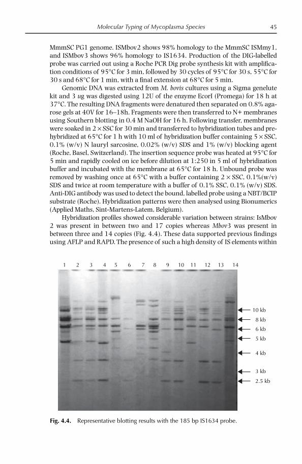
Molecular Typing of Mycoplasma Species 45
MmmSC PG1 genome. ISMbov2 shows 98% homology to the MmmSC ISMmy1, and ISMbov3 shows 96% homology to IS1634. Production of the DIG-labelled probe was carried out using a Roche PCR Dig probe synthesis kit with amplifi ca-tion conditions of 95°C for 3 min, followed by 30 cycles of 95°C for 30 s, 55°C for 30 s and 68°C for 1 min, with a fi nal extension at 68°C for 5 min.
Genomic DNA was extracted from M. bovis cultures using a Sigma genelute kit and 3 ug was digested using 12U of the enzyme EcorI (Promega) for 18 h at 37°C. The resulting DNA fragments were denatured then separated on 0.8% aga-rose gels at 40V for 16–18h. Fragments were then transferred to N+ membranes using Southern blotting in 0.4 M NaOH for 16 h. Following transfer, membranes were soaked in 2 × SSC for 30 min and transferred to hybridization tubes and pre-hybridized at 65°C for 1 h with 10 ml of hybridization buffer containing 5 × SSC, 0.1% (w/v) N lauryl sarcosine, 0.02% (w/v) SDS and 1% (w/v) blocking agent (Roche, Basel, Switzerland). The insertion sequence probe was heated at 95°C for 5 min and rapidly cooled on ice before dilution at 1:250 in 5 ml of hybridization buffer and incubated with the membrane at 65°C for 18 h. Unbound probe was removed by washing once at 65°C with a buffer containing 2 × SSC, 0.1%(w/v) SDS and twice at room temperature with a buffer of 0.1% SSC, 0.1% (w/v) SDS. Anti-DIG antibody was used to detect the bound, labelled probe using a NBT/BCIP substrate (Roche). Hybridization patterns were then analysed using Bionumerics (Applied Maths, Sint-Martens-Latem, Belgium).
Hybridization profi les showed considerable variation between strains: IsMbov2 was present in between two and 17 copies whereas Mbov3 was present in between three and 14 copies (Fig. 4.4). These data supported previous fi ndings using AFLP and RAPD. The presence of such a high density of IS elements within
1 2 3 4 5 6 7 8 9 10 11 12 13 14
10 kb
8 kb
6 kb
5 kb
4 kb
3 kb
2.5 kb
Fig. 4.4. Representative blotting results with the 185 bp IS1634 probe.
Chapter 446
these small genomes indicates an important function in the stability of populations. It is likely that they have a role in genome rearrangement, causing expression of variable proteins.
Other Typing Methods Applied to Mycoplasma Species
Gene sequencing approaches
Recently sequencing methods have been introduced as a new approach for study-ing the molecular epidemiology of bacterial pathogens (Enright and Spratt, 1999). Gene sequencing remains a popular typing method used among a wide range of Mycoplasma species, including M. gallisepticum, M. hyopneumoniae, M. synoviae, M. conjunctivae and others (summarized in Table 4.2). For M. gallisepticum overall, gene targeted sequencing analysis of multiple surface-protein genes was demon-strated to have better discriminatory power than RAPD analysis (Ferguson et al., 2005). Some authors have analysed strains using restriction enzyme analysis without the need for sequencing. 16S sequencing has also proved useful for the genotyping of M. c. capripneumoniae isolates (Pettersson et al., 1998) and has recently been applied to strains from outbreaks of CCPP in the Thrace region of Turkey (Churchward et al., unpublished results).
MLST approaches
Given the rapidly increasing number of sequenced mycoplasma genomes the use of MLST is an increasingly attractive method for typing of Mycoplasma species. MLST is an unambiguous procedure for characterizing isolates of bacterial spe-cies using the sequences of internal fragments of seven housekeeping genes. For each housekeeping gene, the different sequences present within a bacterial species are assigned as distinct alleles and, for each isolate, the alleles at each of the seven loci defi ne the allelic profi le or sequence type (ST). Each isolate of a species is therefore unambiguously characterized by a series of seven integers which cor-respond to the alleles at the seven housekeeping loci. In MLST, the number of nucleotide differences between alleles is ignored and sequences are given different allele numbers whether they differ at a single nucleotide site or at many sites. The rationale is that a single genetic event resulting in a new allele can occur by a point mutation (altering only a single nucleotide site) or by a recombinational replacement (which will often change multiple sites).
MLST is based on the well-established principles of multilocus enzyme elec-trophoresis but differs in that it assigns alleles at multiple housekeeping loci directly by DNA sequencing rather than indirectly via the electrophoretic mobility of their gene products.
More recently molecular epidemiology studies have used multilocus sequence analysis for the typing of MmmSC (Lorenzon et al., 2003). This approach differ-ered from traditional MLST analysis, as the targets for sequencing were not house-keeping genes but non-coding regions or regions adjacent to insertion sequences

Molecular Typing of Mycoplasma Species 47
as it was thought likely that these regions would harbour greater variation than housekeeping genes. This approach was able to differentiate strains to some extent, with isolates being subdivided into 15 different groups (Lorenzon et al., 2003).
Recently an MLST based on the sequence of fi ve housekeeping genes has been developed for members of the M. mycoides cluster (Manso-Silván et al., 2007). This method was initially developed for use in determining phylogeny of the ambiguous mycoides cluster but it may also have limited application for use in the
Table 4.2. Summary of typing methods applied to Mycoplasma species.
Mycoplasma species Typing method Reference
M. mycoides SC VNTRMLSAAFLP / PFGESDS PAGEImmunoblottingIS 1296 analysisIS MyMy analysisBgl REA
McAuliffe et al., 2007Lorenzon et al., 2003Kusiluka et al., 2001Goncalves et al., 1998Poumarat and Solsona, 1995Cheng et al., 1995Westberg et al., 2002Vilei and Frey, 2004
M. mycoides LC LppA REA Monnerat et al., 1999M. sp. bovine group 7 REA of genomic DNA Djordjevic et al., 2001M. c. capripneumoniae 16S rDNA sequencing
H2 sequencingAFLP/PFGE
Pettersson et al., 1998Lorenzon et al., 2003Kusiluka et al., 2001
M. agalactiae PFGE/SDS PAGERAPD/VNTRIS analysisP40 adhesin analysis16S rDNA sequencing
Tola et al., 1996
McAuliffe et al., 2008Pilo et al., 2003Fleury et al., 2002Konigsson et al., 2002
M. bovis AFLP/RAPD/PFGEIS analysis16S rDNA sequencingVsp analysis
McAuliffe et al., 2004Parham et al., 2006Konigsson et al., 2002Beier et al., 1998
M. ovipneumoniae RAPD/PFGE Parham et al., 2006M. gallisepticum RAPD/PFGE
Gene sequencingREA of PvpAAFLP
Ley et al., 1997Ferguson et al., 2005Liu et al., 2001Hong et al., 2005
M. synoviae PFGE/RAPDAFLPSequencing of vlhA
Marois et al., 2001Feberwee et al., 2005Hong et al., 2004
M. hyopneumoniae AFLP/RAPDPFGEP146 sequencingVNTR of adhesion genes
Stakenborg et al., 2006Stakenborg et al., 2005Mayor et al., 2008de Castro et al., 2006
M. conjunctivae LppS sequencing Belloy et al., 2003M. cynos RAPD/PFGE Mannering et al., 2008

Chapter 448
intraspecifi c genotyping of some species. MmmSC and M. species bovine group 7 exhibited very little/no intraspecifi c variation using this approach but some lim-ited variation was seen between strains of M. c. capricolum and M. mycoides LC/M. capri. MLST approaches have many advantages, the data are entirely free from subjective analysis and it is particularly amenable to intra-laboratory comparison as internet-based databases can be constructed.
References
Beier, T., Hotzel, H., Lysnyansky, I., Grajetzki, C., Heller, M., Rabeling, B., Yogev, D. and Sachse, K. (1998) Intraspecies polymorphism of vsp genes and expression profi les of variable surface protein antigens (Vsps) in fi eld isolates of Mycoplasma bovis. Veterinary Microbiology 63, 189–203.
Belloy, L., Vilei, E.M., Giacometti, M. and Frey, J. (2003) Characterization of LppS, an adhesin of Mycoplasma conjunctivae. Microbiology 149, 185–193.
Boettger, C.M. and Dohms, J.E. (2006) Separating Mycoplasma gallisepticum fi eld strains from nonpathogenic avian mycoplasmas. Avian Diseases 50, 605–607.
Byrne, W., McCormack, M., Hotzel, H. and Sachse, K. (2001) Variable surface protein ex-pression profi les and PCR fi ngerprints of clinical isolates of Mycoplasma bovis from Irish cattle. In: Poveda J.B., Fernandez, A., Johansson, K.-E. and Frey, J. (eds) Mycoplas-mas of Ruminants: Pathogenicity, Diagnostics, Epidemiology and Molecular Genetics, vol. 5. European Commission, Brussels, pp. 14–17.
Cheng, X., Nicolet, J., Poumarat, F., Regalla, J., Thiaucourt, F. and Frey, J.R. (1995) Inser-tion element IS1296 in Mycoplasma mycoides subsp. mycoides small colony identifi es a European clonal line distinct from African and Australian strains. Microbiology 141, 3221–3228.
de Castro, L.A., Rodrigues Pedroso, T., Kuchiishi, S.S., Ramenzoni, M., Kich, J.D., Zaha, A., Henning Vainstein, M., and Bunselmeyer Ferreira, H. (2006) Variable number of tandem amino acid repeats in adhesion-related CDS products in Mycoplasma hyopneu-moniae strains. Veterinary Microbiology 116, 258–269.
Djordjevic, S.R., Forbes, W.A., Forbes-Faulkner, J., Kuhnert, P., Hum, S., Hornitzky, M.A., Vilei, E.M. and Frey, J. (2001) Genetic diversity among Mycoplasma species bovine group 7: clonal isolates from an outbreak of polyarthritis, mastitis and abortion in dairy cattle. Electrophoresis 22, 3551–3561.
Dufour-Gesbert, F., Dheilly, A., Marois, C. and Kempf, I. (2006) Epidemiological study on Mycoplasma synoviae infection in layers. Veterinary Microbiology 114, 148–154.
Enright, M.C. and Spratt, B.G. (1999) Multilocus sequence typing. Trends in Microbiology 7, 482–487.
Farlow, J., Smith, K.L., Wong, J., Abrams, M., Lytle, M. and Keim, P. (2001) Francisella tula-rensis strain typing using multiple-locus, variable-number tandem repeat analysis. Journal of Clinical Microbiology 39, 3186–3192.
Farlow, J., Postic, D., Smith, K.L., Jay, Z., Baranton, G. and Keim, P. (2002) Strain typing of Borrelia burgdorferi, Borrelia afzelii, and Borrelia garinii by using multiple-locus variable-number tandem repeat analysis. Journal of Clinical Microbiology 40, 4612–4618.
Feberwee, A., Dijkstra, J.R., von Banniseht-Wysmuller, T.E., Gielkens, A.L. and Wagenaar, J.A. (2005) Genotyping of Mycoplasma gallisepticum and M. synoviae by Amplifi ed Fragment Length Polymorphism (AFLP) analysis and digitalized Random Amplifi ed Polymorphic DNA (RAPD) analysis. Veterinary Microbiology 111, 125–131.
Molecular Typing of Mycoplasma Species 49
Ferguson, N.M., Hepp. D., Sun, S., Ikuta, N., Levisohn, S., Kleven, S.H. and Garcia, M. (2005) Use of molecular diversity of Mycoplasma gallisepticum by gene-targeted sequencing (GTS) and random amplified polymorphic DNA (RAPD) analysis for epidemiological studies. Microbiology 151, 1883–1893.
Fleury, B., Bergonier, D., Berthelot, X., Peterhans, E., Frey, J. and Vilei, E.M. (2002) Charac-terization of P40, a cytadhesin of Mycoplasma agalactiae. Infection and Immunity 70, 5612–5621.
Frothingham, R. and Meeker-O’Connell, W.A. (1998) Genetic diversity in the Mycobacterium tuberculosis complex based on variable numbers of tandem DNA repeats. Microbiology 144, 1189–1196.
Geary, S.J., Forsyth, M.H., Aboul Saoud, S., Wang, G., Berg, D.E. and Berg, C.M. (1994) Mycoplasma gallisepticum strain differentiation by arbitrary primer PCR (RAPD) fingerprinting. Molecular and Cellular Probes 8, 311–316.
Gilmour, J.S., Jones, G.E. and Rae, A.G. (1979) Experimental studies of chronic pneumonia of sheep. Comparative Immunology, Microbiology and Infectious Diseases 1, 285–293.
Goncalves, R., Regalla, J., Nicolet, J., Frey, J., Nicholas, R., Bashiruddin, J., de Santis, P. and Goncalves, A.P. (1998) Antigen heterogeneity among Mycoplasma mycoides subsp. mycoides SC isolates: discrimination of major surface proteins. Veterinary Microbiology 63, 13–28.
Hale, H.H., Helmboldt, C.F., Plastridge, W.N. and Stula, E.F. (1962) Bovine mastitis caused by Mycoplasma species. Cornell Veterinary 52, 582–591.
Hong, Y., Garcia, M., Leiting, V., Bencina, D., Dufour-Zavala, L., Zavala, G. and Kleven, S.H. (2004) Specifi c detection and typing of Mycoplasma synoviae strains in poultry with PCR and DNA sequence analysis targeting the hemagglutinin encoding gene vlhA. Avian Diseases 48, 606–616.
Hong, Y., Garcia, M., Levisohn, S., Savelkoul, P., Leiting, V., Lysnyansky, I., Ley, D.H. and Kleven S.H. (2005) Differentiation of Mycoplasma gallisepticum strains using amplifi ed fragment length polymorphism and other DNA-based typing methods. Avian Diseases 9, 43–49.
Hotzel, H., Schnieder, B. and Sachse, K. (1998) Investigation of Mycoplasma bovis fi eld iso-lates using PCR fi ngerprinting. In: Leori, G., Santini, F., Scanziani, E. and Frey, J. (eds) Mycoplasma of Ruminants: Pathogenicity, Diagnostics, Epidemiology and Molecular Genet-ics, vol. 2. European Commission, Brussels, pp.17–19.
Kokotovic, B., Friis, N.F., Jensen, J.S. and Ahrens, P. (1999) Amplifi ed fragment length poly-morphism fi ngerprinting of Mycoplasma species. Journal of Clinical Microbiology 37, 3300–3307.
Kokotovic, B., Friis, N.F., Nielsen, E.O. and Ahrens, P. (2002) Genomic diversity among Danish fi eld strains of Mycoplasma hyosynoviae assessed by amplifi ed fragment length polymorphism analysis. Veterinary Microbiology 85, 221–231.
Konigsson, M.H., Bolske, G. and Johansson, K.E. (2002) Intraspecifi c variation in the 16S rRNA gene sequences of Mycoplasma agalactiae and Mycoplasma bovis strains. Veterinary Microbiology 85, 209–220.
Kusiluka, L.J.M., Kokotovic, B., Ojeniyi, B., Friis, N.F. and Ahrens, P. (2000) Genetic varia-tions among Mycoplasma bovis strains isolated from Danish cattle. FEMS Microbiology Letters 192, 113–118.
Kusiluka, L.J., Ojeniyi, B., Friis, N.F., Kokotovic, B. and Ahrens, P. (2001) Molecular epide-miology of contagious bovine pleuropneumonia in Tanzania based on amplifi ed frag-ment length polymorphism and pulsed-fi eld gel electrophoresis analysis. Journal of Veterinary Medicicine B, Infectious Diseases and Veterinary Public Health 48, 303–312.
Ley, D.H., Berkhoff, J.E. and Levisohn, S. (1997) Molecular epidemiologic investigations of Mycoplasma gallisepticum conjunctivitis in songbirds by random amplifi ed polymor-phic DNA analyses. Emerging Infectious Diseases 3, 375–380.
Chapter 450
Liu, T., Garcia, M., Levisohn, S., Yogev, D. and Kleven, S.H. (2001) Molecular variability of the adhesin-encoding gene pvpA among Mycoplasma gallisepticum strains and its application in diagnosis. Journal of Clinical Microbiology 39, 1882–1888.
Lorenzon, S., Arzul, I., Peyraud, A., Hendrikx, P. and Thiaucourt, F. (2003) Molecular epi-demiology of contagious bovine pleuropneumonia by multilocus sequence analysis of Mycoplasma mycoides subspecies mycoides biotype SC strains. Veterinary Microbiology 93, 319–333.
Mannering, S.A., McAuliffe, L., Lawes, J.R., Erles, K. and Brownlie, J. (2008) Strain typing of Mycoplasma cynos isolates from dogs with respiratory disease. Veterinary Record (in press).
Manso-Silván, L., Perrier, X. and Thiaucourt, F. (2007) Phylogeny of the Mycoplasma my-coides cluster based on analysis of fi ve conserved protein-coding sequences and possi-ble implications for the taxonomy of the group. International Journal of Systematic and Evolutionary Microbiology 57, 2247–2258.
March, J.B. (1999) Contagious bovine pleuropneumonia. Causative agent: taxonomy and molecular epidemiology. In: Report of the FAO/OIE/OAU CBPP Consultative Group Meeting, Rome, Italy, 5–7 October 1998. FAO, Rome, Italy, pp. 47–51.
March, J.B., Clark, J. and Brodlie, M. (2000) Characterization of strains of Mycoplasma my-coides subspecies mycoides small colony type isolated from recent outbreaks of conta-gious bovine pleuropneumonia in Botswana and Tanzania: evidence for a new biotype. Journal of Clinical Microbiology 38, 1419–1425.
Marois, C., Dufour-Gesbert, F. and Kempf, I. (2001) Molecular differentiation of Mycoplas-ma gallisepticum and Mycoplasma imitans strains by pulsed-fi eld gel electrophoresis and random amplifi ed polymorphic DNA. Journal and Veterinary Medicine B of Infectious Diseases, Veterinary Public Health 48, 695–703.
Mayor, D., Jores, J., Korczak, B.M. and Kuhnert, P. (2008) Multilocus sequence typing (MLST) of Mycoplasma hyopneumoniae: a diverse pathogen with limited clonality. Veterinary Microbiology 127, 63–72.
McAuliffe, L., Kokotovic, B., Ayling, R.D. and Nicholas, R.A.J. (2004) Molecular epidemio-logical analysis of Mycoplasma bovis isolates from the United Kingdom shows two genetically distinct clusters. Journal of Clinical Microbiology 42, 4556–4565.
McAuliffe, L., Ayling, R.D. and Nicholas, R.A.J. (2007) Identifi cation and characterisation of variable-number tandem-repeat markers for the molecular epidemiological analysis of Mycoplasma mycoides subspecies mycoides SC. FEMS Microbiology Letters 276, 181–188.
McAuliffe, L., Churchward, C., Lawes, J., Loria, G.R., Ayling, R.D. and Nicholas R.A.J. (2008) VNTR analysis reveals unexpected genetic diversity within Mycoplasma agalactiae, the main causative agent of contagious agalactia. BMC Microbiology submitted.
Miles, K., McAuliffe, L., Persson, A., Ayling, R.D. and Nicholas, R.A.J. (2005) Insertion se-quence profi ling of UK Mycoplasma bovis fi eld isolates. Veterinary Microbiology 107, 301–306.
Monnerat, M.P., Thiaucourt, F., Nicolet, J. and Frey, J. (1999) Comparative analysis of the lppA locus in Mycoplasma capricolum subsp. capricolum and Mycoplasma capricolum subsp. capripneumoniae. Veterinary Microbiology 69, 157–172.
Musser, J.M. (1996) Molecular population genetic analysis of emerged bacterial pathogens: selected insights. Emerging Infectious Diseases 2, 1–17.
Nicholas, R.A.J. and Ayling, R.D. (2003) Mycoplasma bovis: disease, diagnosis and control. Research in Veterinary Science 74, 105–112.
Parham, K., Churchward, C.P., McAuliffe, L., Nicholas, R.A.J. and Ayling, R.D. (2006) A high level of strain variation within the Mycoplasma ovipneumoniae population of the UK has implications for disease diagnosis and management. Veterinary Microbiology 118, 83–90.

Molecular Typing of Mycoplasma Species 51
Persson, A., Jacobsson, K., Frykberg, L., Johansson, K.E. and Poumarat, F. (2002) Variable surface protein Vmm of Mycoplasma mycoides subspecies mycoides small colony type. Journal of Bacteriology 184, 3712–3722.
Pettersson, B., Bolske, G., Thiaucourt, F., Uhlen, M. and Johansson, K.E. (1998) Molecular evolution of Mycoplasma capricolum subsp. capripneumoniae strains, based on poly-morphisms in the 16S rRNA genes. Journal of Bacteriology 180, 2350–2358.
Pfützner, H. and Sachse, K. (1996) Mycoplasma bovis as an agent of mastitis, pneumonia, arthritis and genital disorders. Revue Scientifique et Technique. Office International Epizooties 15, 1477–1494.
Pilo, P., Fleury, B., Marenda, M., Frey, J. and Vilei, E.M. (2003) Prevalence and distribution of the insertion element ISMag1 in Mycoplasma agalactiae. Veterinary Microbiology 20, 37–48.
Poumarat, F. and Solsona, M. (1995) Molecular epidemiology of Mycoplasma mycoides subsp. mycoides biotype Small Colony, the agent of contagious bovine pleuropneumo-nia. Veterinary Microbiology 47, 305–315.
Pourcel, C., Vidgop, Y., Ramisse, F., Vergnaud, G. and Tram, C. (2003) Characterization of a tandem repeat polymorphism in Legionella pneumophila and its use for genotyping. Journal of Clinical Microbiology 41, 1819–1826.
Sabat, A., Krzyszton-Russjan, J., Strzalka, W., Filipek, R., Kosowska, K., Hryniewicz, W., Travis, J. and Potempa, J. (2003) New method for typing Staphylococcus aureus strains: multiple-locus variable-number tandem repeat analysis of polymorphism and genetic relationships of clinical isolates. Journal of Clinical Microbiology 41, 1801–1804.
Slack, A.T., Dohnt, M.F., Symonds, M.L. and Smythe, L.D. (2005) Development of a multiple-locus variable number of tandem repeat analysis (MLVA) for Leptospira inter-rogans and its application to Leptospira interrogans serovar Australis isolates from Far North Queensland, Australia. Annals of Clinical Microbiology and Antimicrobials 30, 4–10.
Stakenborg, T., Vicca, J., Butaye, P., Maes, D., Peeters, J., de Kruif, A. and Haesebrouck, F. (2005) The diversity of Mycoplasma hyopneumoniae within and between herds using pulsed-fi eld gel electrophoresis. Veterinary Microbiology 109, 29–36.
Stakenborg, T., Vicca, J., Maes, D., Peeters, J., de Kruif, A., Haesebrouck, F. and Butaye, P. (2006) Comparison of molecular techniques for the typing of Mycoplasma hyopneu-moniae isolates. Journal of Microbiological Methods 66, 263–275.
Tola, S., Idini, G., Manunta, D., Casciano, I., Rocchigiani, A.M., Angioi, A. and Leori, G. (1996) Comparison of Mycoplasma agalactiae isolates by pulsed fi eld gel electrophore-sis, SDS-PAGE and immunoblotting. FEMS Microbiology Letters 143, 259–265.
Tyler, K.D., Wang, G., Tyler, S.D. and Johnson, W.M. (1997) Factors affecting reliability and reproducibility of amplifi cation-based DNA fi ngerprinting of representative bacterial pathogens. Journal of Clinical Microbiology 35, 339–346.
van Belkum, A., Scherer, S., van Alphen, L. and Verbrugh, H. (1998) Short-sequence DNA repeats in prokaryotic genomes. Microbiology and Molecular Biology Review 62, 275–293.
Vicca, J., Stakenborg, T., Maes, D., Butaye, P., Peeters, J., de Kruif, A. and Haesebrouck, F. (2003) Evaluation of virulence of Mycoplasma hyopneumoniae fi eld isolates. Veterinary Microbiology 97, 177–190.
Vilei, E.M. and Frey, J. (2004) Differential clustering of Mycoplasma mycoides subsp. mycoides SC strains by PCR-REA of the bgl locus. Veterinary Microbiology 100, 283–288.
Vilei, E.M., Nicolet, J. and Frey, J. (1999) IS1634, a novel insertion element creating long, variable-length direct repeats which is specifi c for Mycoplasma mycoides subspecies mycoides small-colony type. Journal of Bacteriology 181, 1319–1323.

Chapter 452
Vilei, E.M., Abdo, E.M., Nicolet, J., Botelho, A., Goncalves, R. and Frey, J. (2000) Genomic and antigenic differences between the European and African/Australian clusters of Mycoplasma mycoides subspecies mycoides SC. Microbiology 146, 477–486.
Westberg, J., Persson, A., Pettersson, B., Uhlen, M. and Johansson, K.E. (2002) ISMmy1, a novel insertion sequence of Mycoplasma mycoides subspecies mycoides small colony type. FEMS Microbiology Letters 208, 207–213.
Whatmore, A.M., Shankster, S.J., Perrett, L.L., Murphy, T.J., Brew, S.D., Thirlwall, R.E., Cutler, S.J. and MacMillan, A.P. (2006) Identifi cation and characterization of variable-number tandem-repeat markers for typing of Brucella spp. Journal of Clinical Microbiology 44, 1982–1993.

© CAB International 2008. Mycoplasma Diseases of Ruminants(R. Nicholas et al.) 53
5 Antigenic Analysis of Mycoplasmas
Introduction
Western (immuno) blotting analysis has been applied successfully to the assessment of antibody response for several Mycoplasma species such as M. bovis (Sachse et al., 1992; Poumarat et al., 1994), M. mycoides subsp. mycoides SC (Gonçalves et al., 1994; Poumarat and Solsona, 1995; Nicholas et al., 1996), M. agalactiae (Solsona et al., 1996) and several species belonging to other members of the Mycoplasma mycoides cluster (Costas et al., 1987; Olsson et al., 1990). This method allows the analysis of the host humoral immune response based on the electrophoretic profi le of the bacte-rial antigens, thus overcoming problems relating to non-specifi c binding in other immunoassays. This was a particular problem in Portugal during the CBPP eradica-tion campaign, where false positive results in the complement fi xation test often led to the slaughter of healthy cattle. In an effort to overcome this problem, a study was initiated at the Laboratorio Nacional Investigacao Veterinaria in Lisbon to study the immunogenicity of M. mycoides SC to identify specifi c and immunodominant anti-gens which could be applied to confi rmatory diagnostic testing. This led to the devel-opment of an immunoblotting test (IBT) for the detection of the antibody response in sera from cattle affected with CBPP (Regalla et al., 1996). A common immuno-logical pattern consisting of fi ve immunodominant antigens with apparent molec-ular weights of 110, 98, 95, 62/60 and 48 kDa was identifi ed in a preliminary study with sera from cattle experimentally and naturally affected by CBPP.
Materials and Methods
Strain, growth conditions and antigen preparation
A log-phase culture of an appropriate M. mycoides SC isolate (derived from Europe or Africa depending on the source of test sera), growing in modifi ed Gourlay

Chapter 554
medium at 37°C, is harvested by centrifugation at 14,600 g for 1 h at 4°C, followed by three washes in phosphate-buffered saline solution (PBS, 0.1M Na2HPO4, 0.1M NaH2PO4, 0.15M NaCl, pH 7.2). Washed cells are resuspended in the same buffer and stored at –20°C (Goncalves et al., 1998).
Sodium dodecyl sulphate-polyacrylamide gel electrophoresis (SDS-PAGE) and Western blot
The protein content of the thawed mycoplasma cells is estimated by an acceptable method. The samples are mixed with lysis buffer (500 mM Tris/HCl, pH 6.8; 4.6% (w/v) SDS; 20% (v/v) glycerol; 10% (v/v) 2-mercaptoethanol and 0.004% bro-mophenol blue) and boiled for 5 min. The lysates are separated by SDS-PAGE in a 1.5 mm-thick, 4% stacking/5–15% gradient resolving gel and the electrophoresis is run at a constant current of 40 mA. The separated proteins are transferred to 0.45 µm nitrocellulose membranes (NC) at 70 V for 1.5 h.
Preparation of antigen strips
The NC membranes are dried, labelling the side on which the proteins are blotted. The NC membranes are incubated in blocking buffer (PBS containing 5% skimmed milk, 1M glycine and 1% egg albumin) for 2 h at room temperature (about 22°C). The membranes are then washed three times each for 15 min at room tempera-ture in 0.1% (v/v) Tween 20 in PBS and again for 10 min in PBS alone. The NC membranes are dried and cut into 3 mm-wide strips and labelled. These strips contain the antigen used for immunoblotting and should be used immediately, dried or kept at –20°C until they are needed.
Quality control of antigen strips
After performing the Western blot, one 20 mm-wide strip from each side of the membrane is cut and stained with Indian ink to verify the entire range of the transferred cellular proteins and molecular weight standards.
After blocking, the membrane is reacted with the positive control serum, one strip from each edge of the membrane, to identify the specifi c bands of 110, 98, 95, 62/60 and 48 kDa (see also the Figs 5.1 and 5.2 below).
Test procedure
Serum samples to be tested are not heat treated. The serum samples, and 1. positive and negative controls, are diluted 1:3 in dilution buffer (PBS with 0.1% skimmed milk and 0.1% egg albumin) and are applied to the antigen-impregrenated side of the strip before incubating at 37°C for 2 h with continuous agitation.

Antigenic Analysis of Mycoplasmas 55
After washing as above, an appropriate dilution of peroxidase-conjugated 2. anti-bovine-IgG (H+L chains) in dilution buffer is applied to the strip and incu-bated for 1 h at room temperature with continuous agitation.
After another wash the substrate comprising 30 mg 4-chloro-1-naphtol dis-3. solved in 10 ml methanol mixed with 50 ml PBS and 30 ml H2O2 is added and left in the dark with continuous agitation until the protein bands are suitably dark. The reaction is stopped with distilled water.
After drying the strips the reading is carried out based on the presence of the 4. core IgG immunoblot profi le of fi ve specifi c antigenic bands of 110, 98, 95, 62/60 and 48 kDa (Fig. 5.1). Sera showing this core immunological profi le are considered positive.
Sensitivity and Specifi city of the IBT
In a fi eld evaluation of sera from CBPP-affected cattle, the IBT detected 92.5% of infected animals, the CFT detected 77.5%, histology detected 36.9% and myco-plasma culture detected only 31%. All sera from nearly 500 cattle from CBPP-free
kDa
212
170
116
94
7667
53
43
30
Group 1 Group 2 Group 3
kDa
1109895
62/60
48
+C
Group 1 – IBT profile from healthy cattle from CBPP-free regionsGroup 2 – IBT profile from non-infected cattle with cross-reactions at CFTGroup 3 – IBT profile from CBPP-infected cattle
Fig. 5.1. Immunoblot analysis of sera from different groups of cattle in Portugal.

Chapter 556
regions, including 177 sera positive by CFT, were negative by IBT (Fig. 5.1). It was concluded that the IBT was highly specifi c and the most sensitive serological test so far described for the detection of cattle affected by CBPP.
Within the framework of the Portuguese eradication programme, a more com-prehensive and longer-term study beginning in 1998 has tested over 2.5 million sera, of which just over 12,000 (0.5%) were positive by CFT. Of these only 64 (0.6%) from 39 herds were positive by IBT and were located in the infected areas prior to 1999, when the last CBPP outbreaks were seen. Following the last outbreaks and up to to 2001, no positive results have been seen in a total of just over 2000 IBT tests, most of which originated from areas previously identifi ed as infected.
Analysis of M. mycoides SC Strains by Immunoblotting
To assess the antigenic homogeneity of M. mycoides SC strains, 46 isolates from European countries (Portugal (n = 24), Spain (n = 6), France (n = 4) and Italy (n = 12)) were examined. The study was also extended to ten strains from Africa, three strains from Australia and to the type strain PG1 (Gonçalves et al., 1998). The results showed that African and Australian strains could be differentiated from European strains by a strong reaction at the 72/70 kDa level, suggesting for the fi rst time that these strains have evolved separately and that the resurgence of CBPP in Europe was not the result of reinfection from Africa. A distinct antigenic difference was also seen between some of the European strains concerning the 98 kDa protein, with several Italian and Portuguese strains lacking this protein (Fig. 5.2). Therefore, the results indicated that the European strains are antigeni-cally different, suggesting that the Italian strains are a variant clone with the same geographic origin.
94
StdFr
2117
2091
2022
PO2
6364
6363
2059
O69
7B5
26B4
21B3
45B1
03Bf
138
6479
921/
157
/13
170
130/
20L2
Fr Fr Fr Sp Sp Sp Sp Pt Pt Pt Pt It It It It It It It
1109895
6260
48
kDa
67
43
30
Fig. 5.2. Immunoblots of M. mycoides SC strains from France, Spain, Portugal and Italy. Seven strains on the right lack 98 kDa protein.

Antigenic Analysis of Mycoplasmas 57
References
Costas, M., Leach, R.H. and Mitchelmore, D.L. (1987) Numerical analysis of PAGE protein patterns and the taxonomic relationships within the ‘Mycoplasma mycoides cluster’. Journal of General Microbiology 133, 3319–3329.
Gonçalves, R., Regalla, J. and Penha Gonçalves, A. (1994) Immunoblotting and electro-phoretic analysis of Mycoplasma mycoides subsp. mycoides SC strains isolated from bovine and small ruminants. IOM Letters 3, 64–65.
Gonçalves, R., Regalla, J., Nicolet, J., Frey, J., Nicholas, R., Bashiruddin, J., De Santis, P. and Penha Gonçalves, A. (1998) Antigen heterogeneity among Mycoplasma mycoides subsp. mycoides SC isolates: discrimination of major surface proteins. Veterinary Microbiology 63, 13–28.
Nicholas, R.A.J., Santini, F.G., Clark, K.M., Palmer, N.M.A., De Santis, P. and Bashiruddin, J.B. (1996) A comparison of serological tests and gross lung pathology for the detec-tion of contagious bovine pleuropneumonia in two groups of Italian cattle. Veterinary Record 139, 89–93.
Olsson, B., Bölske, G., Bergström, K. and Johansson, K-E. (1990) Analysis of caprine myco-plasmas and mycoplasma infections in goats using two-dimensional electrophoresis and immunoblotting. Electrophoresis 11, 861–869.
Poumarat, F. and Solsona, M. (1995) Molecular epidemiology of Mycoplasma mycoides subsp. mycoides biotype small colony, the agent of contagious bovine pleuropneumonia. Veterinary Microbiology 47, 305–315.
Poumarat, F., Solsona, M. and Boldini, M. (1994) Genomic, protein and antigenic variability of Mycoplasma bovis. Veterinary Microbiology 40, 305–321.
Regalla, J., Caporale, V., Giovannini, A., Santini, F., Martel, J.L. and Penha Goncalves, A. (1996) Manifestation and epidemiology of contagious bovine pleuropneumonia in Europe. Revue Scientifi que et Technique. Offi ce des International Epizooties 15, 1309–1329.
Sachse, K., Grajetzki, C., Pfutzner, H. and Hass, R. (1992) Comparison of Mycoplasma bovis strains based on SDS-PAGE and immunoblot protein patterns. Journal of Veterinary Medicine B 39, 246–252.
Solsona, M., Lambert, M. and Poumarat, F. (1996) Genomic, protein homogeneity and antigenic variability of Mycoplasma agalactiae. Veterinary Microbiology 50, 45–58.
© CAB International 2008. Mycoplasma Diseases of Ruminants58 (R. Nicholas et al.)
6 Antimicrobial Sensitivity Testing
Introduction
Mycoplasma species are the smallest organisms capable of self-replication (Razin et al., 1998). Their lack of a cell wall is thought to make them vulnerable to chang-ing osmotic pressures, temperature changes and desiccation, so they were not thought to survive in the environment for long. Their genomes are extremely small, making them reliant upon their host to provide their required nutrients and amino acids for survival. Based on this it could be anticipated that mycoplas-mas should be extremely vulnerable to host immune defences and antimicrobial treatment. However, many mycoplasmas devote a relatively large percentage of their genomes to mechanisms promoting their survival, including antigenic vari-ation to evade the host. Their lack of a cell wall also means that many antimicro-bials, including the penicillins and cephalosporins, that inhibit the cross-linking of amino acid chains in peptidoglycan synthesis are not effective. Mycoplasmas are also resistant to sulphonamides, which inhibit folic acid synthesis, and those aminoglycosides that inhibit microbial respiration (Barragry, 1994). Mycoplas-mas should be sensitive to antimicrobials such as: tetracyclines, which interrupt amino acid transfer to growing peptide chains at ribosome complexes; quinolones and fl uoroquinolones, which affect DNA gyrase activity; and the macrolides and lincosamides, which inhibit protein synthesis. However, the emergence of strains resistant to these and other potentially effective antimicrobials is well documented (Bébéar, 1996). Some mycoplasmas such as M. bovis appear to have the ability to disseminate widely within the host, which must further limit their effective treat-ment by antimicrobials. In addition, recent work (McAuliffe et al., 2006) has shown that mycoplasmas are able to produce biofi lms, which may enable their survival in the environment and within the host, also reducing their susceptibility to antimicrobial treatment.
The genetic basis of antimicrobial resistance has been described for a num-ber of antimicrobial classes in a number of organisms, mainly the major human
Antimicrobial Sensitivity Testing 59
pathogens. Resistance by M. hominis and M. pneumoniae to the macrolides and fl uoroquinolones is caused by point mutations in the 23S rDNA gene (Pereyre et al., 2007) and in the DNA gyrase and topoisomerase IV, ParC gene (Bébéar et al., 2003), respectively. Undoubtedly, similar mutations are responsible for the anti-microbial resistance but very few studies have been performed. Opportunities therefore exist here to use the information and technologies available from work on other organisms (Fluit et al., 2001) to utilize methods such as PCR, gene sequencing and DNA chip technology to assess and screen for the presence and mechanisms of antimicrobial resistance.
Most antimicrobial studies on mycoplasmas have been carried out in vitro and involve determining minimum inhibition concentration (MIC) values for specifi c isolates against a range of antimicrobials. These may be useful in predicting which antimicrobials are most likely to be effective in vivo and for assessing the develop-ment of antimicrobial resistance. However, the pharmocokinetics and pharmaco-dynamics of drugs are complex and are affected by their behaviour within the host, how well the active ingredient is absorbed into the body, where in the body it concentrates and how long it remains at an effective concentration at the site of infection. It has been reported that some compounds have a preference for local-izing in the lungs, and this may be enhanced in pneumonic lungs with the mac-rolide tilmicosin reaching three times the concentration of that reached in non-pneumonic lungs (Reeve-Johnson, 1999). In man, the newer fl uoroquinolo-nes have been shown to concentrate in lung tissue at levels many times higher than that observed in serum (Wise and Honeybourne, 1999). Other factors such as drug interactions may alter the effectiveness of treatment; some of the mac-rolides, specifi cally erythromycin, may form complexes that affect other drugs (Anado-n and Reeve-Johnson, 1999). It is worth bearing in mind too that adverse reactions to antimicrobials may occur so the manufacturers’ instructions should be followed carefully. Some antimicrobials, for example tilmicosin, are not recom-mended for use in lambs under 15 kg.
Minimum Inhibitory Concentrations
MICs are defi ned as the lowest concentration of an antimicrobial that will inhibit the visible growth of a microorganism after a suitable incubation period. MICs are used by diagnostic laboratories mainly to confi rm resistance but most often as a research tool to determine the in vitro activity of new antimicrobials.
Mycoplasmacidal Activity
While MIC endpoint determination measures the inhibitory effects of antibiotics, it does not assess the killing or cidal effect that the antimicrobials have on the organism, particularly in mycoplasmas. Several methods of determining the min-imum mycoplasmacidal concentration (MMC) have been described (Cooper et al., 1993; Hannan, 2000). We have found the easiest method is to subculture all of the wells of an MIC microtitre plate into a fresh microtitre plate, diluting 1 in 20

Chapter 660
with fresh broth, i.e. 10 µl of MIC culture into 190 µl of fresh media, which effec-tively reduces the concentration of the antimicrobials below the MIC level, enabling an MMC value to be obtained after a suitable incubation period. This method may lack some reproducibility owing to the variation in cell concentra-tion but it is generally reliable, giving replicate results within a few dilutions.
Other methods such as that used by Cooper et al. (1993) rely on prolonged incubation times, where it is assumed that the antimicrobial activity declines with incubation time, such that where growth is initially inhibited antimicrobial con-centrations will fall below MIC values. This approach clearly has limitations in relation to the kinetics of antimicrobial decay, the loss of unstable medium com-ponents and the possible growth of resistant mutants, initially present in very low numbers.
Determination of Breakpoints
Antimicrobial susceptibility testing is a key requirement in enabling veterinarians to have an informed choice about the possible effectiveness of antimicrobials in treating disease. However, in practice, the selection of antimicrobials for treat-ment is often little more than an educated guess as information on breakpoints for the different antimicrobials for the organisms within the different host species is not available. The provision of MIC data will guide the veterinarian away from selecting the apparently less effective antimicrobials. However, the pharmocoki-netics of some antimicrobials, which may concentrate in the lungs or milk for example, may make some antimicrobials suitable for treating some conditions despite having an apparently high MIC. There is therefore a need to determine breakpoints, which have been defi ned as the relationship between MIC values and effective treatment dose for different antimicrobials against mycoplasmas in different host animals.
Mutation Prevention Concentration
Concern about the development of antimicrobial resistance has seen interest grow in mutant prevention concentrations (MPC). MPC is used to determine the capacity of an antimicrobial compound to minimize or limit the selection of fi rst-step resistant mutants in a bacterial population. For a given combination of anti-microbial compound and bacterial strain, MPC is defi ned as the minimum inhibitory concentration (MIC) of the least-susceptible mutant. Thus, bacterial growth in the presence of antimicrobial concentrations greater than the MPC requires the strain to develop two or more spontaneous chromosomal point muta-tions that each cause resistance. MPC is estimated in vitro as the lowest antimicro-bial concentration that prevents colony growth when approximately 1010 cells are applied to agar plates containing the antimicrobial. The choice of 1010 cells as an inoculum is based on the following considerations:
An inoculum of 10 ●10 cells is large enough for mutant subpopulations to be
present and for these to be isolated for further testing if required.

Antimicrobial Sensitivity Testing 61
Clinical infections rarely contain more than 10 ●10 organisms.
Testing an inoculum greater than 10 ●10 is logistically diffi cult.
MPC determinations have used two basic approaches. First, approximately 1010 cells are plated on single agar plates that differ in drug concentration by two-fold increments. This approach allows large numbers of isolates to be tested, but consecutive plates in the series may yield confl uent growth followed by no growth because of the large concentration increment. Furthermore, such a high inoculum density may affect the apparent susceptibility of some strains. Second, cells are applied to multiple agar plates at each antimicrobial concentration such that the total number of cells tested for a given concentration exceeds 1010. If suffi ciently narrow concentration increments are used, isolated colonies can be enumerated to show that their number progressively approaches zero as the antimicrobial concen-tration increases (when colony number is plotted against antimicrobial concen-tration, the ‘mutant selection curve’ becomes steeper as the MPC is approached).
In practice these methods for testing mycoplasmas are diffi cult because it is not easy to obtain the large number of cells required and the mycoplasma media are not usually compatible with the use of large plates, where moisture and agar disruption is a problem. The MPC is then best carried out using a series of small Petri dishes. Our experience has shown that controls grow well to give individual colonies but the antimicrobials may affect the growth so that individual colonies cannot be defi ned without prolonged incubation, which may affect the antimicro-bial being tested or be the result of a mycoplasmastatic effect rather than a true mutation effect.
Factors Affecting Antibiotic Susceptibility Testing
Media
A standard medium does not exist for all mollicutes because of their diverse nutri-tional requirements. Consequently a recognized medium that gives optimal growth for the organism being tested should be used. Using unsuitable media will result in suboptimal growth, giving falsely low MIC values. The medium used should not contain any other antimicrobials such as the penicillins, which are often used to inhibit bacterial contamination, as these may interact with the antimicrobials being tested.
Mycoplasma isolates
It is essential that the purity and identifi cation of the isolate being tested is estab-lished. The inoculum must be standardized and it is recommended that between 103 and 105 colony-forming units (cfu) per ml is used, ideally in the log phase of growth. We have found that the best way to approximate the concentration of cells for use in MIC tests is to measure the optical density and then confi rm the concentration later using a standard dilution count method (Postgate, 1969).
Briefl y, mycoplasmas growing in a broth medium are distributed evenly by shaking, and 1 ml of medium containing the cells is placed into an Eppendorf

Chapter 662
tube. The tube is then micro-centrifuged at 600 g for 3 min. The supernatant is carefully removed and the cells resuspended in 1 ml of PBS. The tube is then cen-trifuged as before, the supernatant removed, the cells resuspended in 1 ml of PBS and the OD450 determined. As a guide an OD450 of 0.1 is equivalent to approxi-mately 1 × 10⁸ cfu per ml. The concentration of the inoculum can then be calcu-lated and adjusted as necessary. 10 µl of this adjusted cell concentration is then dispensed into 190 µl of medium with antibiotic in duplicate and PBS used as a blank. This has been shown to work well for many mycoplasmas but should be evaluated for the species being tested.
Antimicrobial agents
For results to be comparable between tests it is important that the MIC value is based on the active antimicrobial ingredient. Tests should therefore be carried out on the pure antimicrobial compound for which the concentration of active ingre-dient is known. Calculations can then be made to adjust the active concentration to 100%. It is always preferable to use fresh stocks of antimicrobials, although some may be stable if stored under the correct conditions (Hannan, 2000). Many antimicrobials are water-soluble and therefore easy to make into solutions; how-ever, dissolving others may require agitation, mild warming or the use of organic solvents, acids or alkali. If acids or alkali are used, the pH of the solution must be readjusted to pH 7.0 before carrying out the MIC test. It is also essential to ensure that the solvents are not inhibitory to the organisms being tested, so appropriate controls must be used. The dilutions used to determine the MIC are dependent on the test requirements. Often doubling dilutions from 64 to 0.03 µg/ml are used, but it may be necessary to obtain concentrations above 64 µg/ml if an end point is required for some of the antimicrobial-resistant organisms. Essentially, anti-microbial dilutions should be prepared according to the current Clinical and Laboratory Standards Institute (CLSI) guidelines (www.clsi.org).
Incubation conditions
The conditions and times of incubation should be those that enable optimal myco-plasma growth to occur. However, consideration needs to be given to the effect these may have on the antimicrobial being tested, as heat, gases or light may adversely affect their action. In agar dilution assays, drying out can be avoided by using a humidifi ed incubator or by the inclusion of a wet paper towel in the con-tainer with the MIC plates. In liquid microtitre MIC assays, it is essential that each well is sealed to prevent exchange of gases between wells, which may result in false colour changes, leading to erroneous MIC end point values.
Detection of mycoplasma growth in broth
It is essential that adequate controls are used to enable non-specifi c inhibitory fac-tors which may affect mycoplasma growth to be identifi ed. A number of different

Antimicrobial Sensitivity Testing 63
methods for reading the end point have been used. For most ruminant mycoplas-mas, we use visible detection of cell growth for determining the MIC value. At the end of the assay, the microtitre plates are centrifuged at 800 g for 3 min and then examined using an inverted mirror in a light box so that the growth or absence of growth can be clearly observed at the bottom of each microwell. However, for many organisms where visible growth is hard to detect, pH changes can be recorded. A comparison of MIC end points for M. mycoides subsp. mycoides small colony type gave no differences in end point values using pH change or cell growth. For non-glucose-fermenting organisms such as M. bovis, other indicators of growth have been used, including Tween 80 hydrolysis (Devrieuse and Haesebrouck, 1991), tetrazolium (ter Laak et al., 1993) and alarmaBlue (Rosenbusch et al., 2005).
Care should be taken in the interpretation of end points, particularly when meta-bolic inhibition or indicator systems are used, as colour changes may be seen even though the cells are not actively growing as metabolism continues albeit at a low rate. Therefore for standardization purposes, isolates for which MIC values have previously been determined should be used in parallel to enable comparisons to be made.
Other Methods
Antimicrobial sensitivity discs containing a wide range of antimicrobials are used routinely for many bacteria. These can give a comparatively rapid guide to the sensitivity or resistance of an organism to an antimicrobial. However, many vari-ables exist, such as the type of agar used, which can affect antimicrobial diffusion rates. Moreover, the interpretation of the size of the zones of inhibition is rarely precise and does not relate to an MIC value. However, a major advance on disc methods is the E test strips, which use a defi ned gradient technology to give MIC values by reading the zone of growth inhibition against the marked strips. The methodology requires the use of standard media and inocula. Although the range of antimicrobials offered for use on the E test is growing, currently some veteri-nary antimicrobials that are used in treating mollicute infections are not readily available. E tests have been used successfully for M. bovis (Francoz et al., 2005) and M. hominis (Waites et al., 1997, 1999).
An ATP-dependent luminometry method has been described for determining the MICs of highly fastidious species (Robertson and Stemke, 1995; Bébéar and Robertson, 1996), which may be more sensitive than traditional tests. Assunçao et al. (2006) described the use of a rapid fl ow cytometric method using Sybr green-I and propidium iodide stains to determine the MICs for M. agalactiae, which compared favourably with a microbroth dilution method; fl ow cytometry has the additional advantage that it allows the effect of each antibacterial agent to be determined at the single-cell level in real time.
Standardization
Recent work by a CLSI subcommittee on human mycoplasmas has determi-ned standard test conditions for both agar dilution and microbroth dilution for

Chapter 664
M. pneumoniae. They also identifi ed a strain which gave reference ranges for qual-ity control against at least fi ve antimicrobials. Similar work needs to be carried out for the key veterinary mycoplasma pathogens.
References
Anado-n, A. and Reeve-Johnson, L. (1999) Macrolide antibiotics, drug interactions and microsomal enzymes; implications for veterinary medicine. Research in Veterinary Science 66, 197–203.
Assunçao, P., Antunes, N.T., Rosales, R.S., de la Fe, C., Poveda, C., Poveda, J.B. and Davey, H.M. (2006) Flow cytometric method for the assessment of the minimal inhibitory con-centrations of antimicrobial agents to Mycoplasma agalactiae. Cytometry Part A 69A, 1071–1076.
Barragry, T.B. (1994) Veterinary Drug Therapy. Lea and Febinger, Philadelphia, Pennsylva-nia.
Bébéar, C. (1996) Antibiotic sensitivity testing: introductory remarks. In: Tully, J.G. and Razin, S. (eds) Molecular and Diagnostic Procedures in Mycoplasmology, Vol II. Academic Press Limited, London, pp. 181–183.
Bébéar, C. and Robertson, J.A. (1996) Determination of minimum inhibitory concentration. In: Tully, J.G. and Razin, S. (eds) Molecular and Diagnostic Procedures in Mycoplasmology, Vol II. Academic Press, London, pp. 189–197.
Bébéar, C.M., Renaudin, H., Charron, A., Clerc, M., Pereyre, S. and Bébéar, C. (2003) DNA gyrase and topoisomerase IV mutations in clinical isolates of Ureaplasma spp. and Mycoplasma hominis resistant to fl uoroquinolones. Antimicrobial Agents and Chemo-therapy 47, 3323–3325.
Cooper, A.C., Fuller, J.R., Fuller, M.K., Whittlestone, P. and Wise, D.R. (1993) In vitro activity of danofl oxacin, tylosin and oxytetracycline against mycoplasmas of veterinary im-portance. Research in Veterinary Science 54, 329–334.
Devrieuse, L.A. and Haesebrouck, F. (1991) Antibiotic susceptibility testing of Mycoplasma bovis using Tween 80 hydrolysis as an indicator of growth. Journal of Veterinary Medicine B 38, 781–783.
Fluit, A.C., Visser, M.R. and Schmitz, F.-J. (2001) Molecular detection of antimicrobial resistance. Clinical Microbiology Reviews 14, 836–871.
Francoz, D., Fortin, M., Fecteau, G. and Messier, S. (2005) Determination of Mycoplasma bovis susceptibilities against six antimicrobial agents using the E test method. Veterinary Microbiology 105, 57–64.
Hannan, P.C.T. (2000) Guidelines and recommendations for antimicrobial minimum inhi-bition concentration (MIC) testing against veterinary mycoplasma species. Veterinary Research 31, 373–395.
McAuliffe, L., Ellis, R.J., Miles, K., Ayling, R.D. and Nicholas, R.A.J. (2006) Biofi lm forma-tion by mycoplasma species and its role in environmental persistence and survival. Microbiology 152, 913–922.
Pereyre, S., Charron, A., Renaudin, H., Bébéar, C. and Bébéar, C.M. (2007) First report of macrolide resistant strains and description of a novel sequence variation in the P1 adhesin gene in Mycoplasma pneumoniae clinical strains isolated in France over 12 years. Journal of Clinical Microbiology 45, 3534–3539.
Postgate, J.R. (1969) Viable counts and viability. Methods in Microbiology 1, 611–628. Razin, S., Yogev, D. and Nat, Y. (1998) Molecular biology and pathogenicity of mycoplas-
mas. Microbiology and Molecular Biology Reviews 62, 1094–1156.
Antimicrobial Sensitivity Testing 65
Reeve-Johnson, L. (1999) The impact of mycoplasma infections in respiratory diseases of cattle in Europe. In: Stipkovits, L., Rosengarten, R. and Frey, J. (eds) Mycoplasmas of Ruminants: Pathogenicity, Diagnostics, Epidemiology and Molecular Genetics, Vol 3. European Commission, Brussels, pp. 18–31.
Robertson, J.A. and Stemke, G.W. (1995) Measurement of mollicute growth by ATP-dependent luminometry. In: Razin, S. and Tully, J.G. (eds) Molecular and Diagnostic Procedures in Mycoplasmology, Vol I. Academic Press, London, pp. 65–71.
Rosenbusch, R.F., Kinyon, J.M., Apley, M., Funk, N.D., Smith, S. and Hoffman, L.J. (2005) In vitro antimicrobial inhibition profi les of Mycoplasma bovis isolates recovered from various regions of the United States from 2002 to 2003. Journal of Veterinary Diagnos-tic Investigations 17, 436–441.
ter Laak, E.A., Noordergraaf, J.H. and Verschure, M.H. (1993) Susceptibilities of Mycoplas-ma bovis, Mycoplasma dispar and Ureaplasma diversum strains to antimicrobial agents in vivo. Antimicrobial Agents and Chemotherapy 37, 317–321.
Waites, K.B., Crabb, D.M., Duffy, L.B. and Cassel, G.H. (1997) Evaluation of the E test for detection of tetracycline resistance in Mycoplasma hominis. Diagnostic Microbiology and Infectious Diseases 27, 117–122.
Waites, K.B., Canupp, K.C. and Kenny, G.E. (1999) In vitro susceptibilities of Mycoplasma hominis to six fluoroquinolones as determined by E test. Antimicrobial Agents and Chemotherapy 43, 2571–2573.

This page intentionally left blank

II Diseases

This page intentionally left blank

© CAB International 2008. Mycoplasma Diseases of Ruminants(R. Nicholas et al.) 69
7 Contagious Bovine Pleuropneumonia
As for ‘lung sick’ which is a dreadful form of pneumonia, very prevalent in this country, they had all been inoculated against it. This is done by cutting a slit in the tail of an ox, and binding in a piece of the diseased lung of an animal which has died of the sickness. The result is that the ox sickens, takes the disease in a mild form, which causes its tail to drop off, as a rule about a foot from the root, and becomes proof against future attacks. It seems cruel to rob the animal of his tail, especially in a country where there are so many fl ies, but it is better to sacrifi ce the tail and keep the ox than to lose both tail and ox, for a tail without an ox is not much good, except to dust with. (H. Rider Haggard, King Solomon’s Mines, 1888)
Introduction
Contagious bovine pleuropneumonia (CBPP), the only bacterial disease classifi ed as a list A disease by the Offi ce International des Epizooties in 2004, is caused by Mycoplasma mycoides subsp. mycoides small colony (SC) variant. This mycoplasma was isolated in the late 19th century, and, while eradicated from Europe, still pres-ents immense problems in Africa. Following the eradication of rinderpest from most parts of the African continent, CBPP is now the most important transbound-ary disease, along with foot and mouth disease, although its effect on the animals is far more severe.
Geographical Distribution
Africa
In World Animal Health in 2006, the OIE (2008) reported outbreaks of CBPP in 20 countries, with the highest number of cases in Ethiopia, Angola and Cameroon. It is clear that this is not an accurate fi gure as there were no reports from a number of countries, including Tanzania, Somalia, Congo and Burundi, where CBPP is
Chapter 770
believed to be endemic. It is also evident that the number of cases reported by the countries is an underestimate and is highly dependent on the limited resources of many of these countries.
Endemic infection extends throughout the pastoral herds of much of west-ern, central and eastern Africa, with Angola, Zambia and northern Namibia in southern Africa. Newly infected areas in the 1990s include much of Uganda, parts of Kenya, the Democratic Republic of Congo and most of Tanzania, where the disease spread alarmingly, with estimates of losses calculated at US$40 mil-lion (Nicholas et al., 2007). Rwanda (1994), Burundi (1997), Botswana (1995) and Zambia (1997) were the latest countries re-infected, although Botswana remains free after an extensive slaughter campaign in 1995. After 60 years of freedom, the Caprivi region of Namibia became infected twice from Zambia within a 2-year period. Mortality rates in affected herds ranged from 10 to 90% (Nicholas et al., 2007). It is hoped now that with an improving economic and political cli-mate in Angola control measures will be implemented to prevent the continual spread of disease into the neighbouring countries of Namibia and Zambia.
Asia
CBPP has been reported in recent times in the Far East but it is now believed to be absent, with India, Myanmar and Pakistan declaring freedom in 1990, 1995 and 1997, respectively. However, as recently as the late 1970s, high incidences of CBPP were being reported in the Brahmaputra Valley districts of Akajan, Sissibor-gaon, Dhemaji and Dhakuakhana (Choudhary et al., 1987). Little information has emerged from China until the last few years. The disease caused great eco-nomic losses to China’s cattle industry between the 1950s and 1970s, but follow-ing an eradication campaign initially involving mass vaccination, followed by quarantine and slaughter, CBPP was reported to have been eradicated in 1989 (Xin et al., 2007). However, recent serological surveys have reported small numbers of seropositive reactions by both ELISA and CFT in Tibet, Yunnan and Guizhou provinces (Xin et al., 2007); whether these constitute true positives is not clear. Japan, infected in 1910 from Australia, declared freedom in 1941.
Sporadic outbreaks occur in the Middle East, including United Arab Emirates (1990), Kuwait (1991) and Saudi Arabia (1995), largely as a result of importa-tion of cattle from north-east Africa. Suspicions of CBPP in Turkey have not been confi rmed by the small surveys performed in the last few years. In the most com-prehensive study, just under 1000 animals were examined at abattoirs in eastern Turkey. The testing of blood and tissue samples from cattle showing suspicious lung lesions showed less than 0.5% of sera were positive with no isolation of M. mycoides SC from any lesions, strongly suggesting that CBPP was absent or at a very low seroprevalence in Turkey (Çetinkaya et al., 2003).
Europe
The traditional home of European CBPP in the 18th century was Switzerland, in particular the canton of Suabia, where the disease broke out between 1790

Contagious Bovine Pleuropneumonia 71
and 1812, which neatly coincided with the Napoleonic military campaigns which criss-crossed Europe and required large herds to feed the invading forces (Salmon, 1896). Although believed to have been present in the 17th and 18th centuries, Britain became re-infected in 1839 through Ireland from Holland, which had become the ‘hot-bed of pleuropneumonia’ and responsible for an increased spread of CBPP within Europe as trade opened up when the French Wars ended (cited in Fisher, 2003). No real attempt was made to control the disease in Britain in the next 30 years, resulting in 1500 outbreaks being reported in 1870, and 3000 5 years later; a major breakthrough was perhaps inevitably seen in 1878 with the payment of full compensation to owners for slaughtered animals, and the num-ber of outbreaks fell to just over 2000 in 1890 (Hutyra and Marek, 1913). Once responsibility for CBPP control was transferred from local to central government in 1890 there was a fi nal drive for complete eradication and the last case was diagnosed in 1892.
CBPP was largely eradicated from Europe around the end of the 19th century but there was a resurgence after the First World War. The disease reappeared in Eastern Europe, including the Balkans, but was eventually suppressed in Germany (1924), Russia (1928), Georgia (1932) and Poland (1936). As the outbreak of the Second World War a few years later would have prevented any further surveil-lance for this disease, it is not inconceivable that pockets of disease remained in Eastern Europe; indeed, outbreaks occurred in Moldova until 1946. Furthermore, small outbreaks continued in southern Europe during most decades of the 20th century, mainly in the Iberian Peninsula. A resurgence of CBPP occurred in the early 1980s, with reports of outbreaks in southern France on several occasions between 1980 and 1984, while in Italy the disease reappeared in 1990 after an absence of nearly 100 years but was eliminated 3 years later (Nicholas et al., 2000). The last case recorded in Spain was in 1994. In Portugal the disease reap-peared, probably from Spain, in 1983 and became endemic in northern Portugal. With an intensifi cation of the eradication campaign, the number of cases declined rapidly, from nearly 3000 in 1996 to a single case in 1999 (Nicholas et al., 2000). No outbreaks have been reported in the present millennium, although it is still too early to conclude that CBPP has fi nally disappeared from Europe.
Origins
Historically, it is well documented that CBPP was carried to South Africa in 1854 with an imported bull from Holland, where it spread rapidly via trek oxen into the Transvaal, killing 100,000 animals within 2 years. It further spread by cattle movements to countries now known as Namibia, Zimbabwe and Botswana, then eventually to Angola, Zambia and possibly the Congo. Windsor (2001) contends that the infection of north-west and East Africa had a separate origin, probably through Abyssinia (now Ethiopia) with an expeditionary force led by the British commander General Napier in 1868 to free some 60 European hostages held by the Abyssinian Emperor Theodore. This seems highly unlikely as accounts by the journalist/explorer Henry Morton Stanley, who accompanied the £9 million mis-sion, details 13,000 troops, 55,000 camp followers, 18,000 mules, 17,000 camels

Chapter 772
and 44 elephants but no cattle. Moreover, even if a few cattle were part of this force, what is far from clear, however, is whether India, from where Napier’s army originated, was infected at the time. It has long been accepted that India and China became infected in 1910 and 1919, respectively, from Australia, which had itself been infected by a small number of cattle from Britain in 1858 (Hutyra et al., 1938). It is very unlikely that infection could have travelled from Australia to India in such a short time, so some doubt must exist about this second front of infection in Africa. However, there is some molecular epidemiological evidence of two clusters of the causative agent to support Roger Windsor’s ‘two front’ claim, but clearly, amongst many things that General Napier has been held responsible for, introducing CBPP into Africa is not one.
The story of the introduction of CBPP into Australia via a short-horned cow called St Bees after a long sea voyage from Cumbria in England in 1858 and how it took a further 120 years to eradicate has been well told, particularly by Newton (1992). Less well known is that the disease entered New Zealand 5 years later and spread rapidly in cattle in the provinces of Otago and Southland (Fisher, 2006). However, unlike the Australian epidemic, CBPP disappeared 10 years later, prob-ably in part because of the prompt action of stock owners, who were quick to recognize the importance of ‘inoculation’ of crude vaccines in its control.
Similarly, the establishment of CBPP for over 50 years in the USA in the 19th century is not common knowledge. It appears that the disease entered the USA from Britain (Hutyra et al., 1938) or Holland (Fisher, 2003) as early as 1842 in a diseased cow sold in Brooklyn. It then spread to New Jersey in 1847 and Massa-chusetts in 1859. Its importance was recognized and attempts were made to erad-icate it in the east; however, this was too late to prevent a breakout to Ohio, probably with Jersey cattle bought in Baltimore. CBPP reached the limits of its expansion in 1884, with outbreaks in Missouri, Kentucky and Illinois (Fig. 7.1), by which time such were the concerns by stock owners that Congress was forced to extend powers of appropriation to enable stamping out in most states; the biggest diffi culties were, however, seen in New York and New Jersey because of ‘the larger territory infected and the density of population’. The disease was fi nally eliminated from these states in 1891 and 1892, respectively. Salmon writing in 1896 stated that it had been eradicated from the USA and confi dently and correctly predicted that ‘it is not probable that it will ever be seen in this country again’.
Economic Impact
A study of the impact of CBPP on the economy of affected countries was carried out in Tanzania, where the estimated loss to the livestock and livestock products trade was US$136 million over a 14-year period since its introduction in 1990. Described as a national disaster by the Minister for Water and Livestock, CBPP has been responsible for the death of 350,000 cattle up to 2004, resulting in the dis-tribution of some 10 million doses of vaccines. A larger study estimated losses in the whole of Africa at 30 million euros as a result of death and disease but the total losses including control, reduced production and trade made it just under 50 million euros (Tambi et al., 2006).
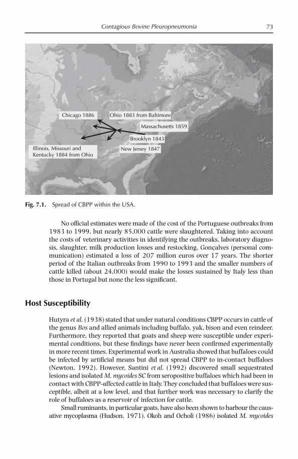
Contagious Bovine Pleuropneumonia 73
No offi cial estimates were made of the cost of the Portuguese outbreaks from 1983 to 1999, but nearly 85,000 cattle were slaughtered. Taking into account the costs of veterinary activities in identifying the outbreaks, laboratory diagno-sis, slaughter, milk production losses and restocking, Gonçalves (personal com-munication) estimated a loss of 207 million euros over 17 years. The shorter period of the Italian outbreaks from 1990 to 1993 and the smaller numbers of cattle killed (about 24,000) would make the losses sustained by Italy less than those in Portugal but none the less signifi cant.
Host Susceptibility
Hutyra et al. (1938) stated that under natural conditions CBPP occurs in cattle of the genus Bos and allied animals including buffalo, yak, bison and even reindeer. Furthermore, they reported that goats and sheep were susceptible under experi-mental conditions, but these fi ndings have never been confi rmed experimentally in more recent times. Experimental work in Australia showed that buffaloes could be infected by artifi cial means but did not spread CBPP to in-contact buffaloes (Newton, 1992). However, Santini et al. (1992) discovered small sequestrated lesions and isolated M. mycoides SC from seropositive buffaloes which had been in contact with CBPP-affected cattle in Italy. They concluded that buffaloes were sus-ceptible, albeit at a low level, and that further work was necessary to clarify the role of buffaloes as a reservoir of infection for cattle.
Small ruminants, in particular goats, have also been shown to harbour the caus-ative mycoplasma (Hudson, 1971). Okoh and Ocholi (1986) isolated M. mycoides
Brooklyn 1843
New Jersey 1847
Massachusetts 1859
Ohio 1883 from Baltimore
Illinois, Missouri andKentucky 1884 from Ohio
Chicago 1886
Fig. 7.1. Spread of CBPP within the USA.

Chapter 774
from an outbreak of disease in sheep in Nigeria. Brandão (1995) isolated M. mycoides SC from the milk of sheep with mastitis as well as from goats with pneu-monia in Portugal outside the then endemic region of CBPP. The pathogenicity of these strains for cattle is unknown at present but has been studied in sheep in Portugal. The milk isolate of M. mycoides SC induced pleuropneumonia, hydrotho-rax and pericarditis in experimentally infected sheep and was isolated from tra-chea, thoracic exudates and lung samples (Machado et al., 1998). The humoral immune response following experimental infection of sheep by the intratracheal–endobronchial route was also monitored over a 3-month period (Gonçalves et al., 2002). A slight cough occurred in some animals and interstitial pneumonia and thickening of pulmonary septa were observed in ten inoculated and six in-contact ewes. One ewe showed hyperplasia of the germinal centres in the lymph nodes and mucosa-associated lymphoid tissue in the lung. M. mycoides SC was detected in frozen samples from one ewe by PCR and RFLP. A specifi c immune response was detected by IBT, starting 2.5 months after inoculation. In all, the clinical and pathological signs appeared too mild for sheep to be considered as models for CBPP infection.
While no evidence has yet been shown of small to large ruminant transmis-sion of CBPP, it is clearly theoretically possible for sheep or goats to harbour the mycoplasma and infect cattle grazing close by.
Transmission/Clinical Signs
Close and repeated contact by expiration of infectious droplets is the principal mode of transmission, although wind-borne and indirect transmission cannot be completely excluded (Regalla et al., 1996). It was felt very strongly in the USA in the late 19th century that infection was possible from previously infected prem-ises, although by what method these fragile mycoplasmas could survive in the environment was until recently quite diffi cult to understand. The discovery that mycoplasmas, including M. mycoides SC, could produce biofi lms enabling increased resistance in the environment may provide an explanation (McAuliffe et al., 2008).
There is considerable variation in the severity of signs observed in cattle affected by CBPP, ranging from hyperacute through acute to chronic and sub-clinical forms. Respiratory distress and coughing, evident on stimulation of rest-ing animals, are the main signs of CBPP. The average incubation period of the natural disease is between 1 and 2 months but can vary from as early as 3 weeks to as much as 4 months. When healthy CBPP-free cattle were placed in contact with naturally affected cattle from a recent outbreak in Namibia, seroconversion was seen after 6 weeks and rose rapidly for the next 2 weeks, by which time 40% of contacts had died (Hubschle et al., 2006). In other experimental aerosol infec-tions, 8% of cattle developed a hyperacute form, 14% acute, 30% mild and 23% inapparent infection with seroconversion; 25% showed no clinical signs and failed to seroconvert (Provost et al., 1987).
The early stages of CBPP are indistinguishable from any severe pneumonia with pleurisy. Animals show dullness, anorexia, irregular rumination with mod-erate fever and may show signs of respiratory disease (Fig. 7.2). Coughing is usually

Contagious Bovine Pleuropneumonia 75
persistent and is slight or dry. Sometimes there is fever with body temperatures of 40–42°C, and the animal prostrates with diffi culty of movement (Fig. 7.3). As the lung lesions develop, the signs become more pronounced, with increased fre-quency of coughing and the animal becomes prostrate or stands with the back arched, head extended and elbows abducted (Fig. 7.4). While classical respiratory signs may be evident in young calves, though pathological lesions are usually slight and restricted to small sequestra, articular localization of the causative agent with attendant arthritis predominates; respiratory disease is the major sign in calves over 6 months.
The clinical signs observed in the acute form are much accelerated. The path-ological signs are usually characteristic, with marked pleural adhesion accompa-nied by exudative pericarditis (Regalla et al., 1996). Affected animals may die within a week, exhibiting classical respiratory signs.
Calves Arthritis/mild pneumonia
Adults (6 months and older) Progression
Acute stage(Maximum 5 days)
Depressed, anorexia, dyspnoeaFall in milk productionTemperature up to 40°C
Sub-acute stage(may last 10–15 days)
May develop cough, increased respiratoryrate, thoracic pain, elbows abductedWide variety of râles, dullnessin lung areas on percussion
Sub-acute to chronic Open-mouthed breathing, severedyspnoea, fluid noisesRumination ceasesExpiratory gruntsReluctance to moveArched back, stands stifflyLarge joints painful, swollenLying in lateral recumbency
Sequelae
Chronic Chronic diseaseNon-thrivingRecurrent low-grade feverExercise precipitates coughing
Death Death (varies 10–70%) dependson strain, stage of outbreak
Recovery Sequestrum or complete resolution
Fig. 7.2. Clinical signs of CBPP (from Done et al., 1995).
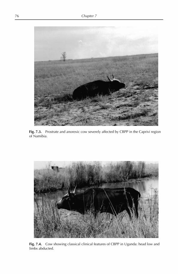
Chapter 776
Fig. 7.3. Prostrate and anorexic cow severely affected by CBPP in the Caprivi region of Namibia.
Fig. 7.4. Cow showing classical clinical features of CBPP in Uganda: head low and limbs abducted.

Contagious Bovine Pleuropneumonia 77
In the subacute form, signs may be limited to a slight cough only noticeable when the animal is exercised. CBPP in Portugal, unlike that caused in Africa, where mortality rates are typically 10–70% in epizootics, was characterized by low mor-bidity and low or non-existent mortality, with the majority of infected cattle show-ing chronic lesions. These differences are, in part, due to the fact that European cattle are healthier in general, better fed, subjected to less physical stress and are often permanently housed throughout the year, requiring little movement; they also probably experience strains of lower virulence than in Africa, although this has never been proved convincingly as animal experiments have been limited to one study using a single African and European strain in very few animals (Abdo et al., 1998). However, signifi cant mortality was reported in the Pyrenees in 1980, including deaths in as much as half of one of the three herds affected, many of which showed clinical signs (ter Laak, 1992). In Italy, during the early 1990s, fewer than 5% of cattle in the fi rst outbreak showed clinical signs, possibly sug-gesting that the disease had been present for several years before it was diag-nosed (Guadagnini et al., 1991). The increased and ready use of antibiotics and anti-infl ammatory drugs may also help to mask clinical signs in affected cattle in Europe. In Africa, it was estimated that up to a third of cases that recover from acute disease become potential carriers (Nicholas et al., 2000). This fi gure was probably higher in Europe, where there is a far more widespread use of antimicrobials.
Pathogenesis/Pathology
In the absence of hard evidence for how the mycoplasma initiates infection, an immunopathological theory has been proposed in which autoimmune complexes are formed as a result of the host response against the pneumogalactan in the lung tissue, stimulated by the reaction to galactan produced by the mycoplasma; this causes thrombi in the lymphatic system, where lesions develop fi rst. As a con-sequence, the lymph coagulates with distension of the interlobular septa and infi ltration of lymphocytes and plasma cells around the blood vessels. The forma-tion of these ‘cuffs’ or ‘organizing centres’ around the arterioles is the only histo-logical pathognomic characteristic of CBPP. The secondary lesion involves the alveoli, which become full of exudates from the previous changes. Necrotic foci, surrounded by polymorphic granulocytes, develop and become sequestra if the lesion is too large to resolve.
Lesions are mostly confi ned to the lungs and thoracic cavity and mostly appear in a single lung. In a study in Portugal, 95% of lesions were unilateral (Egwu et al., 1996), with the diaphragmatic lobes being more commonly affected than cranial lobes. In a small study of recent outbreaks in Namibia, just under 80% of lungs had lesions in a single lung (Aschenborn, personal communica-tion). In Europe the main lesion is the sequestrum but acute lesions are seen, par-ticularly in the early phase of the epidemic; sequestra also predominate in parts of Africa, where the disease has become endemic, suggesting that virulence is cyclic rather than geographic.
Adhesions to the chest causing roughened pleural membranes are common (Fig. 7.5). Many litres of straw-coloured pleural fl uid can be found in acute cases,

Chapter 778
which makes ideal diagnostic material (Fig. 7.6). The interlobular septa are often distended (Fig. 7.7) and lungs show the typical marbled appearance, with lung lobules showing great variations in colour from red, to grey to yellow, depending on the stage of infl ammation (Fig. 7.8). Associated lymph nodes undergo hyper-trophy (Fig. 7.9). In chronic cases the sequestrum is the main lesion type and consists of necrotic material surrounded by a fi brotic capsule ranging from 10 to 100 mm in diameter (Figs 7.10 and 7.11). Necrotic foci have been reported in the kidneys of affected cattle and pericarditis is occasionally seen in severe cases.
Histopathologically, the CBPP lesion comprises a bronchiolar necrosis and oedema, progressing rapidly to an exudative serofi brinous bronchiolitis with extension to the alveoli and uptake of alveolar fl uid into tissue spaces, lymph ves-sels and ultimately septal lymphatics (Done et al., 1995). These rapidly reach sat-uration and the process is extended centrifugally to the tracheobronchial lymph nodes and centripetally to the pleural lymphatics. The mediastinal, sternal, aortic and intercostal lymph nodes may then become enlarged, oedematous or even haemorrhagic. With stasis, lymph vessels become thrombosed and ultimately fi brosed (Nicholas et al., 2000). The pulmonary lobules become consolidated with alveolar oedema, fi brin and infl ammatory cells. Coagulative necrosis is common. M. mycoides SC can be demonstrated in these lobules by immunohistochemistry. Perivascular organization foci or ‘organizing centres’, found in the interlobular septa, are considered pathognomonic for CBPP (Provost et al., 1987). They consist of a centre occupied by a blood vessel with proliferation of connective and infl am-matory cells surrounded by a peripheral zone of necrotic cells.
Fig. 7.5. Adhesion of the lung to the chest wall of a cow affected by CBPP.

Contagious Bovine Pleuropneumonia 79
Fig. 7.6. Pleural fl uid taken from cattle acutely affected with CBPP.
Fig. 7.7. Lung of CBPP-affected cow showing oedematous enlargement of interlobular septa.

Chapter 780
Fig. 7.8. Lung from CBPP-affected cow with classical ‘marbled’ lesion.
Fig. 7.9. Hypertrophic lymph nodes from cow with CBPP.
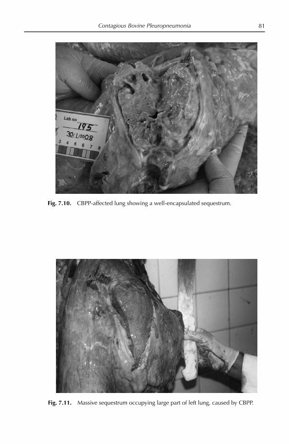
Contagious Bovine Pleuropneumonia 81
Fig. 7.10. CBPP-affected lung showing a well-encapsulated sequestrum.
Fig. 7.11. Massive sequestrum occupying large part of left lung, caused by CBPP.
Chapter 782
Causative Organism
The sequencing of the entire genome of the type strain PG1 in 2004 made much of the early work on characterizing the M. mycoides SC redundant (Westberg et al., 2004). The genome is characterized by a single circular chromosome of 1,211,703 bp with the lowest G + C content (24 mol %) and the highest density of insertion sequences (13% of the genome size) of all sequenced bacterial genomes. The genome contains 985 putative genes with an average length of 982 bp. Only 59% of these have been assigned with a function, while 27% are unique to M. mycoides SC and 14% have unknown function but show signifi cant homology to genes of other Mycoplasma or bacterial species. A variety of potential virulence factors was identifi ed, including genes encoding putative variable surface proteins and enzymes and transport proteins responsible for the production of hydrogen peroxide and the capsule, which is believed to have toxic effects on the animal.
Virulence Factors
Little is known of the pathogenicity factors associated with M. mycoides SC. The galactan capsule is generally considered to contribute to pathogenicity by promot-ing binding to host tissue surfaces and enhancing resistance to phagocytosis. In addition, there is some evidence that the capsule of M. mycoides SC might have a direct toxic effect on host cells, and its structural similarity to bovine pneumo-galactan further suggests that it might induce autoimmune reactions.
The production of hydrogen peroxide and other active oxygen species appears to be an important factor in mycoplasma pathogenicity (Tryon and Baseman, 1992). In mycoplasmas, hydrogen peroxide production may accompany the metabolism of sugars or certain organic acids to acetate plus carbon dioxide. There is a net reduction of NAD+ to NADH during this process and NAD+ is regen-erated via NADH oxidase activity; this oxidation requires molecular oxygen, which is reduced to hydrogen peroxide or water. However, in M. mycoides SC, only traces of hydrogen peroxide are formed (Miles et al., 1991). An alternative means of hydrogen peroxide production in mycoplasmas is via the metabolism of glycerol. African and Australian but not European M. mycoides SC strains oxidized glycerol at high rates which were comparable with those for glucose (Houshaymi et al., 1997). European isolates possessed GP kinase but lacked GP oxidase (Houshaymi et al., 1998). Thus, it appears possible that the apparently reduced virulence of European SC strains, compared with African strains, is specifi cally associated with the lack of GP oxidase activity. Further evidence of the role of hydrogen peroxide has become evident in studies in which strains from acute cases of disease were shown to be more cytotoxic to cultured bovine epithelial cells than those taken from milder outbreaks in Europe (Bischof et al., 2008). The fact that vaccine strains were also shown to be cytotoxic suggests that they are, as many thought, still largely semivirulent preparations (Mbulu et al., 2004); however, Bischof et al. (2008) believe that they may be attenuated in other, though unspecifi ed, ways.
Pilo et al. (2007) have proposed that virulence of mycoplasmas is a factor of their ability to evade the host immune response through reversible antigenic

Contagious Bovine Pleuropneumonia 83
changes, to adhere strongly to the host cell and to persist and disseminate via a blood-borne phase. They believe that damage to the host is caused by the effi cient and competitive scavenging of nutrients and the release of toxic metabolites, lead-ing to infl ammatory processes that can exacerbate disease. That the host response can often do more damage than the mycoplasma itself can be seen partly in the work of Scacchia et al. (2007), where long-term treatment with cyclosporin, which suppresses the cell-mediated immune response, delayed and reduced the size of lesions in some cattle.
In order to evade the host’s immune response, many mycoplasmas express surface proteins that undergo reversible changes to alter their antigenic reper-toire. Some of these variable surface proteins are also involved in adhesion and immunomodulation. Only two are known in M. mycoides SC, one of which, Vmm, is a small lipoprotein of 17 kDa (Persson et al., 2002). Hamsten et al. (2008) showed that antibodies in sera from cattle naturally affected with CBPP recognized Vmm and other Vmm-type proteins, suggesting these may be useful as target antigens for the development of diagnostic tests and recombinant vaccines.
Molecular and Immunological Epidemiology
M. mycoides SC is a relatively homogeneous species and few typing methods have been able to detect signifi cant differences between strains. Many studies have found variation in African strains but have been unable to subdivide European strains (Cheng et al., 1995; Gonçalves et al., 1998; Lorenzon et al., 2003). To date, the consensus on the molecular epidemiology of M. mycoides SC is consistent with there being two main subtypes: those isolated from recent European outbreaks (post-1980) and those isolated from the African and Australian outbreaks, some of which date back to 1936. Initial studies on the molecular typing of M. mycoides SC focused on the restriction analysis of whole DNA (Poumarat and Solsona, 1995) and the chromosomal distribution of insertion sequences (Cheng et al., 1995; Frey et al., 1995). Although these methods could differentiate European from African/Australian strains and distinguish vaccine strains, they were unable to subtype strains further. It must be said that some of the studies performed in the 1990s were often motivated more by a desire to show that the authors’ country was not responsible for a neighbour’s outbreak rather than a desire for good science.
The source of the unexpected outbreaks of CBPP in Italy (1990–1993) was never successfully traced, although France was heavily implicated because it exports large numbers of cattle to Italy and they had experienced disease in the previous decade. Immunological evidence was presented that most strains from Italy differed from those from outbreaks in Western Europe, leading to the sugges-tion that ‘contamination of Italy did not arise from exportation of CBPP from south western Europe’ but from a resurgence in Italy itself or from some unknown foci elsewhere in Europe (Poumarat and Solsona, 1995). However, in the study, two Italian isolates had identical patterns to those from France, Spain and Portu-gal. A more thorough study by Gonçalves et al. (1998), but still lacking suffi cient French and Spanish strains, confi rmed that two Italian strains were similar to other European strains but others lacked the 98 kDa protein.

Chapter 784
Epidemiological evidence compiled during the Italian outbreaks also impli-cated France, as well as, unexpectedly, Germany and Poland, as outbreaks were linked directly to importation from these three countries (Regalla et al., 1996) but it seemed highly unlikely that Italy, which had been free of the disease for nearly 100 years, should be infected from three different countries within the space of 2–3 years, two of which had not experienced outbreaks for over 50 years. It suggested, perhaps, that tracing the origin and movements of these cattle was far from certain. Of the non-infected countries in Europe, only Switzerland and Hungary have recently conducted targeted surveys to demonstrate freedom from CBPP, although the UK and France actively investigate suspicious cases where other causes have been ruled out.
M. mycoides SC strains have also been studied using amplifi ed fragment length polymorphism and pulsed fi eld gel electrophoresis (PFGE); however, these methods only detected limited variation and have not been used on a wide selection of strains (Kusiluka et al., 2001). Based on the polymorphisms of eight housekeeping genes, MLST (see below) differentiated 48 strains into 32 profi les and three main groups and confi rmed the distinction between European and African/Australian strains; some separation was also apparent between strains from West and East Africa from those of Southern Africa, suggesting different origins of African strains (Yaya et al., 2007). A recently unearthed European strain from 1967 which lacks the 8.8 kb deletion of other European strains was located at an intermediate posi-tion between European and certain African strains and is probably much more closely related to ancestral European strains that are thought to be responsible for spreading CBPP into Southern Africa. This work also postulates an introduction of CBPP into the Horn of Africa based on a single isolate from India, although the date of entry, 1868, as discussed earlier, suggested seems highly unlikely.
Later molecular epidemiological studies have focused on multilocus sequence typing (MLST), which aims to fi nd loci in the mycoplasma genome that show suf-fi cient polymorphisms to enable subtyping. This approach was able to differenti-ate strains to some extent, with isolates being subdivided into 15 different groups (Lorenzon et al., 2003). Overall, epidemiological studies to date have shown that most European isolates fall into a single homogenous group, while the African and Australian strains form a separate, more heterogeneous, grouping. This het-erogeneity is most likely due to the widely separated geographical and temporal origins of the African and Australian strains studied in contrast to those from European outbreaks, which have been collected over a relatively short period of time and from a restricted area (March et al., 2000).
Techniques using variable numbers of tandem repeats (VNTR) have been described for a variety of organisms but have not been used to differentiate myco-plasma strains previously. A recent study has found that despite its minimal size the M. mycoides SC genome has 60 VNTRs throughout the M. mycoides SC genome and that there was remarkable genetic homogeneity between strains from differ-ent geographical origins, with only three VNTRs showing variation between strains (McAuliffe et al., 2007). However, these three VNTRs showed promise as an epidemiological tool, fi nding variation with African and Australian strains and generating eight different profi les from the 15 strains tested. There was some cor-relation between the geographic origin of the strains and the VNTR grouping.

Contagious Bovine Pleuropneumonia 85
Vaccine strains were unusual and most were placed in a single group by VNTR analysis, with the exception of KH3J, which was distinct from all other strains.
Little was known about the origin of CBPP in Asia until relatively recently, although the general view was that the disease was imported from Australia via infected cattle into New Zealand, India, China, Mongolia, Hong Kong and Japan. A possible alternative route was through trade with Russia. Reports from China indicate that outbreaks in the second half of the 20th century were characterized by high morbidity and mortality, suggesting that strains were likely to be similar to the African/Australian cluster. Proof of this was provided by Li et al. (2008), who showed that M. mycoides SC strains from China isolated between 1953 and 1960 lacked the 8.4 kb deletion of the European strains, possessed the LppB gene and were identical to African/Australian strains by MLST. This confi rmed the opinion that China became infected via Australia.
Participatory Epidemiology
Farmers, including pastoralists, in Africa have a very well-developed knowledge of many aspects of CBPP, covering recognition of the contagiousness, carrier status, immunity and seasonality; they are also very aware of the dangers of trade both legal and illegal but consider these risks worth taking for the socio-economic benefi ts that arise from these activities. Mariner and Catley (2004), describing their fi ndings from participatory epidemiological surveys of livestock owners in regions endemically affected by CBPP, showed that farmers were aware of the positive effects that antibiotic treatment can have but that these are often ineffec-tive because of under dosage. Mass vaccination programmes on the other hand were seen as having limited or no impact at all. Based on the results of fi eld inves-tigations, mathematical modelling and literature reviews, Mariner and Catley (2004) concluded that mass vaccination alone with the currently available T1/44 vaccine was unlikely to eradicate CBPP, even when applied biannually over a 5-year period, unless linked to a proven treatment regime in which all diseased cattle would be treated while the remainder of the herd was vaccinated; such an option was the only one that approached eradication when modelled.
Diagnosis
While there are plenty of tools available for CBPP diagnosis, including numerous PCR techniques, there is no single test which can detect all infected animals. The OIE-approved serological tests, complement fi xation test (CFT) and the competi-tive (c)ELISA, lack sensitivity. The cELISA is clearly more robust but is often less sensitive than the CFT (Marobela-Raborokgwe et al., 2003). An indirect ELISA based on a recombinant protein LppQ-N’ had similar sensitivity to the CFT and cELISA but was very stable under harsh conditions (Bruderer et al., 2002). Using an ELISA based on this lipoprotein, Xin et al. (2007) screened nearly 4000 sera from Tibet and two provinces of China and showed good agreement between the ELISA and CFT, although the former appeared more sensitive. A more sensitive
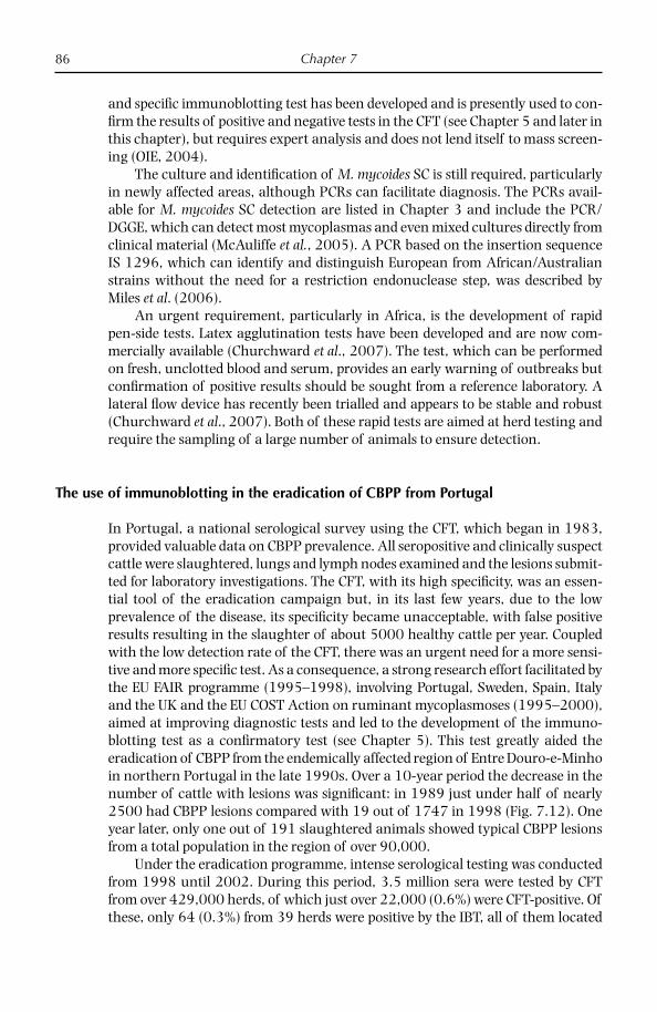
Chapter 786
and specifi c immunoblotting test has been developed and is presently used to con-fi rm the results of positive and negative tests in the CFT (see Chapter 5 and later in this chapter), but requires expert analysis and does not lend itself to mass screen-ing (OIE, 2004).
The culture and identifi cation of M. mycoides SC is still required, particularly in newly affected areas, although PCRs can facilitate diagnosis. The PCRs avail-able for M. mycoides SC detection are listed in Chapter 3 and include the PCR/DGGE, which can detect most mycoplasmas and even mixed cultures directly from clinical material (McAuliffe et al., 2005). A PCR based on the insertion sequence IS 1296, which can identify and distinguish European from African/Australian strains without the need for a restriction endonuclease step, was described by Miles et al. (2006).
An urgent requirement, particularly in Africa, is the development of rapid pen-side tests. Latex agglutination tests have been developed and are now com-mercially available (Churchward et al., 2007). The test, which can be performed on fresh, unclotted blood and serum, provides an early warning of outbreaks but confi rmation of positive results should be sought from a reference laboratory. A lateral fl ow device has recently been trialled and appears to be stable and robust (Churchward et al., 2007). Both of these rapid tests are aimed at herd testing and require the sampling of a large number of animals to ensure detection.
The use of immunoblotting in the eradication of CBPP from Portugal
In Portugal, a national serological survey using the CFT, which began in 1983, provided valuable data on CBPP prevalence. All seropositive and clinically suspect cattle were slaughtered, lungs and lymph nodes examined and the lesions submit-ted for laboratory investigations. The CFT, with its high specifi city, was an essen-tial tool of the eradication campaign but, in its last few years, due to the low prevalence of the disease, its specifi city became unacceptable, with false positive results resulting in the slaughter of about 5000 healthy cattle per year. Coupled with the low detection rate of the CFT, there was an urgent need for a more sensi-tive and more specifi c test. As a consequence, a strong research effort facilitated by the EU FAIR programme (1995–1998), involving Portugal, Sweden, Spain, Italy and the UK and the EU COST Action on ruminant mycoplasmoses (1995–2000), aimed at improving diagnostic tests and led to the development of the immuno-blotting test as a confi rmatory test (see Chapter 5). This test greatly aided the eradication of CBPP from the endemically affected region of Entre Douro-e-Minho in northern Portugal in the late 1990s. Over a 10-year period the decrease in the number of cattle with lesions was signifi cant: in 1989 just under half of nearly 2500 had CBPP lesions compared with 19 out of 1747 in 1998 (Fig. 7.12). One year later, only one out of 191 slaughtered animals showed typical CBPP lesions from a total population in the region of over 90,000.
Under the eradication programme, intense serological testing was conducted from 1998 until 2002. During this period, 3.5 million sera were tested by CFT from over 429,000 herds, of which just over 22,000 (0.6%) were CFT-positive. Of these, only 64 (0.3%) from 39 herds were positive by the IBT, all of them located

Contagious Bovine Pleuropneumonia 87
in the affected areas. However, with the exception of the last outbreak, which occurred in February 1999, no M. mycoides SC isolates were obtained from bacte-riological examinations performed on the slaughtered cattle. The last specifi c pro-fi le was detected in 2000 from an animal with no visible CBPP lesions (Regalla, 2004). All CFT-positive sera originating from disease-free areas gave non-specifi c profi les in the IBT. No positive results were obtained from just under 9000 IBTs performed during 2001 and 2002, most of which came from the previously endemic region. And fi nally, on May 2003, the International Committee of the OIE declared Portugal free from CBPP.
Control
CBPP was one of the fi rst infectious diseases of animals for which vaccines were developed and preventive inoculation of ‘attenuated’ virulent material was prac-tised even before the causative organism was known. Surprisingly, despite being known about for centuries in Europe, it was in Africa where the fi rst attempts at vaccination were made. Virulent material, usually pleural fl uid or lung fragments from cattle dying of the disease, was inserted beneath the skin either on the tip of the tail or on the bridge of the nose. The frequent loss of the tail following vaccina-tion was considered a price worth paying. Even today gross lesions at the site of
1000950900850800750700650600550500450400350300250200150100500 1986 19871988 1989 1990 1991 1992 1993 1994 1995 1996 1997 1998 1999 2000 2001 2002
Years
Num
ber
of o
utbr
eaks
EDM BL TM DFR
239
227
225
498
187
607
527
776
736
880
617
360
240
180
110
28 4617 10 1 23 7
5714 11 1 1 0 0 0 0
Fig. 7.12. Number of CBPP outbreaks seen in the endemic area of Portugal from 1986 to 2002.EDM – Entre Douro e MinhoBL – Beira LitoralTM – Trás-os-MontesDFR – Disease-free regions

Chapter 788
inoculation (Fig. 7.13), often caused by poor vaccine administration, and even occasional death are an accepted side effect of vaccination. Indeed some believe that the production of such lesions is essential for protective immunity (Hudson, 1971).
The OIE-recommended vaccine, which is used throughout Africa, is the T1/44 strain, isolated in Tanzania and attenuated by passaging 44 times in eggs after the Second World War. The vaccine has been in constant use since then and its limitations, including the short duration of immunity, adverse reactions and cold chain dependence, are well documented. It has been proposed that vaccine quality could be improved by simple and inexpensive changes to current vaccines and protocols. Such changes would include the use of HEPES–buffer systems and the inclusion of pH indicators in vaccine media, together with changes to the present vaccine diluent 1 M MgSO4, which is damaging to the mycoplasma (March, 2004). These changes could increase vaccine yields tenfold and stability several 100-fold, increase the ease of production and ultimately produce a vaccine with improved effi cacy.
Two new approaches to vaccination against CBPP were reported by Nicholas et al. (2005): fi rst, the use of a whole cell mycoplasma vaccine inactivated with saponin, an approach which had previously been found to be protective for calf pneumonia, contagious agalactia and CCPP; and second, the use of a recombi-nant subunit vaccine prepared from the highly immunogenic lipoprotein LppQ (Abdo et al., 2000). Results showed that, in spite of two vaccinations at 6-weekly
Fig. 7.13. Adverse reaction caused by poor administration of T1/44 vaccine against CBPP.
Contagious Bovine Pleuropneumonia 89
intervals, there was no evidence in the small number of animals immunized of any protection afforded by either preparation; indeed, there appeared to be an exacerbation of pathology in the vaccinated animals compared with unvacci-nated contact animals. Lesions and fi brin were most extensive and pleural fl uid more abundant in vaccinated animals. In the LppQ group half the cattle died before the end of the experiment while a quarter died in the saponin group. This compared with just under half that died in the control group.
A subunit vaccine based on the capsular polysaccharide (CPS) component of M. mycoides SC was tested in mice (Waite and March, 2002). A signifi cant anti-body response against CPS was elicited only when CPS was conjugated to ovalbu-min. Mice, given a whole cell sonically disrupted vaccine and challenged with live M. mycoides SC, exhibited a signifi cantly reduced degree of mycoplasmaemia when compared with non-vaccinated controls; however, mice immunized with the CPS–ovalbumin conjugate did not show a reduction in mycoplasmaemia (Waite and March, 2002). In addition to the fact that the subunit vaccine did not elicit protection, it seems unlikely that these results can be extrapolated to cattle as the mouse model monitored mycoplasmaemia and not clinical or pathological disease.
Recognizing that, in the fi eld, cattle recovering from CBPP often have long-term immunity, Dedieu-Engelman (2008) analysed the immune response of acute and convalescent animals and showed that the latter were characterized by a stronger and persisting local IgA response and a higher M. mycoides SC-specifi c CD4+ T-cell response, with gamma interferon production detected in the blood and persisting in the respiratory lymph nodes. This suggested that both the local humoral and cell-mediated response were playing a role in protection against CBPP. Thus future vaccines will have to be selected for their ability to elicit or prime the mucosal immunity to inhibit the growth of the pathogen or block its virulence factors and to trigger the specifi c T-cells of the Th1-like type. Ideally these should comprise multicomponent subunit vaccines delivered directly to the mucosal respiratory surface where the mycoplasma initiates infection.
Recently, a novel approach for rapidly testing DNA vaccines using phage library screening was developed for M. mycoides SC (March et al., 2006). This approach enabled rapid, high-throughput screening of genomic libraries so that putative vaccine candidates can be selected and tested. Whole bacteriophage l particles offer many advantages as DNA vaccine delivery vehicles and are cheap and stable. They are also thought to be effective as the antigen-presenting cells are targeted and they have the additional advantage that vaccination produces a highly immunogenic signal against phage coat proteins, therefore enabling easy confi rmation of the vaccination status of the animals. As has been previously dis-cussed, studies such as these that use a mouse model of mycoplasmaemia are not necessarily relevant to the clinical situation in cattle and therefore it is diffi cult to extrapolate fi ndings to the host animal species. More studies in cattle are required to evaluate the potential of these vaccine candidates.
A number of strategies have been used to differentiate vaccine from fi eld strains, which is crucial for epidemiological monitoring. Lorenzon et al. (2000) developed a PCR which amplifi ed a 700 bp fragment specifi c to T1. A PCR based on the 16S rRNA gene coupled with electrophoretic separation of PCR products
Chapter 790
on a denaturing gradient gel could not only differentiate over 70 Mycoplasma species but could clearly differentiate T1 from all other M. mycoides SC strains (McAuliffe et al., 2005).
Treatment
In Europe, apart from a brief period in the second half of the 20th century when vaccines were seriously considered in the Iberian pensinsula following the re-emergence of CBPP, stamping out has been the control method of choice, with the chief detection tools being abattoir surveillance and/or serological monitoring. Control in Africa, where practised, is based on a 60-year-old vaccine which is poorly protective, can cause severe adverse reactions if not performed properly and has residual virulence (Mbulu et al., 2004); movement control is diffi cult to enforce and antibiotics, offi cially frowned upon in some countries, are used widely, although their effectiveness is diffi cult to assess. Evidence from the fi eld has been confusing. Many cattle farmers consider their use essential for keeping affected cattle alive in endemic areas (Mariner and Catley, 2004). Surprisingly then, in a study of CBPP spread within herds in Ethiopia, Lesnoff et al. (2004) could fi nd no evidence that antibiotic treatment reduced case fatality risk and average duration of clinical signs; the authors, however, speculated that the most widely used med-ication, a single intramuscular injection of 10–20 ml of 10% oxytetracycline sus-pension, was unlikely to achieve recovery. Furthermore, it was suggested that the spread of CBPP throughout Tanzania in the mid-1990s may have been due to inadequate dosing of infected cattle with oxytetracyclines, causing a reduction in clinical signs but failing to prevent mycoplasma shedding (Msami et al., 2001).
Despite this, the relentless spread of CBPP throughout Africa, coupled with shortcomings in the vaccine, has required offi cial bodies, in particular the FAO, to re-evaluate the use of antibiotics in the control of this disease, despite the offi cial view in many African countries that their use be prohibited (Benkirane, 1999). This prohibition is testament to the infl uence of the late CBPP expert Alain Pro-vost, who stated that ‘Chemotherapy, including antibiotic therapy, may give excel-lent results from the clinical standpoint but it does not sterilise the infection and favours the creation of chronic carriers’ (Provost et al., 1987). It is timely to review the use of chemotherapy as data have shown that the newer antibiotics, including the fl uoroquinolones, may be mycoplasmacidal, in vitro at least (Ayling et al., 2000).
Hubschle et al. (2006) examined the effectiveness of danofl oxacin (2.5% Advocin, Pfi zer Ltd, New York) on naturally affected cattle and on CBPP transmis-sion to uninfected, seronegative, in-contact cattle. Affected adult cattle, taken from a natural outbreak in the Caprivi region of north-east Namibia, were placed into two groups, one of which was treated with danofl oxacin for 3 days while the other group was saline treated. CBPP-free, seronegative cattle were placed in con-tact with each of the two groups. Although apparently ineffective in improving the clinical and pathological condition of the naturally affected cattle, danofl oxa-cin signifi cantly reduced the transmission of the mycoplasma from the treated cattle to the contact cattle and provided support to the view that chemotherapy may have a role in CBPP control.
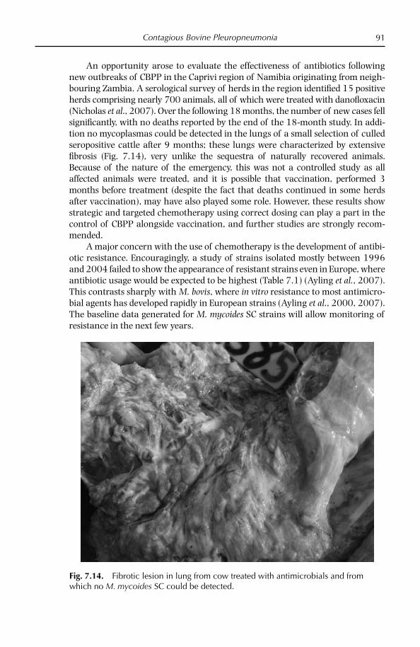
Contagious Bovine Pleuropneumonia 91
An opportunity arose to evaluate the effectiveness of antibiotics following new outbreaks of CBPP in the Caprivi region of Namibia originating from neigh-bouring Zambia. A serological survey of herds in the region identifi ed 15 positive herds comprising nearly 700 animals, all of which were treated with danofl oxacin (Nicholas et al., 2007). Over the following 18 months, the number of new cases fell signifi cantly, with no deaths reported by the end of the 18-month study. In addi-tion no mycoplasmas could be detected in the lungs of a small selection of culled seropositive cattle after 9 months; these lungs were characterized by extensive fi brosis (Fig. 7.14), very unlike the sequestra of naturally recovered animals. Because of the nature of the emergency, this was not a controlled study as all affected animals were treated, and it is possible that vaccination, performed 3 months before treatment (despite the fact that deaths continued in some herds after vaccination), may have also played some role. However, these results show strategic and targeted chemotherapy using correct dosing can play a part in the control of CBPP alongside vaccination, and further studies are strongly recom-mended.
A major concern with the use of chemotherapy is the development of antibi-otic resistance. Encouragingly, a study of strains isolated mostly between 1996 and 2004 failed to show the appearance of resistant strains even in Europe, where antibiotic usage would be expected to be highest (Table 7.1) (Ayling et al., 2007). This contrasts sharply with M. bovis, where in vitro resistance to most antimicro-bial agents has developed rapidly in European strains (Ayling et al., 2000, 2007). The baseline data generated for M. mycoides SC strains will allow monitoring of resistance in the next few years.
Fig. 7.14. Fibrotic lesion in lung from cow treated with antimicrobials and from which no M. mycoides SC could be detected.
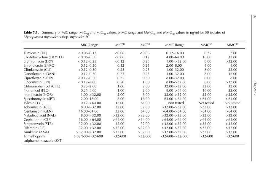
C
hapter 792
Table 7.1. Summary of MIC range, MIC50 and MIC90 values, MMC range and MMC50 and MMC90 values in µg/ml for 50 isolates of Mycoplasma mycoides subsp. mycoides SC.
MIC Range MIC50 MIC90 MMC Range MMC50 MMC90
Tilmicosin (TIL) < 0.06–0.12 < 0.06 < 0.06 0.12–16.00 0.25 2.00Oxytetracycline (OXYTET) < 0.06–0.50 < 0.06 0.12 4.00–64.00 16.00 32.00Erythromycin (ERY) < 0.12–0.25 < 0.12 0.25 1.00–> 32.00 8.00 > 32.00Enrofl oxacin (ENRO) 0.12–0.50 0.12 0.25 2.00–8.00 4.00 8.00Clindamycin (CLI) < 0.12–0.50 0.25 0.25 1.00–32.00 8.00 32.00Danofl oxacin (DAN) 0.12–0.50 0.25 0.25 4.00–32.00 8.00 16.00Ciprofl oxacin (CIP) < 0.12–0.50 0.25 0.50 8.00–32.00 8.00 8.00Lincomycin (LIN) < 0.12–2.00 0.50 1.00 8.00–> 32.00 8.00 > 32.00Chloramphenicol (CHL) 0.25–2.00 1.00 2.00 32.00–> 32.00 32.00 32.00Florfenicol (FLO) 0.25–8.00 1.00 2.00 8.00–> 64.00 16.00 32.00Norfl oxacin (NOR) 1.00–> 32.00 2.00 8.00 32.00–> 32.00 32.00 > 32.00Spectinomycin (SPT) 2.00–16.00 8.00 16.00 64.00–> 64.00 > 64.00 > 64.00Tylosin (TYL) 0.12–> 64.00 16.00 64.00 Not tested Not tested Not testedTobramycin (TOB) 8.00–> 32.00 32.00 32.00 > 32.00–> 32.00 > 32.00 > 32.00Gentamycin (GEN) 16.00–64.00 32.00 64.00 > 64.00–> 64.00 > 64.00 > 64.00Naladixic acid (NAL) 8.00–> 32.00 > 32.00 > 32.00 > 32.00–> 32.00 > 32.00 > 32.00Cephalothin (CEF) 16.00–> 64.00 > 64.00 > 64.00 > 64.00–> 64.00 > 64.00 > 64.00Streptomycin (STR) 32.00–> 32.00 32.00 32.00 > 32.00–> 32.00 > 32.00 > 32.00Rifampin (RIF) 32.00–> 32.00 > 32.00 > 32.00 > 32.00–> 32.00 > 32.00 > 32.00Amikacin (AMK) > 32.00–> 32.00 > 32.00 > 32.00 > 32.00–> 32.00 > 32.00 > 32.00Trimethoprim/ sulphamethoxazole (SXT)
> 32/608–> 32/608 > 32/608 > 32/608 > 32/608–> 32/608 > 32/608 > 32/608

Contagious Bovine Pleuropneumonia 93
References
Abdo, E.M., Nicolet, J., Miserez, R., Gonçalves, R., Regalla, J., Griot, C., Bensaide, A., Krampe, M. and Frey J. (1998) Humoral and bronchial immune responses in cattle experimentally infected with Mycoplasma mycoides subspecies mycoides small colony type. Veterinary Microbiology 59, 109–123.
Abdo, E.M., Nicolet, J. and Frey, J. (2000) Antigenic and genetic characterisation of lipopro-tein LppQ from Mycoplasma mycoides subspecies mycoides small colony type. Clinical Diagnosis and Laboratory Immunology 7, 588–595.
Ayling, R.D., Baker, S.E. Nicholas, R.A.J., Peak, M.L. and Simon, A.J. (2000) Comparison of in vitro activity of danafl oxacin, fl orfenicol, oxytetracycline, spectinomycin and tilmi-cosin against Mycoplasma mycoides subsp. mycoides small colony type. Veterinary Record 146, 243–246.
Ayling, R.D., Godinho, K. and Nicholas, R.A.J. (2007) Comparative studies on the in vitro antimicrobial sensitivities of Mycoplasma mycoides subsp. mycoides SC type and Myco-plasma bovis. CBPP Control: Antibiotics to the Rescue. FAO-OIE-AU/IBAR-IAEA. Consul-tative Group Meeting on CBPP in Africa 2006. FAO, Rome, pp. 51–61.
Benkirane, A. (1999) Role of chemotherapy in CBPP. Report of the First Meeting of the FAO/OIE/OAU-IBAR Consultative Group on CBPP. FAO, Rome, 1998, pp. 97–101.
Bischof, D.F., Janis, C., Vilei, E.M., Bertoni, G. and Frey, J. (2008) Cytotoxicity of Mycoplasma mycoides subsp. mycoides small colony type to bovine epithelial cells. Infection and Immunity 76, 263–269.
Brandão, E. (1995) Isolation and identifi cation of identifi cation of Mycoplasma mycoides subsp. mycoides SC strains in sheep and goats. Veterinary Record 136, 98–99.
Bruderer, U., Regalla, J., Abdo, E.M., Hubschle, O.J.B. and Frey, J. (2002) Serodiagnosis and monitoring of contagious bovine pleuropneumonia (CBPP) with an indirect ELISA based on the specifi c lipoprotein LppQ of Mycoplasma mycoides subsp. mycoides SC. Veterinary Microbiology 84, 195–205.
Çetinkaya, B., Öngör, H., Karahan, M., Kalender, H., Lorenzon, S. and Thiaucourt, F. (2003) Abattoir-based survey of contagious bovine pleuropneumonia in cattle in Turkey. Veterinary Record 152, 254–258.
Cheng, X., Nicolet, J., Poumarat, F., Regalla, J., Thiaucourt, F. and Frey, J. (1995) Insertion element IS1296 in Mycoplasma mycoides subspecies mycoides small colony identifi es a European clonal line distinct from African and Australian strains. Microbiology 141, 3221–3228.
Choudhary, M.R., Datta, B.M., Das, S.K. and Sarma, D.K. (1987) Screening of CBPP in Assam using complement fi xation test. Indian Veterinary Journal 64, 345–346.
Churchward, C.P., Flint, J., Danks, C., Humbert, I., Hashem, E., Nicholas, R.A.J. and Ayling, R.D. (2007) Development and initial evaluation of a lateral fl ow device for the rapid detection of contagious bovine pleuropneumonia. In: Proceedings of the World Association of Veterinary Laboratory Diagnosticians – 13th International Symposium, Melbourne, Australia, 12–14 November 2007.
Dedieu-Engelman, L. (2008) Contagious bovine pleuropneumonia: a rationale for the de-velopment of a mucosal sub-unit vaccine. Comparative Immunology, Microbiology and Infectious Diseases, 31, 227–238.
Done, S., Palmer, N.M.A. and Nicholas, R.A.J. (1995) An emerging disease in Europe: contagious bovine pleuropneumonia. Cattle Practice 3, 13–20.
Egwu, G., Nicholas R.A.J., Ameh, J.A. and Bashiruddin, J.B. (1996) Contagious bovine pleuropneumonia: an update. Veterinary Bulletin 66, 195–208.
Fisher, J. (2003) To kill or not to kill: the eradication of CBPP in Western Europe. Medical History 47, 314–331.
Chapter 794
Fisher, J. (2006) The origin, spread and disappearance of contagious bovine pleuropneu-monia in New Zealand. Australian Veterinary Journal. 84, 439–444.
Frey, J., Cheng, X., Kuhnert, P. and Nicolet, J. (1995) Identifi cation and characterization of IS1296 in Mycoplasma mycoides subsp. mycoides SC and presence in related mycoplas-mas. Gene 160, 95–100.
Gonçalves, M.R ., Regalla, J., Nicolet, J., Frey, J., Nicholas, R.A.J., Bashiruddin, J.B., De Santis, P. and Penha Gonçalves, A. (1998) Antigenic heterogeneity among Mycoplasma my-coides subsp. mycoides SC isolates: discrimination of major surface proteins. Veterinary Microbiology 63, 13–28.
Gonçalves, R., Ferreira-Dias, G., Belo, A., Correia, J., Ferreira, M.L. and Goulão J.V. (2002) Pathological and immunological characteristics of ewes experimentally infected with Mycoplasma mycoides subsp. mycoides SC isolated from cattle and sheep. Small Rumi-nant Research 46, 51–62.
Guadagnini, P.F., De Simone, F., Panini, G.F., Gaffuri, A., Bugnatti, M., Finazzi, M., Madeli, G., Sironi, G. and Belloli, A. (1991) La pleuropneumonite contagiosa dei bovini (PPCB). Selezione Veterinaria 32, 3–31.
Hamsten, C., Westberg, J., Bolske., Ayling, R.D., Uhlen, M. and Persson, A. (2008) Expres-sion and immunogenicity of six putative variable surface proteins in Mycoplasma my-coides subsp. mycoides SC. Microbiology 154, 539–549.
Houshaymi, B.M., Miles, R.J. and Nicholas, R.A.J. (1997) Oxidation of glycerol differenti-ates African from European isolates of Mycoplasma mycoides subspecies mycoides SC (small colony). Veterinary Record 140, 182–183.
Houshaymi, B.M., Miles, R.J. and Nicholas, R.A.J. (1998) Biochemical differentiation of European and African Mycoplasma mycoides subspecies mycoides SC (small colony) isolates. In: Leori, G., Santini, F., Scanziani, E. and Frey, J. (eds) Mycoplasmas of Rumi-nants: Pathogenicity, Diagnostics, Epidemiology and Molecular Genetics, Vol. 2 (ISBN 92-828-1873-X). Offi ce for Offi cial Publications of the European Communities, Luxembourg, pp. 133–136.
Hubschle, O.J.B., Ayling, R.D., Godinho, K., Lukhele, O., Tjipura-Zaire, G., Rowan, T.G. and Nicholas, R.A.J. (2006) Danofl oxacin (AdvocinTM) reduces the spread of contagious bovine pleuropneumonia to healthy in-contact cattle. Research in Veterinary Science 81, 304–309.
Hudson, J.R. (1971) Contagious bovine pleuropneumonia. FAO Agricultural Studies 86, Rome.
Hutyra, F. and Marek, J. (1913) Lungenseuche der rinder. Pleuropneumonia contagiosa bovum. In: Spezielle Pathologie und Therapie der Haustiere. Verlag Von Gustav Fischer, Jena, Germany, pp. 392–406.
Hutyra, F., Marek, J. and Manninger, R. (1938) Special Pathology and Therapeutics of the Diseases of Domestic Animals, 5th edn. Bailliere Tindall, USA, pp. 455–471.
Kusiluka, L.J.M., Ojeniyi, B., Friis, N.F., Kokotovic, B. and Ahrens, P. (2001) Molecular anal-ysis of fi eld strains of Mycoplasma capricolum subspecies capripneumoniae and Myco-plasma mycoides subspecies mycoides, small colony type isolated from goats in Tanzania. Veterinary Microbiology 82, 27–37.
Lesnoff, M., Laval, G., Bonnet, P., Abdicho, S., Workalemahu, A., Kifl e, D., Peyraud, A., Lancelot, R. and Thiaucourt, F. (2004) Within herd spread of contagious bovine pleuropneumonia in Ethiopian highlands. Preventative Veterinary Medicine 64, 27–40.
Li, Y., Xin, J., Gao, Y., Jianhua, Z., Nicholas, R.A.J. and Lin, Y. (2008) Strains of Mycoplasma mycoides subsp. mycoides small colony type isolated in China between 1953 and 1960 show close similarity to strains of the Africa/Australia cluster. The Veterinary Journal doi:10.1016/j.tvjl.2007.08.021.

Contagious Bovine Pleuropneumonia 95
Lorenzon, S., David, A., Nadew, M., Wesonga, H. and Thiaucourt, F. (2000) Specifi c PCR identifi cation of the T1 vaccine strains for contagious bovine pleuropneumonia. Molecular and Cellular Probes 14, 205–210.
Lorenzon, S., Arzul, I., Peyraud, A., Hendrikx, P. and Thiaucourt, F. (2003) Molecular epi-demiology of CBPP by multilocus sequence analysis of Mycoplasma mycoides subsp. mycoides SC strains. Veterinary Microbiology 93, 319–333.
Machado, M., Ferreira, H., Ramos, M.A., Branda-o, E., Tavares, A. and Cardoso, M. (1998) Experimental infection of sheep and goats with Mycoplasma mycoides subsp. mycoides SC isolated from small ruminants. In: Leori, G., Santini, F., Scanziani, E. and Frey, J. (eds) Mycoplasmas of Ruminants: Pathogenicity, Diagnostics, Epidemiology and Molecular Genetics, Vol. 2. COST Action 826, European Commission, EUR 18018 EN, Luxem-bourg, pp. 142–144.
March, J.B. (2004) Improved formulations for existing CBPP vaccines: recommendations for change. Vaccine 22, 4358–4364.
March, J.B., Clark, J. and Brodlie, M. (2000) Characterization of strains of Mycoplasma my-coides subsp mycoides small colony type isolated from recent outbreaks of contagious bovine pleuropneumonia in Botswana and Tanzania: evidence for a new biotype. Journal of Clinical Microbiology 38, 1419–1425.
March, J.B., Jepson, C.D., Clark, J.R., Totsika, M. and Calcutt, M.J. (2006) Phage library screening for the rapid identifi cation and in vivo testing of candidate genes for a DNA vaccine against Mycoplasma mycoides subsp. mycoides small colony biotype. Infection and Immunity 74, 167–174.
Mariner, J.C. and Catley, A. (2004) The dynamics of CBPP endemism and development of effective control strategies. In: Towards Sustainable CBPP Control Programmes for Africa. FAO-OIE-AU/IBAR-IAEA. Third Consultative Group Meeting on CBPP in Africa 2006. FAO, Rome, pp. 76–80.
Marobela-Raborokwge, C., Nicholas, R.A.J., Ayling, R.D. and Bashiruddin, J.B. (2003) Comparison of CFT, immunoblotting, indirect and competitive ELISA for detecting antibodies to Mycoplasma mycoides SC in naturally infected cattle in the 1995 CBPP outbreaks in Botswana. Ondespoort Journal of Veterinary Research 70, 21–27.
Mbulu, R.S., Tjipura-Zaire, G., Lelli, R., Frey, J., Pilo, P., Vilei, E.M., Mettler, F., Nicholas, R.A.J. and Hubschle, O.J.B. (2004) Contagious bovine pleuropneumonia caused by vaccine strain T1/44 of Mycoplasma mycoides subsp. mycoides SC. Veterinary Microbiology 98, 229–234.
McAuliffe, L., Ellis, R., Lawes, J., Ayling, R.D. and Nicholas, R.A.J. (2005) 16S rDNA and DGGE: a single generic test for detecting and differentiating Mycoplasma species. Journal of Medical Microbiology 54, 1–9.
McAuliffe, L., Ellis., R.J., Miles, K., Ayling, R.D. and Nicholas, R.A.J. (2006) Biofi lm formation by mycoplasma species and its role in environment persistence and survival. Microbiology 152, 913–922.
McAuliffe, L., Ayling, R.D. and Nicholas, R.A.J. (2007) Identifi cation and characterisation of variable-number tandem-repeat markers for the molecular epidemiological analysis of Mycoplasma mycoides subspecies mycoides SC. FEMS Microbiology Letters 276, 181–188.
Miles, K., Churchward, C.P., McAuliffe, L., Ayling, R.D. and Nicholas, R.A.J. (2006) Identi-fi cation and differentiation of European and African/Australian strains of Myco-plasma mycoides subspecies mycoides small colony type using PCR analysis. Journal of Veterinary Diagnostic Investigations 18, 168–171.
Miles, R.J., Taylor, R.R. and Varsani, H. (1991) Oxygen uptake and H2O2 production by fermentative Mycoplasma spp. Journal of Medical Microbiology 34, 219–223.
Msami, H.M., Ponela-Mlewa, T., Mtei, B.J. and Kapaga, A.M. (2001) Contagious bovine pleuro-pneumonia in Tanzania: current status. Tropical Animal Health and Production 33, 21–28.
Chapter 796
Newton, L.G. (1992) Contagious bovine pleuropneumonia in Australia: some historic highlights from entry to eradication. Australian Veterinary Journal 69, 306–317.
Nicholas, R.A.J., Bashiruddin, J.B., Ayling, R.D. and Miles, R.J. (2000) Contagious bo-vine pleuropneumonia: a review of recent development. Veterinary Bulletin 70, 827–838.
Nicholas, R.A. J., Tjipura-Zaire, G., Scacchia, M., Frey, J. and Hubschle, O.J.B. (2005) An inactivated vaccine against CBPP exacerbates disease in cattle. Proceedings of FAO/OIE/AU Consultative Meeting on CBPP, November 2004. FAO, Rome, pp. 91–97.
Nicholas, R.A.J., Aschenborn, H.K.O., Ayling, R.D., Loria, G.R., Lukhele, O., Tijpura-Zaire, G., Godinho, K. and Hubschle, O.J.B. (2007) Effect of Advocin on the elimination of CBPP from the Caprivi Region of Namibia. In: CBPP Control: Antibiotics to the Rescue. FAO-OIE-AU/IBAR-IAEA. Consultative Group Meeting on CBPP in Africa 2006. FAO, Rome, pp. 33–40.
OIE (2004) Manual of Diagnostic Tests and Vaccines for Terrestrial Animals. Vols I and II, 5th edn. World Organisation for Animal Health, Paris.
OIE (2008) World Animal Health in 2006. World Organisation for Animal Health, Paris.Okoh, A.E.J. and Ocholi, R.A. (1986) Disease associated with Mycoplasma mycoides subspe-
cies mycoides in sheep in Nigeria. Veterinary Record 118, 212–214.Persson, A., Jacobsson, K., Frykberg, L., Johansson, K.-E. and Poumarat, F. (2002) Variable
surface protein Vmm of Mycoplasma mycoides subsp. mycoides small colony type. Journal of Bacteriology 184, 3712–3722.
Pilo, P., Frey, J. and Vilei, E.M. (2007) Molecular mechanisms of pathogenicity of Myco-plasma mycoides subsp mycoides SC. Veterinary Journal 174, 513–521.
Poumarat, F. and Solsona, M. (1995) Molecular epidemiology of Mycoplasma mycoides subsp. mycoides biotype small colony, the agent of contagious bovine pleuropneumonia. Veterinary Microbiology 47, 305–315.
Provost, A., Perreau, P., Breard, A., le Goff. C., Martel, J.L. and Cottew, G.S. (1987) Contagious bovine pleuropneumonia. Revue Scientifi que et Technique. Offi ce International des Epi-zooties 6, 565–679.
Regalla, J. (2004) Specifi c diagnostic and epidemiological procedures adopted for the CBPP eradication programme (1995–2002) in Portugal: a working model for dis-ease control in African countries. In: Towards Sustainable CBPP Control Programmes for Africa. FAO-OIE-AU/IBAR-IAEA Consultative Group on CBPP. FAO, Rome, pp. 123–126.
Regalla, J., Caporale, V., Giovannini, A., Santini, F., Martel, J.L. and Penha Gonçalves, A. (1996) Manifestation and epidemiology of contagious bovine pleuropneumonia in Europe. Revue Scientifi que et Technique. Offi ce des International Epizooties 15, 1309–1329.
Salmon, D.E. (1896) Infectious diseases of cattle. In: Special Report on Diseases of Cattle and on Cattle Feeding. US Department of Agriculture, Washington, DC, pp. 376–388.
Santini, F.G., D’Angelo, A.R., Scacchia, M., Di Giannatale, E., Visaggio, M.C., Farinelli, G., Di Francesco, G. and Guarducci, M. (1992) Sequestro polmonare in un bufalo domestico da Mycoplasma mycoides subsp mycoides SC: isolamento, quadro anatomo-istopatologico ed immunoistochimico. Veterinaria Italiana 4, 4–10.
Scacchia, M., Pini, A. and Hubschle, O.J.B. (2007) The effect of cyclosporin A on CBPP development. In: CBPP Control: Antibiotics to the Rescue. FAO-OIE-AU/IBAR-IAEA. Consultative Group Meeting on CBPP in Africa 2006. FAO, Rome, pp. 81–82.
Tambi, N.E., Maina, W.O. and Ndi, C. (2006) An estimation of the economic impact on contagious bovine pleuropneumonia in Africa. Scientifi c and Technical Review 25, 991–1011.
ter Laak, E.A. (1992) Contagious bovine pleuropneumonia: a review. The Veterinary Quarterly 14, 104–109.

Contagious Bovine Pleuropneumonia 97
Tryon, V.V. and Baseman, J.B. (1992) Pathogenic mechanisms and determinants. In: Maniloff, J., McElhaney, R.N., Finch, L.R. and Baseman, J.B. (eds) Mycoplasmas: Molecular Biology and Pathogenesis. American Society for Microbiology, Washington, DC, pp. 457–471.
Waite, E.R. and March, J.B. (2002) Capsular polysaccharide conjugate vaccines against contagious bovine pleuropneumonia: immune responses and protection in mice. Journal of Comparative Pathology 126, 171–182.
Westberg, J., Persson, A., Holmberg, A., Goesmann, A., Lundeberg, J., Johansson, K.-E., Pettersson, B. and Uhlén, M. (2004) The genome sequence of Mycoplasma mycoides subsp. mycoides SC type strain PG1T, the causative agent of contagious bovine pleuro-pneumonia (CBPP). Genome Research 14, 221–227.
Windsor, R.S. (2001) Changing patterns of contagious bovine pleuropneumonia in South and Eastern Africa. Report of the Second Meeting of the FAO-OIE-AU/IBAR-IAEA Consultative Group on CBPP. FAO, Rome, pp. 35–37.
Xin, J.-Q., Gao, Y.-I., Li, Y., Wang, Y.-F. and Qian, A.-D. (2007) Development of an indirect ELISA for seromonitoring CBPP using recombinant lipoprotein LppQ of Mycoplasma mycoides subsp. mycoides SC as antigen. Agricultural Sciences in China 6, 100–107.
Yaya, A., Manso-Silvan, L., Peyraud, A. and Thiaucourt, F. (2007) Fine differentiation of Mycoplasma mycoides subsp. mycoides SC by multilocus sequencing. In: CBPP Control: Antibiotics to the Rescue. FAO-OIE-AU/IBAR-IAEA. Consultative Group Meeting on CBPP in Africa 2006. FAO, Rome, pp. 81–82.

© CAB International 2008. Mycoplasma Diseases of Ruminants98 (R. Nicholas et al.)
8 Contagious Agalactia
Fino a quando non saranno disponibilii metodi diagnostici effi caci ed un vaccino protettivo, l’agalassis contagiosa continuera a rappresentare un incubo per I pastori Siciliani. [Until there are effective diagnostics and a protective vaccine, contagious agalactia will remain a nightmare for Sicilian shepherds.] Prof Adelmo Mirri (Director of IZS, Sicily (1952)).
Introduction
Known for nearly 200 years, contagious agalactia (CA) is primarily a disease of dairy sheep and goats, characterized by mastitis, arthritis and keratoconjunctivitis (OIE, 2004). It is mainly caused by Mycoplasma agalactiae, but in recent years, M. capricolum subsp. capricolum, M. mycoides subsp. mycoides LC and M. putrefaciens have also been isolated in many countries from goats with mastitis, arthritis and, occasionally, respiratory disease. The clinical signs of the infections are suffi ciently similar to those of CA for the Offi ce International des Epizooties to include them as causes of this list B disease (OIE, 2004). Furthermore, the consensus of the work-ing group on contagious agalactia of the EC COST Action 826 on ruminant mycoplasmoses (1995–2000), which met in Toulouse in 1999, was that all four mycoplasmas should be considered as causal agents of contagious agalactia. In Italy CA was known as ‘mal di sito’, because of its ability to contaminate the envi-ronment and infect successive fl ocks on a farm. How these wall-less organisms could survive in the environment was unknown until it was shown that they were able to produce biofi lms that increase their resistance to heat, dessication and, probably, host defences (McAuliffe et al., 2006).
Contagious Agalactia 99
Host Susceptibility
Sheep and goats are equally susceptible to M. agalactiae but goats are addition-ally affected by M. c. capricolum, M. mycoides LC and M. putrefaciens. Reports from North Africa indicate that M. c. capricolum may also be a problem in sheep (Nicholas, 2002). In general, clinical disease is more pronounced in goats. Anti-bodies to M. mycoides LC and M. c. capricolum have been detected in South American camelids but no mycoplasmas have yet been isolated (Nicholas, 1998). As alpacas, llamas and vicunas suffer from polyarthritis, pneumonia and pleuritis it is likely that mycoplasmas may eventually be found. Isolations of M. mycoides LC have also been made from calves fed with contaminated goat milk, leading to severe arthritis in New Zealand (OIE, 2004). There is no evidence to date of disease transmission to humans.
Geographic Distribution
CA has been reported in southern Europe, in particular France, Portugal, Spain, Greece and Italy (Bergonier et al., 1997), the Balkans including FYR Macedonia, Bulgaria, Albania (Cokrevski et al., 2001), Turkey (Ozdemir et al., 2005), Israel (Bar-Moshe et al., 1984), Jordan (Al-Momani et al., 2006), India, the USA, South America and North Africa (Nicholas, 2002). Serious problems exist in Iran, where over 1300 cases were reported in 2006 (OIE, 2008). As mentioned above, M. mycoides LC has also been reported from arthritic goats and cattle recently in New Zealand (OIE, 2004).
Epidemiology
CA is predominantly a disease of milking sheep and goats. It often appears in a herd in the spring soon after lactation begins and probably represents the activa-tion of latent infection. The young ruminants become infected directly at suckling, while the adults are contaminated via the milkers’ hands, milking machines or by bedding, which often provides a rich source of mycoplasmas. Transmission by aerosol in infective exudates over short distances and ingestion of contaminated water may also lead to infection (Bergonier et al., 1997). The main reservoir of the mycoplasmas causing CA is the infected animal, in which the organisms can persist for over a year after clinical recovery. The introduction of such carriers into a susceptible fl ock can cause high morbidity and some mortality.
M. mycoides LC is likely to be reclassifi ed as M. mycoides subsp. capri because of its very close similarity to this mycoplasma and will be dealt with in more detail in Chapter 11. Mastitis, arthritis, pleurisy, pneumonia and keratoconjunctivitis may all result from infection with M. mycoides LC, which has one of the widest geographical distributions of ruminant mycoplasmas, being found on all continents where small ruminants are kept and wherever contagious agalactia and caprine pleuropneumonia are reported. However, the lack of diagnostic facilities for mycoplasma diseases in many countries means that it is probably under-reported.

Chapter 8100
M. mycoides LC is mostly confi ned to goats but has occasionally been isolated from sheep with reproductive disease and cattle with arthritis or respiratory disease. Cases usually occur sporadically, but the disease may persist and spread slowly within a herd. After parturition, the opportunity for spread in milking animals increases, and kids ingesting infected colostrum and milk become infected. The resulting septicaemia, with arthritis and pneumonia, causes high mortality in kids.
M. c. capricolum is widely distributed and highly pathogenic, particularly in North Africa, but the frequency of occurrence is low. Goats are more commonly affected than sheep, and clinical signs of fever, septicaemia, mastitis and severe arthritis may be followed rapidly by death. Pneumonia may be seen at necropsy. The severe joint lesions seen in experimental infections with this organism are accompanied by intense periarticular subcutaneous oedema affecting tissues some distance from the joint.
M. putrefaciens, fi rst isolated from mastitic goats in 1955, infl icted severe losses in a goat herd in California in 1987: all but a few died or were destroyed (Bergonier et al., 1997). The outbreak, characterized by mastitis, arthritis and a high rate of abortion, was almost certainly due to unhygienic conditions in the milking parlour and the feeding of infected colostrum to the kids but was com-pounded by inadequate nutrition and shelter. Similar disease, caused by M. putre-faciens, was seen in France in 1982. However, in Spain in 1993, M. putrefaciens was isolated from young goats showing only polyarthritis (Rodriguez et al., 1994). This mycoplasma has also been isolated from apparently healthy sheep in Australia (OIE, 2004). M. putrefaciens is common in milking goat herds in western France, where it can be isolated from animals with and without clinical signs, although milk production is usually severely affected.
Molecular Epidemiology
As M. agalactiae shows differing prevalence across the world, and as it is currently absent from some countries, notably the UK, there is a need for molecular epide-miological techniques which enable a high degree of strain differentiation, allowing the tracing of the source of disease outbreaks. Recently the genome of M. agalactiae was sequenced, making it highly amenable to analysis using sequence-based typ-ing methods such as variable number of tandem repeats (VNTR) analysis and multilocus sequence typing.
Early studies with PFGE analysis showed that M. agalactiae was largely homo-geneous, revealing no variation in over 80 Italian isolates, although some degree of antigenic variation was evident (Solsona et al., 1996; Tola et al., 1996). Insertion sequence analysis of M. agalactiae isolates with wide geographical origin found some degree of variation, with some isolates containing and others lacking the IS element ISMag, although subtyping isolates was not possible (Pilo et al., 2003). McAuliffe et al. (2008) found RAPD analysis to be unreliable for typing M. agalac-tiae but found PFGE to be capable of dividing 88 European and Turkish isolates into four groups. VNTR analysis was, however, far more discriminating, enabling the isolates to be divided into 14 groups, which showed some correlation between

Contagious Agalactia 101
geographical origin and the VNTR profi les of the isolates. The M. agalactiae genome was found to be rich in VNTRs, which was not surprising as, although mycoplas-mas possess a reduced genome with only the genes necessary for replication and survival, they also possess a relatively large proportion of repetitive DNA.
Clinical Signs and Lesions
The course of disease is more likely to be chronic with M. agalactiae, while in goats, M. mycoides LC, M. c. capricolum and M. putrefaciens usually produce acute or hyperacute infection, often with respiratory complications. Animals in the early, acute stage of disease show a general malaise, which probably corresponds to sep-ticaemia. In less than a week the animals can become hyperthermic (over 41°C), prostrated and show inappetence. Pregnant females near full term may abort. While some animals die without showing any other signs, most develop severe mastitis followed by arthritis and keratitis. An unusual feature of outbreaks caused by M. putrefaciens is the lack of pyrexia in the affected does and kids (Mercier et al., 2001).
The main target organ of CA is the mammary gland, where a fall in or complete loss of milk production, sometimes within 2–3 days, and morbidity and occa-sional mortality in young animals can be seen. The milk may become discoloured and granular and take on a thick consistency with milk clots, which may obstruct the teat duct (Fig. 8.1). The causative mycoplasmas can be isolated from milk when mastitis is present. The udder may become hot, swollen and tender (Fig. 8.2). In the later stages of CA, the udder atrophies due to extensive fi brosis of the secretory tissue (Fig. 8.3). The severity of arthritis/polyarthritis may range from
Fig. 8.1. Milk from cases of contagious agalactia, showing discoloration and separation into liquid and solid phases.
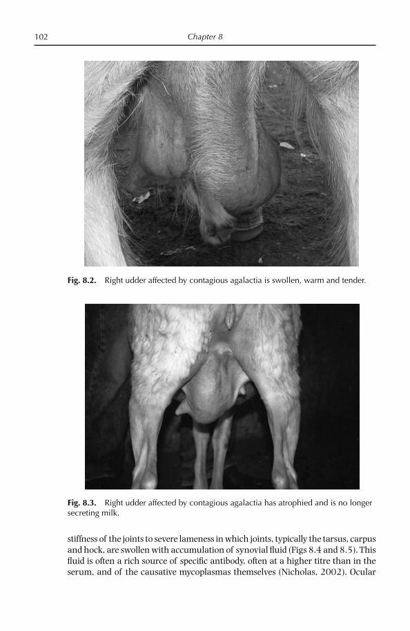
Chapter 8102
stiffness of the joints to severe lameness in which joints, typically the tarsus, carpus and hock, are swollen with accumulation of synovial fl uid (Figs 8.4 and 8.5). This fl uid is often a rich source of specifi c antibody, often at a higher titre than in the serum, and of the causative mycoplasmas themselves (Nicholas, 2002). Ocular
Fig. 8.2. Right udder affected by contagious agalactia is swollen, warm and tender.
Fig. 8.3. Right udder affected by contagious agalactia has atrophied and is no longer secreting milk.

Contagious Agalactia 103
lesions begin with conjunctivitis and congestion, lacrimation and photophobia, followed by vascularization of the cornea, infl ammatory foci and parenchyma-tous keratitis (Fig. 8.6). Severe cases can lead to blindness (Fig. 8.7). Pneumonia has also been reported in cases of CA, especially in young animals, where it may
Fig. 8.4. Ewe naturally affected with contagious agalactia showing swollen joint in front limb.
Fig. 8.5. Ewe naturally affected with contagious agalactia showing swollen joint in rear limb.

Chapter 8104
represent the only external sign. Cheese made from milk from subclinically affected ewes can often be spoiled (Fig. 8.8). The young born from affected females have been reported to be ataxic (Fig. 8.9) (Loria et al., 2007), and this may be related to septicaemia and/or the presence of the mycoplasma in the brain causing a non-purulent encephalomyelitis (Fig. 8.10).
Fig. 8.6. Ewe affected by contagious agalactia showing keratoconjunctivitis.
Fig. 8.7. Ewe naturally affected with contagious agalactia showing severe keratocon-junctivitis, leading to blindness.

Contagious Agalactia 105
Fig. 8.8. Cheese made from mastitic milk of ewe subclinically affected by contagious agalactia.
Fig. 8.9. Lamb born to ewe affected by contagious agalactia, showing lack of coordination.

Chapter 8106
Causative Organisms
M. agalactiae, like other mycoplasmas, has no cell wall and has a very small genome; it is resistant to penicillin and its analogues but sensitive to osmotic shock and the effect of detergents. Its growth is promoted by aerobic conditions at 37°C (Freundt, 1983; Lambert, 1987) but it does not ferment glucose and hydrolyses neither arginine nor urea, obtaining its energy requirements from organic acids (Khan et al., 2004).
The genome, entirely sequenced in 2007, is on the small side for mycoplasma, consisting of 877,438 bp (Sirand-Pugnet et al., 2007), but is a typical mollicute with a low GC content (29.7 moles %), which is slightly higher than the average of the other 16 completely sequenced mollicute genomes to date. It has 751 coding DNA sequences, half of which have unknown functions or encode hypothetical proteins, 66 of which are lipoproteins. The genome has 34 tRNA genes, two (nearly identical) sets of rRNA genes with two 16S–23S operons and two 5S oper-ons cluster in two loci separated by 400 bp. It possesses the following predicted virulence factors: (i) phase variable related surface proteins (Vpma); (ii) the P40 protein, which is involved in host adhesion; (iii) P48, which is probably a mac-rophage stimulatory protein; and (iv) Opp genes, which are known to be impor-tant for biofi lm formation, antimicrobial production and adaptation to specifi c environments. The mycoplasma also has the remnants of an integrative conjuga-tive element, which is a conjugative transposon important for gene transfer. An
Fig. 8.10. Non-purulent encephalitis lesion in the brain of a sheep affected by contagious agalactia.

Contagious Agalactia 107
unusual fi nding was that 18% of the genome was derived from the M. mycoides cluster by horizontal gene transfer, which indicates that it evolved by gene gain from other Mycoplasma species.
Diagnosis
When a fl ock is severely infected, clinical diagnosis is easy as the three major signs, mastitis, arthritis and keratoconjunctivitis, are present within a fl ock, though rarely in the same animal. However, an acute form, in which there is septicaemia without specifi c local signs, can confuse diagnosis.
Laboratory diagnosis provides the only means of confi rming the cause of the disease. Preferred samples from living animals include: nasal swabs and secre-tions, milk from mastitic does or from apparently healthy does where there is a high rate of mortality/morbidity in kids, joint fl uid from arthritic cases, eye swabs from cases of ocular disease, and blood for antibody detection from affected and non-affected animals; the ear canal has been shown to be a rich source of patho-genic mycoplasmas (Nicholas and Baker, 1998). Mycoplasmas may be isolated from the blood during the acute stage of the disease when there is mycoplasmae-mia, although this is rare and transient. From dead animals, samples should include: udder and associated lymph nodes, joint fl uid, lung tissue (at the inter-face between diseased and healthy tissue) and pleural/pericardial fl uid. Samples should be dispatched quickly to a diagnostic laboratory in a moist and cool con-dition. Culturing the organism presents no real problems to a modern laboratory as all causative mycoplasmas grow relatively well in the majority of reported mycoplasma media, although the addition of pyruvate is to be recommended.
M. agalactiae can be distinguished from the other mycoplasmas causing CA by its inability to ferment glucose or to utilize arginine; it also produces phospho-lipids, which appear as an oily fi lm or spots on the surface of the solid or liquid medium. M. putrefaciens, unusually for a mycoplasma, produces a putrid odour in both broth and semi-solid cultures and in milk from affected animals. Final identi-fi cation is usually achieved by the growth inhibition, immunofl uorescent tests using hyperimmune rabbit antiserum, and, increasingly, polymerase chain reaction (PCR) tests, which can be carried out directly on clinical samples including milk.
PCR assays are routinely used in many laboratories and are extremely sensi-tive. They can provide a rapid early warning system when carried out on clinical samples, enabling a full investigation to take place when results are positive. How-ever, negative results should not be considered defi nitive. Several PCRs specifi c for M. agalactiae have been developed and show similar levels of sensitivity, although they are based on different gene sequences (Dedieu et al., 1995; Tola et al., 1997; Subrahamaniam et al., 1998). They can be used directly on nasal, conjunctival, synovial and tissue samples; they have been used on milk samples, where they have been reported to be more sensitive than culture (Tola et al., 1997), although occasionally undefi ned inhibitors may interfere with the test. PCRs can also be used, more reliably, on mycoplasmas growing in culture; a 24-h enrichment of the mycoplasma in the appropriate medium greatly facilitates PCR detection even in the presence of bacterial contamination (Nicholas, 2002). A newly described

Chapter 8108
PCR-based method called denaturing gradient gel electrophoresis (DGGE), which uses mycoplasma-specifi c primers, is capable of identifying the majority of small ruminant mycoplasmas, including all the causative agents of CA, by their migra-tion pattern (McAuliffe et al., 2005). A positive PCR result, particularly in an area previously free of contagious agalactia, should be confi rmed by isolation and identifi cation of the mycoplasma using standard procedures. Individual PCRs have been reported for Mmc and M. c. capricolum (Bashiruddin et al., 1994) and M. putrefaciens (Peyraud et al., 2003).
A rapid and highly convenient biochemical test that exploits the C8-esterase activity of M. agalactiae has been reported (Khan et al., 2004). The mycoplasma forms red colonies on agar media within 1 h of adding the chromogenic substrate, SLPA-octanoate (a newly synthesized ester formed from a C8 fatty acid and a phenolic chromophore). This activity is shared with M. bovis, although this mycoplasma is rarely found in small ruminants. Isolates need not be cloned as M. agalactiae can be detected easily in mixed cultures.
Detection of antibodies in serum by complement fi xation test or enzyme-linked immunosorbent assay (ELISA) provides rapid diagnosis of disease but may not be very sensitive in chronically affected herds and fl ocks. ELISAs using sonicated or Tween-20-treated antigens have been reported to be more sensitive than the CFT for the detection of antibody to M. agalactiae (Bergonier et al., 1997). Problems of non-specifi city have been overcome by the use of monoclonal or protein G conju-gates in the ELISA (Lambert et al., 1998). The use of these conjugates enables the testing of sera from a wide range of mammalian species, including camelids. A number of commercial ELISA kits are now available and these were used for large-scale surveys in France and the UK (Bergonier et al., 1997; Nicholas, 1998). In a ring trial of serological tests for M. agalactiae organized in 1998 under the aus-pices of the EC COST Action 826 on ruminant mycoplasmoses, commercial ELI-SAs performed better than ‘home-made’ kits. In 2006, Kittelberger et al. compared four serological tests for detecting antibody to M. agalactiae and found the ELISA kit made by Bommeli (Switzerland) to be more sensitive than another commercial kit, Institut Pourquier (France); the CFT was the least sensitive, while the Western blot was unreliable because of the variable response of the immunodominant proteins. It is important that the appropriate antigens are used in the Western blot to mitigate this problem.
ELISAs are not widely available for the other three causative mycoplasmas, although ‘home-made’ assays are carried out by some laboratories.
Immunoblotting tests have also been described for M. agalactiae (Tola et al., 1997; Nicholas, 1998). Strong bands at approximately 80 and 55 kDa were seen in sera with antibodies to M. agalactiae, while sera from healthy fl ocks showed no bands or very faint bands of different sizes.
Differential Diagnosis
A number of other mycoplasmas such as M. arginini, M. ovine/caprine serogroup 11 and M. bovis have occasionally been isolated from mastitic milk, eye swabs and joint fl uids but their role in disease is not known (Ayling et al., 2004). Other

Contagious Agalactia 109
bacteria causing mastitis include staphlococci, streptococci, Escherichia coli and klebsiella; while caprine arthritic encephalomyelitis virus and Erysipelothrix rhusiopathiae should also be considered in cases of arthritis.
Treatment/Control
Regular laboratory monitoring of fl ocks/herds and replacement animals may help to prevent spread or introduction of disease, and this can be done on serum and/or on individual or bulk tank milk by serology, culturing or PCR. Culling or isolation of infected animals is generally advised because udder damage is con-sidered permanent. Where this is not possible, hygienic measures such as improved milking hygiene practice and pasteurizing milk before feeding to the young should be implemented. In many disease-free countries and regions, a confi rmed infected herd would be always be slaughtered.
A study was carried out to examine risk factors associated with CA infection in over 100 small sheep fl ocks and goat herds in northern Jordan between 2002 and 2003 (Al-Momani et al., 2007). A total of 31 variables including production and health management practices were tested as risk factors for seropositive fl ocks. Results showed that increasing risk factors were bringing in rams from other farms, improper cleaning of the milking utensils and not separating the young from their clinically affected dams.
Antibiotics such as the penicillins which inhibit cell wall synthesis are not effective against CA. The use of tetracyclines and the macrolides can sometimes bring about clinical improvements but there is always the danger of promoting inapparent carrier animals. Furthermore, the use of erythromycin and tylosin can bring about the destruction of milk-producing tissue in small ruminants. An in vitro assessment of antibiotics for M. agalactiae strains from endemic disease areas of Sicily showed no strong evidence of resistance, although highest mean MIC values were seen for spiramycin (Loria et al., 2003). However, occasional strains isolated from milk showed rather high values for oxytetracyclines. Overall, enrofl oxacin gave the lowest mean values, although there are concerns about the widespread use of these fl uoroquinolones in animals because of their potential impact on human health.
Prevention
Vaccines for the prevention of contagious agalactia due to M. agalactiae are used widely in southern Europe and the Middle East but no single vaccine has been universally adopted and no standard methods of preparation and evaluation have been applied.
Live attenuated vaccines for contagious agalactia caused by M. agalactiae and M. mycoides LC, made at the Pendik Institute, Istanbul, have been used in Turkey for many years and have been reported to provide better protection in ewes and their lambs than inactivated vaccines (Turkaslan, 1990). However, they can produce a

Chapter 8110
transient infection with shedding of mycoplasma so they are not recommended in lactating animals (OIE, 2004).
In Europe, where live M. agalactiae vaccines are not acceptable, attention has focused on the use of killed organisms, mostly using formalin and an adjuvant such as aluminium hydroxide in an oil emulsion. The titres of the preparations, before inactivation, must be very high (108–1010 cfus/ml) and are derived from laboratory strains. A trivalent vaccine, Aglovax™ (Intervet, Boxmeer, Netherlands), combines M. agalactiae, M. mycoides LC and M. c. capricolum, all at 1010 cfus in a 2 ml dose using aluminium hydroxide as the adjuvant. Two subcutaneous treat-ments 2–4 weeks apart followed by a booster vaccination at 4–6 months are rec-ommended. Autogenous vaccines made from milk, brain and mammary gland homogenates from infected sheep material have been used for many years in parts of Italy, although their effi cacy was far from proven. Their use, however, has been discontinued because of their link to severe outbreaks of scrapie in sheep and goats caused by contaminated brains (Agrimi et al., 1999).
A formalized vaccine gave some protection against experimental infection of goats with M. agalactiae in Spain but, despite three vaccinations per year for 6 years, it could not prevent clinical disease following the introduction of naturally infected animals (Leon Vizcaino et al., 1995). Furthermore formalized vaccines did not reduce the excretion of M. agalactiae in the milk compared with challenged con-trols (Pepin et al., 2001). It is possible that in some instances the apparent lack of protection given by vaccines could be the result of animals being infected with one of the other four mycoplasmas involved in the contagious agalactia syndrome (Gil et al., 1999).
Experimental vaccines inactivated with phenol or with saponin have given supe-rior protection against experimental infections compared with formalin, sodium hypochlorite or heat-inactivated vaccines (Tola et al., 1999). An oil-emulsifi ed, inactivated vaccine against M. agalactiae showed promise in fi eld trials, where sheep developed high levels of antibodies in response to vaccination and, when challenged, were protected from disease signs and colonization by M. agalactiae (Greco et al., 2002).
There is little recent published information on the availability of vaccines for M. mycoides LC, although it is believed that inactivated vaccines are widely used in many Mediterranean countries, suggesting that their production and use is localized (Bergonier et al., 1997). In experimental trials in Israel, a preparation of M. mycoides LC, inactivated with formalin and emulsifi ed with mineral oil and sorbitol, gave some protection to 1-day-old and 6-week-old kids against a virulent challenge (Bar-Moshe et al., 1984). Except for a few fi eld trials, the vaccine was never used widely as the occurrence of the disease caused by MmmLC has declined markedly since the mid-1980s.
Although infections with M. mycoides LC and M. putrefaciens can be severe, their prevalence is relatively low and, as might be expected, little or no work appears to have been carried out on preventive vaccination for these infections. A multivalent, formalin-inactivated vaccine incorporating all four mycoplasmas appeared to be benefi cial in fi eld trials, although in practice a vaccine containing only M. agalactiae and M. mycoides would be necessary (Ramirez et al., 2001).
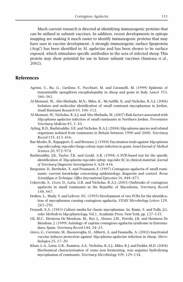
Contagious Agalactia 111
Much current research is directed at identifying immunogenic proteins that can be utilized in subunit vaccines. In addition, recent developments in epitope mapping are making it much easier to identify immunogenic proteins that may have uses in vaccine development. A strongly immunogenic surface lipoprotein (AvgC) has been identifi ed in M. agalactiae and has been shown to be surface exposed, which stimulates specifi c antibodies in the sera of infected sheep. This protein may show potential for use in future subunit vaccines (Santona et al., 2002).
References
Agrimi, U., Ru, G., Cardone, F., Pocchiari, M. and Caramelli, M. (1999) Epidemic of transmissible spongiform encephalopathy in sheep and goats in Italy. Lancet 353, 560–561.
Al-Momani, W., Abo-Shehada, M.N., Miles, K., McAuliffe, K. and Nicholas, R.A.J. (2006) Isolation and molecular identifi cation of small ruminant mycoplasmas in Jordan. Small Ruminant Research 65, 106–112.
Al-Momani, W., Nicholas, R.A.J. and Abo-Shehada, M. (2007) Risk factors associated with Mycoplasma agalactiae infection of small ruminants in Northern Jordan. Preventative Veterinary Medicine 83, 1–10.
Ayling, R.D., Bashiruddin, S.E. and Nicholas, R.A.J. (2004) Mycoplasma species and related organisms isolated from ruminants in Britain between 1990 and 2000. Veterinary Record 155, 413–416.
Bar-Moshe, B., Rapapport, E. and Brenner, J. (1984) Vaccination trials against Mycoplasma mycoides subsp. mycoides (large-colony type) infection in goats. Israel Journal of Medical Sciences 20, 972–974.
Bashiruddin, J.B., Taylor, T.K. and Gould, A.R. (1994) A PCR-based test for the specifi c identifi cation of Mycoplasma mycoides subsp. mycoides SC in clinical material. Journal of Veterinary Diagnostic Investigations 6, 428–434.
Bergonier, D., Bertholet, X. and Poumarat, F. (1997) Contagious agalactia of small rumi-nants: current knowledge concerning epidemiology, diagnosis and control. Revue Scientifi que et Technique. Offi ce International Epizooties 16, 848–873.
Cokrevski, S., Crcev, D., Loria, G.R. and Nicholas, R.A.J. (2001) Outbreaks of contagious agalactia in small ruminants in the Republic of Macedonia. Veterinary Record 148, 667.
Dedieu, L., Mady, V. and Lefevre, P.C. (1995) Development of two PCRs for the identifi ca-tion of mycoplasmas causing contagious agalactia. FEMS Microbiology Letters 129, 243–250.
Freundt, E.A. (1983) Culture media for classic mycoplasmas. In: Razin, S. and Tully, J.G. (eds) Methods in Mycoplasmology, Vol.1. Academic Press, New York, pp. 127–135.
Gil, M.C., Hermosa De Mendoza, M., Rey, J., Alonso, J.M., Poveda, J.B. and Hermosa De Mendoza, J. (1999) Aetiology of caprine contagious agalactia syndrome in Extremu-dura, Spain. Veterinary Record 144, 24–25.
Greco, G., Corrente, M., Buonovoglia, D., Aliberti, A. and Fasanella, A. (2002) Inactivated vaccine induces protection against Mycoplasma agalactiae infection in sheep. Micro-biologica 25, 17–20.
Khan, L.A., Loria, G.R., Ramirez, A.S., Nicholas, R.A.J., Miles, R.J. and Fielder, M.D. (2004) Biochemical characterisation of some non fermenting, non arginine hydrolysing mycoplasmas of ruminants. Veterinary Microbiology 109, 129–134.

Chapter 8112
Kittelberger, R., O’Keefe, J.S., Meynell, R., Sewell, M., Rosati, S., Lambert, M., Dufour, P. and Pepin, M. (2006) Comparison of four diagnostic tests for the identifi cation of serum antibodies in small ruminants infected with Mycoplasma agalactiae. New Zealand Veterinary Journal 54, 10–15.
Lambert, M. (1987) Contagious agalactia of sheep and goats. Revue Scientifi que et Tech-nique. Offi ce International des Epizooties 6, 699–711.
Lambert, M., Calamel, M., Du Four, P., Cabasse, E., Vitu, C. and Pepin, M. (1998) Detection of false-positive sera in contagious agalactia with a multiantigen ELISA and their elimination with a protein G conjugate. Journal of Veterinary Diagnostic Investigations 10, 326–330.
Leon Vizcaino, L., Garrido Abellan, F., Cubero Pablo, M.J. and Perales, A. (1995) Immuno-prophylaxis of caprine contagious agalactia due to Mycoplasma agalactiae with an inactivated vaccine. Veterinary Record 137, 266–269.
Loria, G.R., Sammartino, C., Nicholas, R.A.J. and Ayling, R.D. (2003) In vitro susceptibi-lities of fi eld isolates of Mycoplasma agalactiae to oxytetracycline, tylosin, enrofl oxacin, spiramycin and lincomycin-spectinomycin. Research in Veterinary Science 75, 3–7.
Loria, G.R., Caracappa, S., Monteverde, P. and Nicholas, R.A.J. (2007) Infezione da Myco-plasma agalactiae in cervelli ovini: un nuova sito di infezione. Large Animal Review 13, 65–68.
McAuliffe, L., Ellis, R., Lawes, J., Ayling, R.D. and Nicholas, R.A.J. (2005) 16S rDNA and DGGE: a single generic test for detecting and differentiating Mycoplasma species. Journal of Medical Microbiology 54, 1–9.
McAuliffe, L., Ellis., R.J., Miles, K., Ayling, R.D. and Nicholas, R.A.J. (2006) Biofi lm formation by mycoplasma species and its role in environment persistence and survival. Micro-biology 152, 913–922.
McAuliffe, L., Churchward, C., Lawes, J., Loria, G.R., Ayling, R.D. and Nicholas R.A.J. (2008) VNTR analysis reveals unexpected genetic diversity within Mycoplasma agalactiae, the main causative agent of contagious agalactia. BMC Microbiology submitted.
Mercier, P., Lenfant, D., Poumarat, F. and Perrin, G. (2001) Prevalence of mycoplasma in-fection within French milking caprine herds. In: Poveda, J.B., Fernandez, A., Frey, J. and Johansson, K.-E. (eds) Mycoplasmas of Ruminants: Pathogenicity, Diagnostics, Epide-miology and Molecular Genetics, Vol. 5. European Commission, Brussels, pp. 130–133.
Nicholas, R.A.J. (1998) Surveillance for contagious agalactia in Great Britain. In: Leori, G., Santini, F., Scanziani, E. and Frey, J. (eds) Mycoplasmas of Ruminants: Pathogenicity, Diagnostics, Epidemiology and Molecular Genetics, Vol. 2. European Commission, Brussels, pp. 95–97.
Nicholas, R.A.J. (2002) Improvements in the diagnosis and control of diseases of small ruminants caused by mycoplasmas. Small Ruminant Research 45, 145–149.
Nicholas, R.A.J. and Baker, S.E. (1998) Recovery of mycoplasmas from animals. In: Miles, R.J. and Nicholas, R.A.J. (eds) Mycoplasma Protocols. Humana Press, Totowa, New Jersey, pp. 37–44.
OIE (2004) Contagious caprine pleuropneumonia. In: Manual of Standards for Diagnostic Tests and Vaccines. Offi ce International des Epizooties, Paris, pp. 503–514.
OIE (2008) World Animal Health in 2006. World Organisation for Animal Health, Paris.Ozdemir, U., Ozdemir, S., March, J., Churchwood, C. and Nicholas, R A J. (2005) Outbreaks
of CCPP in the Thrace region of Turkey. Veterinary Record 156, 286–287.Pepin, M., Sanchis, R., Abadie, G., Lambert, M., Dufour, P. and Guibert, J.M. (2001) Experi-
mental vaccination against Mycoplasma agalactiae using an inactivated vaccine. In: Poveda, J.B., Fernandez, A., Frey, J. and Johansson, K.-E. (eds) Mycoplasmas of Rumi-nants: Pathogenicity, Diagnostics, Epidemiology and Molecular Genetics, Vol. 5. European Commission, Brussels, pp. 162–165.

Contagious Agalactia 113
Peyraud, A., Woubit, S., Poveda, J.B., De La Fe, C., Mercier, P. and Thiaucourt, F. (2003) A specifi c PCR for the detection of Mycoplasma putrefaciens, one of the agents of the contagious agalactia syndrome of goats. Molecular and Cellular Probes 17, 289–294.
Pilo, P., Fleury, B., Marenda, M., Frey, J. and Vilei, E.M. (2003) Prevalence and distribution of the insertion element ISMag1 in Mycoplasma agalactiae. Veterinary Microbiology 92, 37–48.
Ramirez, A.S., De La Fe, C., Assuncao, P., Gonzalez, M. and Poveda, J.B. (2001) Preparation and evaluation of an inactivated polyvalent vaccine against Mycoplasma spp. on infected goats. In: Poveda, J.B., Fernandez, A., Frey, J. and Johansson, K.-E. (eds) Myco-plasmas of Ruminants: Pathogenicity, Diagnostics, Epidemiology and Molecular Genetics, Vol. 5. European Commission, Brussels, pp. 154–157.
Rodriguez, J.L., Poveda, J.B., Gutierrez, C., Acosta, B. and Fernandez, A. (1994) Polyarthri-tis in kids associated with Mycoplasma putrefaciens. Veterinary Record 135, 406–407.
Santona, A., Carta, F., Fraghi, P. and Turrini, F. (2002) Mapping antigenic sites of an im-munodominant surface lipoprotein of Mycoplasma agalactiae, AvgC, with the use of synthetic peptides. Infection and Immunity 70, 171–176.
Sirand-Pugnet, P., Lartigue, C., Marenda, M., Jacob, D., Barré, A., Barbe, V., Schenowitz, C., Mangenot, S., Couloux, A., Segurens, B., de Daruvar, A., Blanchard, A. and Citti, C. (2007) Being pathogenic, plastic, and sexual while living with a nearly minimal bacterial genome. PLoS Genetics 3, 74–89.
Solsona, M., Lambert, M. and Poumarat, F. (1996) Genomic, protein homogeneity and antigenic variability of Mycoplasma agalactiae. Veterinary Microbiology 50, 45–58.
Subrahamaniam, S., Bergonier, D., Poumarat, F., Capual, S., Schlatter, Y., Nicolet, J. and Frey, J. (1998) Species identifi cation of Mycoplasma bovis and Mycoplasma agalactiae based on the uvrC gene by PCR. Molecular and Cellular Probes 12, 161–169.
Tola, S., Idini, G., Manunta, D., Casciano, I., Rocchigiani, A.M., Angioi, A. and Leori, G. (1996) Comparison of Mycoplasma agalactiae isolates by pulsed fi eld gel electropho-resis, SDS-PAGE and immunoblotting. FEMS Microbiology Letters 143, 259–265.
Tola, S., Angioi, A., Rocchigiani, A.M., Idini, G., Nanunta, D., Galleri, G. and Leori, G. (1997) Detection of Mycoplasma agalactiae in sheep milk samples by polymerase chain reaction. Veterinary Microbiology 54, 17–22.
Tola, S., Manunta, D., Rocca, S., Rocchigiani, A.M., Idini, G., Angioi, A. and Leori, G. (1999) Experimental vaccination against Mycoplasma agalactiae using different inactivated vaccine. Vaccine 17, 2764–2768.
Turkaslan, J. (1990) Control of important mycoplasma diseases in Turkey with special emphasis on CCPP and contagious agalactia. IOM Letters 1, 184–185.

© CAB International 2008. Mycoplasma Diseases of Ruminants114 (R. Nicholas et al.)
9 Contagious Caprine Pleuropneumonia
The rapidity with which the disease had spread among Mr Nierkert’s fl ock, affecting more than 700 goats within a fourteen day period, induced me to believe that its rapid spread could not be due to contagion alone . . . but after carefully watching the progress of the disease for three days, and noting the uniform character of the pathological appearances in the lungs, I began to realise that I had a specifi c disease to deal with (Duncan Hutcheon, Colonial Veterinary Surgeon, Cape of Good Hope, South Africa, 1881).
Introduction
In the autumn of 2002, a severe respiratory disease characterized by high levels of morbidity and mortality was fi rst seen in goat herds in the Thrace region of Turkey, 250 km west of Istanbul (Ozdemir et al., 2005). The disease, which was diagnosed as contagious caprine pleuropneumonia (CCPP), spread throughout the region to an unknown number of herds, some close to the Greek and Bulgarian borders. Goats of all ages were affected and showed a reluctance to walk, a fever of over 41°C and accelerated respiration with frequent coughing. The lungs of dead and euthanized animals showed characteristic lesions with abundant pleural fl uid, fi brin and unilateral hepatization; in one herd alone nearly 150 of 400 adults and over 100 of 400 kids died in a single year (Ozdemir et al., 2005). This was the fi rst reported outbreak of CCPP on European soil and today threatens the countries of the EU.
CCPP was fi rst described in 1873 in Algeria and known under the local name of ‘bou frida’ because, in the majority of diseased goats, only one lung was affected (McMartin et al., 1980). Its contagiousness was not initially recognized because the disease was endemic in most areas under investigation, so climatic conditions were thought to be responsible for disease outbreaks. However, a major outbreak in South Africa in 1881 following the introduction of goats from Turkey led the

Contagious Caprine Pleuropneumonia 115
colonial veterinary surgeon, Duncan Hutcheon, to conclude that CCPP was highly infectious.
Research into the control of CCPP was initially hampered by the uncertainty over the exact cause of the disease. Two mycoplasmas, M. mycoides subsp. mycoides LC and M. mycoides subsp. capri, were for some time implicated in the aetiology of the disease because they caused a pleuropneumonia in small ruminants that resembled CCPP. It was not until 1976 that a highly fastidious mycoplasma, desig-nated F38 but later renamed M. capricolum subsp. capripneumoniae, was isolated for the fi rst time in vitro by MacOwan and Minette (1976). Once these workers had developed a suitable medium for the mycoplasma and carried out experimental infections, its role as the primary cause of classical CCPP was confi rmed.
However, in spite of this confi rmation, respiratory diseases caused by M. m. capri and M. mycoides LC are still referred to erroneously as CCPP, particularly in the Middle East and India. A condition should only be termed as CCPP when the fol-lowing criteria have been satisfi ed:
M. c. capripneumoniae ● is isolated or there is strong serological evidence of the mycoplasma;Lesions are restricted to lung and pleura and consist of a pleuropneumonia; ●
The condition is highly contagious with high levels of morbidity/mortality; ●
There is no enlargement of the interlobular septa of the lung. ●
Geographic Distribution
While the clinical disease has been reported in nearly 40 countries in Africa and Asia, M. c. capripneumoniae has only been isolated in 13 countries because few have the facilities for isolating and growing mycoplasmas (Nicholas, 2002) (Table 9.1). Serious problems exist in Oman, where nearly 500 outbreaks were reported in 2006 with a mortality rate of nearly 10% from 15,000 cases, and in Iran, where nearly 300 outbreaks have affected over 13,000 goats (OIE, 2008). The 31 reported outbreaks in Ethiopia almost certainly represents an underestimate, as this disease is having a big socio-economic impact here.
Table 9.1. Distribution of CCPP.
Confi rmed by isolation of mycoplasma Clinical disease reported or suspected
Africa Chad, Eritrea, Ethiopia, Kenya, Niger, Sudan, Tunisia, Uganda
Algeria, Benin, Burkina Faso, Cameroon, Central African Republic, Djibouti, Egypt, Libya, Mali, Nigeria, Somalia, Zaire
Asia Nepal, Oman, Turkey, United Arab Emirates, Yemen
Afghanistan, Bangladesh, India, Iran, Iraq, Israel, Jordan, Kuwait, Lebanon, Pakistan, Saudi Arabia, Syria
Europe Thrace (Turkey)

Chapter 9116
In 2003, CCPP was diagnosed in Thrace, the region of Turkey on the Euro-pean mainland bordering Greece and Bulgaria. Prior to this infection, the only previous report of CCPP in Europe dates to the 1920s, when an outbreak occurred in Greece following the seizure of goats from Turkey. Interestingly Greece reported two outbreaks of CCPP in 2006, but it seems likely that this was caused by M. m. capri, which is endemic in Greece, rather than by M. c. capripneu-moniae, as less than 1% of goats died in a herd of over 150. It is probable also that the outbreak of CCPP in the Czech Republic in 1902 was similarly misdiagnosed, although it is clearly impossible to confi rm over a century later. There have been no reports of the isolation of M. c. capripneumoniae on the American continent, although other members of the mycoides cluster have been described there.
Causative Agent
The taxonomic status of F38 has long been unclear, and it was only in 1993 that it became a subspecies of M. capricolum and classifi ed as M. capricolum subsp. capripneumoniae (Leach et al., 1993). Five distinct groups of mollicutes have been identifi ed by phylogenetic analysis of the 16S rRNA sequences, one of which, the spiroplasma group, contains M. c. capripneumoniae, which has been subdivided within the M. mycoides cluster. This cluster contains six important ruminant mycoplasmas, including M. m. mycoides SC, the cause of contagious bovine pleu-ropneumonia, M. m. mycoides LC and M. m. capri, which share immunological and biochemical properties. Their close relationship can lead to problems for diag-nosis. Tables 9.2 and 9.3 summarize the properties of M. c. capripneumoniae and some members of this cluster as well as other mycoplasmas capable of causing diseases in small ruminants.
Host Susceptibility
For a long time goats were believed to be the only susceptible host for CCPP ( Litamoi et al., 1990), although it was reported that sheep could be infected and seroconvert in the face of exposure (Bolske et al., 1995). Following the introduc-tion of CCPP into Eritrea with the livestock of returning refugees from Sudan, sheep mixing with affected goats were reported to be suffering respiratory disease (Houshaymi et al., 2000). More recently there have been confi rmed reports from Qatar of CCPP in captive wild ungulates, including wild goat, Nubian ibex, Larisstan moufl on and gerenuk, kept in animal breeding parks (Arif et al., 2007). Even more surprisingly, however, were the outbreaks of acute respiratory disease in a private collection of captive but free-ranging gazelles and other deer species in the United Arab Emirates, in which over 10% died. The disease was almost cer-tainly introduced via sick goats and spread by close contact with the gazelles at feed stations (Nicholas et al., 2008); it is likely that CCPP is far more widespread in wildlife species in the Middle East as a result of infected escapees from these parks.

Contagious C
aprine Pleuropneumonia
117
Table 9.2. Mycoplasmas, including M. capricolum subsp. capripneumoniae, isolated from small ruminants with respiratory disease.
Mycoplasma Host(s) Primary site of isolation (other) Disease* Pathogenicity In vitro growh
M. c. capripneumoniae Goat (sheep) Lungs CCPP High Slow (5–7days)M. m. mycoides LC Goat (sheep, cattle) Resp. tract (udder, joints) Plp, M, A, C Moderate FastM. m. capri Goat (sheep) Resp. tract (joints) Plp, A, C Moderate FastM. c. capricolum Goat, sheep Joints/resp. tract (udder) Plp, M, A High FastM. ovipneumoniae Sheep, goat Resp. tract P Low ModerateM. conjunctivae Sheep, goat Eyes KC Moderate ModerateM. agalactiae Sheep, goat Udder (joints, eyes) M, A, KC, P High FastM. putrefaciens Goat (sheep) Udder (joints) M, A Moderate FastM. arginini Ubiquitous Resp. tract None Low/non-pathogenic Fast
* CCPP – contagious caprine pleuropneumonia, Plp – pleuropneumonia, P – pneumonia, M – mastitis, A – arthritis, C – conjunctivitis, KC – keratoconjunctivitis

C
hapter 9118
Table 9.3. Major biochemical differences between mycoplasmas of small ruminants.
MycoplasmaGlucose
fermentationArginine
hydrolysisPhosphatase
activityFilm and
spotsCasein
digestionTetrazolium
aerobicReductionanaerobic
M. c. capripneumoniae +/− − − − + varies weak/+M. m. mycoides LC + − − − + + +M. m. capri + − − − + + +M. c. capricolum + + + − + + +M. ovipneumoniae + − − − − varies +M. conjunctivae + − − − − − +M. agalactiae − − + + − + +M. putrefaciens + − + + − varies +M. arginini − + − − − − +
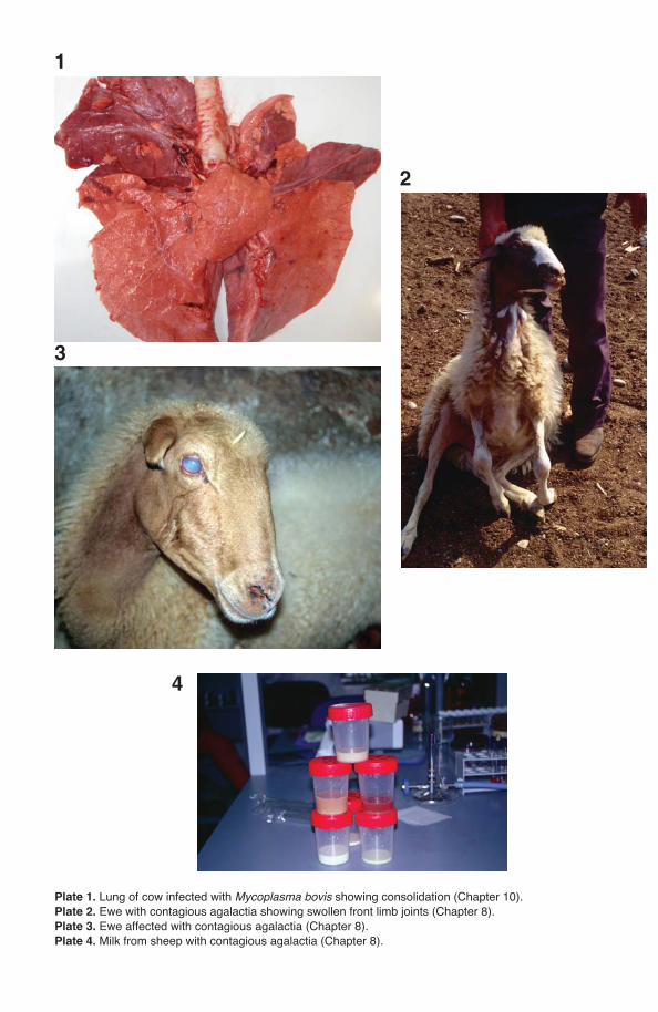
Plate 1. Lung of cow infected with Mycoplasma bovis showing consolidation (Chapter 10).Plate 2. Ewe with contagious agalactia showing swollen front limb joints (Chapter 8).Plate 3. Ewe affected with contagious agalactia (Chapter 8).Plate 4. Milk from sheep with contagious agalactia (Chapter 8).
1
2
4
3
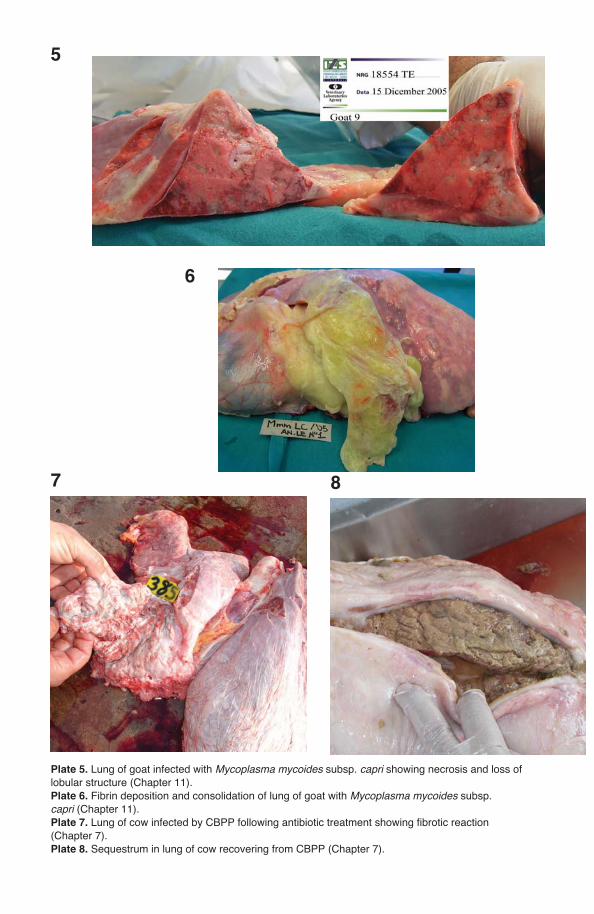
Plate 5. Lung of goat infected with Mycoplasma mycoides subsp. capri showing necrosis and loss of lobular structure (Chapter 11). Plate 6. Fibrin deposition and consolidation of lung of goat with Mycoplasma mycoides subsp. capri (Chapter 11). Plate 7. Lung of cow infected by CBPP following antibiotic treatment showing fibrotic reaction (Chapter 7).Plate 8. Sequestrum in lung of cow recovering from CBPP (Chapter 7).
5
6
87
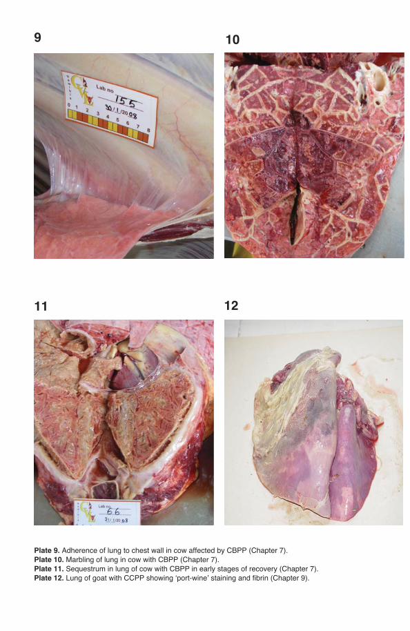
Plate 9. Adherence of lung to chest wall in cow affected by CBPP (Chapter 7).Plate 10. Marbling of lung in cow with CBPP (Chapter 7).Plate 11. Sequestrum in lung of cow with CBPP in early stages of recovery (Chapter 7). Plate 12. Lung of goat with CCPP showing ‘port-wine’ staining and fibrin (Chapter 9).
9
1211
10
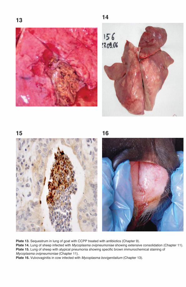
Plate 13. Sequestrum in lung of goat with CCPP treated with antibiotics (Chapter 9).Plate 14. Lung of sheep infected with Mycoplasma ovipneumoniae showing extensive consolidation (Chapter 11).Plate 15. Lung of sheep with atypical pneumonia showing specific brown immunochemical staining of Mycoplasma ovipneumoniae (Chapter 11). Plate 16. Vulvovaginitis in cow infected with Mycoplasma bovigenitalium (Chapter 13).
13
1615
14
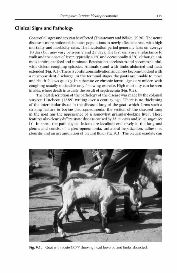
Contagious Caprine Pleuropneumonia 119
Clinical Signs and Pathology
Goats of all ages and sex can be affected (Thiaucourt and Bölske, 1996). The acute disease is more noticeable in naive populations in newly affected areas, with high mortality and morbidity rates. The incubation period generally lasts on average 10 days but may vary between 2 and 28 days. The fi rst signs are a reluctance to walk and the onset of fever, typically 41°C and occasionally 42°C, although ani-mals continue to feed and ruminate. Respiration accelerates and becomes painful, with violent coughing episodes. Animals stand with limbs abducted and neck extended (Fig. 9.1). There is continuous salivation and noses become blocked with a mucopurulent discharge. In the terminal stages the goats are unable to move and death follows quickly. In subacute or chronic forms, signs are milder, with coughing usually noticeable only following exercise. High mortality can be seen in kids, where death is usually the result of septicaemia (Fig. 9.2).
The best description of the pathology of the disease was made by the colonial surgeon Hutcheon (1889) writing over a century ago: ‘There is no thickening of the interlobular tissue in the diseased lung of the goat, which forms such a striking feature in bovine pleuropneumonia; the section of the diseased lung in the goat has the appearance of a somewhat granular-looking liver’. These features also clearly differentiate disease caused by M. m. capri and M. m. mycoides LC. In short, the pathological lesions are localized exclusively in the lung and pleura and consist of a pleuropneumonia, unilateral hepatization, adhesions, pleuritis and an accumulation of pleural fl uid (Fig. 9.3). The pleural exudate can
Fig. 9.1. Goat with acute CCPP showing head lowered and limbs abducted.
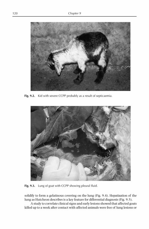
Chapter 9120
solidify to form a gelatinous covering on the lung (Fig. 9.4). Hepatization of the lung as Hutcheon describes is a key feature for differential diagnosis (Fig. 9.5).
A study to correlate clinical signs and early lesions showed that affected goats killed up to a week after contact with affected animals were free of lung lesions or
Fig. 9.2. Kid with severe CCPP probably as a result of septicaemia.
Fig. 9.3. Lung of goat with CCPP showing pleural fl uid.
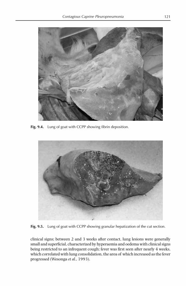
Contagious Caprine Pleuropneumonia 121
clinical signs; between 2 and 3 weeks after contact, lung lesions were generally small and superfi cial, characterized by hyperaemia and oedema with clinical signs being restricted to an infrequent cough; fever was fi rst seen after nearly 4 weeks, which correlated with lung consolidation, the area of which increased as the fever progressed (Wesonga et al., 1993).
Fig. 9.4. Lung of goat with CCPP showing fi brin deposition.
Fig. 9.5. Lung of goat with CCPP showing granular hepatization of the cut section.

Chapter 9122
In an experiment by Ozdemir et al. (2006), it was possible to follow the course of the disease, with and without antibiotic intervention. Healthy Angora goats, confi rmed free of CCPP, were exposed to clinically affected animals from a natural outbreak in Thrace, Turkey. After 14 days' exposure, the majority of contacts showed pyrexia (≥ 41°C). Shortly after, the Angora goats were randomly divided into two groups. Half of these were injected twice over 2 days with danofl oxacin; the remaining animals received saline. Goats which survived were euthanazed at day 42. All danofl oxacin-treated goats showed resolution of clinical disease by the end of the trial. Two saline-treated goats were euthanized after 4 weeks due to severe disease. Danofl oxacin-treated goats showed fewer lung lesions, which were generally sequestrated (Fig. 9.6), and had signifi cantly lower combined clinical scores than saline controls, which showed severe and acute lesions.
Fig. 9.6. Lung of goat with CCPP, treated with antibiotics, showing sequestrum.

Contagious Caprine Pleuropneumonia 123
The outbreaks of CCPP in gazelles in the Middle East had a very similar clinical (Fig. 9.7) and pathological (Fig. 9.8) appearance to that seen in goats.
Histological examination of the lung tissues often reveals an acute serofi brin-ous to chronic fi brino-necrotic pleuropneumonia, with infi ltrates of serofi brinous fl uid and infl ammatory cells, mainly neutrophils, in the alveoli, bronchioles, interstitial septae and subpleural connective tissue. Intralobular oedema is more prominent, but interlobular oedema has also occasionally been reported. Peri-bronchial and peribronchiolar lymphoid hyperplasia with mononuclear cell infi ltration is also present (MacOwan and Minette, 1976; Kibor, 1990; Wesonga et al., 1998).
Ultrastructural examination of the lungs of goats naturally affected with CCPP confi rms histopathological fi ndings of congested septal capillaries, with infl ammatory cells invading thickened septal walls (Johnson et al., 2002). The alveolar lumen contains serous fl uid mixed with neutrophils and lymphocytes, some of which are necrotic. The most signifi cant fi ndings are a widespread hyperplasia of type II pneumocytes that have lost most of their characteristic lamellar ultrastructure and large numbers of mycoplasma-like structures lying close to the microvilli and membranes of these cells. It was proposed that the loss of these lamellae may reduce surfactant production, as well as synthesis of key enzymes, leading to increased surface tension within the alveoli and contributing to the atelectasis often seen at post-mortem examination of CCPP cases.
Conditions which may exacerbate CCPP include concurrent viral infections, in particular orf and pestes petits de ruminant (PPR), and possibly other mycoplasma
Fig. 9.7. Outbreaks of CCPP were seen in semi-captive gazelle species in the Middle East.

Chapter 9124
infections such as M. ovipneumoniae, a cause of disease in its own right; adverse weather conditions and stress caused by transhumance may also compound or accelerate ongoing disease.
Immunology
Little is known of the immunology of CCPP despite a number of reported experi-mental infections (Muthomi and Rurangirwa, 1983; Perreau et al., 1984). More recently March et al. (2000) monitored the humoral response of goats infected with a multipassaged M. c. capripneumoniae strain 19/2 with several serological tests and PCR. While there was little evidence after infection of the infectious agent or clinical or pathological disease, apart from elevated temperatures and a transient cough in one goat, serological responses were detected by latex aggluti-nation test and competitive ELISA. IgG immunodominant bands of 23, 40 and 44 kDa were seen by immunoblotting in all experimentally infected animals, as well as in some sera from a natural outbreak of CCPP in Eritrea, which additionally showed bands of 62, 70 and 108 kDa.
Transmission
Outbreaks follow the introduction of an infected animal into a susceptible herd. The mycoplasma is transmitted over short distances through the expulsion of
Fig. 9.8. Gazelle affected by CCPP showed similar pathological features to those of goats.
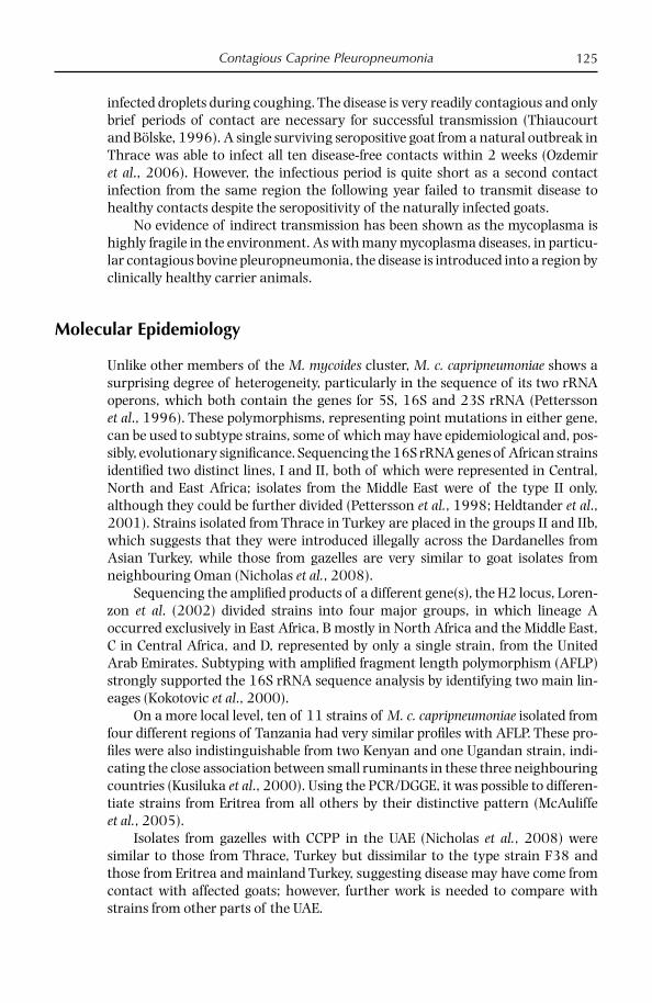
Contagious Caprine Pleuropneumonia 125
infected droplets during coughing. The disease is very readily contagious and only brief periods of contact are necessary for successful transmission (Thiaucourt and Bölske, 1996). A single surviving seropositive goat from a natural outbreak in Thrace was able to infect all ten disease-free contacts within 2 weeks (Ozdemir et al., 2006). However, the infectious period is quite short as a second contact infection from the same region the following year failed to transmit disease to healthy contacts despite the seropositivity of the naturally infected goats.
No evidence of indirect transmission has been shown as the mycoplasma is highly fragile in the environment. As with many mycoplasma diseases, in particu-lar contagious bovine pleuropneumonia, the disease is introduced into a region by clinically healthy carrier animals.
Molecular Epidemiology
Unlike other members of the M. mycoides cluster, M. c. capripneumoniae shows a surprising degree of heterogeneity, particularly in the sequence of its two rRNA operons, which both contain the genes for 5S, 16S and 23S rRNA (Pettersson et al., 1996). These polymorphisms, representing point mutations in either gene, can be used to subtype strains, some of which may have epidemiological and, pos-sibly, evolutionary signifi cance. Sequencing the 16S rRNA genes of African strains identifi ed two distinct lines, I and II, both of which were represented in Central, North and East Africa; isolates from the Middle East were of the type II only, although they could be further divided (Pettersson et al., 1998; Heldtander et al., 2001). Strains isolated from Thrace in Turkey are placed in the groups II and IIb, which suggests that they were introduced illegally across the Dardanelles from Asian Turkey, while those from gazelles are very similar to goat isolates from neighbouring Oman (Nicholas et al., 2008).
Sequencing the amplifi ed products of a different gene(s), the H2 locus, Loren-zon et al. (2002) divided strains into four major groups, in which lineage A occurred exclusively in East Africa, B mostly in North Africa and the Middle East, C in Central Africa, and D, represented by only a single strain, from the United Arab Emirates. Subtyping with amplifi ed fragment length polymorphism (AFLP) strongly supported the 16S rRNA sequence analysis by identifying two main lin-eages (Kokotovic et al., 2000).
On a more local level, ten of 11 strains of M. c. capripneumoniae isolated from four different regions of Tanzania had very similar profi les with AFLP. These pro-fi les were also indistinguishable from two Kenyan and one Ugandan strain, indi-cating the close association between small ruminants in these three neighbouring countries (Kusiluka et al., 2000). Using the PCR/DGGE, it was possible to differen-tiate strains from Eritrea from all others by their distinctive pattern (McAuliffe et al., 2005).
Isolates from gazelles with CCPP in the UAE (Nicholas et al., 2008) were similar to those from Thrace, Turkey but dissimilar to the type strain F38 and those from Eritrea and mainland Turkey, suggesting disease may have come from contact with affected goats; however, further work is needed to compare with strains from other parts of the UAE.
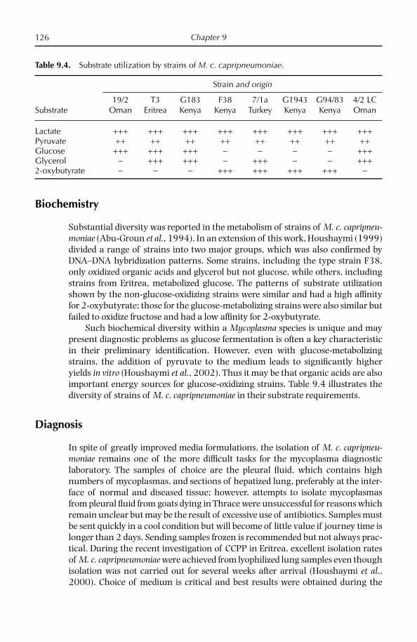
Chapter 9126
Biochemistry
Substantial diversity was reported in the metabolism of strains of M. c. capripneu-moniae (Abu-Groun et al., 1994). In an extension of this work, Houshaymi (1999) divided a range of strains into two major groups, which was also confi rmed by DNA–DNA hybridization patterns. Some strains, including the type strain F38, only oxidized organic acids and glycerol but not glucose, while others, including strains from Eritrea, metabolized glucose. The patterns of substrate utilization shown by the non-glucose-oxidizing strains were similar and had a high affi nity for 2-oxybutyrate; those for the glucose-metabolizing strains were also similar but failed to oxidize fructose and had a low affi nity for 2-oxybutyrate.
Such biochemical diversity within a Mycoplasma species is unique and may present diagnostic problems as glucose fermentation is often a key characteristic in their preliminary identifi cation. However, even with glucose-metabolizing strains, the addition of pyruvate to the medium leads to signifi cantly higher yields in vitro (Houshaymi et al., 2002). Thus it may be that organic acids are also important energy sources for glucose-oxidizing strains. Table 9.4 illustrates the diversity of strains of M. c. capripneumoniae in their substrate requirements.
Diagnosis
In spite of greatly improved media formulations, the isolation of M. c. capripneu-moniae remains one of the more diffi cult tasks for the mycoplasma diagnostic laboratory. The samples of choice are the pleural fl uid, which contains high numbers of mycoplasmas, and sections of hepatized lung, preferably at the inter-face of normal and diseased tissue; however, attempts to isolate mycoplasmas from pleural fl uid from goats dying in Thrace were unsuccessful for reasons which remain unclear but may be the result of excessive use of antibiotics. Samples must be sent quickly in a cool condition but will become of little value if journey time is longer than 2 days. Sending samples frozen is recommended but not always prac-tical. During the recent investigation of CCPP in Eritrea, excellent isolation rates of M. c. capripneumoniae were achieved from lyophilized lung samples even though isolation was not carried out for several weeks after arrival (Houshaymi et al., 2000). Choice of medium is critical and best results were obtained during the
Table 9.4. Substrate utilization by strains of M. c. capripneumoniae.
Strain and origin
Substrate19/2
OmanT3
EritreaG183Kenya
F38Kenya
7/1aTurkey
G1943Kenya
G94/83Kenya
4/2 LCOman
Lactate +++ +++ +++ +++ +++ +++ +++ +++Pyruvate ++ ++ ++ ++ ++ ++ ++ ++Glucose +++ +++ +++ − − − − +++Glycerol − +++ +++ − +++ − − +++2-oxybutyrate − − − +++ +++ +++ +++ −
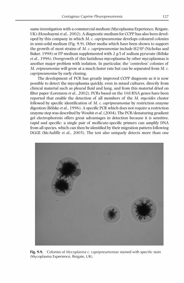
Contagious Caprine Pleuropneumonia 127
same investigation with a commercial medium (Mycoplasma Experience, Reigate, UK) (Houshaymi et al., 2002). A diagnostic medium for CCPP has also been devel-oped by this company in which M. c. capripneumoniae develops coloured colonies in semi-solid medium (Fig. 9.9). Other media which have been shown to support the growth of most strains of M. c. capripneumoniae include H25P (Nicholas and Baker, 1998) or FP medium supplemented with 2 g/l of sodium pyruvate (Bölske et al., 1996). Overgrowth of this fastidious mycoplasma by other mycoplasmas is another major problem with isolation. In particular, the ‘centreless’ colonies of M. ovipneumoniae will grow at a much faster rate but can be separated from M. c. capripneumoniae by early cloning.
The development of PCR has greatly improved CCPP diagnosis as it is now possible to detect the mycoplasma quickly, even in mixed cultures, directly from clinical material such as pleural fl uid and lung, and from this material dried on fi lter paper (Lorenzon et al., 2002). PCRs based on the 16S RNA genes have been reported that enable the detection of all members of the M. mycoides cluster followed by specifi c identifi cation of M. c. capripneumoniae by restriction enzyme digestion (Bölske et al., 1996). A specifi c PCR which does not require a restriction enzyme step was described by Woubit et al. (2004). The PCR/denaturing gradient gel electrophoresis offers great advantages in detection because it is sensitive, rapid and specifi c: a single pair of mollicute-specifi c primers can amplify DNA from all species, which can then be identifi ed by their migration pattern following DGGE (McAuliffe et al., 2005). The test also uniquely detects more than one
Fig. 9.9. Colonies of Mycoplasma c. capripneumoniae stained with specifi c stain (Mycoplasma Experience, Reigate, UK).

Chapter 9128
Mycoplasma species in a sample. However, because of the close similarity of the sequence of the 16S rDNA gene, the target of the PCR/DGGE, in the M. mycoides cluster members, a confi rmatory PCR such as that described by Woubit et al. (2004) should be performed.
Serodiagnosis of CCPP, on the other hand, is a relatively easy task, thanks to a rapid, specifi c and relatively sensitive test developed initially in Kenya. The latex agglutination test (LAT) uses a carbohydrate extracted from M. c. capripneumoniae linked to latex particles which agglutinate in the presence of specifi c antibodies in the blood of affected goats (Rurangirwa et al., 1987). The test, which takes minutes to complete, is more sensitive than the complement fi xation test and easier to perform than the CFT (OIE, 2004) or indirect or competitive ELISA, which should be used for confi rmation (Houshaymi et al., 2002). An LAT has also been described for circulating antigen and could provide earlier detection in affected animals before antibodies have appeared (March et al., 2000). The test also provides a convenient means of monitoring growth of the mycoplasma in vitro, which is often quite diffi cult to detect because of the small pH changes produced by some strains; the test requires just a few drops of culture fl uid.
Disease Prevention and Control
Protection against CCPP was shown to be possible more than a century ago when Hutcheon subcutaneously inoculated goats with lung extract from affected ani-mals (McMartin et al., 1980). Later, goats vaccinated with an attenuated broth culture of F38 did not succumb to contact infection (MacOwan and Minette, 1978). This clearly demonstrated control was possible. Since then a number of different preparations have been produced which are reported to produce strong immunity even after 1 year. These include a vaccine composed of sonicated anti-gens emulsifi ed with incomplete Freund’s adjuvant (Rurangirwa et al., 1984) and another in which lyophilized F38 was inactivated with saponin immediately before immunization (Rurangirwa et al., 1987). The latter vaccine has been in use in Kenya for the last few years but this somewhat dangerous practice was modifi ed so that the mycoplasma was inactivated with saponin for at least 12 h at 4°C, enabling successful inactivation to be checked (OIE, 2004). Kids older than 10 weeks of age are vaccinated to avoid maternal antibody, although there is little evidence to show such interference.
In countries where vaccination is not practised, other control measures are used. Antibiotics such as the tetracyclines, fl uoroquinolones and the macrolide family are generally effective clinically if used early enough (Onovarian, 1974; Hassan et al., 1984; Ozdemir et al., 2006). However, the complete elimination of the mycoplasma is rarely achieved and treated animals should always be consid-ered as potential carriers.
Movement restrictions and slaughtering of infected and contact animals are recommended for countries or regions that are newly infected.
The risk of introducing CCPP to the USA is probably very small as it does not import small ruminants from Asia or Africa. There is, however, a risk of introduc-ing CCPP to those EU states bordering countries like Turkey, where disease is

Contagious Caprine Pleuropneumonia 129
endemic, or from Eastern Europe, where surveillance for CCPP rarely occurs. Italy may also be threatened from North Africa, particularly Tunisia and Libya, which are only a short boat ride away. Once introduced into the EU, the disease could theoretically spread throughout the member states, including the UK, via animal trade. The widespread use of antibiotics would suppress the overt clinical signs, reducing the direct economic consequences, with the disease remaining undetectable for several months or years, as was the case in Italy following the introduction of contagious bovine pleuropneumonia in 1990. However, the costs of eradication would be signifi cant. The key to control of this disease, therefore, given an outbreak in Europe, would be the rapid identifi cation of the disease, enabling the destruction of affected and contact animals.
References
Abu-Groun, E.A.M., Taylor, R.R., Varsani, H., Wadher, B.J., Leach, R.H. and Miles, R.J. (1994) Biochemical diversity within the ‘Mycoplasma mycoides cluster’. Microbiology 140, 2033–2042.
Arif, A., Schulz, J., Thiaucourt, F., Taha, A. and Hammer, S. (2007) Contagious caprine pleuropneumonia outbreak in captive wild ungulates at Al Wabra Wildlife Preserva-tion, State of Qatar. Journal of Zoo Wildlife Medicine 38, 93–96.
Bölske, G., Johansson, K.E., Heinonen, R., Panvuga, P.A. and Twinamasiko, E. (1995) Con-tagious caprine pleuropneumonia in Uganda and isolation of Mycoplasma capricolum subspecies capripneumoniae from goats and sheep. Veterinary Record 137, 594–599.
Bölske, G., Mattsson, J.G., Bascunana, C.R., Bergström, K., Wesonga, H. and Johansson, K.E. (1996) Diagnosis of contagious caprine pleuropneumonia by detection and identifi cation of Mycoplasma capricolum subsp. capripneumoniae by PCR and restriction enzyme analysis. Journal of Clinical Microbiology 34, 785–791.
Hassan, S., Harbi, M.S.M.A. and Bakr, M.I.A. (1984) Treatment of contagious caprine pleuropneumonia. Veterinary Research Communications 8, 65–67.
Heldtander, M., Wesonga, H. and Bölske, G. (2001) Genetic diversity and evolution of Mycoplasma capricolum subsp. capripneumoniae strains from eastern Africa assessed by 16S rDNA sequence analysis. Veterinary Microbiology 78, 13–28.
Houshaymi, B.M. (1999) Studies on Mycoplasma mycoides subsp. mycoides SC and Myco-plasma capricolum subsp. capripneumoniae. PhD thesis, King’s College London.
Houshaymi, B.M., Miles, R.J. and Nicholas, R.A.J. (2000) Studies on strains of M. capricolum subsp. capripneumoniae isolated from outbreaks of contagious caprine pleuropneu-monia. Small Ruminant Research 45, 139–143.
Houshaymi, B., Tekleghiorghis, T., Wilsmore, A.J., Miles, R.J. and Nicholas, R.A.J. (2002) Investigations of outbreaks of contagious caprine pleuropneumonia in Eritrea. Tropical Animal Health and Production 34, 383–389.
Hutcheon, D. (1889) Contagious pleuropneumonia in goats at Cape Colony, South Africa. Veterinary Journal 29, 399–404.
Johnson, E.H., Muirhead, D.E. and King, G.J. (2002) Ultrastructual changes in caprine lungs infected with Mycoplasma capricolum subsp. capripneumoniae. Journal of Veteri-nary Medicine B 49, 206–208.
Kibor, A.C. (1990) Methods for the laboratory diagnosis of contagious carprine pleuro-pneumonia (CCPP). In: Alton, G.G., Carter, G.R., Kibor, A.C. and Pesti, L. (eds) Veterinary Diagnostic Microbiology. A Manual of Laboratory Procedures for Selected Diseases of Live-stock. Food and Agricultural Organisation of the United Nations, Rome, pp. 169–200.
Chapter 9130
Kokotovic, B., Bölske, G., Ahrens, P. and Johansson, K.-E. (2000) Genomic variations of Mycoplasma capricolum subsp. capripneumoniae detected by amplifi ed fragment length polymorphism (AFLP) analysis. FEMS Microbiology Letters 184, 63–68.
Kusiluka, L.J.M., Semuguruka, W.D. and Kazwala, R.R. (2000) Demonstration of Myco-plasma capricolum subsp. capripneumoniae and Mycoplasma mycoides subsp. mycoides, small colony type in outbreaks of caprine pleuropneumonia in eastern Tanzania. Acta Veterianaria Scandinavia 41, 331–339.
Leach, R.H., Ernø, H. and MacOwan, K.J. (1993) Proposal for designation of F38-type caprine mycoplasmas as Mycoplasma capricolum subsp. capripneumoniae subsp. nov. and consequent obligatory relegation of strains currently classifi ed as M. capricolum Tully, Braile, Edward, Theodore and Ernø 1974 to an additional new subspecies M. capricolum subsp. capricolum subsp. nov. International Journal of Systematic Bacteriology 43, 603–605.
Litamoi, J.K., Wanyangu, S.W. and Siman, P.K. (1990) Isolation of Mycoplasma biotype F38 from sheep in Kenya. Tropical Animal Health and Production 22, 260–262.
Lorenzon, S., Wesonga, H., Ygesu, L., Teklioghiorgis, T., Maikano, Y., Angaya, M., Hendrikx, P. and Thiaucourt, F. (2002) Genetic evolution of Mycoplasma capri-colum subsp. capripneumoniae strains and molecular epidemiology of contagious caprine pleuropneumonia by sequencing of locus H2. Veterinary Microbiology 85, 111–123.
MacOwan, K.J. and Minette, J.E. (1976) A mycoplasma from acute contagious caprine pleuropneumonia in Kenya. Tropical Animal Health and Production 8, 91–95.
MacOwan, K.J. and Minette, J.E. (1978) The effect of high passage mycoplasma strain F38 on the course of contagious caprine pleuropneumonia (CCPP). Tropical Animal Health and Production 10, 31–35.
March, J.B., Gammack, C. and Nicholas, R.A.J. (2000) Rapid detection of contagious caprine pleuropneumonia using a Mycoplasma capricolum subsp. capripneumoniae capsular polysaccharide-specifi c antigen detection latex agglutination test. Journal of Clinical Microbiology 38, 4152–4159.
McAuliffe, L., Ellis, R., Lawes, J., Ayling, R.D. and Nicholas, R.A.J. (2005) 16S rDNA and DGGE: a single generic test for detecting and differentiating Mycoplasma species. Journal of Medical Microbiology 54, 1–9.
McMartin, D.A., MacOwan, K.J. and Swift, L.L. (1980) A century of classical contagious pleuropneumonia: from original description to aetiology. British Veterinary Journal 136, 507–515.
Muthomi, E.K. and Rurangirwa, F.R. (1983) Passive haemagglutination and complement-fi xation as diagnostic tests for contagious caprine pleuropneumonia caused by F-38 strain of mycoplasma. Research in Veterinary Science 35, 1–4.
Nicholas, R.A.J. (2002) Improvements in the diagnosis and control of diseases of small ruminants caused by mycoplasmas. Small Ruminant Research 45, 145–149.
Nicholas, R.A.J. and Baker, S.E. (1998) Recovery of mycoplasmas from animals. In: Miles, R.J. and Nicholas, R.A.J. (eds) Mycoplasma Protocols. Humana Press, Totowa, New Jersey, pp. 37–44.
Nicholas, R.A.J., Churchward, C. and Furniss, G. (2008) First outbreak of CCPP in gazelle species in the Middle East. In: Abstracts of the AVTRW meeting, Scarborough, March, 2008, p. 52.
OIE (2004) Contagious caprine pleuropneumonia. In: Manual of Standards for Diagnostic Tests and Vaccines. Offi ce International des Epizooties, Paris, pp. 503–514.
OIE (2008) World Animal Health in 2006. World Organisation for Animal Health, Paris.Onovarian, O. (1974) The comparative effi cacy of some antibiotics used to treat experi-
mentally induced mycoplasma infection in goats. Veterinary Record 94, 418–420.
Contagious Caprine Pleuropneumonia 131
Ozdemir, U., Ozdemir, S., March, J., Churchwood, C. and Nicholas, R.A.J. (2005) Outbreaks of CCPP in the Thrace region of Turkey. Veterinary Record 156, 286–287.
Ozdemir, U., Churchward, C., Ayling, R.D., Samson, R., Rowan, T., Godinho, K. and Nicholas, R.A.J. (2006) Effect of danafl oxacin on goats affected with CCPP. Tropical Animal Health and Production 38, 533–540.
Perreau, P., Breard, A. and le Goff, C. (1984) Experimental infection of goats with type F38 mycoplasma strains (CCPP). Annales of Microbiology 135A, 119–124.
Pettersson, B., Leitner, T. and Ronaghi, M. (1996) Phylogeny of the Mycoplasma mycoides cluster as determined by sequence analysis of 16S rRNA genes from the two rRNA operons. Journal of Bacteriology 178, 4131–4142.
Pettersson, B., Bölske, G. and Thiaucourt, F. (1998) Molecular evolution based on Myco-plasma capricolum subsp. capripneumoniae strains based on polymorphisms in the 16S rRNA genes. Journal of Bacteriology 180, 2350–2358.
Rurangirwa, F.R., Masiga, W.N. and Muthomi, E.K. (1984) Immunisation of goats against contagious caprine pleuropneumonia using sonicated antigens of F-38 strain of my-coplasma. Research in Veterinary Science 36, 174–176.
Rurangirwa, F.R., McGuire, T.C., Kibor, A. and Chema, S. (1987) An inactivated vaccine for contagious caprine pleuropneumonia. Veterinary Record 121, 397–402.
Thiaucourt, F. and Bölske, G. (1996) Contagious caprine pleuropneumonia and other pulmonary mycoplasmoses of sheep and goats. Revue Scientifi que et Technique. Offi ce International des Epizooties 15, 1397–1414.
Wesonga, H.O., Litamoi, J.K., Kagumba, M. and Wakhusama, E. (1993) Relationship be-tween clinical signs and early lesions of contagious caprine pleuropneumonia caused by Mycoplasma strain F38. Small Ruminant Research 10, 45–54.
Wesonga, H.O., Lindberg, R., Litamoi, J.K. and Bölske, G. (1998) Late lesions of experi-mental contagious caprine pleuropneumonia caused by Mycoplasma capricolum subsp. capripneumoniae. Journal of Veterinary Medicine Series B 45, 105–114.
Woubit, S., Lorenzon, S., Peyraud, A., Manso-Silvan, L. and Thiaucourt, F. (2004) A specifi c PCR for the identifi cation of Mycoplasma capricolum subsp. capripneumoniae, the caus-ative agent of contagious caprine pleuropneumonia (CCPP). Veterinary Microbiology 104, 125–132.

© CAB International 2008. Mycoplasma Diseases of Ruminants132 (R. Nicholas et al.)
10 Bovine Respiratory Disease
The outbreak of respiratory disease started during December 1974 . . . to date 27 calves have died out of the 102 calves in the barn (L.H.Thomas et al. describing the fi rst outbreak of respiratory disease caused by Mycoplasma bovis in the UK).
OVERVIEW
Bovine respiratory disease (BRD) is the most common illness affecting housed cattle worldwide and is a major impediment to animal production. It is primarily a problem in calves less than 6 months old but may be seen in calves up to 1 year of age. BRD may also be referred to as ‘shipping fever’ pneumonia, which has a multifactorial aetiology and is associated with the assembly into feedlots of large groups of calves from diverse geographical, nutritional and genetic backgrounds. Cell-walled bacteria, in particular Mannheimia haemolytica, Pasteurella multocida and Histophilus somni, are some of the infectious agents involved. Morbidity can approach 35%, while mortality can be as much as 5–10%. In addition to the huge economic losses, there are increasing welfare considerations to be taken into account. BRD is a multifactorial disease which is infl uenced by:
Host factors; ●
Type and number of infectious agents involved; ●
Environmental factors such as crowding and inadequate ventilation; ●
Management factors. ●
The major pathogens involved include the viruses: bovine respiratory syncy-tial virus (BRSV), parainfl uenza virus type 3 (PI3), bovine herpes virus 1 (BHV1), infectious bovine rhinotracheitis (IBR) and bovine viral disease virus (BVDV). While virulent strains of BRSV and PI3 have been shown to cause primary lung damage in calves, their immunosuppressive effects on the host’s pulmonary defences may also enable the colonization of other pathogens such as pathogenic
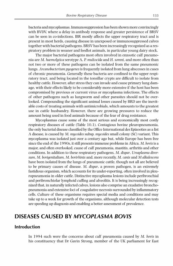
Bovine Respiratory Disease 133
bacteria and mycoplasmas. Immunosuppression has been shown more convincingly with BVDV, where a delay in antibody response and greater persistence of BRSV can be seen in co-infections. IBR mostly affects the upper respiratory tract and is present in most herds, causing disease in unexposed or immunosuppressed calves together with bacterial pathogens. BRSV has been increasingly recognized as a res-piratory problem in weaner and feedlot animals, in particular young dairy stock.
The major bacterial pathogens most often involved in enzootic calf pneumo-nia are M. haemolytica serotype A, P. multocida and H. somni, and more often than not two or more of these pathogens can be isolated from the same pneumonic lungs. Arcanobacterium pyogenes is frequently isolated from lung abscesses in cases of chronic pneumonia. Generally these bacteria are confi ned to the upper respi-ratory tract, and being located in the tonsillar crypts are diffi cult to isolate from healthy cattle. However, after stress they can invade and cause primary lung dam-age, with their effects likely to be considerably more extensive if the host has been compromised by previous or current virus or mycoplasma infections. The effects of other pathogens such as lungworm and other parasites should not be over-looked. Compounding the signifi cant animal losses caused by BRD are the inevit-able costs of treating animals with antimicrobials, which amounts to the greatest use in cattle husbandry. However, there are growing pressures to reduce the amount being used in food animals because of the fear of drug resistance.
Mycoplasmas cause some of the most serious and economically most costly respiratory diseases of cattle (Table 10.1). Contagious bovine pleuropneumonia, the only bacterial disease classifi ed by the Offi ce International des Epizooties as a list A disease, is caused by M. mycoides subsp. mycoides small colony (SC) variant. This mycoplasma was isolated just over a century ago but, while Europe has been free since the end of the 1990s, it still presents immense problems in Africa. M. bovis is a major, and often overlooked, cause of calf pneumonia, mastitis, arthritis and other conditions. In addition to these respiratory pathogens, M. dispar, Ureaplasma diver-sum, M. bovigenitalium, M. bovirhinis and, more recently, M. canis and M alkalescens have been isolated from the lungs of pneumonic cattle, though not all are believed to be primary causes of disease. M. dispar, a proven pathogen, is an extremely fastidious organism, which accounts for its under-reporting, often involved in pleu-ropneumonia in older cattle. Distinctive mycoplasma lesions include peribronchial and peribronchiolar lymphoid cuffi ng and alveolitis. It is being increasingly recog-nized that, in naturally infected calves, lesions also comprise an exudative broncho-pneumonia and extensive foci of coagulative necrosis surrounded by infl ammatory cells. Culture of these organisms requires special media and conditions and may take up to a week for growth of the organisms, although molecular detection tests are speeding up diagnosis and enabling a better assessment of prevalence.
DISEASES CAUSED BY MYCOPLASMA BOVIS
Introduction
In 1994 such were the concerns about calf pneumonia caused by M. bovis in his constituency that Dr Gavin Strong, member of the UK parliament for East

Chapter 10134
Edinburgh and Musselburgh, asked the then Minister of Agriculture what plans there were to combat the disease (Anon, 1994). The Minister replied that there were no plans to make the disease, which was believed to have been introduced into the UK after the introduction of the EU single market, notifi able. In fact, M. bovis was fi rst reported in the UK nearly 20 years earlier, probably introduced from the USA. A second invasion of perhaps more virulent strains entered with imported cattle herds from mainland Europe at the time of the single market, as evidenced by the signifi cant impact it had on Ireland (Byrne et al., 2001).
It is now well accepted that M. bovis is a major contributor to calf pneumonia. The evidence for this involvement includes the frequent isolation from the lungs of pneumonic calves, high seroprevalence in affected herds and the signifi cant impact the organism has had since its arrival in Ireland in the 1990s (Nicholas and Ayling, 2003). The effect of M. bovis on adult cattle is less well known in the UK and Ireland, although it has been reported as a major and uncomplicated cause of mastitis, arthritis and otitis in North America (Lamm et al., 2004). Recent isolations of M. bovis from the aborted fetuses and brains of cattle showing neuro-logical signs and otitis in the UK and Ireland suggest an even wider role in disease than previously thought (Ayling et al., 2005).
In some countries the lesions caused by M. bovis are thought to resemble those seen in CBPP, so the comparative properties of M. bovis and M. mycoides SC, the cause of CBPP, are given in Table 10.1.
Table 10.1. Comparative properties of the two most important cattle mycoplasmas.
Properties M. mycoides subsp. mycoides SC M. bovis
Diseases Contagious bovinepleuropneumonia in cattle, occasionally arthritis in calves
Calf pneumonia, mastitis, arthritis, abortion, keratoconjunctivitis
Distribution Sub-Saharan Africa, probably in parts of the Middle East, Central Asia
Worldwide
Hosts Cattle, goats (sheep) Cattle Histopathological lesions
Fibrinous pleuropneumonia with necrosis
Interstitial pneumonia, lympho-histiocytic bronchitis, catarrhal bronchopneumonia
Clinical signs Few signs, respiratory distress evident after exercise
Respiratory distress, mastitis, arthritis
Diagnosis Isolation, serology, PCR, abattoir surveillance
Serology, isolation, PCR
Treatment Recommendation of chemotherapy is contra-indicated because carrier status has been revised (FAO, 2007)
Chemotherapy
Control Vaccination, movement control, slaughter
Management, e.g. improved ventilation, reduced stocking density. Some auto genous vaccines available in USA and UK

Bovine Respiratory Disease 135
Distribution
M. bovis was fi rst isolated in 1961 in the USA from a case of severe mastitis in cattle (Hale et al., 1962) and, because of its close serological and biochemical similarity with the small ruminant pathogen M. agalactiae, was originally desig-nated as M. agalactiae var. bovis. It then appears to have spread via animal move-ments worldwide to many countries including Israel (1964), Spain (1967), Australia (1970), France (1974), mainland Britain (1974), Czechoslovakia (1975), Germany (1977), Denmark (1981), Switzerland (1983), Morocco (1988), South Korea (1989), Brazil (1989), Northern Ireland (1993), Republic of Ireland (1994), Chile (2000) (Nicholas, 2002a), South Africa (2005) and the Czech Republic (Zendulkova, 2007).
The prevalence of M. bovis is almost certainly underestimated, as M. haemo-lytica, P. multocida and H. somni are invariably isolated fi rst from pneumonic calves; in arthritis and mastitis, where M. bovis is a primary and uncomplicated cause, it is often overlooked as few laboratories routinely monitor for mycoplasmas. How-ever, unlike these conventional bacteria, the occurrence of M. bovis in a herd is invariably linked to cases of disease, in particular pneumonia, mastitis and/or arthritis (Pfützner and Sachse, 1996).
Economic Losses
In the UK, it is estimated that nearly 2 million cattle are affected annually by respi-ratory disease (Reeve-Johnson, 1999). Furthermore approximately 157,000 calves, which would have a potential market value of £99 million, die annually as a result of pneumonia and related illnesses. Across Europe, with approximately 90 million cattle, this extrapolates to total losses of 576 million euros. It is likely that M. bovis is responsible for at least a quarter to a third of these losses, although this is likely to be an underestimate (Nicholas et al., 2000). In the USA, the cost of M. bovis infections as a result of loss of weight gain and carcass value has been estimated at $32 million per year (Rosengarten and Citti, 1999). The losses due to bovine mastitis caused by M. bovis may be higher than that for respiratory disease, with estimates from the USA of up to $108 million per year, with infection rates of up to 70% of a herd (Rosengarten and Citti, 1999).
Host Range
M. bovis is highly adapted to cattle but occasional isolations have been made in chickens, buffaloes, small ruminants and even humans (Pitcher and Nicholas, 2005). In one case, M. bovis was isolated from the throat of a woman who had developed bronchopneumonia after heavy exposure to cow manure (Madoff et al., 1979). The disease subsided following tetracycline therapy with the development of antibodies to M. pneumoniae, but not to M. bovis, during convalescence, sug-gesting that M. bovis may not have been the primary aetiological agent. In a

Chapter 10136
second case, M. bovis was isolated from a patient with respiratory disease who suc-cessfully responded to tetracycline treatment (Pitcher and Nicholas, 2005).
M. bovis was isolated from a mastitic goat on a smallholding in Kent, UK in 1999 (Ayling et al., 2004). This was the fi rst case of M. bovis from a goat in Britain and caused some concern on its initial isolation because of the close biochemical and immunological similarity to M. agalactiae, which causes contagious agalac-tia and is exotic to Britain. The highly specifi c PCR of Subramaniam et al. (1998) confi rmed identifi cation of this isolate as M. bovis. In Turkey, multiple isolates of M. bovis were detected in the trachea of clinically healthy broiler chickens from a farm where poultry and cattle mixed freely (Ongor et al., 2008).
Epidemiology
M. bovis is not ubiquitous but widely spread within the bovine population in enzootically infected areas. The infection is usually introduced to M. bovis-free herds by clinically healthy calves or young cattle shedding the mycoplasma and, once established on multi-age sites, it becomes very diffi cult to eradicate. Its appearance on some farms suffering low-grade respiratory disease can lead to increased morbidity and mortality (Gourlay et al., 1989). Infected cattle shed the mycoplasma via the respiratory tract for many months and even years, when they act as reservoirs of infection (Pfützner, 1990). Contact animals become infected via the respiratory tract, the teat canal or genital tract; artifi cial insemination with infected semen is another common route (Pfützner, 1990). The male genital tract can become infected with M. bovis through contact with other animals or, possibly, via a heavily contaminated environment. Infection of the prepuce or urethra by M. bovis leads to an ascending infection of the testes, causing orchi-tis, vesiculitis, decrease of semen quality and ultimately shedding in the semen (Kreusel et al., 1989).
In a study of calves in the Netherlands, ter Laak et al. (1992a,b) found M. bovis in 20% of pneumonic lungs from fattening herds but only in a small number of apparently healthy calves. Following its introduction into the north and south of Ireland in 1994 from mainland Europe, M. bovis has been consis-tently isolated from 13 to 23% of pneumonic lungs (Brice et al., 2000; Byrne et al., 2001; Blackburn et al., 2007). In France, M. bovis was isolated from 30% of calf herds with pneumonia (Le Grand et al., 2001) while in Britain, about 20–25% of pneumonic herds contain animals with antibodies to M. bovis (Ayling et al., 2004). In the UK, signifi cant antibody titres to M. bovis were detected in just under half of 55 pneumonic herds examined, of which only seven had rising titres to viral pathogens: respiratory syncytial virus, infectious bovine rhinotracheitis or bovine viral diarrhoea virus (Nicholas et al., 2001).
Clearly other factors play a role in bovine respiratory disease, such as con-current viral and bacterial diseases as well as stress and environmental factors, but it is increasingly believed that M. bovis is the predisposing factor in the infec-tious process, leading to invasion by other bacterial pathogens possibly by com-promising host defences (Rebhun et al., 1995; Poumarat et al., 2001). In a study of calves which had died of pneumonia, Buchvarova and Vesselinova (1989)

Bovine Respiratory Disease 137
showed that over a third of lungs were infected only with M. bovis while the rest contained a combination of M. bovis with P. multocida and/or H. somni. These workers concluded that alterations in the lungs were chiefl y due to mycoplasma infection, with the remaining bacteria contributing to complications in the pneu-monic process.
A major epidemiology survey of M. bovis infection in Britain covering the years 1995–2005 was conducted by Lawes et al. (2008). At the herd level this investiga-tion showed that the proportion of pneumonic herds infected with M. bovis in England and Wales was very high (42.4%). Other studies have found that the pro-portion of infected herds is often higher than the percentage of serologically posi-tive animals. Le Grand et al. (2001) found that the average number of seropositive herds was 60%, yet the percentage of serologically positive animals ranged from 2 to 13%. This unexpected distribution was also seen in dairy herds in Switzerland (Burnens et al., 1999). Previously published work analysing data between 1994 and 1999 from the UK showed that 22% of herds were seropositive (Nicholas et al., 2000). This showed that the number of infected herds had risen and that this increase could be due to either the pathogen becoming even more widespread or the fact that there was a greater awareness of the infection in the fi eld, refl ecting a more accurate picture of the infection status in England and Wales.
Surprisingly, the analysis did not fi nd any seasonality of M. bovis infection, which is contrary to other pneumonia-causing pathogens, which are thought to cause higher infection rates in the winter months. On average the seroprevalence was highest in May, June and August (Lawes et al., 2008). Previous studies in the UK have found that outbreaks of M. bovis continue in herds where animals are housed all year round and that during the summer months the percentage of herds affected by M. bovis increases in relation to the number tested (Nicholas et al., 2000).
The results of spatial analysis in this study showed that M. bovis infection is not distributed evenly across England and Wales but that there are areas of very high seroprevalence (hotspots) (Fig. 10.1), which are continually changing on a yearly basis (Lawes et al., 2008). It is possible that the areas of high seropositiv-ity could be related to areas where the numbers of cattle are high. Using census data from 2005, Cornwall was an area with high cattle numbers (greater than 200,000) and in this year the seropositivity for this county was 41–50%. York-shire also showed a similar correlation. It is perhaps unsurprising that the ‘hotspot’ areas are not continually associated with one area, considering the dynamics of the UK cattle industry. Cattle move extensively in their lifetime and cover large distances. The animals may become infected when they move to new premises or they may shed the mycoplasma and consequently infect other herds through the stress of movement. Animals travel further distances now due to the closure of many livestock markets and therefore those remaining may be supplied by animals from many more counties and hence the spread of infection increases. However, despite the organism’s persistence on a farm, it is clear that some are able to eliminate the infection, otherwise the prevalence in the UK would continue to increase. Farms operating ‘all in, all out systems’ are less likely to suffer persis-tent infection, but some mixed-aged farms also seem to lose infection after a period, although it is not known how. The data set used in this study had some

Chapter 10138
limitation in that there appear to be regional differences in the awareness of M. bovis as a cause of pneumonia, and this factor therefore affects the number of samples that are taken for diagnosis.
Molecular Epidemiology
The genetic diversity of 53 isolates collected in the UK between 1996 and 2002 was studied using pulsed fi eld gel electrophoresis (PFGE), amplifi ed fragment length polymorphism (AFLP) and random amplifi ed polymorphic DNA (RAPD) analysis (McAuliffe et al., 2004). In addition, the infl uence of variable surface pro-tein (Vsp) profi les on the profi les generated using molecular typing techniques was studied. Both AFLP and RAPD separated the isolates into two distinct groups
Fig. 10.1. Map of England and Wales to show seroprevalence of M. bovis by area for 1995 to 2005 inclusive.
Mycoplasma bovisseroprevalence
Average percentage(1995–2005)
2–1011–2021–3031–4041–50>50No data

Bovine Respiratory Disease 139
comprising over 30 different profi les; however, PFGE showed less congruence with the other techniques. It was possible to speculate that the two distinct groups identifi ed by RAPD and AFLP in this study may represent two clonal lines of descent. It was hypothesized that group A may have entered the UK from mainland Europe around the time of the creation of the European Union single market in 1993, when large numbers of cattle came into the UK from continental Europe (McAuliffe et al., 2004). Group B could have a different and possibly older ancestral origin and have derived from the fi rst strains of M. bovis isolated in the UK in 1975, which were thought to have originated in the USA. A later study using RAPD showed a closer similarity between Italian strains and the type strain of M. agalactiae than with the type strain of M. bovis which originated from the USA (Loria et al., 2007). These two species are very similar and were originally considered subspecies, so molecular typing could provide fresh insights into the evolution of these organisms.
McAuliffe et al. (2004) noted a lack of correlation between geographical origin, time of isolation and genomic profi les of the UK strains, and in some instances, strains from farms over 400 km apart had identical profi les. These fi nd-ings are similar to a study of European M. bovis strains using restriction enzyme analysis, which indicated that there was no correlation between the geographical origin, genetic heterogeneity and isolation date (Poumarat et al., 1994) and refl ects the extensive cattle movement that occurs within the UK and Europe.
Causative Organism
M. bovis was elevated to species rank and given its present name in 1976 (Askaa and Erno, 1976). However, in spite of the fact that the two mycoplasmas cause different diseases in different animals and have a DNA–DNA reassociation value of only 40%, they show a very close 16S rRNA similarity of 99.8%. M. bovis is also biochemically similar to M. agalactiae, as neither ferment glucose nor hydrolyse arginine but instead use organic acids, lactate and pyruvate for their energy source for growth (Miles et al., 1988); they also give fi lm and spot reactions in vitro, indicating the possession of lipolytic activity. Table 10.2 shows comparative biochemical characteristics of bovine mycoplasmas.
Like all mollicutes, M. bovis is small, pleomorphic and lacks a cell wall (Hermann, 1992). It had a genome size of 1080 kbp estimated by PFGE but the complete genome of the type strain M. bovis (PG45) was sequenced through a collaboration between the University of Missouri and the J.C. Venter Institute. Traditional shotgun cloning and Sanger sequencing technologies were employed to generate 8X sequence coverage of the genome of a clonal isolate. Analysis of the fully assembled sequence revealed a single circular chromosome of 1,003,404 base pairs, with a G+C content of 29.3%. The current annotation of the genome contained 868 genes, of which 827 are predicted to encode open reading frames (Wise, Calcutt and Methe, personal communication, 2008). Future analysis of the genetic blueprint of this important cattle pathogen is anticipated to reveal features that are critical for virulence and survival, in addition to establishing a database of targets that might be utilized in diagnosis, treatment and control.
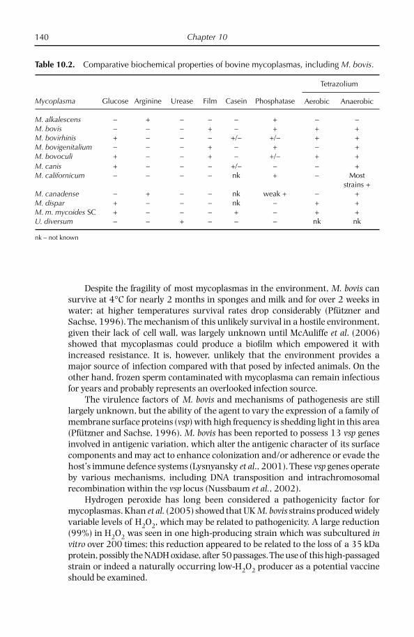
Chapter 10140
Despite the fragility of most mycoplasmas in the environment, M. bovis can survive at 4°C for nearly 2 months in sponges and milk and for over 2 weeks in water; at higher temperatures survival rates drop considerably (Pfützner and Sachse, 1996). The mechanism of this unlikely survival in a hostile environment, given their lack of cell wall, was largely unknown until McAuliffe et al. (2006) showed that mycoplasmas could produce a biofi lm which empowered it with increased resistance. It is, however, unlikely that the environment provides a major source of infection compared with that posed by infected animals. On the other hand, frozen sperm contaminated with mycoplasma can remain infectious for years and probably represents an overlooked infection source.
The virulence factors of M. bovis and mechanisms of pathogenesis are still largely unknown, but the ability of the agent to vary the expression of a family of membrane surface proteins (vsp) with high frequency is shedding light in this area (Pfützner and Sachse, 1996). M. bovis has been reported to possess 13 vsp genes involved in antigenic variation, which alter the antigenic character of its surface components and may act to enhance colonization and/or adherence or evade the host’s immune defence systems (Lysnyansky et al., 2001). These vsp genes operate by various mechanisms, including DNA transposition and intrachromosomal recombination within the vsp locus (Nussbaum et al., 2002).
Hydrogen peroxide has long been considered a pathogenicity factor for mycoplasmas. Khan et al. (2005) showed that UK M. bovis strains produced widely variable levels of H2O2, which may be related to pathogenicity. A large reduction (99%) in H2O2 was seen in one high-producing strain which was subcultured in vitro over 200 times; this reduction appeared to be related to the loss of a 35 kDa protein, possibly the NADH oxidase, after 50 passages. The use of this high-passaged strain or indeed a naturally occurring low-H2O2 producer as a potential vaccine should be examined.
Table 10.2. Comparative biochemical properties of bovine mycoplasmas, including M. bovis.
Mycoplasma Glucose Arginine Urease Film Casein Phosphatase
Tetrazolium
Aerobic Anaerobic
M. alkalescens − + − − − + − −M. bovis − − − + − + + +M. bovirhinis + − − − +/− +/− + +M. bovigenitalium − − − + − + − +M. bovoculi + − − + − +/− + +M. canis + − − − +/− − − +M. californicum − − − − nk + − Most
strains +M. canadense − + − − nk weak + − +M. dispar + − − − nk − + +M. m. mycoides SC + − − − + − + +U. diversum − − + − − − nk nk
nk – not known
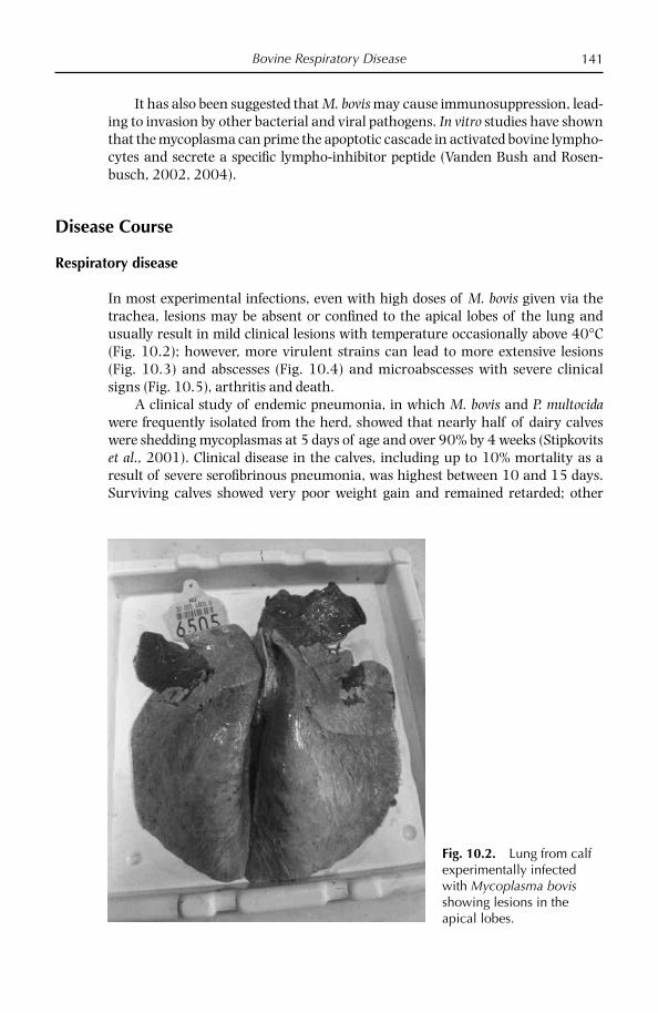
Bovine Respiratory Disease 141
It has also been suggested that M. bovis may cause immunosuppression, lead-ing to invasion by other bacterial and viral pathogens. In vitro studies have shown that the mycoplasma can prime the apoptotic cascade in activated bovine lympho-cytes and secrete a specifi c lympho-inhibitor peptide (Vanden Bush and Rosen-busch, 2002, 2004).
Disease Course
Respiratory disease
In most experimental infections, even with high doses of M. bovis given via the trachea, lesions may be absent or confi ned to the apical lobes of the lung and usually result in mild clinical lesions with temperature occasionally above 40°C (Fig. 10.2); however, more virulent strains can lead to more extensive lesions (Fig. 10.3) and abscesses (Fig. 10.4) and microabscesses with severe clinical signs (Fig. 10.5), arthritis and death.
A clinical study of endemic pneumonia, in which M. bovis and P. multocida were frequently isolated from the herd, showed that nearly half of dairy calves were shedding mycoplasmas at 5 days of age and over 90% by 4 weeks (Stipkovits et al., 2001). Clinical disease in the calves, including up to 10% mortality as a result of severe serofi brinous pneumonia, was highest between 10 and 15 days. Surviving calves showed very poor weight gain and remained retarded; other
Fig. 10.2. Lung from calf experimentally infected with Mycoplasma bovis showing lesions in the apical lobes.
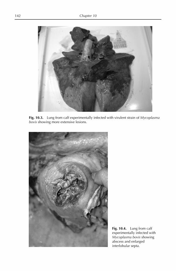
Chapter 10142
Fig. 10.3. Lung from calf experimentally infected with virulent strain of Mycoplasma bovis showing more extensive lesions.
Fig. 10.4. Lung from calf experimentally infected with Mycoplasma bovis showing abscess and enlarged interlobular septa.

Bovine Respiratory Disease 143
signs included fever, depression, hyperpnoea, dyspnoea, nasal discharge, mild to continuous coughing and loss of appetite. In the UK, calf pneumonia usually begins in November and peaks around January, but deaths due to M. bovis con-tinue to occur in some herds in the spring at pasture, representing relapses because of unresolved lung lesions (Nicholas et al., 2001).
Clinical signs of M. bovis infection are not characteristic, but some observers consider the following to be key features of this disease:
Harsh hacking cough; ●
Low-grade fever; ●
Mild depression; ●
Runny eyes; ●
Ear droop; ●
Arthritis . ●
Pulmonary lesions in naturally infected calves comprise an exudative bron-chopneumonia and extensive foci of coagulative necrosis surrounded by infl am-matory cells. In studies involving experimental infections of gnotobiotic calves with M. bovis, signifi cant pneumonia was induced, involving up to 30% of the lung surface, and was of suffi cient severity to cause clinical respiratory disease in some calves (Thomas et al., 1986). Distinctive areas of coagulative necrosis were prominent within the lesions. Chronic infections are often associated with a lymphocytic ‘cuffi ng’ pneumonia, where there is marked hyperplasia of peribron-chial lymphoid tissue, causing stenosis of airway lumina and compression and collapse of adjacent pulmonary prenchyma. M. bovis antigen is mainly detected at
Fig. 10.5. Calf experimentally infected with Mycoplasma bovis showing clinical signs of respiratory distress.
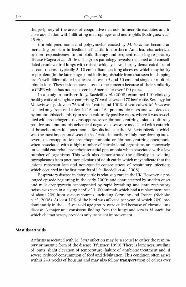
Chapter 10144
the periphery of the areas of coagulative necrosis, in necrotic exudates and in close association with infi ltrating macrophages and neutrophils (Rodriguez et al., 1996).
Chronic pneumonia and polysynovitis caused by M. bovis has become an increasing problem in feedlot beef cattle in northern America, characterized by non-responsiveness to antibiotic therapy and frequent relapsing respiratory disease (Gagea et al., 2006). The gross pathology reveals: reddened and consoli-dated cranioventral lungs with raised, white–yellow, sharply demarcated foci of caseous necrosis typically 2–10 cm in diameter; lung abcesses, which may be dry or purulent (in the later stages) and indistinguishable from that seen in ‘shipping fever’; well-differentiated sequestra between 5 and 30 cm; and single or multiple joint lesions. These lesions have caused some concern because of their similarity to CBPP, which has not been seen in America for over 100 years.
In a study in northern Italy, Raedelli et al. (2008) examined 140 clinically healthy cattle at slaughter, comprising 70 veal calves and 70 beef cattle. Serology for M. bovis was positive in 76% of beef cattle and 100% of veal calves. M. bovis was isolated only from veal calves in 16 out of 64 pneumonic cases and was detected by immunohistochemistry in seven culturally positive cases, where it was associ-ated with bronchogenic necrosuppurative or fi brinonecrotizing lesions. Culturally positive and immunohisto chemical negative cases were associated with catarrh-al–bronchointerstitial pneumonia. Results indicate that M. bovis infection, which was the most important disease in beef cattle in northern Italy, may develop into a severe necrosuppurative bronchopneumonia or fi brinonecrotizing pneumonia when associated with a high number of intralesional organisms or, conversely, into a mild catarrhal–bronchointerstitial pneumonia when associated with a low number of organisms. This work also demonstrated the diffi culty in isolating mycoplasmas from pneumonic lesions of adult cattle, which may indicate that the lesions represent late and non-specifi c consequences of respiratory infections which occurred in the fi rst months of life (Raedelli et al., 2008).
Respiratory disease in dairy cattle is relatively rare in the UK. However, a pro-longed episode beginning in the early 2000s and characterized by sudden onset and milk drop/pyrexia accompanied by rapid breathing and hard respiratory noises was seen in a ‘fl ying herd’ of 1400 animals which had a replacement rate of about 20% from various sources, including Germany and France (Nicholas et al., 2006). At least 10% of the herd was affected per year, of which 20%, pre-dominantly in the 4–5-year-old age group, were culled because of chronic lung disease. A major and consistent fi nding from the lungs and sera is M. bovis, for which chemotherapy provides only transient improvement.
Mastitis/arthritis
Arthritis associated with M. bovis infection may be a sequel to either the respira-tory or mastitic form of the disease (Pfützner, 1990). There is lameness, swelling of joints, slight elevation of temperature, failure of antibiotic treatment and, if severe, reduced consumption of feed and debilitation. This condition often arises within 2–3 weeks of housing and may also follow transportation of calves over
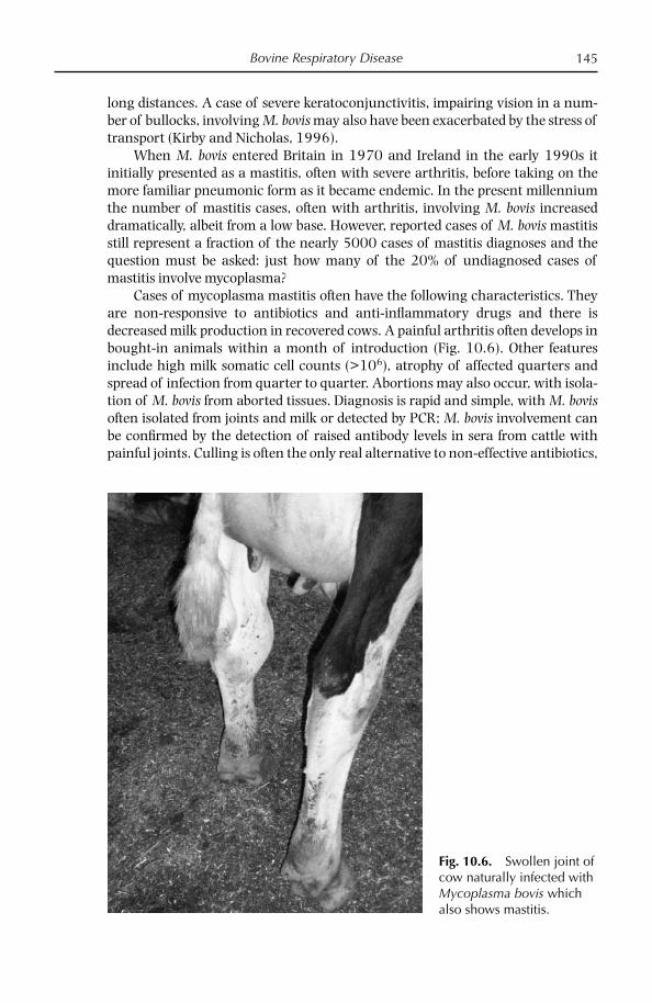
Bovine Respiratory Disease 145
long distances. A case of severe keratoconjunctivitis, impairing vision in a num-ber of bullocks, involving M. bovis may also have been exacerbated by the stress of transport (Kirby and Nicholas, 1996).
When M. bovis entered Britain in 1970 and Ireland in the early 1990s it initially presented as a mastitis, often with severe arthritis, before taking on the more familiar pneumonic form as it became endemic. In the present millennium the number of mastitis cases, often with arthritis, involving M. bovis increased dramatically, albeit from a low base. However, reported cases of M. bovis mastitis still represent a fraction of the nearly 5000 cases of mastitis diagnoses and the question must be asked: just how many of the 20% of undiagnosed cases of mastitis involve mycoplasma?
Cases of mycoplasma mastitis often have the following characteristics. They are non-responsive to antibiotics and anti-infl ammatory drugs and there is decreased milk production in recovered cows. A painful arthritis often develops in bought-in animals within a month of introduction (Fig. 10.6). Other features include high milk somatic cell counts (>106), atrophy of affected quarters and spread of infection from quarter to quarter. Abortions may also occur, with isola-tion of M. bovis from aborted tissues. Diagnosis is rapid and simple, with M. bovis often isolated from joints and milk or detected by PCR; M. bovis involvement can be confi rmed by the detection of raised antibody levels in sera from cattle with painful joints. Culling is often the only real alternative to non-effective antibiotics,
Fig. 10.6. Swollen joint of cow naturally infected with Mycoplasma bovis which also shows mastitis.

Chapter 10146
for humane reasons and to prevent the spread of disease from these affected animals to healthy contacts.
Otitis and related neurological disorders
Otitis in calves may be associated with a variety of bacterial agents, including H. somni, M. haemolytica, P. multocida, streptococcal spp., Actinomyces spp., Arcanobac-terium pyogenes and M. bovis (Duarte and Hamdan, 2004; Francoz et al., 2005). The pathogenesis of otitis due to M. bovis is not fully understood; infection may arise by extension from the pharynx via the Eustachian tube to the middle ear and may extend into the inner ear (Walz et al., 1997; Maeda et al., 2003; Lamm et al., 2004). Clinical signs associated with infection may include head tilt and neuro-logical signs in dairy and beef calves from 2 to 18 weeks of age (Lamm et al., 2004; Francoz et al., 2005). Previous reports of otitis associated with M. bovis infection have been from North America and Japan (Maeda et al., 2003; Lamm et al., 2004; Francoz et al., 2005).
In 2007, signs of severe otitis media in 20% of dairy calves on one farm were associated with M. bovis infection, based on isolation from the external ear canal and nares (Foster et al., 2008) (Fig. 10.7). Affected calves also seroconverted to M. bovis and no other signifi cant bacteria were isolated. Infection was considered likely to have originated from cows in the milking herd showing low-grade
Fig. 10.7. Cow with otitis caused by Mycoplasma bovis.

Bovine Respiratory Disease 147
mastitis, based on evidence of seroconversion and detection of infection in milk samples.
Two separate cases involving M. bovis from calf brains were reported in 2005 in England and Ireland (Ayling et al., 2005). In England, calves presented with head tilt and convulsions, suggesting central nervous system (CNS) involvement. One of the affected heifer calves had previously shown signs of head tilt, which appeared to have been treated successfully but after rehousing in the autumn the animal had become ataxic, recumbent and had developed CNS signs including convulsions. Following euthanasia, post-mortem fi ndings included cloudiness and scarring of the left cornea and a purulent meningitis affecting the left side of the cerebellum in the area adjacent to the left middle ear and the ventral part of the brain near the pituitary. M. bovis, the only organism isolated, was cultured from the brain close to the left middle ear (Fig. 10.8). Histology indicated a focus of otitis media which had spread to the cranial vault. Serological examination of 13 samples for M. bovis showed a range of titres from negative to very high posi-tive samples. Interestingly, the highest titres were from the two affected calves, their dams also being positive.
The Irish case occurred on a farm experiencing outbreaks of respiratory disease. One of the affected and treated calves responded but did not thrive and later developed clinical signs of lethargy, depression, apparent blindness, severe weight loss and was also observed to grind its teeth. Post-mortem examination
Fig. 10.8. Immunoperoxidase technique for Mycoplasma bovis antigen showing large numbers of intralesional organisms (dark staining), particularly at the advancing margin of a cerebellopontine abscess in a 7-month-old calf (photo courtesy of Sandra Scholes (VLA) using monoclonal antibody made by Dr Hywell Ball).

Chapter 10148
revealed a large spheroidal, fi brinous lesion in the heart, which was attached to the wall of the left atrium. There were also multiple areas of necrosis within the cerebral hemispheres of the brain. Only M. bovis colonies were isolated from the brain and endocarditis lesions. A second calf showed arthritis in one hock joint but also failed to respond to treatment, resulting in impaired movement and was euthanized. Severe chronic arthritic changes in addition to chronic pneumonic lesions were observed during necropsy examination, from which M. bovis was isolated.
These fi ndings suggest an important and possibly under-reported role for M. bovis in neurological disease and consideration should be given to its diagnosis when investigating spongiform encephalopathies.
Diagnosis
Clinical and pathological signs are not always characteristic for M. bovis, so labo-ratory diagnosis is necessary for identifi cation. Samples of choice are listed in Table 10.3. In a study, Thomas et al. (2002a) showed that sampling by broncho-alveolar lavage was more predictive of lower respiratory airway pathogens, including M. bovis, than nasal swabs, although clearly not as convenient. M. bovis grows well in a variety of media, producing ‘centred’ colonies within 3–5 days (Nicholas and Baker, 1998). In an appropriate medium (such as Eaton’s) M. bovis produces fi lms and spots and gives an orange colour to the broth. Other biochem-ical characteristics are shown in Table 10.2 alongside other bovine mycoplasmas. Growth inhibition, fi lm inhibition, fl uorescent antibody or metabolic inhibitions tests can be used to identify the mycoplasma using hyperimmune rabbit serum (Poveda and Nicholas, 1998).
Ball and Findlay (1998) described a sandwich ELISA for M. bovis in which specifi c monoclonal antibodies, fi xed to the microplate, captured M. bovis anti-gen from the medium; the sensitivity of the test, similar to that of conventional culture diagnosis, can be improved by a short enrichment stage. This test is now available in a commercial format. M. bovis can be easily outgrown by
Table 10.3. Samples of choice for diagnosis of diseases caused by M. bovis.
Disease
Nasal swabs/ BAL*/affected lung Milk
Joint fl uid/ synovia
Eye swabs
Semen/genital discharge/preputial washings
Serum for antibody detection
Pneumonia ✓ ✓Mastitis ✓ ✓Arthritis ✓ ✓KC** ✓ ✓Infertility ✓ ✓
*bronchial alveolar lavage**keratoconjunctivitis
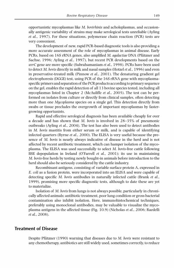
Bovine Respiratory Disease 149
opportunistic mycoplasmas like M. bovirhinis and acholeplasmas, and occasion-ally antigenic variability of strains may make serological tests unreliable (Ayling et al., 1997). For these situations, polymerase chain reaction (PCR) tests are very convenient.
The development of new, rapid PCR-based diagnostic tools is also providing a more accurate assessment of the role of mycoplasmas in animal disease. Early PCRs, based on 16S rRNA genes, also amplifi ed M. agalactiae DNA (Pfützner and Sachse, 1996; Ayling et al., 1997), but recent PCR developments based on the uvrC gene are more specifi c (Subrahamaniam et al., 1998). PCRs have been used to detect M. bovis directly in milk and nasal samples (Hotzel et al., 1996) and even in preservative-treated milk (Pinnow et al., 2001). The denaturing gradient gel electrophoresis (DGGE) test, using PCR of the 16S rRNA gene with mycoplasma-specifi c primers and separation of the PCR products according to primary sequence on the gel, enables the rapid detection of all 13 bovine species tested, including all mycoplasmas listed in Chapter 2 (McAuliffe et al., 2005). The test can be per-formed on isolates from culture or directly from clinical samples, often detecting more than one Mycoplasma species on a single gel. This detection directly from swabs or tissue precludes the overgrowth of important mycoplasmas by faster-growing opportunists.
Rapid and effective serological diagnosis has been available cheaply for over a decade and has shown that M. bovis is involved in 28–35% of pneumonic outbreaks (Ayling et al., 2004). The test has also been used to detect antibodies in M. bovis mastitis from either serum or milk, and is capable of identifying infected quarters (Byrne et al., 2000). The ELISA is very useful because the pre-sence of M. bovis is nearly always indicative of disease in the herd and is not affected by recent antibiotic treatment, which can hamper isolation of the myco-plasma. The ELISA was used successfully to select M. bovis-free cattle following BSE depopulation in Ireland (O’Farrell et al., 2001); its use in maintaining M. bovis-free herds by testing newly bought-in animals before introduction to the herd should also be seriously considered by the cattle industry.
Recombinant antigens, consisting of variable surface protein A, expressed in E. coli as a fusion protein, were incorporated into an ELISA and were capable of detecting specifi c M. bovis antibodies in naturally infected cattle (Brank et al., 1999), promising more specifi c diagnostic tests, although to date these are yet to materialize.
Isolation of M. bovis from lungs is not always possible, particularly in chroni-cally affected animals; antibiotic treatment, poor lung condition or gross bacterial contamination also inhibit isolation. Here, immunohistochemical techniques, preferably using monoclonal antibodies, may be valuable to visualize the myco-plasma antigens in the affected tissue (Fig. 10.9) (Nicholas et al., 2006; Raedelli et al., 2008).
Treatment of Disease
Despite Pfützner (1990) warning that diseases due to M. bovis were resistant to any chemotherapy, antibiotics are still widely used, sometimes correctly, to reduce
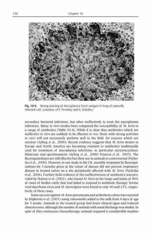
Chapter 10150
secondary bacterial infections, but often ineffectively to treat the mycoplasma infections. Many in vitro studies have compared the susceptibility of M. bovis to a range of antibiotics (Table 10.4). While it is clear that antibiotics which are ineffective in vitro are unlikely to be effective in vivo, those with strong activities in vitro will not necessarily perform well in the fi eld, for reasons which are unclear (Ayling et al., 2000). Recent evidence suggests that M. bovis strains in Europe and North America are becoming resistant to antibiotics traditionally used for treatment of mycoplasma infections, in particular oxytetracyclines, tilmicosin and spectinomycin (Ayling et al., 2000; Francoz et al., 2005). The fl uoro quinolones are still effective but their use in animals is controversial (Nicho-las et al., 2000). However, in one study in the UK, monthly treatment by fl uoroqui-nolones for 3 months given at the outset of disease did not prevent respiratory disease in treated calves on a site persistently affected with M. bovis (Nicholas et al., 2006). Further field evidence of the ineffectiveness of antibiotics was pro-vided by Haines et al. (2001), who found M. bovis in the lungs and joints of 80% of cases of feedlot cattle that had failed to respond to antibiotic therapy; bovine viral dia rrhoea virus and M. haemolytica were found in only 40 and 23%, respec-tively, of these cases.
Some success against M. bovis pneumonia and arthritis in calves was reported by Stipkovits et al. (2001) using valnemulin added to the milk from 4 days of age for 3 weeks. Animals in the treated group had fewer clinical signs and reduced clinical scores, although the number of animals with nasal discharge was similar. In spite of this continuous chemotherapy, animals required a considerable number
Fig. 10.9. Strong staining of Mycoplasma bovis antigen in lung of naturally infected calf. (courtesy of F. Twomey and S. Scholes.)

Bovine R
espiratory Disease
151
Table 10.4. Effectiveness of antibiotics in vitro against fi eld strains of M. bovis (minimum inhibitory concentrations, µg/ml).
Country(source)
No. of strains
Enrofl oxacin/Danofl oxacin Lincomycin Spectinomycin Tylosin Tilmicosin Oxytetracycline
The Netherlands (ter Laak et al., 1993)
19 0.5–2.0/ND
0.25–1.00 1–4 0.06–4.00 ND 8–> 64
N. Ireland (Ball et al., 1995) 23 1–2/ND
0.125–2.000 4–6 ND 0.06–0.5.00 (9 strains)4–> 32 (14 strains)
ND
Italy (Mazzolini et al., 1997) 23 0.06–0.25/ND
0.25–1.00 0.12–2.00 0.12–4.00 16–> 32 0.12–4.00
Britain(Ayling et al., 2000)
62 ND/0.125–2.000
ND 1–8 (50 strains)128–> 128 (12 strains)
ND 4–< 128 1–128
(Ayling et al., 2007) 40 0.12–1.00/0.12–0.50
1–> 32 2–> 64 ND < 0.06–> 64.00 0.12–32.00
Interpretation (ter Laak et al., 1993): 1 µg/ml – mycoplasmas susceptible 2–4 µg/ml – mycoplasma show intermediate susceptibility > 8 µg/ml – mycoplasmas resistantND – not determined
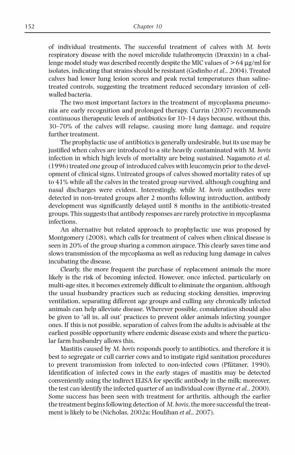
Chapter 10152
of individual treatments. The successful treatment of calves with M. bovis respiratory disease with the novel microlide tulathromycin (Draxxin) in a chal-lenge model study was described recently despite the MIC values of > 64 µg/ml for isolates, indicating that strains should be resistant (Godinho et al., 2004). Treated calves had lower lung lesion scores and peak rectal temperatures than saline-treated controls, suggesting the treatment reduced secondary invasion of cell-walled bacteria.
The two most important factors in the treatment of mycoplasma pneumo-nia are early recognition and prolonged therapy. Currin (2007) recommends continuous therapeutic levels of antibiotics for 10–14 days because, without this, 30–70% of the calves will relapse, causing more lung damage, and require further treatment.
The prophylactic use of antibiotics is generally undesirable, but its use may be justifi ed when calves are introduced to a site heavily contaminated with M. bovis infection in which high levels of mortality are being sustained. Nagamoto et al. (1996) treated one group of introduced calves with leucomycin prior to the devel-opment of clinical signs. Untreated groups of calves showed mortality rates of up to 41% while all the calves in the treated group survived, although coughing and nasal discharges were evident. Interestingly, while M. bovis antibodies were detected in non-treated groups after 2 months following introduction, antibody development was signifi cantly delayed until 8 months in the antibiotic-treated groups. This suggests that antibody responses are rarely protective in mycoplasma infections.
An alternative but related approach to prophylactic use was proposed by Montgomery (2008), which calls for treatment of calves when clinical disease is seen in 20% of the group sharing a common airspace. This clearly saves time and slows transmission of the mycoplasma as well as reducing lung damage in calves incubating the disease.
Clearly, the more frequent the purchase of replacement animals the more likely is the risk of becoming infected. However, once infected, particularly on multi-age sites, it becomes extremely diffi cult to eliminate the organism, although the usual husbandry practices such as reducing stocking densities, improving ventilation, separating different age groups and culling any chronically infected animals can help alleviate disease. Wherever possible, consideration should also be given to ‘all in, all out’ practices to prevent older animals infecting younger ones. If this is not possible, separation of calves from the adults is advisable at the earliest possible opportunity where endemic disease exists and where the particu-lar farm husbandry allows this.
Mastitis caused by M. bovis responds poorly to antibiotics, and therefore it is best to segregate or cull carrier cows and to instigate rigid sanitation procedures to prevent transmission from infected to non-infected cows (Pfützner, 1990). Identifi cation of infected cows in the early stages of mastitis may be detected conveniently using the indirect ELISA for specifi c antibody in the milk; moreover, the test can identify the infected quarter of an individual cow (Byrne et al., 2000). Some success has been seen with treatment for arthritis, although the earlier the treatment begins following detection of M. bovis, the more successful the treat-ment is likely to be (Nicholas, 2002a; Houlihan et al., 2007).
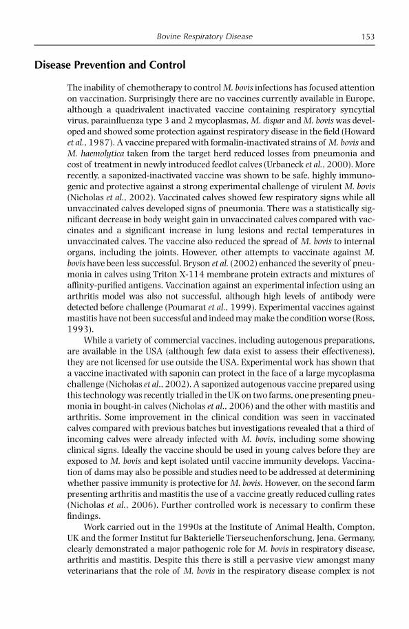
Bovine Respiratory Disease 153
Disease Prevention and Control
The inability of chemotherapy to control M. bovis infections has focused attention on vaccination. Surprisingly there are no vaccines currently available in Europe, although a quadrivalent inactivated vaccine containing respiratory syncytial virus, parainfl uenza type 3 and 2 mycoplasmas, M. dispar and M. bovis was devel-oped and showed some protection against respiratory disease in the fi eld (Howard et al., 1987). A vaccine prepared with formalin-inactivated strains of M. bovis and M. haemolytica taken from the target herd reduced losses from pneumonia and cost of treatment in newly introduced feedlot calves (Urbaneck et al., 2000). More recently, a saponized-inactivated vaccine was shown to be safe, highly immuno-genic and protective against a strong experimental challenge of virulent M. bovis (Nicholas et al., 2002). Vaccinated calves showed few respiratory signs while all unvaccinated calves developed signs of pneumonia. There was a statistically sig-nifi cant decrease in body weight gain in unvaccinated calves compared with vac-cinates and a signifi cant increase in lung lesions and rectal temperatures in unvaccinated calves. The vaccine also reduced the spread of M. bovis to internal organs, including the joints. However, other attempts to vaccinate against M. bovis have been less successful. Bryson et al. (2002) enhanced the severity of pneu-monia in calves using Triton X-114 membrane protein extracts and mixtures of affi nity-purifi ed antigens. Vaccination against an experimental infection using an arthritis model was also not successful, although high levels of antibody were detected before challenge (Poumarat et al., 1999). Experimental vaccines against mastitis have not been successful and indeed may make the condition worse (Ross, 1993).
While a variety of commercial vaccines, including autogenous preparations, are available in the USA (although few data exist to assess their effectiveness), they are not licensed for use outside the USA. Experimental work has shown that a vaccine inactivated with saponin can protect in the face of a large mycoplasma challenge (Nicholas et al., 2002). A saponized autogenous vaccine prepared using this technology was recently trialled in the UK on two farms, one presenting pneu-monia in bought-in calves (Nicholas et al., 2006) and the other with mastitis and arthritis. Some improvement in the clinical condition was seen in vaccinated calves compared with previous batches but investigations revealed that a third of incoming calves were already infected with M. bovis, including some showing clinical signs. Ideally the vaccine should be used in young calves before they are exposed to M. bovis and kept isolated until vaccine immunity develops. Vaccina-tion of dams may also be possible and studies need to be addressed at determining whether passive immunity is protective for M. bovis. However, on the second farm presenting arthritis and mastitis the use of a vaccine greatly reduced culling rates (Nicholas et al., 2006). Further controlled work is necessary to confi rm these fi ndings.
Work carried out in the 1990s at the Institute of Animal Health, Compton, UK and the former Institut fur Bakterielle Tierseuchenforschung, Jena, Germany, clearly demonstrated a major pathogenic role for M. bovis in respiratory disease, arthritis and mastitis. Despite this there is still a pervasive view amongst many veterinarians that the role of M. bovis in the respiratory disease complex is not
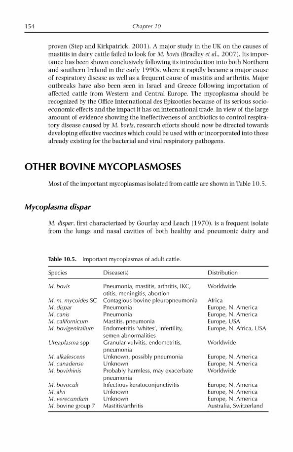
Chapter 10154
proven (Step and Kirkpatrick, 2001). A major study in the UK on the causes of mastitis in dairy cattle failed to look for M. bovis (Bradley et al., 2007). Its impor-tance has been shown conclusively following its introduction into both Northern and southern Ireland in the early 1990s, where it rapidly became a major cause of respiratory disease as well as a frequent cause of mastitis and arthritis. Major outbreaks have also been seen in Israel and Greece following importation of affected cattle from Western and Central Europe. The mycoplasma should be recognized by the Offi ce International des Epizooties because of its serious socio-economic effects and the impact it has on international trade. In view of the large amount of evidence showing the ineffectiveness of antibiotics to control respira-tory disease caused by M. bovis, research efforts should now be directed towards developing effective vaccines which could be used with or incorporated into those already existing for the bacterial and viral respiratory pathogens.
OTHER BOVINE MYCOPLASMOSES
Most of the important mycoplasmas isolated from cattle are shown in Table 10.5.
Mycoplasma dispar
M. dispar, fi rst characterized by Gourlay and Leach (1970), is a frequent isolate from the lungs and nasal cavities of both healthy and pneumonic dairy and
Table 10.5. Important mycoplasmas of adult cattle.
Species Disease(s) Distribution
M. bovis Pneumonia, mastitis, arthritis, IKC, otitis, meningitis, abortion
Worldwide
M. m. mycoides SC Contagious bovine pleuropneumonia AfricaM. dispar Pneumonia Europe, N. AmericaM. canis Pneumonia Europe, N. AmericaM. californicum Mastitis, pneumonia Europe, USAM. bovigenitalium Endometritis ‘whites’, infertility,
semen abnormalitiesEurope, N. Africa, USA
Ureaplasma spp. Granular vulvitis, endometritis,pneumonia
Worldwide
M. alkalescens Unknown, possibly pneumonia Europe, N. AmericaM. canadense Unknown Europe, N. AmericaM. bovirhinis Probably harmless, may exacerbate
pneumoniaWorldwide
M. bovoculi Infectious keratoconjunctivitis Europe, N. AmericaM. alvi Unknown Europe, N. AmericaM. verecundum Unknown Europe, N. AmericaM. bovine group 7 Mastitis/arthritis Australia, Switzerland

Bovine Respiratory Disease 155
fattening calves. It has been isolated from most parts of the world where cattle are kept, including Europe, Australia, the USA, Canada, Korea and Japan. In Europe there have been reports from the UK, Denmark, Belgium, Holland and France (Nicholas, 2002b). It has been shown to cause pneumonia following experimen-tal infection, although the disease produced is not severe; it has, occasionally, been reported in cases of mastitis (Jasper, 1981). It is certainly under-reported because of its extreme fastidiousness in in vitro culture.
It is not possible to determine the signifi cance of M. dispar infections as it is rarely the sole cause of disease. Viruses, bacteria and other mycoplasmas are major contributors to calf pneumonia, which has been estimated to cost the UK up to £50 million per year (Rebhun et al., 1995) in terms of set-back costs, calf mortality and treatment; in Europe this fi gure may exceed 560 million euros (Reeve-Johnson, 1999).
Like all mycoplasmas, M. dispar is small with a genome size of 803 kbp (Her-mann, 1992), pleomorphic, lacks a cell wall and has a low G+C ratio (28.5–29.3 mol%). However, it is very fastidious in culture, requiring a specialist medium for growth and does not produce typical ‘fried egg’ colonies, particularly in early pas-sages of fresh isolates. Furthermore these are not always inhibited by hyperim-mune serum, making conventional identifi cation diffi cult in the early stages (ter Laak and Noordergraaf, 1987). M. dispar grows slowly in media, requiring 7–14 days to produce colonies. It ferments glucose and reduces tetrazolium chloride under aerobic and anaerobic conditions but does not hydrolyse arginine, digest serum or possess phosphatase activity (Table 10.2).
M. dispar has an external capsule composed of a polysaccharide identifi ed as a polymer of galacturonic acid and which can be stained by ruthenium red and polycationic ferritin and observed by electron microscopy (Almeida and Rosen-busch, 1991). It appears to be produced during infection in response to mam-malian host cells and may exert an inhibitory effect on the activity of bovine alveolar macrophages by preventing their activation; however, non-encapsulated mycoplasmas may also have an inhibitory effect on neutrophils, so the role of these polysaccharides is unclear (Howard et al., 1980). Antibodies against this capsule are reported to be necessary to enable killing of the mycoplasma by alveolar macrophages (Almeida and Rosenbusch, 1991).
Phylogenetically, M. dispar has been assigned to the hominis group on the basis of its 16S rRNA sequence and, within, this close to the M. neurolyticum cluster.
M. dispar is found widely in the healthy bovine respiratory tract, even in the lower tract, without causing disease. In studies by ter Laak et al. (1992a,b) in the Netherlands, M. dispar was isolated from 92% of pneumonic lungs from 148 calves and from only 40% of healthy lungs from 270 calves. In Denmark, Tegtmeier et al. (1999) isolated M. dispar from 13 of 31 lungs showing fi brino-necrotizing bron-chopneumonia, 15 of 31 lungs with supperative bronchopneumonia and from 3 of 31 lungs with embolic pneumonia; in all cases other bacteria, including H. somni, P. multicoda, M. haemolytica and A. pyogenes, were also present. In Canada, M. dispar was isolated from just under half of over 300 pneumonic calf lungs (van Donkersgoed et al., 1993). Results from a study of the development of respiratory disease in dairy calves indicated a possible initiating role for M. dispar leading to

Chapter 10156
invasion of P. multicoda (Virtala et al., 1996). In the UK, M. dispar is detected frequently from pneumonic calves and was believed to be the cause of a severe pleuropneumonia similar to CBPP in adult cattle which resulted in several deaths, thus requiring differential tests to be readily available (Ayling et al., 2004).
M. dispar colonizes the epithelial lining of the respiratory tract, where it may be ciliostatic and even cytopathic to cells of the distal bronchi and bronchioles, leading to severely reduced tracheobronchial clearance of bacteria (Almeida and Rosen-busch, 1991). Immunosuppressive effects have been shown on the response of bovine lymphocytes to mitogens and mixed leucocytes in vivo (Nicholas, 2002b).
M. dispar causes purple to red consolidation mainly in the cranioventral areas of the lung (Ross, 1993). In experiments with gnotobiotic calves M. dispar caused a mild subclinical bronchiolitis with lymphoid cuffi ng (Howard et al., 1980). Other authors report M. dispar causing an alveolitis in which neutrophils, macrophages and oedema fl uid accumulated in the alveolar walls and spaces (Gourlay et al.,1979; Friis, 1980). Field cases of subclinical pneumonia from which M. dispar has been isolated have similar lesions (St George et al., 1973).
Clinical or pathological signs are not characteristic for M. dispar, so labora-tory diagnosis is necessary for diagnosis. Isolation of M. dispar from the dead calf is best achieved by aseptically taking tissue from the interface between affected and non-affected lung and isolating the mycoplasma. Serial dilutions of the lung homogenate should be made to at least 10–3 in a medium containing a bactericide like thallium acetate and appropriate antibiotics. Aliquots of the homogenate should be inoculated in broth and solid medium. From the living animal, samples should be taken from nasal secretions or ideally from lower respiratory secretions by bronchial–alveolar lavage. A number of medium formulations have been reported which support the growth of this mycoplasma (Gourlay and Leach, 1970; Nicholas and Baker, 1998). A commercial medium is also available from Mycoplasma Experience (Reigate, UK), in which the mycoplasma is reported to grow well.
Identifi cation of M. dispar can be achieved by growth or metabolic inhibition or immunofl uorescence using hyperimmune serum prepared in rabbits. Ter Laak and Noordergraaf (1987) described an immunoperoxidase test which could be conveniently carried out on fresh isolates on nitrocellulose membranes. Formalin-fi xed tissue can also be examined by immunocytochemistry or immunofl uores-cence using hyperimmune antiserum or preferably monoclonal antibodies.
The introduction of PCR has greatly facilitated detection of this fastidious mycoplasma, although it was not until 2004 that a PCR specifi c for M. dispar was reported (Miles et al., 2005). In this reliable and sensitive test, specifi c and universal oligonucleotides were used in combination to detect the presence of single polymorphisms within the 16S ribosomal DNA sequence. The presence of mycoplasma 16S rDNA was indicated by the production of a single control fragment while M. dispar samples generated an additional smaller, specifi c product over the same region; a similar test was developed for M. bovirhinis, which is gen-erally believed to be avirulent but may confound diagnosis because of over-growth of more important pathogens like M. dispar. Alternatively, McAuliffe et al. (2003, 2005) reported a PCR/DGGE technique which detects and identifi es over 70 different mycoplasmas, including M. dispar.

Bovine Respiratory Disease 157
Serological tests are not widely available for M. dispar, although single radial haemolysis, enzyme immunoassays (Howard, 1983) and passive haemagglutina-tion (Martin et al., 1989, 1990) have been reported in the past. It has been reported that antibodies to M. dispar are not very prevalent in cattle, possibly because the mycoplasma is restricted to the lung surface (Howard, 1983); however, this may be more a refl ection of the poorly sensitive serological test used than the humoral response of the animal. ELISAs have not been reported upon, which may be due partly to the lack of awareness of this mycoplasma as well as to the diffi culty in growing suffi ciently large quantities of antigen needed in this test.
Calf pneumonias are usually treated with antibiotics, not all of which are effective for those pneumonias caused or exacerbated by mycoplasmas. Two studies carried out on the in vitro susceptibility of M. dispar to antibiotics showed that oxytetracyclines (often used for mycoplasma infections) were ineffective (ter Laak et al., 1993; Reeve-Johnson, 1999). Tilmicosin, tylosin and enrofl oxacin, on the other hand, were effective. Friis and Szancer (1994) also showed the potency of enrofl oxacin for M. dispar.
Awareness of the importance of this mycoplasma in respiratory disease was suffi cient in the 1980s for it to be included as a component of a multivalent vaccine produced at the Institute of Animal Health at Compton, UK, and incorpo-rating respiratory syncytial virus, parainfl uenza type 3 virus, M. bovis and M. dis-par, showing some levels of protective immunity (Howard et al., 1987). This vaccine was never widely available.
As there are no widely available serological tests for M. dispar it is not easy (although advisable) to prevent the introduction of M. dispar with affected calves. Cultural or PCR detection of M. dispar is not practical or effective. Control of calf pneumonia (including those caused by mycoplasmas) should include measures to reduce environmental stress and to ensure adequate housing with good circu-lation of air. Wherever possible consideration should be given to ‘all in, all out’ practices to prevent older animals infecting younger ones. If this is not possible separation of calves from the adults is advisable at the earliest possible opportunity. Little is known of the persistence of M. dispar in the environment but transmission is mostly by close and repeated contact.
Mycoplasma bovirhinis
M. bovirhinis was fi rst reported in 1967 from the upper respiratory tract of cattle (Leach, 1967). Since then it has been found in the upper and lower respiratory tracts of both healthy and diseased cattle and buffaloes all over the world. It is not believed to be a primary pathogen but may exacerbate existing disease conditions caused by other pathogens, including M. bovis or M. dispar. Its main signifi cance may be that of obscuring or overgrowing more fastidious myco-plasma pathogens found in the same tissues, as it grows rapidly in most media. In a study of the mycoplasma populations in the respiratory tract of calves, ter Laak et al. (1992a,b) found M. bovirhinis to be present in 66% of healthy calves and 88% of pneumonic calves, being isolated equally in the upper and lower respiratory tract of diseased animals. Many other studies have shown it to be

Chapter 10158
widespread in the respiratory tract of pneumonic cattle (Tegtmeier et al., 1999; Nicholas et al., 2001).
This is very little evidence to indicate that M. bovirhinis is capable of initiating disease; it is often associated with infections caused by M. bovis, M. haemolytica, P. multocida, H. somni or viruses such as respiratory syncytial virus, infectious bovine rhinotracheitis or parainfl uenza virus 3. Experimental infections of M. bovirhinis were not able to successfully reproduce pneumonia in gnotobiotic calves (Gourlay et al., 1979). However, evidence to suggest that M. bovirhinis may have some opportunistic role was presented by Komoda et al. (1988), who showed that signifi -cant increases in its isolation were seen following sampling of pneumonic calves later in the disease process compared with samples taken before clinical signs were apparent. Gourlay et al. (1979) reported that M. bovirhinis was pathogenic for the mammary tissue, although its role in mastitis has never been considered.
In addition to the respiratory tract, M. bovirhinis has been isolated from the preputial washings and sperm samples of bulls in Turkey (Ozdemir and Turkarlsan, 1998), the vaginal mucus of cows in Brazil (Nascimento et al., 1998), the milk of cattle and buffaloes with and without mastitis (El Shabiny, 1994), joints from cattle with arthritis (Reeve-Johnson, 1999), conjunctival swabs from cattle with ocular lesions in Croatia (Naglic et al., 1996) and the kidneys of a bull with urinary obstruction and subacute nephritis (Panagala et al., 1990). Immunohisto-chemical evidence to enable a link between the mycoplasma and tissue damage is lacking from these studies.
Like all mycoplasmas, M. bovirhinis is small, with a genome size of 955 kbp, pleomorphic, lacks a cell wall and has a low G+C ratio (24.5–27.3 mol%) (Hermann, 1992). M. bovirhinis grows rapidly in most mycoplasma media and ferments glucose, producing large quantities of acid. It does not hydrolyse arginine, possess phosphatase activity or digest serum but reduces tetrazolium chloride under aerobic and anaerobic conditions (Table 10.2). Phylogenetically, M. bovirhinis has been assigned to the hominis group on the basis of its 16S rRNA sequence and, within this, to the M. synoviae cluster.
No clinical or pathological signs have been reported for M. bovirhinis infections, so laboratory diagnosis is necessary for identifi cation. Isolation of M. bovirhinis from the upper and lower respiratory tracts, eyes, joints and reproductive system is easily carried out in most mycoplasma media (Nicholas and Baker, 1998). It grows rapidly within 2–3 days, producing small, ‘centred’ colonies and can be identifi ed by immunological techniques using hyperimmune rabbit serum (Poveda and Nicholas, 1998). A fi lter immunobinding assay has been described for detecting and identifying mycoplasmas including M. bovirhinis and can detect about 104–105 colony-forming units (CFU)/ml (Takahata et al., 1997). PCR is not thought necessary to aid identifi cation but a PCR based on the 16S ribosomal RNA gene was described which can detect as few as 103 cfu/ml in pure culture and spiked clinical material (Kobayashi et al., 1998). Miles et al. (2005) developed a reliable and more sensitive test where specifi c and universal oligonucleotides were used in combination to detect the presence of single polymorphisms within the 16S ribosomal DNA sequence. The presence of mycoplasma 16S rDNA was indicated by the production of a single control fragment, while M. bovirhinis DNA generated a smaller, specifi c product over the same region. Alternatively,
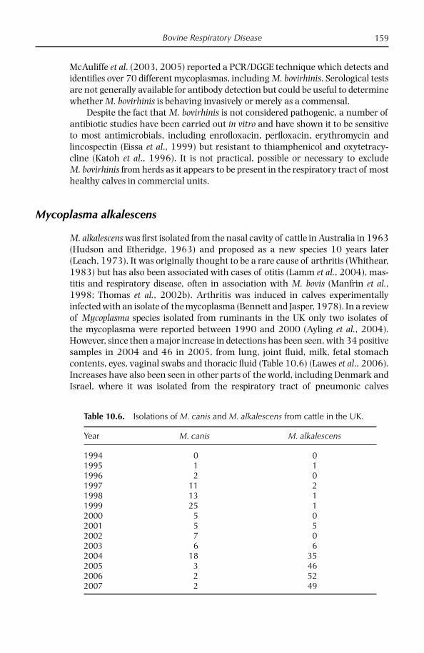
Bovine Respiratory Disease 159
McAuliffe et al. (2003, 2005) reported a PCR/DGGE technique which detects and identifi es over 70 different mycoplasmas, including M. bovirhinis. Serological tests are not generally available for antibody detection but could be useful to determine whether M. bovirhinis is behaving invasively or merely as a commensal.
Despite the fact that M. bovirhinis is not considered pathogenic, a number of antibiotic studies have been carried out in vitro and have shown it to be sensitive to most antimicrobials, including enrofl oxacin, perfl oxacin, erythromycin and lincospectin (Eissa et al., 1999) but resistant to thiamphenicol and oxytetracy-cline (Katoh et al., 1996). It is not practical, possible or necessary to exclude M. bovirhinis from herds as it appears to be present in the respiratory tract of most healthy calves in commercial units.
Mycoplasma alkalescens
M. alkalescens was fi rst isolated from the nasal cavity of cattle in Australia in 1963 (Hudson and Etheridge, 1963) and proposed as a new species 10 years later (Leach, 1973). It was originally thought to be a rare cause of arthritis (Whithear, 1983) but has also been associated with cases of otitis (Lamm et al., 2004), mas-titis and respiratory disease, often in association with M. bovis (Manfrin et al., 1998; Thomas et al., 2002b). Arthritis was induced in calves experimentally infected with an isolate of the mycoplasma (Bennett and Jasper, 1978). In a review of Mycoplasma species isolated from ruminants in the UK only two isolates of the mycoplasma were reported between 1990 and 2000 (Ayling et al., 2004). However, since then a major increase in detections has been seen, with 34 positive samples in 2004 and 46 in 2005, from lung, joint fl uid, milk, fetal stomach contents, eyes, vaginal swabs and thoracic fl uid (Table 10.6) (Lawes et al., 2006). Increases have also been seen in other parts of the world, including Denmark and Israel, where it was isolated from the respiratory tract of pneumonic calves
Table 10.6. Isolations of M. canis and M. alkalescens from cattle in the UK.
Year M. canis M. alkalescens
1994 0 01995 1 11996 2 01997 11 21998 13 11999 25 12000 5 02001 5 52002 7 02003 6 62004 18 352005 3 462006 2 522007 2 49

Chapter 10160
in 2007 (Kokotovic et al., 2007) and 2008 (Lysnyansky et al., 2008). Immuno-histochemical studies for M. alkalescens performed on fi xed lung samples from these cases showed good agreement with mycoplasma isolation. In a number of cases, M. alkalescens was identifi ed in alveolar exudates and in the cytoplasm of macrophages. The isolation of M. alkalescens as the single pathogen, together with localization of M. alkalescens antigen in pneumonic lesions of clinical samples, strongly suggests a contributory role in the disease.
An increase in the prevalence of M. alkalescens is possibly the result of the use of a more sensitive and discriminatory test in the UK: the PCR/DGGE (Lawes et al., 2006). The culture method of identifi cation used before this favoured rapidly growing mycoplasmas, and therefore fastidious species may have been under- reported. This could be the case for M. alkalescens, which was possibly outgrown by M. bovis and M. bovirhinis, resulting in a lower isolation rate. Comparison of the 16S rDNA sequences of Mycoplasma species shows that M. alkalescens is closely related to other arginine-hydrolysing mycoplasmas such as M. arginini and M. canadense. An advantage of DGGE is that mixed cultures can be identifi ed because DNA from all Mycoplasma species present will be amplifi ed. Alternatively, the prev-alence of M. alkalescens could be increasing in real terms in the UK cattle popula-tion and may be developing into an important cause of disease. The increases in isolation rate seen in the UK could be the result of restocking with imported cattle from mainland Europe following the foot-and-mouth outbreaks in 2001.
Mycoplasma canis
In 1993, ter Laak reported the isolation of M. canis from pneumonic calves in the Netherlands. M. canis had also been isolated from cattle in Canada. In 1995 the fi rst isolation of M. canis was reported from pneumonic cattle in Britain (Nicholas et al., 1995). Two further isolations were made in 1996 from similar conditions. In 1997 and 1998 there were small increases, but in 1999, however, 25 isolations of M. canis were seen in cases of calf pneumonia (Table 10.7). Many outbreaks involved animals below the age of 5 months, with some mortality. Most involve other known pathogens such as M. haemolytica, P. multicoda, H. somni, RSV or M. bovis. However, in a number of cases, no other pathogens were implicated (Table 10.7).
During experimental infections, all calves inoculated by aerosol with M. canis showed clinical signs of pneumonia, including two deaths within 2 weeks of infection (Nicholas et al., 2000). Post-mortem examination revealed lung lesions consistent with pneumonia in all animals. M. canis was recovered from the lungs with characteristic mycoplasma lesions, including congested apical and median lobes; mycoplasma was also isolated from the liver, blood and kidneys. There was, however, evidence of secondary infection with M. bovis and Pasteurella but no viral involvement.
The increasing reports of M. canis, although relatively small, represent a real increase rather than greater surveillance because the mycoplasma grows readily in most media. So far four countries (Canada 1974, Netherlands 1993, the UK 1995, Belgium 2002) have reported its isolation and there may be others which

Bovine Respiratory Disease 161
have not yet considered the organism signifi cant. It is possible that surveillance has not been extended to this mycoplasma or that it has been misidentifi ed as other glucose fermenters such as M. bovirhinis. Biochemically the two are similar, apart from the inability of M. canis to reduce tetrazolium salts, but immuno-logically they are quite distinct (Table 10.2).
The occurrence of M. canis in cattle may have originally been due to close contact with dogs, in which the organism was fi rst isolated. However, the wide-spread distribution of M. canis in the UK suggests that the organism should now be considered part of the mycoplasma fl ora of cattle and there is good evidence to suggest involvement in respiratory disease.
References
Almeida, R.A. and Rosenbusch, R.F. (1991) Capsule-like surface material of Mycoplasma dispar induced by in vitro growth in culture with bovine cells is antigenically related to similar structures in vivo. Infection and Immunity 59, 3119–3125.
Anon. (1994) In: Hansard 17 March 1994, House of Commons, London, pp. 816–818.Askaa, G. and Erno, H. (1976) Elevation of Mycoplasma agalactiae subsp. bovis to species
rank Mycoplasma bovis. International Journal of Systematic Bacteriology 26, 323–325.Ayling, R.D., Nicholas, R.A.J. and Johansson, K.-E. (1997) Application of the polymerase
chain reaction for the routine identifi cation of Mycoplasma bovis. Veterinary Record 141, 307–308.
Ayling, R.D., Baker, S.E., Peek, M.L., Simon, A.J. and Nicholas, R.A.J. (2000) Comparison of in vitro activity of danofl oxacin, fl orfenicol, oxytetracycline, spectinomycin and tilmicosin against recent fi eld isolates of Mycoplasma bovis. Veterinary Record 146, 745–747.
Table 10.7. Disease data from herds where M. canis was isolated.
Date Disease Age (months) Number affected Other fi ndings
1/95 Pneumonia 3–5 3/10* H. somni4/97 Pneumonia 1–5 2/10* None5/97 Pneumonia 2 3/20 None7/97 Pneumonia 1–2 1/? Clostridium perfringens7/97 Pneumonia 4–5 10/20 RSV10/97 Pneumonia < 6 150/150 RSV/Pm/M. bovis11/97 Pneumonia 2 ~20 None11/97 Pneumonia 3 30/100* None11/97 Pneumonia 3 not known H. somni/Mh12/97 Pneumonia 4 5/10 Mh12/97 Pneumonia 2 1 Staphylococcus
typhimurium/Pm3/98 Pneumonia 3 2/10* None3/98 Pneumonia 5 2/10 M. bovis/H. somni
*mortality seenMh – M. haemolyticaPm – P. multicoda

Chapter 10162
Ayling, R.D., Bashiruddin, S.E. and Nicholas, R.A.J. (2004) Mycoplasmas and related organisms identifi ed from ruminants 1990–2000. Veterinary Record 155, 413–416.
Ayling, R.D., Nicholas, R.A.J., Wessells, J., Hogg, R. and Byrne, W. (2005) Isolation of Mycoplasma bovis from the brain tissue of calves. Veterinary Record 156, 391–392.
Ayling, R.D., Godinho, K. and Nicholas, R.A.J. (2007) Comparative studies on the in vitro anitmocrobial sensitivities of Mycoplasma mycoides subsp. mycoides SC and Mycoplas-ma bovis. In: CBPP Control: Antibiotics to the Rescue. FAO-OIE-AU/IBAR-IAEA Con-sulative Group Meeting on CBPP in Africa, FAO, Rome, pp. 51–61.
Ball, H.J. and Findlay, D. (1998) Diagnostic applications of monoclonal antibody-based sandwich ELISAs. In: Miles, R.J. and Nicholas, R.A.J. (eds) Mycoplasma Protocols. Humana Press, Totowa, New Jersey, pp. 127–132.
Ball, H.J., Craig Reilly, G.A. and Bryson, D.G. (1995) Antibiotic susceptibility of Mycoplasma bovis strains in Northern Ireland. Irish Veterinary Journal 48, 316–318.
Bennett, R.H. and Jasper, D.E. (1978) Mycoplasma alkalescens-induced arthritis in dairy calves. Journal of the American Veterinary Medical Association 172, 484–488.
Blackburn, P., Brooks, C., McConnell, W. and Ball, H.J. (2007) Isolation of Mycoplasma bovis from cattle in Northern Ireland from 1999 to 2005. Veterinary Record 161, 452–453.
Bradley, A.J., Leach, K.A., Breen, J.E., Green, L.E. and Green, M.J. (2007) Survey of the incidence and aetiology of mastitis on dairy farms in England and Wales. Veterinary Record 160, 253 –258.
Brank, M., Le Grand, D., Poumarat, F., Bezille, P., Rosengarten, R. and Citti, C. (1999) Development of a recombinant antigen for antibody based diagnosis of Mycoplasma bovis infection in cattle. Clinical and Diagnostic Laboratory Immunology 6, 861–867.
Brice, N., Finlay, D., Bryson, D.G., Henderson, J., McConnell, W. and Ball, H.J. (2000) Isolation of Mycoplasma bovis from cattle in Northern Ireland 1993–1998. Veterinary Record 146, 643–644.
Bryson, T.D.G., Ball, H.J., Foster, F. and Brice, N. (2002) Enhanced severity of induced Mycoplasma bovis pneumonia in calves following immunization with different anti-genic extracts. Research in Veterinary Science Supplement A 72, 19.
Buchvarova, Y. and Vesselinova, A. (1989) On the aetiopathogenesis of mycoplasma pneumonia in calves. Archiv fur Experimentelle Veterinarmedizin 43, 685–689.
Burnens, A.P., Bonnemain, P., Bruderer, U., Schalch, L., Audige, L., Le Grand, D., Poumarat, F. and Nicolet, J. (1999) The seroprevalence of Mycoplasma bovis in lactating cows in Switzerland, particularly in the republic and canton of Jura. Schweiz Arch Tierheilkd. 141, 455–460.
Byrne, W.J., Ball, H.J., Brice, N., McCormack, R., Baker, S.E., Ayling, R.D. and Nicholas R.A.J. (2000) Application of an indirect ELISA to milk samples to identify cows with Mycoplasma bovis mastitis. Veterinary Record 146, 368–369.
Byrne, W.J., McCormack, R., Brice, N., Markey, B. and Ball, H.J. (2001) Isolation of Myco-plasma bovis from bovine clinical samples in the Republic of Ireland. Veterinary Record 148, 331–333.
Currin, J.F. (2007) Mycoplasma in beef cattle. Virginia Co-operative Extension, Virginia Tech. USA. www.ext.vt.edu/pubs/beef/400-304.html
Duarte, E.R. and Hamdan, J.S. (2004) Otitis in cattle, an aetiological review. Journal of Veterinary Medicine Series B – Infectious Diseases and Veterinary Public Health 51, 1–7.
Eissa, S.I., El-Shater, S.A. and Dardeer, M.A.A. (1999) Effi cacy of quinolones and amino-glycosides against bovine and ovine mycoplasma. Egyptian Journal of Agricultural Research, 77, 1361–1369.
El-Shabiny, L.M. (1994) Enzyme linked immunosorbent assay for the diagnosis of myco-plasmal mastitis in cows and buffaloes. Veterinary Medical Journal-Giza 42, 51–53.
Bovine Respiratory Disease 163
FAO (2007) CBPP Control: Antibiotics to the Rescue. FAO-OIE-AU/IBAR-IAEA. Consultative Group Meeting on CBPP in Africa 2006. FAO, Rome, pp. 7–11.
Foster, A P., Naylor, R.D., Howie, N.M., Nicholas, R.A.J. and Ayling, R.D. (2008) Mycoplasma bovis and otitis in dairy calves in the UK. The Veterinary Journal doi:10.1016/j.tvjl.2007.10.020.
Francoz, D., Fortin, M., Fecteau, G. and Messler, S. (2005) Determination of Mycoplasma bovis susceptibilities against six antimicrobial agents using the E test method. Veterinary Microbiology 105, 57–64.
Friis, N.F. (1980) Mycoplasma dispar as a causative agent in pneumonia in calves. Acta Veterinaria Scandinavica 21, 34–42.
Friis, N.F. and Szancer, J. (1994) Sensitivity of certain porcine and bovine mycoplasmas to antimicrobial agents in a liquid medium test compared to a disc assay. Acta Veterinaria Scandinavica 35, 389–394.
Gagea, M.I., Bateman, K.G., Shamahan, R.A., van Dreumel, T., McEwen, B.J., Carmen, S., Archambault, M. and Caswell, J.L. (2006) Naturally occurring Mycoplasma bovis associated pneumonia and polyarthritis in feedlot beef calves. Journal of Veterinary Diagnostic Investigations 18, 29–40.
Godinho, K.S., Rae, A., Windsor, G.D., Tilt, N., Rowan, T.G. and Sunderland, S.J. (2004) Effi cacy of tulathromycin in the treatment of bovine respiratory disease associated with induced Mycoplasma bovis infections in young dairy calves. Veterinary Therapy 6, 96–102.
Gourlay, R.N. and Leach, R.H. (1970) A new mycoplasma species isolated from pneumonic lungs of calves (Mycoplasma dispar sp. Nov). Journal of Medical Microbiology 3, 111–123.
Gourlay, R.N., Howard, C.J., Thomas, L,H. and Wyld, S.G. (1979) Pathogenicity of some mycoplasmas and acholeplasma species in the lungs of of gnotobiotic calves. Research in Veterinary Science 27, 233–237.
Gourlay, R.N., Thomas, I.H. and Wyld, S.G. (1989) Increased severity of calf pneumonia associated with the appearance of Mycoplasma bovis in a rearing herd. Veterinary Record 124, 420–422.
Haines, D.M., Martin, K.M., Clark, E.G., Jim, G.K. and Janzen, E.D. (2001) The immuno-histochemical detection of feedlot cattle with chronic unresponsive respiratory disease and/or arthritis. Canadian Veterinary Journal 42, 857–860.
Hale, H.H., Helmboldt, C.F., Plastridge, W.N. and Stula, E.F. (1962) Bovine mastitis caused by Mycoplasma species. Cornell Veterinarian 52, 582–591.
Hermann, R. (1992) Genome structure and organization. In: Maniloff, J. (ed.) Mycoplasmas: Molecular Biology and Pathogenesis. American Society of Microbiology, Washington, DC, pp. 157–168.
Hotzel, H., Sachse, K. and Pfützner, H. (1996) Rapid detection of Mycoplasma bovis in milk samples and nasal swabs using the polymerase chain reaction. Journal of Applied Bac-teriology 80, 505–510.
Houlihan, M.G., Veenstra, B., Christian, M.K., Nicholas, R.A.J. and Ayling, R.D. (2007) Mastitis and arthritis in two dairy herds caused by Mycoplasma bovis. Veterinary Record 160, 126–127.
Howard, C.J. (1983) Mycoplasmas and bovine respiratory disease: studies related to patho-genicity and the immune response – a selective review. Yale Journal of Biological Medicine 56, 789–797.
Howard, C.J., Gourlay, R.N. and Collins, J. (1980) Immunity to mycoplasma infections of the calf respiratory tract. Advances in Experimental Medicine and Biology 137, 711–726.
Howard, C.J., Stott, E.J., Thomas, I.H., Gourlay, R.N. and Taylor, G. (1987) Protection against respiratory disease in calves induced by vaccines containing respiratory

Chapter 10164
syncytial virus, parinfl uenza type 3 virus, Mycoplasma bovis and M. dispar. Veterinary Record 121, 372–376.
Hudson, J.R. and Etheridge, J.R. (1963) A new type of pleuropneumonia-like microorganism (PPLO) from the nose of cattle. Australian Veterinary Journal 39, 1–5.
Jasper, D.E. (1981) Bovine mycoplasma mastitis. Advances in Veterinary Science 25, 121–159.Katoh, T., Koya, M., Watanabe, E., Sakai, J.I., Ogata, Y. and Hikinuma, T. (1996) Antibiotic
susceptibility of microbial isolates from nasal swabs of bovine pneumonic cases. Journal of the Japanese Veterinary Medical Association. 49, 81–84.
Khan, A., Loria, G., Ramirez, S., Fielder, M., Miles, R.J. and Nicholas, R.A.J. (2005) Biochemical characterisation of some non fermenting, non arginine hydrolysing mycoplasmas of ruminants. Veterinary Microbiology 109, 129–134.
Kirby, F.D. and Nicholas, R.A.J. (1996) Isolation of Mycoplasma bovis from bullocks’ eyes. Veterinary Record 138, 552.
Kobayashi, H., Hirose, K., Worarach, A., Paugtes, P., Ito, N., Morozumi, T. and Yamamoto, K. (1998). In vitro amplifi cation of the 16S rRNA genes from Mycoplasma bovirhinis, Mycoplasma alkalescens and Mycoplasma bovigenitalium by PCR. Journal of Veterinary Medical Science 60, 1299–1303.
Kokotovic, B., Friis, N.F. and Ahrens, P. (2007) Mycoplasma alkalescens demonstrated in the broncholavage of cattle in Denmark. Acta Veterinaria Scandinavica 49, 2.
Komoda, M., Kozai, Y., Ito, H., Noro, A., Yamada, T., Kimura, Y., Noguchi, M. and Koizumi, S. (1988) Outbreak of shipping fever associated with several pathogens in grazing calves. Journal of the Japanese Veterinary Medical Association 41, 408–411.
Kreusel, S., Bocklisch, H., Pfützner, H., Brys, A., Leirer, R. and Ziegenhals, U. (1989) Experimentelle infektionen von bullen mit Mycoplasma bovis und Mycoplasma bovi-genitalium. Archiv fur Experimentelle Veterinarmedizin 43, 705–712.
Lamm, C.G., Munson, I., Thurmond, M.C., Barr, B.C. and George, L.W. (2004) Mycoplasma otitis in California calves. Journal of Veterinary Diagnostic Investigation 16, 397–402.
Lawes, J., Bisgaard-Frantzen, S., Bashiruddin, S.E., McAuliffe, L., Ayling, R.D. and Nicholas, R.A.J. (2006) Emergence of Mycoplasma alkalescens in cattle in the UK. In: Proceedings of the 16th International Congress of the International Organisation for Mycoplasmology, Cambridge, UK, July 2006, p. 129.
Lawes, J.R., McAuliffe, L., Nicholas, R.A.J. and Ayling, R.D. (2008) Epidemiological analysis of Mycoplasma bovis ELISA data from 1995–2005. Veterinary Record submitted
Leach, R.H. (1967) Comparative studies of mycoplasmas of bovine origin. Annals of the New York Academy of Sciences 143, 305–306.
Leach, R.H. (1973) Further studies on classifi cation of bovine strains of mycoplasmatales, with proposals for new species, Acholeplasma modicum and Mycoplasma alkalescens. Journal of General Microbiology 75, 135–153.
Le Grand, D., Phillippe, S., Calavalas, D., Bezille, P. and Poumarat, F. (2001) Prevalence of Mycoplasma bovis infection in France. In: Poveda, J.B., Fernandez, A., Frey, J. and Johansson, K.-E. (eds) Mycoplasmas of Ruminants: Pathogenicity, Diagnostics, Epidemio-logy and Molecular Genetics, Vol. 5. European Commission, Brussels, pp. 106–109.
Loria, G., Scanziani, E., Lelli, R., Caracappa, S., Nicholas, R.A.J. and Lillini, E. (2007) Myco-plasma bovis: la malattia in Italia. Altti della Societa di Buiatria Vol. XXXVII, 315–322.
Lysnyansky, I., Ron, Y. and Yogev, D. (2001) Juxtaposition of an active promoter to vsp genes via site-specifi c DNA inversions generates antigenic variation in Mycoplasma bovis. Journal of Bacteriology 183, 5698–5708.
Lysnyansky, I., Levisohn, S., Perl, S., Malone, F. and Ball, H.J. (2008) Immunohistochemical detection of Mycoplasma alkalescens in naturally infected calves. Proceedings of the 17th International Congress of the International Organisation for Mycoplasmology, Tianjin, China, in press.

Bovine Respiratory Disease 165
Madoff, S., Pixley, B.Q., Del Guidice, R.A. and Moellering, R.C. (1979) Isolation of Myco-plasma bovis from a patient with systemic illness. Journal of Clinical Microbiology 9, 709–711.
Maeda, T., Shibahara, T., Kimura, K., Wada, Y., Sato, K., Imada, Y., Ishikawa, Y. and Kadota, K. (2003) Mycoplasma bovis-associated suppurative otitis media and pneumonia in bull calves. Journal of Comparative Pathology 129, 100–110.
Manfrin, A., Friso, S., Perin, R. and Cirelli, L. (1998) Isolation of Mycoplasma sp. in the North East of Italy. In: Leori, G., Santini, F., Scanziani, E., and Frey, J. (eds) Mycoplasmas of Ruminants: Pathogenicity, Diagnostics, Epidemiology and Molecular Genetics, Vol. 2. European Commission, Brussels, pp. 84–87.
Martin, S.W., Bateman, K.G., Shewen, P.E., Rosendal, S. and Bohac, J.E. (1989) The fre-quency, distribution and effects of antibodies, to seven putative respiratory pathogens, on respiratory disease and weight gain in feedlot calves on Ontario. Canadian Journal of Veterinary Research 53, 355–362.
Martin, S.W., Bateman, K.G., Shewen, P.E., Rosendal, S., Bohac, J.G. and Thorburn, M. (1990) A group level analysis of the associations between antibodies to seven putative pathogens and respiratory disease and weight gain in Ontario feedlot calves. Canadian Journal of Veterinary Research 54, 337–342.
Mazzolini, E., Agnoletti, F. and Friso, S. (1997) Farmaco-sensibilita dei Micoplasmi respi-ratori del bovino. Obviettivi and Documenti Veterinari 1, 61–66.
McAuliffe, L., Ellis, R., Ayling, R.D. and Nicholas, R.A.J. (2003) Detection and differentia-tion of Mycoplasma species by rDNA PCR and DGGE fi ngerprinting. Journal of Clinical Microbiology 41, 4844–4847.
McAuliffe, L., Kokotovic, B. and Nicholas, R.A.J. (2004) Molecular epidemiologic analysis of Mycoplasma bovis isolates from the UK shows two genetically distinct clusters. Journal of Clinical Microbiology 42, 4556–4565.
McAuliffe, L., Ellis, R., Lawes, J., Ayling, R.D. and Nicholas, R.A.J. (2005) 16S rDNA and DGGE: a single generic test for detecting and differentiating Mycoplasma species. Journal of Medical Microbiology 54, 1–9.
McAuliffe, L., Ellis., R.J., Miles, K., Ayling, R.D. and Nicholas, R.A.J. (2006) Biofi lm formation by mycoplasma species and its role in environment persistence and survival. Micro-biology 152, 913–922.
Miles, K., McAuliffe, L., Ayling, R.D. and Nicholas, R.A.J. (2005) Rapid detection for Mycoplasma dispar and Mycoplasma bovirhinis using an allele specifi c PCR. Veterinary Research Communications 241, 103–107.
Miles, R.J., Wadher, C.L., Henderson, C.L. and Mohan, K. (1988) Increased yields of Mycoplasma spp in the presence of pyruvate. Letters in Applied Microbiology 7, 149–151.
Montgomery, A. (2008) Nowe strategie w leczeniu syndrome oddechowy bydla (BRD). In: Bednarek, D. (ed.) Najwazniejsze Czynniki Etiologiczne, Patogeneza I Najnowsze Trendy w Profi lataktyce I Terapii Syndrome Oddechowego Bylda. PIWet, Pulawy, Poland, pp. 121–129.
Nagatomo, H., Shimizu, T., Higashiyama, Y., Yano, Y., Kuroki, H. and Hamana, K. (1996) Antibody response to Mycoplasma bovis of calves introduced in a farm contaminated with the organism. Journal of Veterinary Medical Science 58, 919–920.
Naglic, T., Sankovic, F., Madic, J., Hajsig, D., Seol, B. and Busch, K. (1996) Mycoplasmas associated with bovine conjunctivitis and keratoconjunctivitis. Acta Veterinaria Hunga-rica 44, 21–24.
Nascimento, M.G.F., d’Angelis, F.H.F., Nascimento, E.R., Resende, O.A. and Lignon, G.B. (1998) Mycoplasma bovirhinis em muco vaginal de novilhas. Revista Brasileira de Medicina Veterinaria 20, 210–214.
Chapter 10166
Nicholas, R.A.J. (2002a) Mycoplasma bovis. Compendium of Animal Health. CABI, Wallingford, UK. www.cabi.org.
Nicholas, R.A.J. (2002b) Mycoplasma dispar. Animal Health Compendium. CABI, Wallingford, UK. www.cabi.org.
Nicholas, R.A.J. and Ayling, R.D. (2003) Mycoplasma bovis, disease, diagnosis and control. Research in Veterinary Science 74, 105–112.
Nicholas, R.A.J. and Baker, S.E. (1998) Recovery of mycoplasmas from animals. In: Miles, R.J. and Nicholas, R.A.J. (eds) Mycoplasma Protocols. Humana Press, Totowa, New Jersey, pp. 37–44.
Nicholas, R.A.J., Hannam, D.A.R., Baker, S.E., Weaver, C.R. and ter Laak, E.A. (1995) Mycoplasma canis in a calf. Veterinary Record 137, 443–444.
Nicholas, R.A.J., Baker, S.E., Ayling, R.D. and Stipkovits, L. (2000) Mycoplasma infections in growing cattle. Cattle Practice 8, 115–118.
Nicholas, R.A.J., Wood, E., Baker, S.E. and Ayling, R.D. (2001) Mycoplasmas isolated from ruminants in Britain 1995-2000. In: Poveda, J.B., Fernandez, A., Frey. J. and Johansson, K.-E. (eds) Mycoplasmas of Ruminants: Pathogenicity, Diagnostics, Epidemiology and Molecular Genetics, Vol. 5. European Commission, Brussels, pp. 116–120.
Nicholas, R.A.J., Ayling, R.D. and Stipkovits, L. (2002) An experimental vaccine for calf pneumonia caused by Mycoplasma bovis. Vaccine 20, 3569–3575.
Nicholas, R.A.J., Ayling, R D., Houlihan, M. and Woodger, N. (2006) Mycoplasmas of adult cattle: bugs worth bothering with? Irish Veterinary Journal 59, 301–304.
Nussbaum, S., Lysnyansky, I., Sachse, K., Levisohn, S. and Yogev, D. (2002) Extended repertoire of genes encoding variable surface lipoproteins in Mycoplasma bovis strains. Infection and Immunity 70, 2220–2225.
O’Farrell, K., Dillon, P., Mee, J., Crosse, S., Nolan, M., Byrne, N., Reidy, M., Flynn, F. and Condon, T. (2001) Strategy for restocking Moorepark after depopulation following BSE. Irish Veterinary Journal 54, 70–75.
Ongor, H., Kalin, R., Karahan, M., Cetinkaya, B. and Nicholas, R.A.J. (2008) Isolation of Mycoplasma bovis from broiler chickens in Turkey. Avian Pathology in press
Ozdemir, U. and Turkarslan, J. (1998) Damizlik bogalardan izole edilen genital miko-plazmalar. Pendik Veteriner-Mikrobiyoloji Dergisi 30, 37–39.
Panagala, V.S., Stringfellow, J.S., Oliver, J.L. and Wolfe, D.F. (1990) Isolation of Mycoplasma bovirhinis from the kidneys of a bull with urinary obstruction and subacute nephritis. Journal of the American Veterinary Medical Association 197, 381–382.
Pettersson, B., Uhlen, M. and Johansson, K.-E., (1996) Phylogeny of some mycoplas-mas from ruminants based on 16S rRNA sequences and defi nition of new cluster within the hominis group. International Journal of Systematic Bacteriology 46, 1093–1098.
Pfützner, H. (1990) Epizootiology of the Mycoplasma bovis infection of cattle. Zentralblatt fur Bakteriologie Supplement 20, 394–399.
Pfützner, H. and Sachse, K. (1996) Mycoplasma bovis as an agent of mastitis, pneumonia, arthritis and genital disorders. Revue Scientifi que and Technique. Offi ces International Des Epizooties 15, 1477–1494.
Pinnow, C.C., Butler, J.A., Sachse, K., Hotzel, H., Timms, L.L. and Rosenbusch, R.F. (2001) Detection of Mycoplasma bovis in preservative treated milk samples. Journal of Dairy Science 84, 1640–1645.
Pitcher, D. and Nicholas, R.A.J. (2005) Mycoplasma host specifi city: fact or fi ction? The Veterinary Journal 170, 300–306.
Poumarat, F., Solsona, M. and Boldini, M. (1994) Genomic, protein and antigenic variability of Mycoplasma bovis. Veterinary Microbiology 40, 305–321.

Bovine Respiratory Disease 167
Poumarat, F., Belli, P. and Calavalas, D. (1999) Immunoprophylaxis of experimental Myco-plasma bovis arthritis in calves. International Symposium Mycoplasmas of Ruminants. COST Action 826, Toulouse, France, June 2–4. Abstracts, 65.
Poumarat, F., Le Grand, D., Philippe, S., Calavas, D., Schelcher, F., Cabanie, P., Tessier, P. and Navetat, H. (2001) Effi cacy of spectinomycin against Mycoplasma bovis induced pneumonia in conventionally reared calves. Veterinary Microbiology 80, 23–35.
Poveda, J.B. and Nicholas, R.A.J. (1998) Serological identifi cation by growth and metabo-lism inhibition tests. In: Miles, R.J. and Nicholas, R.A.J. (eds) Mycoplasma Protocols. Humana Press, Totowa, New Jersey, pp. 105–112.
Raedelli, E., Luini, M., Loria, G.R., Nicholas, R.A.J. and Scanziani, E. (2008) Studies of Mycoplasma bovis infections in veal calves and adult cattle. Research in Veterinary Science doi:10.1016/j.rvsc.2007.11.012.
Rebhun, W.C., Guard, C. and Richards, C.M. (1995) Respiratory diseases. In: Divers, T.J. and Peek, S.F. (eds) Diseases of Dairy Cattle. Williams and Wilkins, Baltimore, Maryland, pp. 79–80.
Reeve-Johnson, L. (1999) The impact of mycoplasma infections in respiratory disease in cattle in Europe. In: Stipkovits, L., Rosengarten, R. and Frey, J. (eds) Mycoplasmas of Ruminants: Pathogenicity, Diagnostics, Epidemiology and Molecular Genetics, Vol. 3. European Commission, Brussels, pp. 18–31.
Rodriguez, F., Bryson, D.G., Ball, H.J. and Forster, F. (1996) Pathological and immuno-histochemical studies of natural and experimental Mycoplasma bovis pneumonia in calves. Journal of Comparative Pathology 115, 151–162.
Rosengarten, R. and Citti, C. (1999) The role of ruminant mycoplasmas in systemic infec-tion. In: Stipkovits, L., Rosengarten, R. and Frey, J. (eds) Mycoplasmas of Ruminants: Pathogenicity, Diagnostics, Epidemiology and Molecular Genetics, Vol. 3. European Commission, Brussels, pp. 14–17.
Ross, R.F. (1993) Mycoplasmas – animal pathogens. In: Kahane, I. and Adoni, A. (eds) Rapid Diagnosis of Mycoplasmas. Plenum Press, New York, pp. 69–110.
Step, D.L. and Kirkpatrick, J.G. (2001) Mycoplasma infection in cattle. I Pneumonia–arthritis syndrome. The Bovine Practitioner 35, 149–155.
St George, T.D., Horsfall, N. and Sullivan, N.D. (1973) A subclinical pneumonia of calves associated with Mycoplasma dispar. Australian Veterinary Journal 49, 580–589.
Stipkovits, L., Ripley, P.H., Varga, J. and Palfi , V. (2001) Use of valnemulin in the control of Mycoplasma bovis infection under fi eld conditions. Veterinary Record 148, 399–402.
Subramaniam, S., Bergonier, D., Poumarat, F., Capual, S., Schlatter, Y., Nicolet, J. and Frey, J. (1998) Species identifi cation of Mycoplasma bovis and Mycoplasma agalactiae based on the uvrC gene by PCR. Molecular and Cellular Probes 12, 161–169.
Takahata, T., Kato, M., Nagatomo, H. and Shimizu, T. (1997) A fi lter immunobinding technique for the rapid detection and simultaneous identifi cation of avian and bovine mycoplasmas. Journal of Veterinary Medical Science 59, 965–969.
Tegtmeier, C., Uttenthal, A., Friis, N.F., Jensen, N.E. and Jensen, H.E. (1999) Pathological and microbiological studies on pneumonic lungs from Danish calves. Journal of Veterinary Medicine, Series B 46, 693–700.
ter Laak, E.A. and Noordergraaf, J.H. (1987) An improved method for the identifi cation of Mycoplasma dispar. Veterinary Microbiology 14, 25–31.
ter Laak, E.A., Noordergraaf, J.H. and Dieltjes, R.P.J.W. (1992a) Prevalence of mycoplas-mas in the respiratory tracts of pneumonic calves. Journal of Veterinary Medicine, Series B 39, 553–562.
ter Laak, E.A., Noordergraaf, J.H. and Boomsluiter, E. (1992b) The nasal mycoplasmal fl ora of healthy calves and cows. Journal of Veterinary Medicine, Series B 39, 610–616.

Chapter 10168
ter Laak, E.A., Noordergraaf, J.H. and Verschure, M.H. (1993) Susceptibilities of Myco-plasma bovis, Mycoplasma dispar and Ureaplasma diversum strains to antimicrobial agents in vitro. Antimicrobial Agents and Chemotherapy 37, 317–321.
Thomas, A., Dizier, I., Trolin, A., Mainil, J. and Linden, A. (2002a) Comparison of sam-pling procedures for isolating pulmonary mycoplasmas in cattle. Veterinary Research Communications 26, 333–339.
Thomas, A., Ball, H., Dizier, I., Trolin, A., Bell, J., Mainil, A. and Zinden, A. (2002b) Isola-tion of Mycoplasma species from the lower respiratory tract of healthy cattle and cattle with respiratory disease in Belgium. Veterinary Record 151, 472–476.
Thomas, L.H., Howard, C.I., Stott, E.J. and Parsons, K.A. (1986) Mycoplasma bovis infec-tions in gnotobiotic calves and combined infection with respiratory syncytial virus. Veterinary Pathology 23, 571–578.
Urbaneck, D., Leibig, F., Forbrig, T.H. and Stache, B. (2000) Erfahrungsbericht zur Anwedung bestandsspezifi scher impfstoffe gegen respiratorische infektionen mit beteiligung von Mykoplasma bovis in einem mastrindergrossbestand. Der Praktische Tierarzt 81, 756–763.
Vanden Bush, T.J. and Rosenbusch, R.F. (2002) Mycoplasma bovis induces apoptosis of bovine lymphocytes. FEMS Immunology and Medical Microbiology 32, 97–103.
Vanden Bush, T.J. and Rosenbusch, R.F. (2004) Characterization of a lympho-inhibitory peptide produced by Mycoplasma bovis. Biochemical and Biophysical Research Com-munications 315, 336–341.
van Donkersgoed, J., Ribble, C.S., Boyer, L.G. and Townsend, H.G.G. (1993) Epidemiological study of enzootic pneumonia in dairy calves in Saskatchewan. Canadian Journal of Veterinary Research 57, 247–254.
Virtala, A.M.K., Mechor, G.D., Grohn, Y.T., Erb, H.N. and Dubovi, E.J. (1996) Epidemiologic and pathologic characteristics of respiratory tract disease in dairy heifers during the fi rst three months of life. Journal of the American Veterinary Medical Association 208, 2035–2042.
Walz, P.H., Mullaney, T.P., Render, J.A., Walker, R.D., Mosser, T. and Baker, J.C. (1997) Otitis media in preweaned Holstein dairy calves in Michigan due to Mycoplasma bovis. Journal of Veterinary Diagnostic Investigation 9, 250–254.
Whithear, K.G. (1983) Isolation of Mycoplasma alkalescens from cases of polyarthritis in embryo-transplant calves. Australian Veterinary Journal 60, 191–192.
Zendulkova, D. (2007) Mycoplasma infections in cattle and their meanings. Veterinarstvi 57, 590–597.

© CAB International 2008. Mycoplasma Diseases of Ruminants(R. Nicholas et al.) 169
11 Respiratory Diseases of Small Ruminants
The evidence from abbatoir surveys… suggest that we may be underestimating the effect that pneumonic diseases have on our sheep fl ocks. Pneumonic lesions are seen at necropsy in sheep of all ages and clinical signs are frequently evident when fl ocks are driven. (University of Sydney, 2005)
OVERVIEW
Respiratory disease in small ruminants is responsible for enormous fi nancial losses worldwide, estimated at over one billion dollars in the USA. It may result in sudden death or in a protracted illness causing much suffering to affected animals. In addition to death and sickness, reduced feed effi ciency, slaughter condemnations, prevention and treatment measures contribute substantially to losses. It is believed by some that this condition is dominated by pneumonic pasteurellosis caused by Mannheimia haemolytica, particularly in newborn and feedlot lambs as well as in mature ewes. Goats, on the other hand, appear suscep-tible to a wider range of respiratory diseases, many of them caused by infectious agents including mycoplasmas; the role of viruses, however, is largely unknown. It is clear with few exceptions that the aetiology of respiratory disease is multi-factorial and complex.
The most common cause of pneumonia, particularly in sheep, is pasteurel-losis, which occurs worldwide. Although fi rst described in 1961, pneumonic pas-teurellosis became unravelled only with the introduction of serotyping and biotyping in the 1960s, leading, to date, with the recognition of two morphologi-cally identical species, M. haemolytica and Pasteurella trehalosi, comprising a total of 17 serotypes. M. haemolytica (A biotypes) strains are responsible for disease in sheep of all ages, while P. trehalosi (T biotypes) strains cause a systemic disease in 6–10-month lambs, often resulting in sudden death. Serotype A2 is most

Chapter 11170
frequently found in both pneumonic sheep and goats, with A1, A6, A7 and A9 being restricted to sheep. Outbreaks of disease are most common in the summer with a second peak in late autumn. Secondary infections leading to chronic abscess formation are common. The A biotypes are carried in the nasopharynx and many factors such as stress, dipping, castration, transport and other infec-tions, including PI3 and mycoplasmoses, may predispose animals to outbreaks of pneumonia.
Other bacteria which are occasionally isolated from pneumonic small rumi-nants include Actinomyces pyogenes, staphlococcal spp., streptococcal spp., Haemo-philus spp. and Klebsiella pneumoniae. These bacteria are rarely primary causes of disease but may exacerbate an existing condition.
Infection with parainfl uenza virus type 3 (PI3) mainly causes a mild respira-tory disease, sometimes without clinical signs, but occasionally acute infections can result in high morbidity in lambs under 1 year old. Mainly a threat to livestock in West Africa, Asia and the Middle East, the OIE B-listed peste des petits rumi-nants (PPR), caused by a paramyxovirus, shows a range of clinical signs, includ-ing necrotic stomatitis, foaming at the mouth and ocular discharge; respiratory signs are seen during the acute phase, when a mucopurulent nasal discharge, frequent sneezing and increased respiratory rate accompanied by extended head and mouth breathing are seen. The goats may die several weeks later of a second-ary bronchopneumonia or sooner if co-infected with contagious caprine pleuro-pneumonia. Some goats infected with caprine arthritis encepahalitis (CAE) virus, a lentivirus of the Retroviridae family, develop a progressive interstitial pneumo-nia characterized by a chronic dry cough and weight loss. This ‘slow’ disease is very similar to that caused by another lentivirus, Maedi-visna, which is wide-spread throughout the world except Australia and New Zealand. The two viruses are genetically but not serologically distinct.
Husk, or parastic bronchitis, caused by the adult lungworm Dictyocaulus fi laria, is seen in young sheep and goats, where it causes coughing, tachypnoea and reduced growth rates. Although not as important as its equivalent in cattle, husk is associated with just under 4% of ovine pneumonias in the UK, appearing late in summer and early autumn.
Goats are highly susceptible to a number of mycoplasmas, including the highly pathogenic M. capricolum subsp. capripneumoniae, the cause of the OIE B-listed contagious caprine pleuropneumonia (CCPP) (Chapter 9, this volume). Mortality and morbidity rates may exceed 80% in naive herds, with animals dying within days of infection. The disease is present in the Middle East, North and East Africa and Asia but is almost certainly under-reported because of the diffi culty in isolating and culturing the mycoplasma. It has been isolated from the respiratory tracts of pneumonic and healthy sheep, suggesting a role for this species as a car-rier. M. ovipneumoniae is often isolated with M. c. capripneumoniae, which can often result in the overgrowth of the latter; early cloning of the two morphologically distinct mycoplasmas may overcome this, as does the use of the PCR directly on lung tissue. M. ovipneumoniae is a cause of a non-progressive (atypical) pneumo-nia in its own right, which may then enable the invasion of M. haemolytica with far more serious results. Interestingly, reproduction of atypical pneumonia is far more successful with infected tissue homogenates than with isolated cultures of
Respiratory Disease of Small Ruminants 171
M. ovipneumoniae. In recent years the term CCPP has been restricted to infections caused by M. c. capripneumoniae; however, some workers still refer to infections caused by the very closely related M. mycoides subsp. capri and M. mycoides subsp. mycoides LC as CCPP. Classical CCPP is highly contagious in goats, and its lesions are severe with fi brinous or fi brinonecrotic pneumonia giving a granular appear-ance when the lung is cut. The enlargement of the interlobular septa, a feature of M. mycoides LC infection, is rarely seen in classical CCPP. M. mycoides LC is much more widely distributed, occuring in France and countries bordering the Mediter-ranean, Asia and America, where it may be seen in goats suffering from conta-gious agalactia. Pneumonia and/or contagious agalactia are also a feature of infections with M. capricolum subsp. capricolum, which can be seen sporadically in goats in Europe and in sheep and goats in North Africa. Many other mycoplasmas are occasionally isolated from the lungs of small ruminants, in particular M. argi-nini, but there is little evidence to connect them primarily with respiratory disease. M. mycoides subsp. mycoides SC, the cause of contagious bovine pleuropneumo-nia, is occasionally isolated from goats with pneumonia but rarely from sheep.
ATYPICAL PNEUMONIA OF SHEEP AND GOATS
Introduction
Respiratory disease in sheep and goats may result in sudden death or in protracted illness, causing suffering to affected animals as well as high fi nancial losses to owners. In addition to death and sickness, reduced feed effi ciency, slaughter con-demnations, prevention and treatment measures contribute substantially to losses. M. haemolytica is the most obvious cause of pneumonic pasteurellosis but the role of mycoplasmas, in particular M. ovipneumoniae, is often overlooked. As well as causing disease in its own right, M. ovipneumoniae predisposes animals to pasteurellosis and viral infections (Ayling and Nicholas, 2007). Sheep are gener-ally less susceptible to mycoplasmas than goats but a reassessment of the myco-plasma fl ora of sheep is necessary, particularly in the Middle East, where mixed fl ocks of small ruminants are kept (Al-Momani et al., 2006a).
Epidemiology and Transmission
M. ovipneumoniae is mainly transmitted via the respiratory route by close and repeated contact. Mycoplasmas frequently occur in the upper respiratory tract of healthy sheep, which may act as a major source of infection to lambs. Lambs are thought to become infected within a few days of birth but disease is slowly pro-gressive and often occurs with secondary bacterial infections from 5 to 10 weeks of age. The pneumonia may be severe, but some lambs recover within a few weeks whilst it persists in other lambs for much longer. Outbreaks can occur when groups of lambs from different sources are housed together and may be a result of mixing uninfected lambs with infected lambs or possibly the effect of encounter-ing different strains of M. ovipneumoniae. High summer temperatures may be a
Chapter 11172
precipitating factor in exacerbating a mild condition and principally involves M. ovipneumoniae with Actinobacillus pleuropneumoniae biovar A (Hervas et al., 1996).
Extensive heterogeneity has been shown amongst M. ovipneumoniae strains by DNA and protein analysis, revealing 58 different profi les in 60 isolates (Mew et al., 1985; Ionas et al., 1991). Molecular typing with RAPD and PFGE of 43 dif-ferent strains in the UK also showed heterogeneity, with 40 different profi les including different profi les within the same fl ocks (Miles et al., 2006). No link could be made between strains and geographic origin, refl ecting extensive animal movements within the UK. The same work also confi rmed the highly variable pro-tein expression seen within M. ovipneumoniae strains. The observed heterogeneity between this panel of isolates, which were all associated with disease, may repre-sent cell lineages resulting from a much larger population of non-pathogenic pre-cursor clones. Once strains have become established within a host, close contact and animal movement facilitate their spread. This potentially results in a mixture of strains being present on the same farm, or even co-existing within the same host. The presence of such wide variation results in a large gene pool, allowing the population as a whole to respond to changes in its environment, facilitating its survival. Under specifi c selection pressures it may be expected that the numbers of those isolates best suited to the new conditions will be elevated signifi cantly (Miles et al., 2006).
M. ovipneumoniae infections lead to a variable pattern of morbidity and gen-erally cause low mortality. The observed differences might be a result of the existence of variable strain virulence, host response or co-infecting organisms. Bacterial pneumonia in sheep is multifactorial, resulting in conditions that vary in severity and may be chronic or acute. It is believed that primary infection by M. ovipneumoniae exacerbates clinical signs resulting from the colonization of tissue by other microorganisms.
Early investigations (Ionas et al., 1991) have indicated that the presence of multiple M. ovipneumoniae strains within the same fl ock leads to more severe dis-ease signs. Although horizontal transfer and recombination of DNA between strains have not been shown, it has been suggested that the unique combination of virulence genes required for successful colonization might only be obtained from a mixture of strains acting together. Such cooperation has been observed for other bacterial species where cells within colonies show spatial organization and cellular differentiation (Shapiro, 1998).
There are few reports of M. ovipneumoniae as a cause of severe respiratory disease in goats. In the UK high levels of morbidity, up to 20%, were seen, in which coughing, dyspnoea, pyrexia, inappetance and profound depression were wide-spread in home-bred goats on a farm where there was sudden mixing with goats from France (Nicholas, 2000). Lesions, which resembled those of CCPP, were con-fi ned to the thoracic cavity and included a fi brinopurulent pleurisy and pericardi-tis with copious amounts of yellow oily fl uid in the pericardial sac and thoracic cavity. Lung consolidation (grey hepatization and lung marbling) was particularly noticeable in the anteroventral lung lobes, with up to 70% of the total lung area affected in some goats, often more prominent in a single lung. Well-encapsulated lung abcesses were also evident. The lesions were consistent with an infection
Respiratory Disease of Small Ruminants 173
involving M. haemolytica and M. ovipneumoniae, both of which were isolated pro-fusely. The severity of the infection was linked to overcrowding, sudden mixing of goats from different sources and concurrent infections with caseous lymphadeni-tis and caprine arthritic encephalitis brought in with the imported animals.
An outbreak of severe respiratory disease, which was restricted to kids and associated with M. ovipneumoniae, M. arginini, M. haemolytica and P. multocida, was reported recently in Portugal (Gonçalves et al., 2008). The mortality rates excee ded 20% in kids, with severe pleuropneumonia, lung consolidation, large quantities of pleural fl uid and pericarditis seen. Antibiotic treatment with enrofl oxa-cin and fl or fenicol was administered for 5 days and clinical signs resolved within 10–14 days.
Clinical Signs
Clinical signs may be mild, with dull lambs showing increased respiratory rates; in contrast, infections may also result in mortality, acute fi brinous pneumonia, con-solidated lesions, pulmonary abscesses and pleurisy, depending on exacerbating circumstances. The signs initially present as coughing, temperature rise and depression of appetite and growth rate, with a drop in milk yield. A variable per-centage of lambs will be affected but, over a period of weeks, the problem may involve the majority of the lambs. Growth rates may be slowed and carcass weights reduced. The main clinical signs are chronic, persistent and irregular cough, which may lead to rectal prolapses, and a mucopurulent nasal discharge; other bacterial infections may also be involved that cause a more severe infl ammatory response and clinical signs (Niang et al., 1998).
Reports from Australia described outbreaks of pneumonia in housed lambs in the fi rst year of life, with high morbidity and low mortality and with poor growth rates and exercise tolerance (Cottew, 1971; St George et al., 1971; St George, 1972). M. ovipneumoniae has also been isolated from cases of mastitis and kerato-conjunctivitis but its role in these diseases is unknown (Ayling et al., 2004).
Pathology
Typically, lesions begin with dull red ventral areas of collapse which are accompa-nied by bronchiolitis in associated airways. They progress to fi rm red–grey areas of consolidation over 2–3 weeks (Fig. 11.1) but may continue as grey areas of con-solidation, often with attached localized pleural adhesions (Alley et al., 1999).
Histologically, a wide range of lung changes are seen, including hyperplasia of the lymphoid nodules and bronchiolar epithelium and a mononuclear cell reaction in the alveolar septa. Also evident are lymphocytic cuffs around bronchi-oles and vessels, collapsed alveoli and others with exudates containing mononu-clear cells. Lymphoid hyperplasia may result in extensive cuffs around airways with consequent compression of the passages. Ultrastructurally M. ovipneumoniae has been reported to attach to cilia in airways of lambs and to colonize the upper respiratory tract of sheep (Niang et al., 1998).
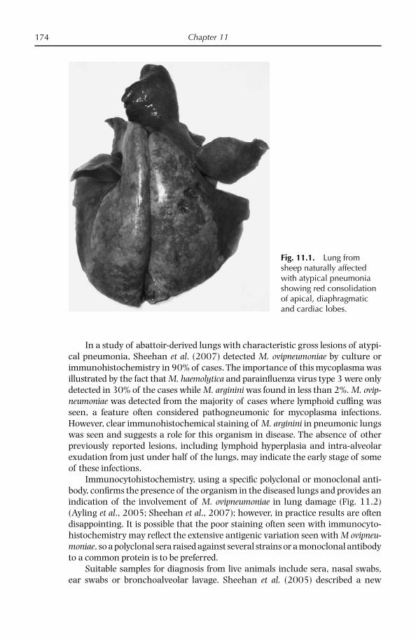
Chapter 11174
In a study of abattoir-derived lungs with characteristic gross lesions of atypi-cal pneumonia, Sheehan et al. (2007) detected M. ovipneumoniae by culture or immunohistochemistry in 90% of cases. The importance of this mycoplasma was illustrated by the fact that M. haemolytica and parainfl uenza virus type 3 were only detected in 30% of the cases while M. arginini was found in less than 2%. M. ovip-neumoniae was detected from the majority of cases where lymphoid cuffi ng was seen, a feature often considered pathogneumonic for mycoplasma infections. However, clear immunohistochemical staining of M. arginini in pneumonic lungs was seen and suggests a role for this organism in disease. The absence of other previously reported lesions, including lymphoid hyperplasia and intra-alveolar exudation from just under half of the lungs, may indicate the early stage of some of these infections.
Immunocytohistochemistry, using a specifi c polyclonal or monoclonal anti-body, confi rms the presence of the organism in the diseased lungs and provides an indication of the involvement of M. ovipneumoniae in lung damage (Fig. 11.2) (Ayling et al., 2005; Sheehan et al., 2007); however, in practice results are often disappointing. It is possible that the poor staining often seen with immunocyto-histochemistry may refl ect the extensive antigenic variation seen with M ovipneu-moniae, so a polyclonal sera raised against several strains or a monoclonal antibody to a common protein is to be preferred.
Suitable samples for diagnosis from live animals include sera, nasal swabs, ear swabs or bronchoalveolar lavage. Sheehan et al. (2005) described a new
Fig. 11.1. Lung from sheep naturally affected with atypical pneumonia showing red consolidation of apical, diaphragmatic and cardiac lobes.

Respiratory Disease of Small Ruminants 175
transtracheal bronchoalveolar lavage technique which successfully recovered M. ovipneumoniae and M. haemolytica. However, its general use in the fi eld may not be widely accepted. From dead animals, lung taken from the interface between healthy and diseased tissue and pleural fl uid is desirable. An alternative approach is to take lung washings using a syringe containing PBS and a sterile tube which can be passed into the lung and withdrawn. The washings can then be examined by culture or molecular methods.
It is important to differentiate atypical pneumonia from the progressive pneu-monias maedi and pulmonary adenomatosis. The age, pattern of disease, clinical signs and histological changes are useful distinguishing features. Furthermore, invasion of the lower respiratory tract by Chlamydia psittaci can also produce lung damage similar to that seen in atypical pneumonia.
Causative Organism
M. ovipneumoniae, fi rst isolated in New Zealand in 1974, is the cause of atypical or ovine non-progressive pneumonia. Mycoplasma pneumonia is well recognized in both Australia and New Zealand, where it is known as ‘summer pneumonia’ because of an increase in the prevalence of the disease in the hotter weather. A coughing syndrome identifi ed in the USA in which there was persistent and long-term cough-ing as well as rectal prolapse was believed to be caused by a combination of M. ovip-neumoniae and M. arginini (Niang et al., 1998). It is believed that a primary infection
Fig. 11.2. Immunoperoxidose staining of M. ovipneumoniae in the lung of a sheep with atypical pneumonia.
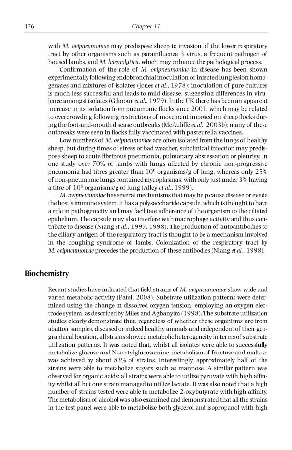
Chapter 11176
with M. ovipneumoniae may predispose sheep to invasion of the lower respiratory tract by other organisms such as parainfl uenza 3 virus, a frequent pathogen of housed lambs, and M. haemolytica, which may enhance the pathological process.
Confi rmation of the role of M. ovipneumoniae in disease has been shown experimentally following endobronchial inoculation of infected lung lesion homo-genates and mixtures of isolates (Jones et al., 1978); inoculation of pure cultures is much less successful and leads to mild disease, suggesting differences in viru-lence amongst isolates (Gilmour et al., 1979). In the UK there has been an apparent increase in its isolation from pneumonic fl ocks since 2001, which may be related to overcrowding following restrictions of movement imposed on sheep fl ocks dur-ing the foot-and-mouth disease outbreaks (McAuliffe et al., 2003b); many of these outbreaks were seen in fl ocks fully vaccinated with pasteurella vaccines.
Low numbers of M. ovipneumoniae are often isolated from the lungs of healthy sheep, but during times of stress or bad weather, subclinical infection may predis-pose sheep to acute fi brinous pneumonia, pulmonary abscessation or pleurisy. In one study over 70% of lambs with lungs affected by chronic non-progressive pneumonia had titres greater than 106 organisms/g of lung, whereas only 25% of non-pneumonic lungs contained mycoplasmas, with only just under 3% having a titre of 106 organisms/g of lung (Alley et al., 1999).
M. ovipneumoniae has several mechanisms that may help cause disease or evade the host’s immune system. It has a polysaccharide capsule, which is thought to have a role in pathogenicity and may facilitate adherence of the organism to the ciliated epithelium. The capsule may also interfere with macrophage activity and thus con-tribute to disease (Niang et al., 1997, 1998). The production of autoantibodies to the ciliary antigen of the respiratory tract is thought to be a mechanism involved in the coughing syndrome of lambs. Colonization of the respiratory tract by M. ovipneumoniae precedes the production of these antibodies (Niang et al., 1998).
Biochemistry
Recent studies have indicated that fi eld strains of M. ovipneumoniae show wide and varied metabolic activity (Patel, 2008). Substrate utilization patterns were deter-mined using the change in dissolved oxygen tension, employing an oxygen elec-trode system, as described by Miles and Agbanyim (1998). The substrate utilization studies clearly demonstrate that, regardless of whether these organisms are from abattoir samples, diseased or indeed healthy animals and independent of their geo-graphical location, all strains showed metabolic heterogeneity in terms of substrate utilization patterns. It was noted that, whilst all isolates were able to successfully metabolize glucose and N-acetylglucosamine, metabolism of fructose and maltose was achieved by about 83% of strains. Interestingly, approximately half of the strains were able to metabolize sugars such as mannose. A similar pattern was observed for organic acids: all strains were able to utilize pyruvate with high affi n-ity whilst all but one strain managed to utilize lactate. It was also noted that a high number of strains tested were able to metabolize 2-oxybutyrate with high affi nity. The metabolism of alcohol was also examined and demonstrated that all the strains in the test panel were able to metabolize both glycerol and isopropanol with high

Respiratory Disease of Small Ruminants 177
affi nity. Indeed, the metabolism of isopropanol was so consistently vigorous that this substrate was used as a marker substrate for M. ovipneumoniae metabolism. Often mycoplasma metabolic profi les are not generally useful in strain character-ization owing to limited metabolic diversity. However, with M. ovipneumoniae it may be possible that useful metabolic profi ling may be of value because of the variabil-ity observed. This apparent heterogeneity is not limited to the metabolic profi les of this organism; Miles et al. (2006) showed genetic variability in a similar panel of strains. Taken together these two observations might be important in the identifi ca-tion of the presence of subspecies within the genera. The reasons for this diversity, and the pronounced use of certain substrates such as isopropanol, have yet to be determined; however, it has been speculated that the metabolism of isopropanol to acetone might be important in terms of pathology. These metabolic observations are similar to that seen by Abu-Amero et al. (1996); further studies are required to fully ascertain the importance of the metabolic profi les established.
One of the putative virulence factors for mycoplasmas is the production of hydrogen peroxide. Studies by Patel (2008) following on from the metabolic pro-fi les have shown that M. ovipneumoniae is no different, producing high levels of hydrogen peroxide following oxidative metabolism of substrates such as NADH and/or α-glycerol phosphate (αGP). M. ovipneumoniae metabolism of NADH or αGP showed an interesting departure from what is normally observed. It was clear that the oxidation of either of these two substrates was very vigorous but short lived and produced high levels of hydrogen peroxide, especially when compared with other mycoplasma strains. It is tempting to speculate that this high level of hydrogen peroxide production might be an important contributory component to the respiratory pathology observed. Further study of the biochemistry of organ-isms such as M. ovipneumoniae is of potential importance, as a sound understand-ing of these characteristics and metabolomic analysis help, in the development of discriminating diagnostic tests, strain differentiation and culture.
Diagnosis
Gross lung pathology is not pathognomic for mycoplasma pneumonia, so labora-tory diagnosis is essential to confi rm mycoplasma involvement in disease. Good serological responses have been detected in experimentally infected lambs; how-ever, in the fi eld titres can be much lower and clearly refl ect the antigenic hetero-geneity of strains. Because of its wide prevalence, examining paired sera for a rising titre gives a better indication of an active M. ovipneumoniae infection. The isolation or detection of M. ovipneumoniae is only an additional aid to diagnosis of the disease, as many healthy sheep carry low levels of M. ovipneumoniae; however, quantifi cation of the mycoplasma concentration in the lungs may be useful. Unlike most mycoplasmas, M. ovipneumoniae produces small centreless colonies (Fig. 11.3) but the identity must be confi rmed by serological tests like growth inhi-bition or, increasingly, PCR. Newer molecular methods such as PCR (McAuliffe et al., 2003b) and the 16S rDNA PCR and denaturing gradient gel electrophoresis (McAuliffe et al., 2003a, 2005) offer a rapid method for detecting mycoplasma and can detect multiple mycoplasma infections.
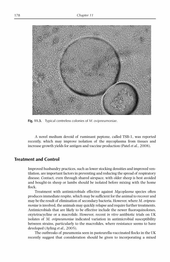
Chapter 11178
A novel medium devoid of ruminant peptone, called TSB-1, was reported recently, which may improve isolation of the mycoplasma from tissues and increase growth yields for antigen and vaccine production (Patel et al., 2008).
Treatment and Control
Improved husbandry practices, such as lower stocking densities and improved ven-tilation, are important factors in preventing and reducing the spread of respiratory disease. Contact, even through shared airspace, with older sheep is best avoided and bought-in sheep or lambs should be isolated before mixing with the home fl ock.
Treatment with antimicrobials effective against Mycoplasma species often produces immediate respite, which may be suffi cient for the animal to recover and may be the result of elimination of secondary bacteria. However, where M. ovipneu-moniae is involved, the animals may quickly relapse and require further treatments. Antimicrobials that are likely to be effective include the newer fl uoroquinolones, oxytetracycline or a macrolide. However, recent in vitro antibiotic trials on UK isolates of M. ovipneumoniae indicated variation in antimicrobial susceptibility between strains, particularly to the macrolides, where resistance seems to have developed (Ayling et al., 2005).
The outbreaks of pneumonia seen in pasteurella-vaccinated fl ocks in the UK recently suggest that consideration should be given to incorporating a mixed
Fig. 11.3. Typical centreless colonies of M. ovipneumoniae.

Respiratory Disease of Small Ruminants 179
selection of M. ovipneumoniae strains into these vaccines to provide greater protection against respiratory disease (McAuliffe et al., 2003b).
Other Mycoplasmas Associated with Disease
Other Mycoplasma species are occasionally associated with respiratory disease in sheep. M. arginini, a ubiquitous mycoplasma of many animal species, is frequently isolated from the respiratory tracts of lambs with disease, sometimes mixed with M. ovipneumoniae. While it is not thought to be a major pathogen it may increase pathological damage.
M. agalactiae is a major cause of contagious agalactia in sheep and goats but has also been isolated from pneumonic lungs (Loria et al., 2003), although its role in respiratory disease is not clear. The cattle pathogen M. bovis has also been infre-quently isolated from the respiratory tracts of sheep and goats (Ayling et al., 2004). In a recent study in the Middle East the goat pathogens M. putrefaciens, M. mycoides subsp. mycoides large colony variant and M. capricolum subsp. capri-colum were isolated from both nasal swabs and milk samples of pneumonic sheep and goats in mixed herds (Al-Momani et al., 2006a). In North Africa, M. c. capri-colum appears to be the major cause of pneumonia and contagious agalactia in sheep (Benkirane et al., 1993). Similarly, M. capricolum subsp. capripneumoniae, the cause of contagious caprine pleuropneumonia, has been isolated from respiratory tracts of sheep in contact with affected goats (Nicholas, 2000).
Whilst only a few Mycoplasma species have been reported to cause respira-tory disease in sheep, it is apparent that sheep may be equally susceptible to infection by a range of Mycoplasma species if the opportunity presents itself. Sheep may also act as carriers of Mycoplasma species for other susceptible host species.
DISEASES CAUSED BY MYCOPLASMA MYCOIDES SUBSP. CAPRI AND MYCOPASMA MYCOIDES SUBSP. MYCOIDES LC
Introduction
While M. mycoides subsp. mycoides large colony type (LC) produces severe disease in goats, its main signifi cance has been its close similarity to the small colony type, M. mycoides subsp. mycoides SC, the cause of one of the most serious diseases in cattle, contagious bovine pleuropneumonia (CBPP). It was originally thought that strains of M. mycoides subsp. mycoides isolated from goats produced large colonies in vitro whereas cattle strains gave small colonies; as a result Cottew and Yeats (1978) used this feature to distinguish the two mycoplasmas: M. mycoides LC and M. mycoides SC. Later, however, Valdiviesio-Garcia and Rosendal (1982) found this to be unreliable as both small and large types could be recovered from goats infected with M. mycoides LC. Although serologically related, recent evidence
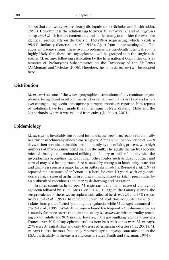
Chapter 11180
shows that the two types are clearly distinguishable (Nicholas and Bashiruddin, 1995). However, it is the relationship between M. mycoides LC and M. mycoides subsp. capri which is more contentious and has led many to consider the two to be identical, particularly on the basis of 16S rRNA sequencing, which reveals a 99.9% similarity (Pettersson et al., 1996). Apart from minor serological differ-ences with some strains, these two mycoplasmas are genetically identical, so it is highly likely that these two mycoplasmas will be grouped into the single sub-species M. m. capri following ratifi cation by the International Committee on Sys-tematics of Prokaryotes Subcommittee on the Taxonomy of the Mollicutes (Al-Momani and Nicholas, 2006). Therefore, the name M. m. capri will be adopted here.
Distribution
M. m. capri has one of the widest geographic distributions of any ruminant myco-plasma, being found in all continents where small ruminants are kept and wher-ever contagious agalactia and caprine pleuropneumonia are reported. New reports of isolations have been made this millennium in New Zealand, Chile and the Netherlands, where it was isolated from calves (Nicholas, 2004).
Epidemiology
M. m. capri is invariably introduced into a disease-free farm/region via clinically healthy or subclinically affected carrier goats. After an incubation period of 2–28 days, it then spreads to the kids, predominantly by the milking process, with high numbers of mycoplasmas being shed in the milk. The adults themselves become infected through contaminated milking machinery or milkers’ hands, with the mycoplasmas ascending the teat canal; other routes such as direct contact and aerosol may also be important. Stress caused by changes in husbandry, nutrition and climate is seen as a major factor in outbreaks in adults. Rosendal et al. (1979) reported maintenance of infection in a herd for over 10 years with only occa-sional clinical cases of arthritis in young animals, almost certainly precipitated by an outbreak of coccidiosis and later by de-horning and castration.
In most countries in Europe, M. agalactiae is the major cause of contagious agalactia followed by M. m. capri (Loria et al., 1999); in the Canary Islands, the seroprevalence of these two mycoplasmas in affected herds was 23 and 16% respec-tively (Real et al., 1994). In mainland Spain, M. agalactiae accounted for 83% of isolates from goats affected by contagious agalactia, while M. m. capri accounted for 7% (Gil et al., 1999). While M. m. capri is found less frequently, the disease it causes is usually far more severe than that caused by M. agalactiae, with mortality reach-ing 25% in adults and 90% in kids. However, in the goat-milking regions of western France, over 50% of mycoplasma isolates from bulk milk tanks were M. m. capri, 27% were M. putrefaciens and only 6% were M. agalactiae (Mercier et al., 2001). M. m. capri is also the most frequently reported caprine mycoplasma infection in the USA, particularly in the eastern and coastal states (Smith and Sherman, 1994).

Respiratory Disease of Small Ruminants 181
M. m. capri, like other mycoplasmas of small ruminants, has been found in the ear canals, often associated with ear mites (Da Massa and Brooks, 1991) or with fl eas (Nayak and Bhowmik, 1990), possibly representing a means of main-taining the carrier state and transmitting new infections to susceptible animals.
Hosts
M. m. capri has been isolated mainly from goats and occasionally from sheep with balanoposthitis and vulvovaginitis (Trichard et al., 1993). There have been a few reports from cattle; Perreau (1979) isolated M. m. capri from one of four cases of pericarditis, pleurisy and bronchopneumonia in calves in France and Kapoor et al. (1989) detected M. m. capri in aborted bovine fetuses. In the Nether-lands, the mycoplasma was detected in calves showing CBPP-like disease (Nicholas, 2004) and in healthy cattle during the CBPP eradication campaigns in Portugal and Italy, where it was reported to cause serological cross-reactions with M. mycoides SC (Machado et al., 2000; De Santis, 2005). Perrin et al. (1994) isolated M. m. capri from captive wild goats in Switzerland.
Antibody responses to M. m. capri were detected in 5–15% of Peruvian cam-elids, including alpacas and llamas, by passive haemagglutination tests (Hung et al., 1991) but no mycoplasmas have been isolated to date (Nicholas, 2004). Alpacas, llamas and vicunas suffer from pneumonia, pleuritis and polyarthritis, so it is not unlikely that mycoplasmas are causative agents.
Economic Impact
Economic losses caused by decreased production, diagnosis and treatment costs can be very high in severe outbreaks, particularly in intensive commercial goat dairy operations. Unfortunately little mycoplasma diagnosis is carried out in countries where these diseases are most prevalent. In one outbreak in the Canary Islands, 150 of 700 goats died of respiratory disease caused by several strains of M. m. capri (Rodriguez et al., 1996a).
Causative Organism
M. m. capri is a member of the M. mycoides cluster, which comprises pathogenic ruminant mycoplasmas that share immunological, biochemical and genetic prop-erties. They include M. m. capri/LC, M. capricolum subsp. capricolum, M. capricolum subsp. capripneumoniae, M. mycoides SC and the unclassifi ed M. bovine group 7 (which have all been isolated from small ruminants). Table 11.1 shows how M. m. capri compares clinically with other mycoplasmas of small ruminants.
Like most mollicutes, M. m. capri is small, simple, self-replicating and distin-guishable from walled bacteria and L-form bacteria by a number of unique prop-erties, including lack of a cell wall and the genetic machinery to synthesize one; its DNA has a low guanine plus cytosine content, typically less than 30 mol %; it has

C
hapter 11182
Table 11.1. Comparative characteristics of some mycoplasmas of small ruminants, including M. mycoides subsp. capri.
Mycoplasma Host Primary site of isolation (other) Disease* Pathogenicity
M. m. mycoides LC Goat (sheep, cattle) Resp. tract (udder, joints) Plp, M, A, C ModerateM. m. capri Goat (sheep) Resp. tract (joints) Plp, A, C ModerateM. c. capricolum Goat, sheep Joints/resp. tract (udder) Plp, M, A HighM. c. capripneumoniae Goat (sheep) Lungs CCPP HighM. ovipneumoniae Sheep, goat Resp. tract Pneumonia LowM. conjunctivae Sheep and goat Eyes KC ModerateM. agalactiae Sheep and goat Udder (joints, eyes) M, A, KC, P HighM. putrefaciens Goat (sheep) Udder (joints) M, A ModerateM. arginini Ubiquitous Resp. tract None Low/non-pathogenic
*Plp – pleuropneumonia, P – pneumonia, M – mastitis, A – arthritis, C – conjunctivitis, KC – keratoconjunctivitis, CCPP – contagious caprine pleuropneumonia
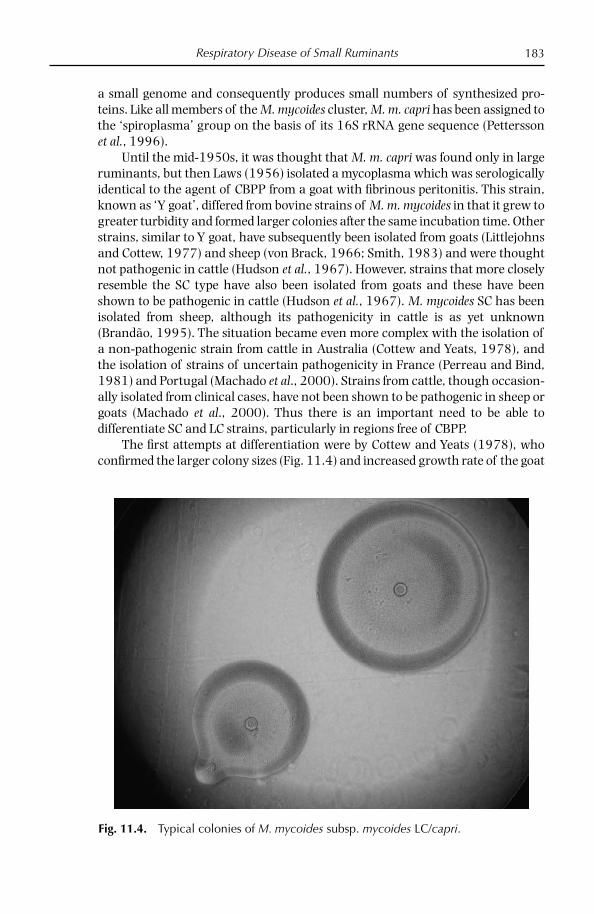
Respiratory Disease of Small Ruminants 183
a small genome and consequently produces small numbers of synthesized pro-teins. Like all members of the M. mycoides cluster, M. m. capri has been assigned to the ‘spiroplasma’ group on the basis of its 16S rRNA gene sequence (Pettersson et al., 1996).
Until the mid-1950s, it was thought that M. m. capri was found only in large ruminants, but then Laws (1956) isolated a mycoplasma which was serologically identical to the agent of CBPP from a goat with fi brinous peritonitis. This strain, known as ‘Y goat’, differed from bovine strains of M. m. mycoides in that it grew to greater turbidity and formed larger colonies after the same incubation time. Other strains, similar to Y goat, have subsequently been isolated from goats (Littlejohns and Cottew, 1977) and sheep (von Brack, 1966; Smith, 1983) and were thought not pathogenic in cattle (Hudson et al., 1967). However, strains that more closely resemble the SC type have also been isolated from goats and these have been shown to be pathogenic in cattle (Hudson et al., 1967). M. mycoides SC has been isolated from sheep, although its pathogenicity in cattle is as yet unknown (Brandão, 1995). The situation became even more complex with the isolation of a non-pathogenic strain from cattle in Australia (Cottew and Yeats, 1978), and the isolation of strains of uncertain pathogenicity in France (Perreau and Bind, 1981) and Portugal (Machado et al., 2000). Strains from cattle, though occasion-ally isolated from clinical cases, have not been shown to be pathogenic in sheep or goats (Machado et al., 2000). Thus there is an important need to be able to differentiate SC and LC strains, particularly in regions free of CBPP.
The fi rst attempts at differentiation were by Cottew and Yeats (1978), who confi rmed the larger colony sizes (Fig. 11.4) and increased growth rate of the goat
Fig. 11.4. Typical colonies of M. mycoides subsp. mycoides LC/capri.
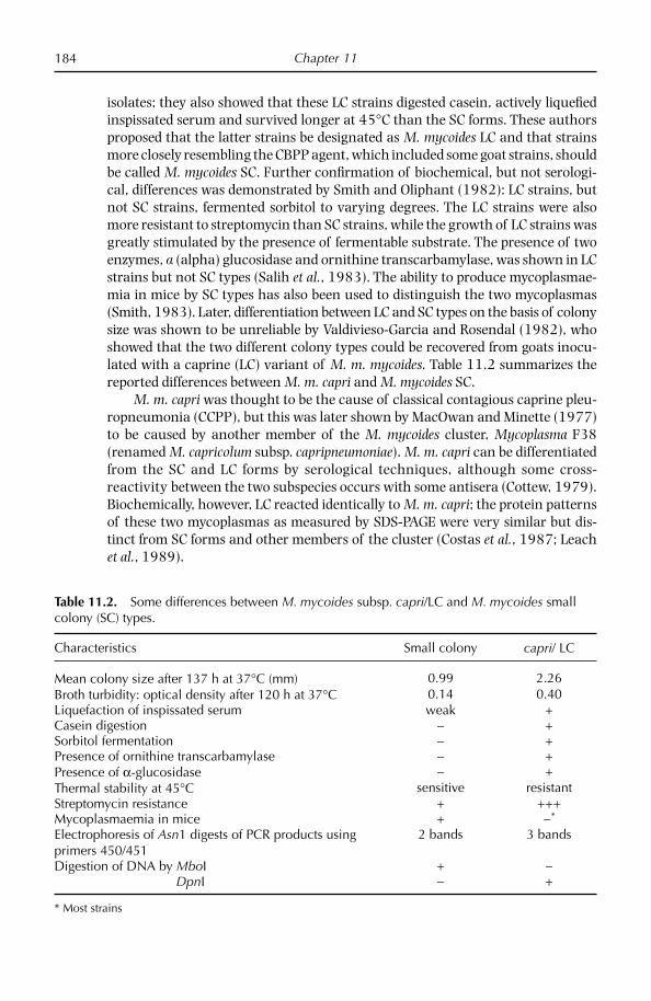
Chapter 11184
isolates; they also showed that these LC strains digested casein, actively liquefi ed inspissated serum and survived longer at 45°C than the SC forms. These authors proposed that the latter strains be designated as M. mycoides LC and that strains more closely resembling the CBPP agent, which included some goat strains, should be called M. mycoides SC. Further confi rmation of biochemical, but not serologi-cal, differences was demonstrated by Smith and Oliphant (1982): LC strains, but not SC strains, fermented sorbitol to varying degrees. The LC strains were also more resistant to streptomycin than SC strains, while the growth of LC strains was greatly stimulated by the presence of fermentable substrate. The presence of two enzymes, a (alpha) glucosidase and ornithine transcarbamylase, was shown in LC strains but not SC types (Salih et al., 1983). The ability to produce mycoplasmae-mia in mice by SC types has also been used to distinguish the two mycoplasmas (Smith, 1983). Later, differentiation between LC and SC types on the basis of colony size was shown to be unreliable by Valdivieso-Garcia and Rosendal (1982), who showed that the two different colony types could be recovered from goats inocu-lated with a caprine (LC) variant of M. m. mycoides. Table 11.2 summarizes the reported differences between M. m. capri and M. mycoides SC.
M. m. capri was thought to be the cause of classical contagious caprine pleu-ropneumonia (CCPP), but this was later shown by MacOwan and Minette (1977) to be caused by another member of the M. mycoides cluster, Mycoplasma F38 (renamed M. capricolum subsp. capripneumoniae). M. m. capri can be differentiated from the SC and LC forms by serological techniques, although some cross-reactivity between the two subspecies occurs with some antisera (Cottew, 1979). Biochemically, however, LC reacted identically to M. m. capri; the protein patterns of these two mycoplasmas as measured by SDS-PAGE were very similar but dis-tinct from SC forms and other members of the cluster (Costas et al., 1987; Leach et al., 1989).
Table 11.2. Some differences between M. mycoides subsp. capri/LC and M. mycoides small colony (SC) types.
Characteristics Small colony capri/ LC
Mean colony size after 137 h at 37°C (mm) 0.99 2.26Broth turbidity: optical density after 120 h at 37°C 0.14 0.40Liquefaction of inspissated serum weak +Casein digestion − +Sorbitol fermentation − +Presence of ornithine transcarbamylase − +Presence of α-glucosidase − +Thermal stability at 45°C sensitive resistantStreptomycin resistance + +++Mycoplasmaemia in mice + −*
Electrophoresis of Asn1 digests of PCR products using primers 450/451
2 bands 3 bands
Digestion of DNA by MboI + − DpnI − +
* Most strains
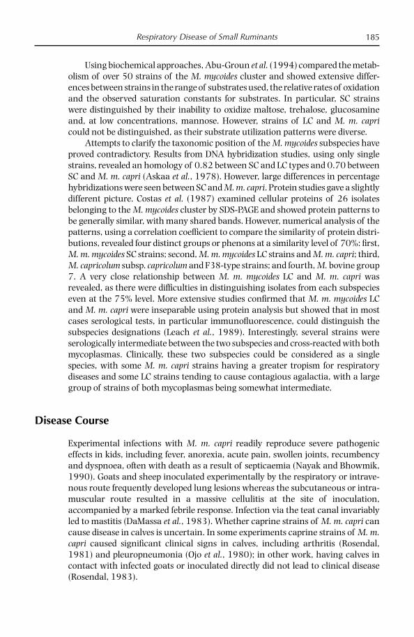
Respiratory Disease of Small Ruminants 185
Using biochemical approaches, Abu-Groun et al. (1994) compared the metab-olism of over 50 strains of the M. mycoides cluster and showed extensive differ-ences between strains in the range of substrates used, the relative rates of oxidation and the observed saturation constants for substrates. In particular, SC strains were distinguished by their inability to oxidize maltose, trehalose, glucosamine and, at low concentrations, mannose. However, strains of LC and M. m. capri could not be distinguished, as their substrate utilization patterns were diverse.
Attempts to clarify the taxonomic position of the M. mycoides subspecies have proved contradictory. Results from DNA hybridization studies, using only single strains, revealed an homology of 0.82 between SC and LC types and 0.70 between SC and M. m. capri (Askaa et al., 1978). However, large differences in percentage hybridizations were seen between SC and M. m. capri. Protein studies gave a slightly different picture. Costas et al. (1987) examined cellular proteins of 26 isolates belonging to the M. mycoides cluster by SDS-PAGE and showed protein patterns to be generally similar, with many shared bands. However, numerical analysis of the patterns, using a correlation coeffi cient to compare the similarity of protein distri-butions, revealed four distinct groups or phenons at a similarity level of 70%: fi rst, M. m. mycoides SC strains; second, M. m. mycoides LC strains and M. m. capri; third, M. capricolum subsp. capricolum and F38-type strains; and fourth, M. bovine group 7. A very close relationship between M. m. mycoides LC and M. m. capri was revealed, as there were diffi culties in distinguishing isolates from each subspecies even at the 75% level. More extensive studies confi rmed that M. m. mycoides LC and M. m. capri were inseparable using protein analysis but showed that in most cases serological tests, in particular immunofl uorescence, could distinguish the subspecies designations (Leach et al., 1989). Interestingly, several strains were serologically intermediate between the two subspecies and cross- reacted with both mycoplasmas. Clinically, these two subspecies could be considered as a single species, with some M. m. capri strains having a greater tropism for respiratory diseases and some LC strains tending to cause contagious agalactia, with a large group of strains of both mycoplasmas being somewhat intermediate.
Disease Course
Experimental infections with M. m. capri readily reproduce severe pathogenic effects in kids, including fever, anorexia, acute pain, swollen joints, recumbency and dyspnoea, often with death as a result of septicaemia (Nayak and Bhowmik, 1990). Goats and sheep inoculated experimentally by the respiratory or intrave-nous route frequently developed lung lesions whereas the subcutaneous or intra-muscular route resulted in a massive cellulitis at the site of inoculation, accompanied by a marked febrile response. Infection via the teat canal invariably led to mastitis (DaMassa et al., 1983). Whether caprine strains of M. m. capri can cause disease in calves is uncertain. In some experiments caprine strains of M. m. capri caused signifi cant clinical signs in calves, including arthritis (Rosendal, 1981) and pleuropneumonia (Ojo et al., 1980); in other work, having calves in contact with infected goats or inoculated directly did not lead to clinical disease (Rosendal, 1983).

Chapter 11186
The most common clinical fi ndings in adult goats with natural infections are fever, mastitis, pleuropneumonia, polyarthritis and occasionally abortion; in kids, arthritis, septicaemia and, occasionally, meningitis and abortion. Keratoconjunc-tivitis may also be seen in both. Kids are most frequently and more severely affected than adults but mastitis may affect 25–30% of does. The most severe outbreaks occur amongst young kids. Arthritis, more common in kids, always occurs as a result of septicaemia, frequently involving multiple joints and accompanied by generalized malaise and fever. It rarely occurs in isolation and in adults usually follows mastitis, abortion, agalactia, pneumonia and keratoconjunctivitis. In extreme cases septicaemic goats may die without manifestation of localized signs, which may be the result of galactan production by M. m. capri (Smith and Sherman, 1994).
Pathology
Lesions caused by M. m. capri are characterized by lungs which are enlarged and fi rm with hepatization of cardiac and diaphragmatic lobes and marked pleural effusion and fi brinous pleuritis. There is extensive pulmonary oedema and inter-lobular septa are usually distended and pale. Histopathologically, arterial and arteriolar vasculitis with necrosis of vessel walls and thrombi are common.
Severe respiratory signs and high mortality were the most signifi cant fi ndings in one goat herd with pleuropneumonia caused by M. m. capri in Spain (Rodriguez et al., 1995). The adults presented mainly with respiratory disease and/or masti-tis, whereas the young animals died showing arthritis and/or keratoconjunctivi-tis. The most signifi cant lesions were found in the thoracic cavity. Macroscopically, a focal extensive fi brinonecrotic pleuropneumonia was seen and histopathologi-cal analysis confi rmed a fi brinopurulent and necrotic pleuropneumonia with areas of acute pyogenic bronchopneumonia and fi brinous pericarditis associated with a mucopurulent mastitis and/or a fi brinopurulent arthritis.
An unusual mycoplasma, isolated from a goat presenting contagious agalac-tia in northern Jordan and identifi ed as M. mycoides subsp. mycoides LC/M. mycoides subsp. capri by PCR and DGGE, was used to infect healthy seronegative goats by either aerosol or intracheal routes (Di Provvido et al., 2008). While ELISA and latex agglutination tests detected antibody after about 2 weeks, the comple-ment fi xation test remained negative throughout the experiment. After 2 weeks, intubated goats became febrile, showing nasal discharge and slight conjunctivitis, which was followed by respiratory distress and polyarthritis 1 week later. Clinical signs were much milder in the aerosol group, consisting of nasal discharge and slight articular swelling of the carpal joints in one goat. Lesions were seen in intu-bated goats at post-mortem, including coagulative necrotic pneumonia, fi brinous pleurisy, pleural fl uid in the thoracic cavity (Figs 11.5 and 11.6) and infl amma-tory exudates, necrosis and fi brosis in the joints (Fig. 11.7). Seroconversion was fi rst seen in a group of contacts after 7 weeks by both ELISA and latex agglutination test, which was accompanied by coughing and laboured respiration. Pulmonary lesions in the contacts consisted of fi brinous pleuropneumonia, with focal areas of necrosis and abundant pleural fl uid in the thoracic cavity. This work demonstrated

Respiratory Disease of Small Ruminants 187
the pathogenicity of this unusual serotype in both infected and contact goats. It was interesting to note that the CFT using the type strain as antigen would fail to detect this serotype.
Diagnosis
Clinical signs and pathological changes caused by M. m. capri are not characteris-tic and it is very important to distinguish between infections caused by M. m. capri and those caused by M. c. capripneumoniae, the agent of classical contagious pleuropneumonia (CCPP), which is not present in the EU. The key features in CCPP infections are its highly infectious nature, the lack of distention of the inter-lobular septa in the lungs and its restriction to the lungs. Laboratory diagnosis must always be performed to determine the true cause of the disease (Nicholas and Baker, 1998).
Samples of choice from living animals include nasal swabs and secretions, milk from mastitic does or from apparently healthy does where there is a high rate of mortality/morbidity in kids, joint fl uid from arthritic cases, eye swabs from cases of ocular disease, blood for antibody detection from affected and non-affected animals, and ear swabs. From dead animals samples should include lung tissue (at the interface between diseased and healthy tissue) and pleural/
Fig. 11.5. Lung of goat experimentally infected with M. mycoides subsp. capri, showing fi brin deposition.
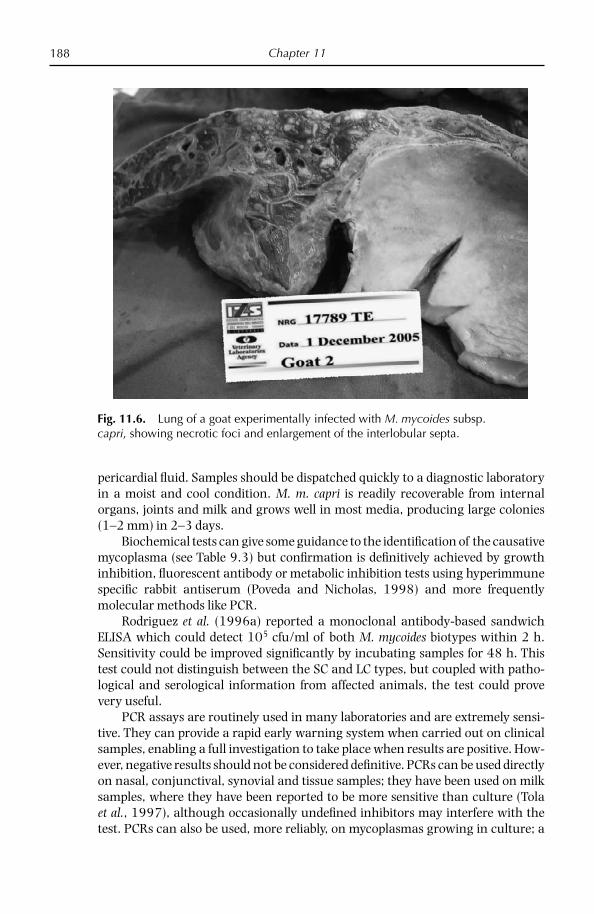
Chapter 11188
pericardial fl uid. Samples should be dispatched quickly to a diagnostic laboratory in a moist and cool condition. M. m. capri is readily recoverable from internal organs, joints and milk and grows well in most media, producing large colonies (1–2 mm) in 2–3 days.
Biochemical tests can give some guidance to the identifi cation of the causative mycoplasma (see Table 9.3) but confi rmation is defi nitively achieved by growth inhibition, fl uorescent antibody or metabolic inhibition tests using hyperimmune specifi c rabbit antiserum (Poveda and Nicholas, 1998) and more frequently molecular methods like PCR.
Rodriguez et al. (1996a) reported a monoclonal antibody-based sandwich ELISA which could detect 105 cfu/ml of both M. mycoides biotypes within 2 h. Sensitivity could be improved signifi cantly by incubating samples for 48 h. This test could not distinguish between the SC and LC types, but coupled with patho-logical and serological information from affected animals, the test could prove very useful.
PCR assays are routinely used in many laboratories and are extremely sensi-tive. They can provide a rapid early warning system when carried out on clinical samples, enabling a full investigation to take place when results are positive. How-ever, negative results should not be considered defi nitive. PCRs can be used directly on nasal, conjunctival, synovial and tissue samples; they have been used on milk samples, where they have been reported to be more sensitive than culture (Tola et al., 1997), although occasionally undefi ned inhibitors may interfere with the test. PCRs can also be used, more reliably, on mycoplasmas growing in culture; a
Fig. 11.6. Lung of a goat experimentally infected with M. mycoides subsp. capri, showing necrotic foci and enlargement of the interlobular septa.
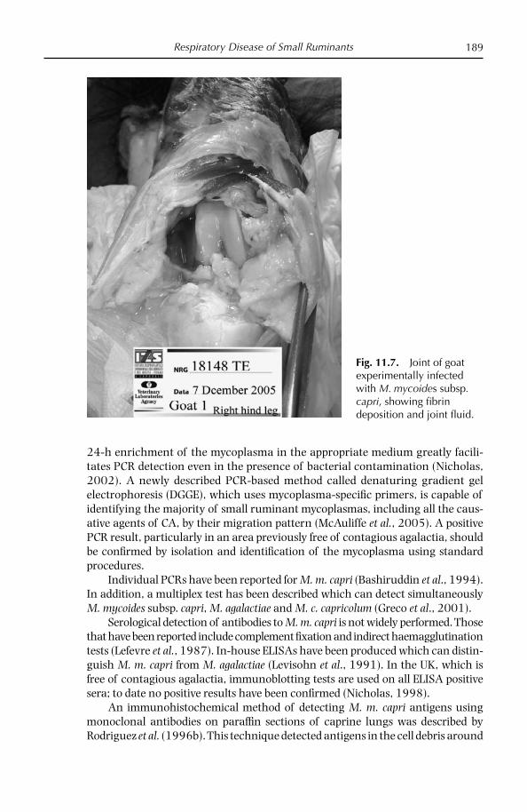
Respiratory Disease of Small Ruminants 189
24-h enrichment of the mycoplasma in the appropriate medium greatly facili-tates PCR detection even in the presence of bacterial contamination (Nicholas, 2002). A newly described PCR-based method called denaturing gradient gel electrophoresis (DGGE), which uses mycoplasma-specifi c primers, is capable of identifying the majority of small ruminant mycoplasmas, including all the caus-ative agents of CA, by their migration pattern (McAuliffe et al., 2005). A positive PCR result, particularly in an area previously free of contagious agalactia, should be confi rmed by isolation and identifi cation of the mycoplasma using standard procedures.
Individual PCRs have been reported for M. m. capri (Bashiruddin et al., 1994). In addition, a multiplex test has been described which can detect simultaneously M. mycoides subsp. capri, M. agalactiae and M. c. capricolum (Greco et al., 2001).
Serological detection of antibodies to M. m. capri is not widely performed. Those that have been reported include complement fi xation and indirect haemagglutination tests (Lefevre et al., 1987). In-house ELISAs have been produced which can distin-guish M. m. capri from M. agalactiae (Levisohn et al., 1991). In the UK, which is free of contagious agalactia, immunoblotting tests are used on all ELISA positive sera; to date no positive results have been confi rmed (Nicholas, 1998).
An immunohistochemical method of detecting M. m. capri antigens using monoclonal antibodies on paraffi n sections of caprine lungs was described by Rodriguez et al. (1996b). This technique detected antigens in the cell debris around
Fig. 11.7. Joint of goat experimentally infected with M. mycoides subsp. capri, showing fi brin deposition and joint fl uid.
Chapter 11190
necrotic areas and in macrophages, neutrophils and epithelial cells in lungs from both natural and experimental infections.
Disease Treatment
Like most mycoplasma diseases, those caused by M. m. capri do not respond well to chemotherapy, in particular in cases of mastitis. Although clinical improvements may be seen, the mycoplasma may remain viable in the goats. Infected but appar-ently healthy does may continue to produce milk containing large numbers of mycoplasmas, causing problems in vulnerable kids. In such circumstances pas-teurization of milk may help to control outbreaks; in one case morbidity and mortality was reduced from 90% to less than 5% when only mycoplasma-free milk was fed (Da Massa et al., 1983). Tylosin and oxytetracycline have both been reported to be effective against pneumonia, but all animals in a herd must be treated, including apparently healthy goats, although it should be noted that tylo-sin is not recommended for treatment of animals under 15 kg. For arthritis, a combination of spectinomycin and lincomycin given intramuscularly for 3 days generated some recovery in sheep and goats (Smith and Sherman, 1994).
Disease Prevention and Control
No vaccines are currently available for M. m. capri as the disease is thought not to be suffi ciently widespread in Europe; in Greece, Turkey and the Middle East clini-cal disease is more prevalent but differential diagnosis is not routinely performed. Vaccines inactivated by saponin have been reported in India, these provoking a strong antibody response and showing some protection. As most disease-free herds become infected following the introduction of a carrier animal, knowledge of the health status of the herd of origin is essential; serious consideration should also be given to maintaining the bought-in animals in quarantine until serologi-cal testing has been carried out. Stress such as that triggered by movement or parturition should be minimized. Where mastitis is present, affected animals should be milked last and machines/hands should be disinfected. Where the situ-ation warrants, culling of clinically affected animals should also be encouraged.
The possible involvement of ear mites and fl eas in transmission suggests that insecticides such as ivermectin may be worth considering.
IN VITRO SUSCEPTIBILITY OF SMALL RUMINANT MYCOPLASMAS TO ANTIMICROBIALS
Relatively few studies have been carried out on the in vitro effi cacy of antimicrobi-als against the mycoplasmas that cause disease in sheep and goats. Bergonier et al. (1997) reviewed data on some antimicrobials used for treating CA, but as with all mycoplasma MIC studies they should be examined carefully as the methods used
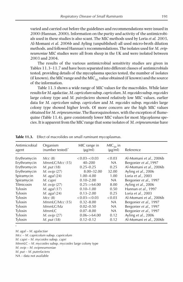
Respiratory Disease of Small Ruminants 191
varied and carried out before the guidelines and recommendations were issued in 2000 (Hannan, 2000). Information on the purity and activity of the antimicrobi-als used in these studies is also scant. The MIC methods used by Loria et al. 2003, Al-Momani et al. 2006b and Ayling (unpublished) all used micro-broth dilution methods, and followed Hannan’s recommendations. The isolates used for M. ovip-neumoniae MIC studies were all from sheep in the UK and were isolated between 2003 and 2004.
The results of the various antimicrobial sensitivity studies are given in Tables 11.3–11.7 and have been separated into different classes of antimicrobials tested, providing details of the mycoplasma species tested, the number of isolates (if known), the MIC range and the MIC50 value obtained (if known) and the source of the information.
Table 11.3 shows a wide range of MIC values for the macrolides. While later results for M. agalactiae, M. capricolum subsp. capricolum, M. mycoides subsp. mycoides large colony type and M. putrefaciens showed relatively low MIC values, earlier data for M. capricolum subsp. capricolum and M. mycoides subsp. mycoides large colony type showed higher levels. Of more concern are the high MIC values obtained for M. ovipneumoniae. The fl uoroquinolones, with the exception of fl ume-quine (Table 11.4), gave consistently lower MIC values for most Mycoplasma spe-cies. It is apparent from the MIC range that some isolates of M. ovipneumoniae have
Table 11.3. Effect of macrolides on small ruminant mycoplasmas.
Antimicrobial agent
Organism(number tested)*
MIC range in(µg/ml)
MIC50 in(µg/ml) Reference
Erythromycin Mcc (8) < 0.03–< 0.03 < 0.03 Al-Momani et al., 2006bErythromycin MmmLC/Mcc (15) 40–200 NA Bergonier et al.,1997Erythromycin M. put (18) 0.25–0.25 0.25 Al-Momani et al., 2006bErythromycin M. ovip (27) 8.00–32.00 32.00 Ayling et al., 2006Spiramycin M. agal (24) 1.00–4.00 1.00 Loria et al., 2003Spiramycin M. capri 0.10–2.00 NA Bergonier et al., 1997Tilmicosin M. ovip (27) 0.25–> 64.00 8.00 Ayling et al., 2006Tylosin M. agal (17) 0.10–1.00 0.50 Hannan et al., 1997Tylosin M. agal (24) 0.13–2.00 0.25 Loria et al., 2003Tylosin Mcc (8) < 0.03–< 0.03 < 0.03 Al-Momani et al., 2006bTylosin MmmLC/Mcc (15) 0.32–8.00 NA Bergonier et al., 1997Tylosin MmmLC/Ma 0.02–0.50 NA Bergonier et al., 1997Tylosin MmmLC 0.07–8.00 NA Bergonier et al., 1997Tylosin M. ovip (27) 0.06–> 64.00 0.12 Ayling et al., 2006Tylosin M. put (18) 0.12–0.12 0.12 Al-Momani et al., 2006b
*
M. agal – M. agalactiaeMcc – M. capricolum subsp. capricolumM. capri – M. mycoides subsp. capri MmmLC – M. mycoides subsp. mycoides large colony typeM. ovip – M. ovipneumoniaeM. put – M. putrefaciensNA – data not available

Chapter 11192
given high MIC values of 4, 8 and 32 µg/ml for enrofl oxacin, danofl oxacin and norfl oxacin, respectively. Most species and isolates appear to show low MIC results for the tetracyclines (Table 11.5), but again a few isolates have higher MIC values. Generally the chloramphenicols and derivatives (Table 11.6) show MIC values on
Table 11.6. Effect of chloramphenicol and derivatives on mycoplasmas of small ruminants.
Antimicrobial agentOrganism(number tested)
MIC range in(µg/ml)
MIC50 in(µg/ml) Reference
Chloramphenicol Mcc (8) 8.00–8.00 8.00 Al-Momani et al., 2006bChloramphenicol MmmLC (6) 8.00–8.00 8.00 Al-Momani et al., 2006bChloramphenicol M. ovip (27) 1.00–32.00 8.00 Ayling et al., 2006Chloramphenicol M. put (18) 1.00–8.00 1.00 Al-Momani et al., 2006bFlorfenicol Mcc (8) 4.00–8.00 4.00 Al-Momani et al., 2006bFlorfenicol MmmLC (6) 2.00–4.00 2.00 Al-Momani et al., 2006bFlorfenicol M. ovip (27) 0.50–16.00 4.00 Ayling et al., 2006Florfenicol M. put (18) 0.50–4.00 1.00 Al-Momani et al., 2006b
Table 11.5. Effect of tetracyclines on mycoplasmas of small ruminants.
Antimicrobial agent
Organism(number tested)
MIC range in(µg/ml)
MIC50 in(µg/ml) Reference
Oxytetracycline M. agal (17) 0.10–0.25 0.10 Hannan et al., 1997Oxytetracycline Mcc (8) 0.12–0.25 0.25 Al-Momani et al., 2006bOxytetracycline MmmLC/Mcc (15) 0.32–8.00 NA Bergonier et al., 1997Oxytetracycline MmmLC 0.50–10.0 NA Bergonier et al., 1997Oxytetracycline M. ovip (27) 0.06–8.00 0.12 Ayling et al., 2006Oxytetracycline M. put (18) 0.25–1.00 0.25 Al-Momani et al., 2006bTetracycline M. agal (24) 0.13–4.00 0.25 Loria et al., 2003
Table 11.4. Effect of fl uoroquinolones on mycoplasmas of small ruminants.
Antimicrobial agent
Organism(number tested)*
MIC range in(µg/ml)
MIC50 in(µg/ml) Reference
Ciprofl oxacin M. ovip (27) < 0.12–2.00 0.12 Ayling et al., 2006Danofl oxacin M. agal (17) 0.05–2.50 0.25 Hannan et al., 1997Danofl oxacin M. ovip (27) < 0.06–8.00 0.12 Ayling et al., 2006Enrofl oxacin M. agal 0.17–0.22 NA Bergonier et al., 1997Enrofl oxacin M. agal (17) 0.05–1.00 0.25 Hannan et al., 1997Enrofl oxacin M. agal (24) 0.13–0.50 0.13 Loria et al., 2003Enrofl oxacin MmmLC (6) 0.12–0.12 0.12 Al-Momani et al., 2006bEnrofl oxacin M. ovip (27) 0.03–4.00 0.06 Ayling et al., 2006Enrofl oxacin M. put (18) 0.25–0.25 0.25 Al-Momani et al., 2006bFlumequine M. agal (17) 5.00–100 25.00 Hannan et al., 1997Norfl oxacin M. ovip (27) < 0.12–32.00 0.25 Ayling et al., 2006
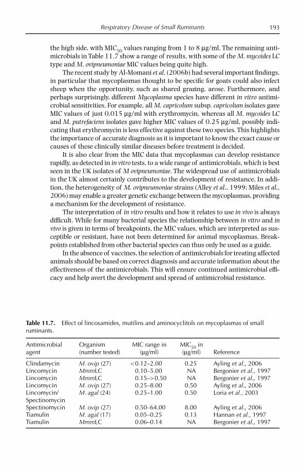
Respiratory Disease of Small Ruminants 193
the high side, with MIC50 values ranging from 1 to 8 µg/ml. The remaining anti-microbials in Table 11.7 show a range of results, with some of the M. mycoides LC type and M. ovipneumoniae MIC values being quite high.
The recent study by Al-Momani et al. (2006b) had several important fi ndings, in particular that mycoplasmas thought to be specifi c for goats could also infect sheep when the opportunity, such as shared grazing, arose. Furthermore, and perhaps surprisingly, different Mycoplasma species have different in vitro antimi-crobial sensitivities. For example, all M. capricolum subsp. capricolum isolates gave MIC values of just 0.015 µg/ml with erythromycin, whereas all M. mycoides LC and M. putrefaciens isolates gave higher MIC values of 0.25 µg/ml, possibly indi-cating that erythromycin is less effective against these two species. This highlights the importance of accurate diagnosis as it is important to know the exact cause or causes of these clinically similar diseases before treatment is decided.
It is also clear from the MIC data that mycoplasmas can develop resistance rapidly, as detected in in vitro tests, to a wide range of antimicrobials, which is best seen in the UK isolates of M ovipneumoniae. The widespread use of antimicrobials in the UK almost certainly contributes to the development of resistance. In addi-tion, the heterogeneity of M. ovipneumoniae strains (Alley et al., 1999; Miles et al., 2006) may enable a greater genetic exchange between the mycoplasmas, providing a mechanism for the development of resistance.
The interpretation of in vitro results and how it relates to use in vivo is always diffi cult. While for many bacterial species the relationship between in vitro and in vivo is given in terms of breakpoints, the MIC values, which are interpreted as sus-ceptible or resistant, have not been determined for animal mycoplasmas. Break-points established from other bacterial species can thus only be used as a guide.
In the absence of vaccines, the selection of antimicrobials for treating affected animals should be based on correct diagnosis and accurate information about the effectiveness of the antimicrobials. This will ensure continued antimicrobial effi -cacy and help avert the development and spread of antimicrobial resistance.
Table 11.7. Effect of lincosamides, mutilins and aminocyclitols on mycoplasmas of small ruminants.
Antimicrobial agent
Organism(number tested)
MIC range in(µg/ml)
MIC50 in(µg/ml) Reference
Clindamycin M. ovip (27) < 0.12–2.00 0.25 Ayling et al., 2006Lincomycin MmmLC 0.10–5.00 NA Bergonier et al., 1997Lincomycin MmmLC 0.15–> 0.50 NA Bergonier et al., 1997Lincomycin M. ovip (27) 0.25–8.00 0.50 Ayling et al., 2006Lincomycin/Spectinomycin
M. agal (24) 0.25–1.00 0.50 Loria et al., 2003
Spectinomycin M. ovip (27) 0.50–64.00 8.00 Ayling et al., 2006Tiamulin M. agal (17) 0.05–0.25 0.13 Hannan et al., 1997Tiamulin MmmLC 0.06–0.14 NA Bergonier et al., 1997

Chapter 11194
References
Abu-Amero, K.K., Halablab, M.A. and Miles, R.J. (1996) Nisin resistance distinguishes My-coplasma species from Acholeplasma species and provides a basis for selective growth media. Applied and Environmental Microbiology 62, 3107–3111.
Abu-Groun, E.A., Taylor, R.R., Varsani, H., Leach, R.H. and Miles, R.J. (1994) Biochemical diversity within the Mycoplasma mycoides cluster. Microbiology 140, 2033–2042.
Alley, M.R., Ionas, G. and Clarke, J.K. (1999) Chronic non-progressive pneumonia of sheep in New Zealand – a review of the role of Mycoplasma ovipneumoniae. New Zealand Veterinary Journal 47, 155–160.
Al-Momani, W. and Nicholas, R.A.J. (2006) Small ruminant mycoplasmas with particular reference to the Middle East. Veterinary Bulletin 76, 1–11.
Al-Momani, W., Halablab, M.A., Abo-Shehada, M.N., Miles, K., McAuliffe, L. and Nicholas, R.A.J. (2006a) Isolation and molecular identifi cation of small ruminant mycoplasmas in Jordan. Small Ruminant Research 65, 106–112.
Al-Momani, W., Nicholas, R. A. J., Janakat, S., Abu-Basha, E. and Ayling, R.D. (2006b) The in vitro effect of six antimicrobials against Mycoplasma putrefaciens, Mycoplasma my-coides subsp. mycoides LC and Mycoplasma capricolum subsp. capricolum isolated from sheep and goats in Jordan. Tropical Animal Health and Production 38, 1–7.
Askaa G, Erno, H. and Ojo, M.O. (1978) Bovine mycoplasmas: classifi cation of groups related to Mycoplasma mycoides. Acta Veterinaria Scandinavica 19, 166–178.
Ayling, R.D., Bisgaard-Frantzen, S., Nicholas, R.A.J. (2006) The use of antimicrobials in controlling mycoplasma infections. SIPAOC 2006 XVII Edizione 25/28 Ottobre 2006, Lamezia Terme (CZ), pp. 112-117.
Ayling, R.D. and Nicholas, R.A.J. (2007) Mycoplasma respiratory infections. In: Aitken, I.D (ed.) Sheep Diseases, 4th edition. Blackwell Publishing, Oxford, UK, pp. 231–235.
Ayling, R.D., Bashiruddin, S.E. and Nicholas, R.A.J. (2004) Mycoplasma species and related organisms isolated from ruminants in Britain between 1990 and 2000. Veterinary Record 155, 413–416.
Ayling, R.D, McAuliffe, L., Bisgaard-Frantzen, S., Bashiruddin, S.E, Miles, K. and Nicholas, R.A.J. (2005) Mycoplasma ovipneumoniae: recent developments in diagnosis and in vitro susceptibility to antimicrobials. In: Proceedings of the International Sheep Veterinary Congress, Crete, pp. 134–135.
Bashiruddin, J.B., Taylor, T.K. and Gould A.R. (1994) A PCR-based test for the specifi c iden-tifi cation of Mycoplasma mycoides subsp. mycoides SC. Journal of Veterinary Diagnostic Investigation 6, 428–434.
Benkirane, A., Amghar, A. and Kirchhoff, H. (1993) Analysis of membrane proteins of My-coplasma capricolum strains by SDS-PAGE and immunoblotting. Journal of Veterinary Medicine B 40, 119–124.
Bergonier, D., Berthelot, X. and Poumarat, F. (1997) Contagious agalactia of small ruminants: current knowledge concerning epidemiology, diagnosis and control. Revue Scientifi que et Technique. Offi ce International Epizooties 16, 848–873.
Brandão, E. (1995) Isolation and identifi cation of Mycoplasma mycoides subsp. mycoides SC strains in sheep and goats. Veterinary Record 136, 98–99.
Costas, M., Leach, R.H. and Mitchelmore, D.L. (1987) Numerical analysis of PAGE protein patterns and the taxonomic relationships within the ‘Mycoplasma mycoides cluster’. Journal of General Microbiology 133, 3319–3329.
Cottew, G.S. (1971) Characterisation of mycoplasmas isolated from sheep with pneumo-nia. Australian Veterinary Journal 47, 591–596.
Cottew, G.S. (1979) Caprine-ovine mycoplasmas. In: Tully, J.G. and Whitcomb, R.F. (eds) The Mycoplasmas, vol. 2. Academic Press, London, pp. 103–132.

Respiratory Disease of Small Ruminants 195
Cottew, G.S. and Yeats, F.R. (1978) Subdivision of Mycoplasma mycoides subsp. mycoides from cattle and goats into two types. Australian Veterinary Journal 54, 293–296.
Da Massa, A.J. and Brooks, D.L. (1991) The external ear canal of goats and other animals as a mycoplasma habitat. Small Ruminant Research 4, 85–93.
Da Massa, A.J., Brooks, D.L. and Adler, H.E. (1983) Caprine mycoplasmosis infection in goats with Mycoplasma mycoides subsp. mycoides (large colony type). American Journal of Veterinary Research 44, 322–325.
De Santis, P. (2005) Contagious bovine pleuropneumonia: studies on the disease and causative agent Mycoplasma mycoides subsp. mycoides SC. PhD thesis, King’s College, London.
Di Provvido, A., Scacchia, M., Varasano, V., Churchward, C., De Caro, C., Al-Momani, W., Di Francesco, G., Ayling, R.D., Lelli, R. and Nicholas, R.A.J. (2008) Experimental infec-tion of goats with an unusual strain of Mycoplasma mycoides subsp. capri isolated in Jordan. Journal of Comparative Pathology, in press.
Gil, M.C., Hermosa De Mendoza, M., Rey, J., Alonso, J.M., Poveda, J.B. and Hermosa De Men-doza, J. (1999) Aetiology of caprine contagious agalactia syndrome in Extremudura, Spain. Veterinary Record 144, 24–25.
Gilmour, J.S., Jones, G.E. and Rae, A.G. (1979) Experimental studies of chronic pneumonia of sheep. Comparative Immunology, Microbiology and Infectious Diseases 1, 285–293.
Gonçalves, R., Mariano, I., Fairfoul, G. and Nicholas, R.A.J. (2008) Atypical non progres-sive pneumonia in goats associated with Mycoplasma ovipneumoniae in Portugal. The Veterinary Journal, in press.
Greco, G., Corrente, M., Martella, V., Pratelli, A. and Bounovoglia, D. (2001) A mulitiplex PCR for the diagnosis of contagious agalactia of sheep and goats. Molecular and Cellular Probes 15, 21–25.
Hannan, P.C.T. (2000) Guidelines and recommendations for antimicrobial minimum inhibi-tory concentration (MIC) testing against veterinary mycoplasma species. Veterinary Research 31, 373–395.
Hannan, P.C., Windsor, G.D., de Jong, A., Schmeer, N. and Stegemann, M. (1997) Compara-tive susceptibilities of various animal-pathogenic mycoplasmas to fl uoroquinolones. Antimicrobial Agents and Chemotherapy 41, 2037–2040.
Hervas, J., Mendez, A., Gomez-Villamandos, J.C., Villalba, E., Diaz, E., Cano, T., Carrasco, L., Padro, J.M., Fernandez, A. and Sierra, M.A. (1996) Etiologic and pathologic study of respiratory disease in intensively bred lambs in southern Spain. Zentralblatt fur Veteri-narmedizin B 43, 221–231.
Hudson, J.R., Cottew, G.S. and Adler, H.E. (1967) Diseases of goats caused by Mycoplasma: a review of the subject with some new fi ndings. Annals of the New York Academy of Sciences 143, 285–297.
Hung, A., Alvarado, A., Lopez, T., Perales, R., Li, O. and Garcia, E. (1991) Detection of an-tibodies to mycoplasmas in South American camelids. Research in Veterinary Science 51, 250–253.
Ionas, G., Norman, N.G., Clarke, J.K. and Marshall, R.B. (1991) A study of heterogeneity of isolates of Mycoplasma ovipneumoniae from sheep in New Zealand. Veterinary Microbi-ology 29, 339–347.
Jones, G.E., Gilmour, J.S. and Rae, A.G. (1978) Endobronchial inoculation of sheep with pneumonic lung tissue suspensions and with bacteria and mycoplasmas isolated from them. Journal of Comparative Pathology 88, 85–96.
Kapoor, P.K., Garg, D.N. and Mahajan, S.K. (1989) Isolation of Mycoplasma mycoides subsp. mycoides (LC variant, Y-goat) from naturally aborted bovine fetuses. Theriogenology, 32, 683–691.
Laws, L. (1956) A pleuropneumonia-like organism causing peritonitis in goats. Australian Veterinary Journal 32, 326–329.

Chapter 11196
Leach, R.H., Costas, M. and Mitchelmore, D.L. (1989) Relationship between Mycoplasma mycoides subsp. mycoides (large colony strains) and M. mycoides subsp. capri as indi-cated by numerical analysis of one dimensional SDS-PAGE protein patterns. Journal of General Microbiology 135, 2993–3000.
Lefevre, P.-C., Jones, G.E. and Ojo, M.O. (1987) Les mycoplasmoses pulmonaires des petits ruminants. Mycoplasmoses of ruminants. Revue Scientifi que et Technique. Offi ce Inter-national des Epizooties 6, 713–757.
Levisohn, S., Davidson, I., Caro Vergara, M.R. and Rappoport, E. (1991) Use of an ELISA for differential diagnosis of Mycoplasma agalactiae and M. mycoides subspecies mycoides (LC) in naturally infected goat herds. Research in Veterinary Science 51, 66–71.
Littlejohns, I.R. and Cottew, G.S. (1977) The identifi cation of Mycoplasma mycoides subsp. capri from goats in Australia. Australian Veterinary Journal 53, 297.
Loria, G.R., Pirano, C., Worth, D.R., Caracappa, S. and Nicholas, R.A.J. (1999) Identifi cation and antibiotic susceptibility of mycoplasmas associated with contagious agalactia in Sicily. In: Stipkovits, L., Rosengarten, R. and Frey, J. (eds) Mycoplasmas of Ruminants: Pathogenicity, Diagnostics, Epidemiology and Molecular Genetics, Vol. 3. European Com-mission, Brussels, pp. 116–118.
Loria, G.R., Sammartino, C., Nicholas, R.A.J. and Ayling, R.D. (2003) In vitro susceptibili-ties of fi eld isolates of Mycoplasma agalactiae to oxytetracycline, tylosin, enrofl oxacin, spiramycin and lincomycin-spectinomycin. Research in Veterinary Science 75, 3–7.
Machado, M., Ferreira, H. and Brandao, E. (2000) Restriction endonuclease pattern of large colony biotype from bovine and caprine with particular reference to methylation of GATC sequence. In: Bergonier, D., Berthelot, X. and Frey, J. (eds) Mycoplasmas of Ruminants: Pathogenicity, Diagnostics, Epidemiology and Molecular Genetics, Vol. 4. European Commission, Brussels, pp. 186–189.
MacOwan, K.J. and Minette, J.E. (1977) The role of mycoplasma F38 in contagious caprine pleuropneumonia (CCPP) in Kenya. Veterinary Record 101, 380–381.
McAuliffe, L., Ellis, R.J., Ayling, R.D. and Nicholas, R.A.J. (2003a) Differentiation of Myco-plasma species by 16S rDNA PCR and DGGE fi ngerprinting. Journal of Clinical Micro-biology 41, 4844–4847.
McAuliffe, L., Hatchell, F.M., Ayling, R.D., King, A.I.M. and Nicholas, R.A.J. (2003b) Detection of Mycoplasma ovipneumoniae in Pasteurella-vaccinated sheep fl ocks with respiratory disease in England. Veterinary Record 153, 687–688.
McAuliffe, L., Ellis, R.J., Lawes, J.R., Ayling, R.D. and Nicholas, R.A.J. (2005) 16S rDNA PCR and DGGE, a single generic test for detecting and differentiating Mycoplasma species. Journal of Medical Microbiology 54, 1–9.
Mercier, P., Lenfant, D., Poumarat, F. and Perrin, G. (2001) Prevalence of mycoplasmal in-fection within French milking caprine herds In: Poveda, J.B., Fernandez, A., Frey, J. and Johnansson, K.-E. (eds) Mycoplasmas of Ruminants: Pathogenicity, Diagnostics, Epi-demiology and Molecular Genetics, Vol. 5. European Commission, Brussels, pp. 130–133.
Mew, A.J., Ionas, G., Clarke, J.K., Robinson, A.J. and Marshall, R.B. (1985) Comparison of Mycoplasma ovipneumoniae isolates using bacterial restriction endonuclease DNA analysis and SDS-PAGE. Veterinary Microbiology 10, 541–548.
Miles, K., Churchward, C.P., McAuliffe, L., Nicholas, R.A.J. and Ayling, R.D. (2006) A high level of strain variation within the Mycoplasma ovipneumoniae population of the UK has signifi cant implications for disease diagnosis and management. Veterinary Micro-biology 118, 83–90.
Miles, R.J. and Agbanyim, C. (1998) Determination of substrate utilisation rate by myco-plasmas. In: Miles, R.J. and Nicholas, R.A.J. (eds) Mycoplasma Protocols. Humana Press, Totowa, New Jersey, pp. 95–104.

Respiratory Disease of Small Ruminants 197
Nayak, N.C. and Bhowmik, M.K. (1990) Goat fl ea (order Siphonaptera) as a possible vector for the transmission of caprine mycoplasmal polyarthritis with septicaemia. Preventive Veterinary Medicine 9, 259–266.
Niang, M., Rosenbusch, R.F., Lopez-Virella, J. and Kaeberle, M.L. (1997) Expression of functions by normal sheep alveolar macrophages and their alteration by interaction with Mycoplasma ovipneumoniae. Veterinary Microbiology 58, 31–43.
Niang, M., Rosenbusch, R.F., Andrews, J.J. and Kaeberle, M.L. (1998) Demonstration of a capsule on Mycoplasma ovipneumoniae. American Journal of Veterinary Research 59, 557–562.
Nicholas, R.A.J. (1998) Surveillance for contagious agalactia in Great Britain. In: Leori, G., San-tini, F., Scanziani, E. and Frey, J. (eds) Mycoplasmas of Ruminants: Pathogenicity, Diagnostics, Epidemiology and Molecular Genetics, Vol. 2. European Commission, Brussels, pp. 95–97.
Nicholas, R.A.J. (2002) Improvements in the diagnosis and control of diseases of small ruminants caused by mycoplasmas. Small Ruminant Research 45, 145–149.
Nicholas, R.A.J. (2004) Contagious agalactia. In: Manual of Standards for Diagnostic Tests and Vaccines for Terrestrial Animals, Vol. II. OIE, Paris, pp. 607–614.
Nicholas, R.A.J. and Baker, S.E. (1998) Recovery of mycoplasmas from animals. In: Miles, R.J. and Nicholas, R.A.J. (eds) Mycoplasma Protocols. Humana Press, Totowa, New Jersey, pp. 37–44.
Nicholas, R.A.J. and Bashiruddin, J.B. (1995) Mycoplasma mycoides subsp. mycoides SC variant: the agent of contagious bovine pleuropneumonia and member of the Myco-plasma mycoides cluster. Journal of Comparative Pathology 113, 1–27.
Ojo, M.O., Kasali, O.B. and Ozoya, S.E. (1980) Pathogenicity of a caprine strain of Mycopolasma mycoides subsp. mycoides for cattle. Journal of Comparative Pathology 90, 209–215.
Patel, H. (2008) Biochemical and genetic studies on Mycoplasma ovipneumoniae. PhD thesis, Kingston University, UK.
Patel, H., Mackintosh, D., Ayling, R.D., Nicholas, R.A.J. and Fielder, M.D. (2008) A novel medium devoid of ruminant peptone for high yield growth of Mycoplasma ovipneumo-niae. Veterinary Microbiology 127, 309–314.
Perreau, P. (1979) Les mycoplasmoses de la chevre. Cahiers de Médecine Vétérinaire 48, 71–85.Perreau, P. and Bind, J.L. (1981) Infection naturelle du veau par Mycoplasma mycoides sub-
sp. mycoides (biotype chèvre). Bulletin Academie Vétérinaire de France 52, 575–581.Perrin, J., Muller, M., Zangger, N. and Nicolet, J. (1994) Mycoplasma mycoides subsp. my-
coides LC (large colony type) infection in wild goat kids (Capra aegagrus cretica) in Bern Zoo (Switzerland). Schweizer Archiv fur Tierheilkunde 136, 270–274.
Pettersson, B., Leitner, T., Ronaghi, M., Bolske, G., Uhlen, M. and Johansson, K.-E. (1996) Phylogeny of the Mycoplasma mycoides cluster as determined by sequence analysis of the 16S rRNA genes from the two rRNA operons. Journal of Bacteriology 178, 4131–4142.
Poveda, J.B. and Nicholas, R.A.J. (1998) Serological identifi cation by growth and metabo-lism inhibition tests. In: Miles, R.J. and Nicholas, R.A.J. (eds) Mycoplasma Protocols. Humana Press, Totowa, New Jersey, pp. 105–112.
Real, F., Deniz, S., Acosta, B., Ferrer, O. and Poveda, J.B. (1994) Caprine contagious agalactia caused by Mycoplasma agalactiae in the Canary Islands. Veterinary Record 135, 15–16.
Rodriguez, J.L., Poveda, J.B., Oros, J., Herraez, P., Sierra, M.A. and Fernandez, A. (1995) High mortality in goats associated with the isolation of a strain of Mycoplasma mycoides subsp. mycoides (Large Colony Type). Journal of Veterinary Medicine, Series B 42, 587–593.
Rodriguez, F., Ball, H.J., Finlay, D., Campbell, C. and Mackie, D.P. (1996a) Detection of Mycoplasma mycoides subspecies mycoides by monoclonal antibody-based sandwich ELISA. Veterinary Microbiology 51, 69–76.
Chapter 11198
Rodriguez, F., Kennedy, S., Bryson, T.D.G., Fernandez, A., Rodriguez, J.L. and Ball, H.J. (1996b) An immunohistochemical method of detecting mycoplasma species antigens by use of monoclonal antibodies on paraffi n sections of pneumonic bovine and caprine lungs. Journal of Veterinary Medicine, Series B 43, 429–438.
Rosendal, S. (1981) Experimental infection of goats, sheep and calves with Mycoplasma mycoides subsp. mycoides. Veterinary Pathology 18, 71–81.
Rosendal, S. (1983) Susceptibility of goats and calves after experimental inoculation or contact exposure to a Canadian strain of Mycoplasma mycoides subsp. mycoides isolated from a goat. Canadian Journal of Comparative Medicine 47, 484–490.
Rosendal, S., Erno, H. and Wyand, D.S. (1979) Mycoplasma mycoides subsp. mycoides as a cause of polyarthritis in goats. Journal of the American Veterinary Medical Association 175, 378–380.
Salih, M.M., Erno, H. and Simonsen, V. (1983) Electrophoretic analysis of isoenzymes of Mycoplasma species. Acta Veterinaria Scandinavica 24, 14–33.
Shapiro, J.A. (1998) Thinking about bacterial populations as multicellular organisms. Annual Reviews in Microbiology 52, 81–104.
Sheehan, M., Markey, B., Cassidy, J., Ball, H., Duane, M. and Doherty, M.L. (2005) New tran-stracheal bronchoalveolar lavage technique for the diagnosis of respiratory disease in sheep. Veterinary Record 157, 309–313.
Sheehan, M., Cassidy, J.P., Brady, J., Ball, H., Doherty, M.L., Quinn, P.J., Nicholas, R.A.J. and Markey, B K. (2007) An aetiopathological study of chronic bronchopneumonia in lambs in Ireland. The Veterinary Journal 173, 630–637.
Smith, G.R. (1983) Examination of the subspecies and types of M. mycoides by of small ruminants. Mycoplasmaemia and cross-immunisation tests in mice. In: Hall, S.A. (ed.) The Diagnosis of Contagious Bovine Pleuropneumonia and other Infections with Mycoplasma mycoides subsp. mycoides. Commission of the European Communities, Luxembourg, EUR 8654, pp. 47–53.
Smith, G.R. and Oliphant, J.C. (1982) Some in vitro characters of the subspecies of Myco-plasma mycoides. Journal of Hygiene Cambridge 89, 521–527.
Smith, M.C. and Sherman, D.M. (1994) Bacterial diseases. In: Goat Medicine. Lea and Febiger, Philadelphia, USA, p. 86.
St George, T.D. (1972) Investigations of respiratory disease of sheep in Australia. Australian Veterinary Journal 48, 318.
St George, T.D., Sullivan, N.D., Love, J.A. and Horsfall, N. (1971) Experimental transmission of pneumonia in sheep with a mycoplasma isolated from pneumonic sheep lung. Aus-tralian Veterinary Journal 47, 282–283.
Tola, S., Angioi, A., Rocchigiani, A.M., Idini, G., Manunta, D., Galleri, G. and Leori, G. (1997) Detection of Mycoplasma agalactiae in sheep milk samples by polymerase chain reaction. Veterinary Microbiology 54, 17–22.
Trichard, C.J.V., Jordan, P., Prozesky, L., Jacobsz, E.P. and Henton, M.M. (1993) The identi-fi cation of Mycoplasma mycoides mycoides LC as the aetiological agent of balanoposthi-tis and vulvovaginitis in sheep in South Africa. Onderstepoort Journal of Veterinary Research 60, 29–37.
Valdivieso-Garcia, A. and Rosendal, S. (1982) Variation in colony size of Mycoplasma mycoides subsp. mycoides isolated from goats. Veterinary Record 110, 470–471.
von Brack, M. (1966) Mycoplasmose bei jungen Mahnenspringern (Ammotragus lervia) Berliner und Munchener. Tierarztliche Wochenschrift 79, 169–174.

© CAB International 2008. Mycoplasma Diseases of Ruminants(R. Nicholas et al.) 199
12 Eye Infections of Ruminants
“A total of 409 chamois and 33 ibex with clinical signs of IKC were reported. Most of the chamois were shot, primarily because they were blind or in poor general condition. Almost a quarter were observed alive, and 16.9% died as a result of IKC.” Tschopp et al. describing the cases of infectious keratoconjunctivitis in wild goats in the Swiss Alps in 2005.
Introduction
Once the traumatic causes of eye injury such as foreign bodies and feedstuffs have been discounted then infectious agents should be considered. Infectious kerato-conjunctivitis (IKC), ‘pink-eye’ or ‘New Forest eye’, is the most frequent and costly eye disease of ruminants. A gram-negative bacterium Moraxella bovis is consid-ered to be the major cause in cattle but Mycoplasma bovoculi Pasteurella spp., Histo-philus somni, Neisseria spp. and infectious bovine rhinotracheitis (IBR) virus have also been associated with conjunctivitis. Ureaplasmas are also frequently isolated from eyes during epidemics of conjunctivitis, a condition which has been repro-duced experimentally (Ross, 1993). The role of M. bovis in this condition is not entirely clear but it may act as secondary invader following Moraxella infection. Other mycoplasmas, M. bovigenitalium, Acholeplasma and M. bovirhinis, have also been isolated from cases of ocular disease with profuse lacrimation but are thought to be opportunistic. The economic impact of all forms of pink-eye was estimated to be $150 million in 1993 because of the negative effect it has on weight gain and milk production as well as treatment and veterinary costs.
In small ruminants M. conjunctivae is recognized as the single major cause of IKC in domestic and wild ovine and caprine species (Giacometti et al., 1999; Hosie, 2007), but M. agalactiae, M. mycoides LC, M. arginini and Acholeplasma oculi have also been isolated from sheep with ocular infections (Baker et al., 2001). Bacterial

Chapter 12200
causes of a clinically similar disease include Moraxella ovis, E. coli, Chlamydia spp. and Rickettsia spp., which may also be involved in co-infections with M. conjuncti-vae (Lysnyansky et al., 2007). It is invariably brought into a fl ock via sheep with mild or inapparent infection and is spread by close contact and/or possibly by fl ies, although fi rm evidence is so far lacking for this insect vector. Generally the disease is milder or inapparent in young animals.
MYCOPLASMA BOVOCULI
Introduction
While research into M. bovoculi has been neglected of late, a substantial body of infor-mation was acquired in the 1980s in the USA by Rosenbusch and co-workers, which defi nitively linked it to eye disease and provided valuable data on its behaviour.
Distribution and Epidemiology
The mycoplasma has been reported in the UK, Denmark, Switzerland, Croatia, Germany, the Netherlands, Israel, Nigeria, Canada, the USA and India but is prob-ably present wherever cattle are kept.
In a large study in Germany, nearly all mycoplasmas isolated from both natural cases of conjunctivitis and healthy animals were identifi ed as M. bovoculi (Schöttker-Wegner et al., 1990) but no attempt was made to correlate the presence of myco-plasma with increased disease. Mycoplasmas could be isolated from the infected eyes for up to 6 months after the appearance of clinical signs. It was noticed that mycoplasmas were more frequently isolated in the autumn, although the reasons for this were unknown; this is puzzling as others have linked IKC with fl y infesta-tion, which would be at its worst in the summer months. A serological survey using an ELISA of cattle and buffaloes with and without IKC showed antibody to M. bovoculi to be present in 44% of cattle with conjunctivitis and in 15% of healthy cattle; no seroconversion could be detected in buffaloes (Bansal et al., 2002).
In Israel, both M. bovoculi and M. bovis, but not Moraxella bovis, were isolated from conjunctival swabs from young calves with IKC (Levisohn et al., 2004). The authors postulated that the disease was a sequel to pneumonia caused by M. bovis and possibly infectious bovine syncitial virus.
Causative Organism
M. bovoculi grows relatively well in most mycoplasma media, fermenting glucose but not hydrolysing arginine or urea, and produces centred colonies. Little work has been done on characterizing strains, although a small study of six isolates showed similar but not identical protein profi les and some antigenic differences detected by immunoblotting with post-exposure calf antiserum, which also revealed a common 94 kDa protein (Salih and Rosenbusch, 1998). Further work with this monoclonal antibody showed some cross-reaction with M dispar.

Eye Infections of Ruminants 201
Phylogenetic analysis of the 16S rDNA gene places M. bovoculi in the Myco-plasma neurolyticum cluster of the hominis group.
Clinical Signs
Cattle affected with M. bovoculi do not appear ill but 10–50% of the herd may have a unilateral or bilateral conjunctivitis with an ocular discharge, initially serous then later mucopurulent, and conjunctival hyperaemia (Fig. 12.1). Photophobia may also accompany these signs. The disease can be produced experimentally by intraconjunctival inoculation of live cultures, appearing within 3–4 days after infection and persisting for over 1 month; the mycoplasma can be recovered from the conjunctival mucosa for at least 2 months (Rosenbusch and Knudtson, 1980). In addition to it being able to cause disease, M. bovoculi also has a role in facilitat-ing colonization by other pathogens, including Moraxella bovis (Rosenbusch and Ostle, 1986).
Diagnosis
Disease produced by M. bovoculi is not defi nitive, so laboratory diagnosis is neces-sary to enable identifi cation. The mycoplasma can be isolated relatively easily, although mixed infections are likely to be encountered, requiring cloning of isolates before the growth inhibition test using hyperimmune serum can be performed. Immunofl uorescence tests probably offer a better alternative but inter-pretation can be subjective (Poveda and Nicholas, 1998). PCR tests are clearly the
Fig. 12.1. Severe infectious keratoconjunctivitis in bovine eye from which M. bovoculi and M. bovis were isolated.

Chapter 12202
quickest and most sensitive tests but to date no specifi c tests have been described. The PCR/DGGE using mollicute-specifi c primers, on the other hand, can detect M. bovoculi and over 70 other mycoplasmas, including mixed mycoplasma cultures (McAuliffe et al., 2005).
Specifi c antibodies can be detected by ELISA in the serum, nasal fl uids and lacrimal fl uids of infected calves (Salih and Rosenbusch, 1986). In nasal and lacrimal fl uids, IgA appeared as early as the fi rst week following exposure to M. bovoculi and was present in both of these fl uids and lasted for at least 9 weeks. Sera from fi eld cases showed high IgG and IgM activities. However, these tests are not commercially available.
Treatment and Control
Most cases resolve without chemotherapeutic intervention but not before the mycoplasma has spread, and may affect up to half the herd (Riis, 2008). Topical applications of tetracycline ointments are believed to facilitate recovery.
Until studies are conducted to determine the prevalence of M. bovoculi in IKC, no decision can be made as to the need for a vaccine. Theoretically vaccines should be effective because it is known that animals recovered from fi eld infections are protected from reinfection. Furthermore, animals experimentally or naturally infected mount both a signifi cant cell-mediated and humoral response. However, no protective immunity was seen after immunization with three antigen prepara-tions consisting of two non-ionic detergent extracts and a killed whole organism (Salih et al., 1987). In view of the possible transmission by fl ies, methods which reduce infestations should be encouraged.
Other Mycoplasmas Involved in Eye Infections
An outbreak of severe IKC reported in the UK was probably exacerbated by the stress of transport, as only two of the yearling bullocks presented with signs on arrival at a Welsh farm; however, within 2 days it spread to 13 of the group of 20 and to all but two after several weeks (Kirby and Nicholas, 1996). The disease varied in sever-ity from watering of the eyes to a severe conjunctivitis, with the eyelids so badly swollen that vision was impaired. The worst cases showed a purulent discharge from the eyelids with corneal opacity, some ulceration and transient blindness in three bullocks for 7–10 days. Overt conjunctivitis took about 2 weeks to resolve but some animals still showed corneal scarring 4–6 weeks later. Early cases were treated ineffectively with ampicillin and topical treatment of chlortetracycline powder and benzocaine. While the disease was clearly highly infectious within this newly intro-duced group, it did not spread to cattle in an adjoining shed or to some animals that had transient contact with some of the infected bullocks. The lack of spread may be due to the fact that the outbreak occurred in winter when there was no fl y infesta-tion. M. bovis was isolated throughout the outbreak and strong serocoversion was shown a month from onset. The role of Morexella bovis in this outbreak was unclear as it was isolated from only one of four swabs taken.

Eye Infections of Ruminants 203
MYCOPLASMA CONJUNCTIVAE
Introduction
Infectious keratoconjunctivitis is a disease of worldwide distribution in all small ruminant species. It is important to differentiate IKC caused by M. conjunctivae and that caused by M. agalactiae or M. mycoides LC, which are the aetiological agents of the important OIE-listed disease, contagious agalactia.
Clinical Signs and Epidemiology
In the early stages, the disease may present as a unilateral or bilateral conjuctivitis with hyperaemia of the vessels, but untreated cases may progress to mucopuru-lent keratitis and corneal ulceration, resulting in opaque or perforated corneas and reversible blindness (Fig. 12.2). Permanent corneal lesions are rare and the occurrence of corneal ulceration is thought to be due to secondary bacterial infection.
Seroprevalence studies in Switzerland have shown it to be present in nearly 90% of 123 sheep fl ocks in which over half the animals tested were positive (Janovsky et al., 2001). Slightly lower overall fl ock prevalence (70%) was seen in sheep fl ocks in New Zealand, although the percentage of seropositive animals
Fig. 12.2. Severe infectious keratoconjunctivitis in a sheep in Sicily and from which M. conjunctivae was detected.

Chapter 12204
(65%) in a fl ock was higher (Motha, 2003). A recent report has implicated M. conjunctivae as a cause of conjunctivitis in a human patient following a visit to a zoo (Lysnyansky et al., 2007).
The common features of IKC are persistence of the organism, spontaneous recovery from disease and relapses with incubation times varying between 1 and 21 days, depending on the degree of infection. M. conjunctivae may be viable in the conjunctival sac and the nares for several months, providing opportunities for reinfections within a fl ock (Baker et al., 2001). Severe outbreaks of IKC were seen in wild small ruminants, notably ibex, moufl on and chamois, in Switzerland in the early 1990s, resulting in blind animals falling from cliffs or dying of starvation, with mortality rates estimated at 30% in some areas (Belloy et al., 2003). Out-breaks increased during the summer months as a result of the mixing of wild species with domestic sheep on summer pasture. With individual seroprevalence levels estimated at 53% in sheep throughout the whole country, it seemed highly likely that the wild ruminants were being infected by their domestic relatives. Experimental infections showed that ibexes and chamois were susceptible to sheep strains, and molecular typing based on variations in the lpps gene proved convinc-ingly that strains were being transmitted between the wild and domestic species (Belloy et al., 2003). Furthermore, studies showed that the disease was not being maintained in the wild animals but resulted in reinfections from the sheep during shared grazing.
In England a fl ock of Suffl ok sheep was investigated in which animals pre-sented with disorientation as a result of unilateral and transient blindness; some had conjunctivitis or corneal oedema, none of which responded to topical or intramuscular tetracycline treatment (Baker et al., 2001). IKC was diagnosed but the nature of the ocular lesions, which was reminiscent of those seen in sheep infected with M. agalactiae and some members of the M. mycoides cluster, prompted farm visits for further studies to determine the exact cause. In the fi rst visit 20 sheep were examined and in the second visit, 3 months later, 19 sheep (11 of which were repeat examinations from the fi rst visit) were sampled from this farm. At the onset of the outbreak two rams which had clinical signs of IKC previously were isolated from the fl ock, but despite this measure, it was evident that the agent had spread throughout the fl ock and signs appeared in the fl ock sporadically, which is typical of this epizootic disease. Of the animals which were sampled again, four of 11 had developed pink-eye over the 3-month period. M. conjunctivae was detected in 5% of the animals by culture and in 10% of cases by PCR in the samples from the fi rst visit. The detection rate from the second visit rose to 16% by culture and 36% by PCR.
A more severe case of IKC was reported in central Sicily, where about 80% of a milking sheep fl ock were showing severe IKC affecting about 120 ewes and lambs (Loria et al., 2005). The condition was painful, with milk production and feed consumption greatly reduced. The condition had not been improved by the use of tylosin. Long-acting oxytetracycline reduced the clinical signs but did not prevent relapses and slow spread throughout the herd. In the laboratory, M. con-junctivae was detected by specifi c PCR and a few strains were isolated from recently affected ewes but not from sheep which had been affected for longer than 1 week,

Eye Infections of Ruminants 205
despite clinical signs. Flies were a particular problem on this farm and may have been responsible for the rapid spread of disease.
Diagnosis
Culture of the mycoplasma is often unsuccessful due to its fastidiousness in vitro and may also be due to antimicrobial therapy. The detection of M. conjunctivae is better achieved by PCR or PCR/DGGE, which is recommended for diagnosis after gently sampling the eye and fornex behind the third eyelid with saline-dampened swabs (Baker et al., 2001; McAuliffe et al., 2003, 2005). Grattarola et al. (1999) also reported improved detection rates with PCR carried out on ocular samples from chamois and ibexes. An immunofl uoresent test used to be performed but required local anaesthesia before scraping cells from the conjunctiva for staining and examination.
A strong humoral response is detectable in affected animals, making sero-logical detection convenient, but while ELISAs using antigens consisting of Tween 20-soluble mycoplasma proteins have been reported for serological detection (Belloy et al., 2001), they are not commercially available.
Immunoblot analysis of the humoral and local immune response of alpine chamois, ibex and domestic sheep naturally and experimentally affected with IKC revealed the major immunogenic proteins to be at 83, 68, 60 and 42 kDa, although the 83 and 42 kDa was shared with other small ruminant mycoplasmas (Degiorgis et al., 2000). Naturally infected animals showed much stronger immune reactions than those experimentally infected with specifi c antibodies.
Treatment and Control
Treatment is rarely completely effective and the disease spreads slowly through the fl ock, mainly in adults, despite topical and intramuscular injections of long-acting oxytetracyclines. Isolation of treated animals is recommended because treatment does not prevent spread, and sporadic outbreaks continue for some time. Intramuscular injection of oxytetracycline dehydrate given at the onset of clinical signs prevents further clinical disease but if treatment is delayed until IKC is at its most severe, then clinical cure is delayed for 4 days (Hosie, 2007). For severe cases topical application of auromycin was recom-mended and often effective for hill sheep, but intensively managed sheep may require repeated doses. Veterinary advice should be sought before using this product.
The reported development of a strong immune response in affected sheep may suggest that a vaccine could be effective, particularly for pedigree sheep, which often acquire infections after visiting shows, or indeed for sheep which may share pasture with rare wild ruminants. Quarantining incoming animals for at least 2 weeks while checking for ocular disease is an effective though often not a practical method of control.

Chapter 12206
References
Baker, S., Bashiruddin, J.B., Ayling, R.D. and Nicholas, R.A.J. (2001) Molecular detection of Mycoplasma conjunctivae in English sheep affected by infectious keratoconjunctivi-tis. Veterinary Record 148, 240–241.
Bansal, V.K., Garg, D.N. and Singh, Y. (2002) Seroprevalence of Mycoplasma bovoculi antibodies by ELISA in conjunctivitis affected and healthy bovines. Haryana Veterinarian 41, 33–37.
Belloy, L., Giacometti, M., Abdo, E.-M., Nicolet, J., Krawinkler, M., Janovsky, M., Bruderer, U. and Frey, J. (2001) Detection of specifi c Mycoplasma conjunctivae antibodies in the sera of sheep with infectious keratoconjunctivitis. Veterinary Research 32, 155–164.
Belloy, L., Janovsky, M., Vilei, E.M., Pilo, P., Giacometti, M. and Frey, J. (2003) Molecular epidemiology of Mycoplasma conjunctivae in Caprinae: transmission across species in natural outbreaks. Applied and Environmental Microbiology 69, 1913–1919.
Degiorgis, M.-P., Abdo, E.M., Nicolet , J., Frey, J., Mayer, D. and Giacometti, M. (2000) Immune responses to Mycoplasma conjunctivae in alpine ibex, alpine chamois and domestic sheep in Switzerland. Journal of Wildlife Diseases 36, 265–271.
Giacometti, M., Nicolet, J., Johansson, K.E., Naglic, T., Degiorgis, M.P. and Frey, J. (1999) Detection and identification of Mycoplasma conjunctivae in infectious keratocon-junctivitis by PCR based on the 16S rRNA gene. Journal of Veterinary Medicine B 46, 173–180.
Grattarola, C., Frey, J., Abdo, E.M., Orusa, R., Nicolet, J. and Giacometti, M. (1999) Myco-plasma conjunctivae infections in chamois and ibexes affected with keratoconjunctivitis in the Italian alps. Veterinary Record 145, 588–589.
Hosie, B.D. (2007) Ocular diseases. In: Aitken, I.D. (ed.) Diseases of Sheep. Blackwell Publishing, Oxford, UK, pp. 342–346.
Janovsky, M., Frey, J., Nicolet, J., Belloy, L., Goldschmidt-Clermont, E. and Giacometti, M. (2001) Mycoplasma conjunctivae infection is self maintained in the Swiss domestic sheep population. Veterinary Microbiology 83, 11–22.
Kirby, F.D. and Nicholas, R.A.J. (1996) Isolation of Mycoplasma bovis from bullocks’ eyes. Veterinary Record 138, 552.
Levisohn, S., Garazi, S., Gerchmann, I. and Brenner, J. (2004) Diagnosis of a mixed myco-plasma infection associated with a severe outbreak of bovine pinkeye in young calves. Journal of Veterinary Diagnostic Investigations 16, 579–581.
Loria, G., Oliveri, F., Orusa, R. and Nicholas, R.A.J. (2005) Laboratory evidence of Myco-plasma conjunctivae as a cause of IKC (pink-eye) in sheep in Sicily. In: Proceedings of the International Sheep Veterinary Congress, Crete, 17–21 June 2005, pp. 215–216.
Lysnyansky, I., Levisohn, S., Bernstein, M., Kenigswald, G., Blum, S., Gerchman, I., Elad, D. and Brenner, J. (2007) Diagnosis of Mycoplasma conjunctivae and chlamydophila spp. in an episode of conjunctivitis/keratoconjunctivitis in lambs. Israel Journal of Veterinary Medicine 62, 79–82.
McAuliffe, L., Ellis, R.J., Ayling, R.D. and Nicholas, R.A.J. (2003) Differentiation of Myco-plasma species by 16S rDNA PCR and DGGE fi ngerprinting. Journal of Clinical Microbi-ology 41, 4844–4847.
McAuliffe, L., Ellis, R.J., Lawes, J.R., Ayling, R.D. and Nicholas, R.A.J. (2005) 16S rDNA PCR and DGGE, a single generic test for detecting and differentiating Mycoplasma species. Journal of Medical Microbiology 54, 1–9.
Motha, J. (2003) A serological survey for Mycoplasma conjunctivae in sheep and goats. Surveillance 30, 9–10.

Eye Infections of Ruminants 207
Poveda, J.B. and Nicholas, R.A.J. (1998) Serological identifi cation of mycoplasmas by growth and metabolism inhibition tests. In: Miles, R.J. and Nicholas, R.A.J. (eds) Mycoplasma Protocols. Humana Press, Totowa, New Jersey, pp. 105–112.
Riis, R. (2008) Ocular diseases. In: Divers, T.J. and Peek, S.F. (eds) Rebhun’s Diseases of Dairy Cattle. Saunders Elsevier, St Louis, Missouri, pp. 561–589.
Rosenbusch, R.F. and Knudtson, W.U. (1980) Bovine mycoplasmal conjunctivitis: exper-imental reproduction and characterization of the disease. Cornell Veterinarian 70, 307–320.
Rosenbusch, R.F. and Ostle, A.G. (1986) Mycoplasma bovoculi infection increases ocular colonization by Moraxella bovis in calves. American Journal of Veterinary Research 47, 1214–1216.
Ross, R.F. (1993) Mycoplasmas – animal pathogens. In: Kahane, I. and Adoni, A. (eds) Rapid Diagnosis of Mycoplasmas. Plenum Press, New York, pp. 69–110.
Salih, B.A. and Rosenbusch, R.F. (1986) Antibody response in calves experimentally or naturally exposed to Mycoplasma bovoculi. Veterinary Microbiology 11, 93–102.
Salih, B.A. and Rosenbusch, R.F. (1998) Identifi cation and localization of a 94 kDa mem-brane protein found in Mycoplasma bovoculi strains. Comparative Immunology, Microbi-ology and Infectious Diseases 21, 281–290.
Salih, B.A., Ostle, A.G. and Rosenbusch, R.F. (1987) Vaccination of cattle with Mycoplasma bovoculi antigens: evidence for fi eld immunity. Comparative Immunology, Microbiology and Infectious Diseases 10, 109–116.
Schöttker-Wegner, H.H., Binder, A. and Kirchhoff, H. (1990) Detection of mycoplasmas in eye swab specimens of cattle. Zentralblatt fur Veterinarmedizin B 37, 436–441.
© CAB International 2008. Mycoplasma Diseases of Ruminants208 (R. Nicholas et al.)
13 Reproductive Diseases of Cattle
Introduction
Mycoplasmas are commonly isolated from the genital tract of cattle and often from apparently healthy animals, so their role in disease has been unclear. Evidence for their role in infertility, endometritis and the granular vulvitis complex, which is characterized by a chronic cloudy or muco-purulent discharge and raised lesions on the vulvar mucosa, is, however, strong and may be the result of infec-tion with Ureaplasma diversum, M. bovis or M. bovigenitalium. Other possible causes include the gram-negative coccobacillus H. somni, campylobacteriosis and trichomoniasis.
MYCOPLASMA BOVIGENITALIUM
M. bovigenitalium, fi rst characterized by Freundt (1955), is mainly found in the reproductive tract of cattle and buffaloes but has also been isolated from pneu-monic, arthritic and mastitic cattle as well as aborted fetuses (Ruhnke, 1994). Although experimental infections show it is capable of inducing a range of clini-cal conditions, published evidence from the fi eld that it is a major pathogen is still lacking, with the fastidious ureaplasmas often considered to be the causative agents of reproductive diseases. Reduced fertility and endometritis are conditions often described on farms, but samples are rarely taken for diagnosis of myco-plasma diseases. Even when M. bovigenitalium is isolated, other conditions such as mineral defi ciency are frequently and erroneously diagnosed. In recent years in the UK, M. bovigenitalium has been isolated on numerous occasions from cattle with reduced fertility, endometritis (‘whites’) (Fig. 13.1) and granular vulvitis (Fig. 13.2), from semen samples and from the respiratory tract.

Reproductive Diseases of Cattle 209
Fig. 13.1. Endometritis (‘whites’) seen in a cow from which M. bovigenitalium was isolated.
Fig. 13.2. Vulvovaginitis in a cow from which M. bovigenitalium was isolated.
There have been isolations of an unclassifi ed mycoplasma, M. ovine/caprine serogroup 11, which is very similar to M. bovigenitalium and causes similar repro-ductive problems in sheep and goats (Nicholas et al., 1999). Proposals have been made to amalgamate these two mycoplasmas into a single species called M. bovigenitalium (Nicholas et al., 2008).
Chapter 13210
Geographical Distribution
M. bovigenitalium has been found worldwide, being reported from the following countries: UK, USA, Egypt, India, Germany, Austria, Croatia, Brazil, Denmark, Nigeria, Italy Japan, Turkey, The Netherlands, Switzerland, South Africa, France, Canada and Morocco (Nicholas, 2002a).
Epidemiology
M. bovigenitalium is commonly found in semen samples or sheath washings from cattle, often along with Acholeplasma laidlawii, Ureaplasma species and M. bovis (Jasper, 1981; Ball, 1990). It has also been associated with sporadic cases of epidy-dimitis, orchitis, urethritis and seminal vesiculitis, leading to pain on ejaculation or following palpation; the mycoplasma may impair the motility of spermatozoa (Ross, 1993; Ruhnke, 1994). Experimental infections with artifi cially contaminated semen have shown that it can be isolated easily from washed embryos, where it forms a close association with the surface of the zona pellucida, intact embryos and sperm cells (Bielanski et al., 2000); this close attachment of the mycoplasma makes it diffi cult to eliminate with approved antibiotics.
M. bovigenitalium was isolated from dairy herds with vulvovaginitis and infer-tility, though less frequently than ureaplasmas (Reid et al., 1989). In an outbreak of granulomatous vulvitis in feedlot heifers, M. bovigenitalium was isolated from affected animals along with bovine herpesvirus type 1, Moraxella bovis and M. bovis, suggesting a multifactorial aetiology (Gilbert and Oettle, 1990). Serological proof of its involvement in disease has been provided in several reports where antibodies to M. bovigenitalium have been detected more frequently in cattle with reproductive disorders than in healthy cattle (Ashwani et al., 1996; Garg et al., 1999).
M. bovigenitalium, like M. bovis and M. californicum, is commonly isolated from mastitic milk and udder secretions (Jackson and Boughton, 1991; Baumgartner, 1999). It was fi rst isolated in the UK in 1960 then later reported in a more severe form in the USA (Davidson and Stuart, 1960; Boughton, 1979). In one outbreak, M. bovigenitalium was isolated from over 50% of a large dairy herd suffering clinical mastitis (Ahmed, 1987). Ter Laak et al. (1992a) isolated M. bovigenitalium from the respiratory tract of pneumonic calves but not from healthy calves. Similar fi ndings were reported by Naglic et al. (1996), who also isolated M. bovigenitalium from eye swabs of the same pneumonic cattle, some of which were also showing conjunctivitis.
Chima et al. (1995) isolated M. bovigenitalium from ewes with vaginal dis-charge and swollen vulvas. Reports of the biochemically and immunologically similar M. ovine/caprine serogroup 11 from identical clinical conditions (Pou-marat et al., 1992; Nicholas et al., 1999) suggest a very close relationship between this mycoplasma and M. bovigenitalium. Nicholas et al. (1999) consistently iso-lated M. ovine/caprine serogroup 11 from a fl ock of infertile ewes, some of which were showing vaginal discharge; it was likely that the mycoplasma was introduced by infertile rams showing abnormal sperm morphology and motility.
Reproductive Diseases of Cattle 211
M. bovigenitalium was isolated from the joints of arthritic cattle but was thought not to be a primary cause (Mazzolini et al., 1993). M. bovigenitalium and M. canis were isolated from dogs with and without reproductive disorders, suggesting they played little role in disease (Zoldag et al., 1993).
Hosts
Cattle, buffalo, sheep (Chima et al., 1995), goats (Pathak et al., 1993) and dogs (Zoldag et al., 1993) may harbour M. bovigenitalium.
Causative Organism
Like all mycoplasmas, M. bovigenitalium is small, pleomorphic, lacks a cell wall and has a low G+C ratio (28.1–32.6 mol%) (Hermann, 1992). Phylogenetically, M. bovigenitalium belongs to the M. fermentans group based on 16S ribosomal gene sequence analysis, showing a very close similarity to M. californicum. A strong immunological relationship between M. bovigenitalium and M. ovine/caprine serogroup 11 was fi rst seen by Poumarat et al. (1992), who found bovine and ovine isolates to cross-react strongly in immunological tests. This was con-fi rmed by Nicholas et al. (1999, 2002), who isolated M. ovine/caprine serogroup 11 from infertile sheep. M. bovigenitalium does not hydrolyse arginine or ferment glucose but possesses phosphatase activity and reduces tetrazolium chloride under anaerobic conditions. It produces hydrogen peroxide, which may account for its pathogenicity (Cole et al., 1968). Two major studies concluded that these two mycoplasmas should be considered a single species called M. bovigenitalium because of their identical biochemical, genetic and immunological characteristics (Nicholas et al., 2002, 2008). Strains isolated from infertile sheep were compared with the type strain, 2D, and with strains of the cattle pathogen M. bovigenitalium, including the type strain, PG11. Examination by growth inhibition and immuno-fl uorescence tests showed strong serological cross-reactivity between M. serogroup 11 and M. bovigenitalium but not with other ruminant mycoplasmas. Substrate oxidation and growth studies did not show any consistent differences between M. serogroup 11 and M. bovigenitalium strains; all strains assigned were adapted to the utilization of a small range of organic acids as energy sources. DNA:DNA hybrid-ization, carried out between DIG-labelled reference strains of M. serogroup 11 and M. bovigenitalium and fi eld isolates of these two mycoplasmas, showed a particu-larly close relationship, with hybridization rates all greater than 70% and, mostly, closer to 90% (Table 13.1). Sequencing of the 16S ribosomal RNA gene region of the M. serogroup 11 and M. bovigenitalium strains as well as the respective type strains revealed very high overall homologies of 99.5%. In summary, the results showed a very close phenotypic and genotypic relatedness between these two ruminant mycoplasmas, which justifi es their classifi cation into a single species.
Some differences in virulence between M. bovigenitalium strains for the bovine mammary gland have been detected following intrauterine infection, although little evidence of disease was seen during the experiment (Ball, 1990).
Chapter 13212
Disease Course
There is plenty of experimental evidence to link M. bovigenitalium with a number of clinical conditions. Mastitis was reproduced in all cows experimentally infected via the udder with M. bovigenitalium; interestingly, one cow also developed arthri-tis, although Bacillus cereus was also isolated (Roberts, 1968). The mycoplasma persisted in the udder for at least 18 weeks. Only a small number of the bulls experimentally infected with M. bovigenitalium via the prepuce showed mild infl ammatory lesions of the genital tract, although mycoplasmas were recovered at various times from most animals (Kreusel et al., 1989). Experimental infections of the ovine mammary gland with M. bovigenitalium resulted in pathogenic effects, including high milk cell counts and mycoplasma excretion, leading in some cases to subclinical infections (Ball, 1990). Gourlay et al. (1979) showed that, when given intratracheally to gnotobiotic calves, M. bovigenitalium colonized the lungs and induced pneumonic lesions, producing microscopic cuffi ng lesions.
Pathology
Affected seminal vesicles are enlarged and, when sectioned, appear brownish with decreased secretions (Ross, 1993). Respiratory disease associated with M. bovi-genitalium consisted of cranioventral consolidation of the lungs, although some doubt existed as to whether this was the result of infection with M. bovigenitalium
Table 13.1. DNA:DNA hybridization between labelled reference M. bovigenitalium /M. serogroup 11 DNA and DNA from other strains of M. bovigenitalium,M. serogroup 11 and other mycoplasmas.
% hybridization
Species/strains M. bovigenitalium10122
M. serogroup 11 2-D
M. bovigenitalium strains10122 Type strain 100 94434/81 94 9857B00 87 96
M. serogroup 11 strains2D Type strain 90 10047 SR99 80 913 SR99 98 97126 SR99 87 9895 SR00 88 9252 SR98 87 76Salmon sperm DNA < 10 < 10M. mycoides subsp. mycoides (LC) Y-goat < 30 < 30M. bovine serogroup 7 PG50 (10133) < 30 < 30
Data shown are the mean % hybridization values from duplicate experiments.
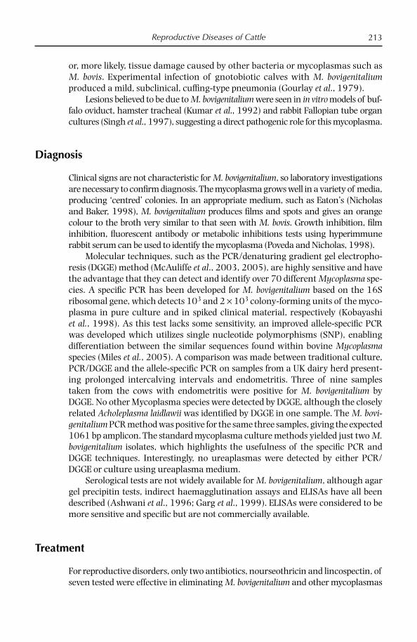
Reproductive Diseases of Cattle 213
or, more likely, tissue damage caused by other bacteria or mycoplasmas such as M. bovis. Experimental infection of gnotobiotic calves with M. bovigenitalium produced a mild, subclinical, cuffi ng-type pneumonia (Gourlay et al., 1979).
Lesions believed to be due to M. bovigenitalium were seen in in vitro models of buf-falo oviduct, hamster tracheal (Kumar et al., 1992) and rabbit Fallopian tube organ cultures (Singh et al., 1997), suggesting a direct pathogenic role for this mycoplasma.
Diagnosis
Clinical signs are not characteristic for M. bovigenitalium, so laboratory investigations are necessary to confi rm diagnosis. The mycoplasma grows well in a variety of media, producing ‘centred’ colonies. In an appropriate medium, such as Eaton’s (Nicholas and Baker, 1998), M. bovigenitalium produces fi lms and spots and gives an orange colour to the broth very similar to that seen with M. bovis. Growth inhibition, fi lm inhibition, fl uorescent antibody or metabolic inhibitions tests using hyperimmune rabbit serum can be used to identify the mycoplasma (Poveda and Nicholas, 1998).
Molecular techniques, such as the PCR/denaturing gradient gel electropho-resis (DGGE) method (McAuliffe et al., 2003, 2005), are highly sensitive and have the advantage that they can detect and identify over 70 different Mycoplasma spe-cies. A specifi c PCR has been developed for M. bovigenitalium based on the 16S ribosomal gene, which detects 103 and 2 × 10³ colony-forming units of the myco-plasma in pure culture and in spiked clinical material, respectively (Kobayashi et al., 1998). As this test lacks some sensitivity, an improved allele-specifi c PCR was developed which utilizes single nucleotide polymorphisms (SNP), enabling differentiation between the similar sequences found within bovine Mycoplasma species (Miles et al., 2005). A comparison was made between traditional culture, PCR/DGGE and the allele-specifi c PCR on samples from a UK dairy herd present-ing prolonged intercalving intervals and endometritis. Three of nine samples taken from the cows with endometritis were positive for M. bovigenitalium by DGGE. No other Mycoplasma species were detected by DGGE, although the closely related Acholeplasma laidlawii was identifi ed by DGGE in one sample. The M. bovi-genitalium PCR method was positive for the same three samples, giving the expected 1061 bp amplicon. The standard mycoplasma culture methods yielded just two M. bovigenitalium isolates, which highlights the usefulness of the specifi c PCR and DGGE techniques. Interestingly, no ureaplasmas were detected by either PCR/DGGE or culture using ureaplasma medium.
Serological tests are not widely available for M. bovigenitalium, although agar gel precipitin tests, indirect haemagglutination assays and ELISAs have all been described (Ashwani et al., 1996; Garg et al., 1999). ELISAs were considered to be more sensitive and specifi c but are not commercially available.
Treatment
For reproductive disorders, only two antibiotics, nourseothricin and lincospectin, of seven tested were effective in eliminating M. bovigenitalium and other mycoplasmas

Chapter 13214
without adversely affecting the semen (Pfützner, 1989). An assessment of some photosensitive agents, haematopophyrin, a derivative of this and thiopronine, to disinfect bovine semen spiked with various microbial agents including M. bovigen-talium showed little effectiveness for mycoplasma at concentrations harmless to the sperm (Eaglesome et al., 1994).
In a later study, 11 M. bovigenitalium isolates from UK herds were tested in vitro to assess their sensitivity to 20 different antimicrobials using a broth dilu-tion method (Ayling et al., 2005). The results, summarized in Table 13.2, showed that tilmicosin, clindamycin, oxytetracyclines and the fl uoroquinalones were effective in vitro but all or some strains were resistant or developing resistance to spectinomycin, chloramphenicol and lincomycin, among many others.
Prevention and Control
With the exception of the reproductive disorders, there is still a weak association between M. bovigenitalium and disease, including respiratory, arthritic and mas-titic diseases. The only feasible and practical means of control should be directed towards ensuring that mycoplasma-free semen is used for artifi cial insemination.
Table 13.2. In vitro antimicrobial sensitivity data for 11 strains of M. bovigenitalium (µg/ml).
Antibiotic MIC range MIC50 MIC90
Tilmicosin 0.06–0.12 0.06 0.12Enrofl oxacin 0.06–0.25 0.12 0.25Clindamycin < 0.12–0.25 0.12 0.25Ciprofl oxacin < 0.12–0.50 0.12 0.50Danofl oxacin 0.12–0.50 0.25 0.50Oxytetracycline 0.06–2.00 0.12 0.25Florfenicol 0.50–2.00 2.00 2.00Norfl oxacin 0.25–8.00 0.50 2.00Lincomycin 0.12–> 32.00 0.25 0.50Gentamycin 2.00–8.00 8.00 8.00Spectinomycin 2.00–32.00 4.00 8.00Chloramphenicol 1.00–32.00 2.00 32.00Rifampin 32.00–> 32.00 32.00 > 32.00Erythromycin < 0.12–> 32.00 > 32.00 > 32.00Amikacin 32.00–> 32.00 > 32.00 > 32.00Naladixic acid 32.00–> 32.00 > 32.00 > 32.00Streptomycin 32.00–> 32.00 > 32.00 > 32.00Tobramycin > 32.00–> 32.00 > 32.00 > 32.00Trimethoprim/sulfamethoxazole > 32/608–> 32/608 > 32/608 > 32/608Cephalothin >64.00–> 64.00 >64.00 >64.00
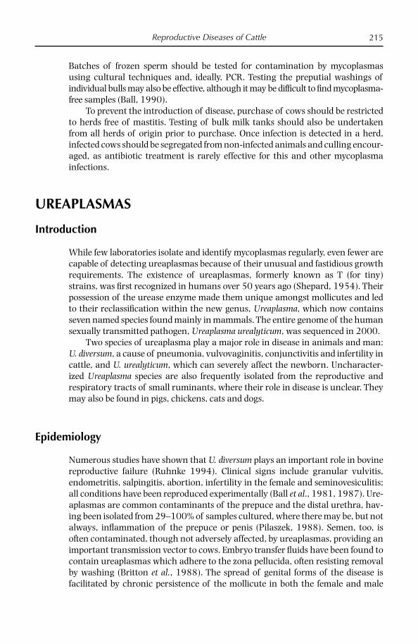
Reproductive Diseases of Cattle 215
Batches of frozen sperm should be tested for contamination by mycoplasmas using cultural techniques and, ideally, PCR. Testing the preputial washings of individual bulls may also be effective, although it may be diffi cult to fi nd mycoplasma-free samples (Ball, 1990).
To prevent the introduction of disease, purchase of cows should be restricted to herds free of mastitis. Testing of bulk milk tanks should also be undertaken from all herds of origin prior to purchase. Once infection is detected in a herd, infected cows should be segregated from non-infected animals and culling encour-aged, as antibiotic treatment is rarely effective for this and other mycoplasma infections.
UREAPLASMAS
Introduction
While few laboratories isolate and identify mycoplasmas regularly, even fewer are capable of detecting ureaplasmas because of their unusual and fastidious growth requirements. The existence of ureaplasmas, formerly known as T (for tiny) strains, was fi rst recognized in humans over 50 years ago (Shepard, 1954). Their possession of the urease enzyme made them unique amongst mollicutes and led to their reclassifi cation within the new genus, Ureaplasma, which now contains seven named species found mainly in mammals. The entire genome of the human sexually transmitted pathogen, Ureaplasma urealyticum, was sequenced in 2000.
Two species of ureaplasma play a major role in disease in animals and man: U. diversum, a cause of pneumonia, vulvovaginitis, conjunctivitis and infertility in cattle, and U. urealyticum, which can severely affect the newborn. Uncharacter-ized Ureaplasma species are also frequently isolated from the reproductive and respiratory tracts of small ruminants, where their role in disease is unclear. They may also be found in pigs, chickens, cats and dogs.
Epidemiology
Numerous studies have shown that U. diversum plays an important role in bovine reproductive failure (Ruhnke 1994). Clinical signs include granular vulvitis, endometritis, salpingitis, abortion, infertility in the female and seminovesiculitis; all conditions have been reproduced experimentally (Ball et al., 1981, 1987). Ure-aplasmas are common contaminants of the prepuce and the distal urethra, hav-ing been isolated from 29–100% of samples cultured, where there may be, but not always, infl ammation of the prepuce or penis (Pilaszek, 1988). Semen, too, is often contaminated, though not adversely affected, by ureaplasmas, providing an important transmission vector to cows. Embryo transfer fl uids have been found to contain ureaplasmas which adhere to the zona pellucida, often resisting removal by washing (Britton et al., 1988). The spread of genital forms of the disease is facilitated by chronic persistence of the mollicute in both the female and male
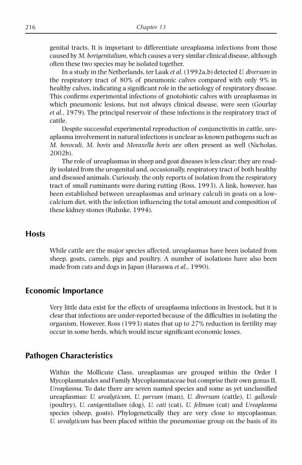
Chapter 13216
genital tracts. It is important to differentiate ureaplasma infections from those caused by M. bovigenitalium, which causes a very similar clinical disease, although often these two species may be isolated together.
In a study in the Netherlands, ter Laak et al. (1992a,b) detected U. diversum in the respiratory tract of 80% of pneumonic calves compared with only 9% in healthy calves, indicating a signifi cant role in the aetiology of respiratory disease. This confi rms experimental infections of gnotobiotic calves with ureaplasmas in which pneumonic lesions, but not always clinical disease, were seen (Gourlay et al., 1979). The principal reservoir of these infections is the respiratory tract of cattle.
Despite successful experimental reproduction of conjunctivitis in cattle, ure-aplasma involvement in natural infections is unclear as known pathogens such as M. bovoculi, M. bovis and Moraxella bovis are often present as well (Nicholas, 2002b).
The role of ureaplasmas in sheep and goat diseases is less clear; they are read-ily isolated from the urogenital and, occasionally, respiratory tract of both healthy and diseased animals. Curiously, the only reports of isolation from the respiratory tract of small ruminants were during rutting (Ross, 1993). A link, however, has been established between ureaplasmas and urinary calculi in goats on a low-calcium diet, with the infection infl uencing the total amount and composition of these kidney stones (Ruhnke, 1994).
Hosts
While cattle are the major species affected, ureaplasmas have been isolated from sheep, goats, camels, pigs and poultry. A number of isolations have also been made from cats and dogs in Japan (Haraswa et al., 1990).
Economic Importance
Very little data exist for the effects of ureaplasma infections in livestock, but it is clear that infections are under-reported because of the diffi culties in isolating the organism. However, Ross (1993) states that up to 27% reduction in fertility may occur in some herds, which would incur signifi cant economic losses.
Pathogen Characteristics
Within the Mollicute Class, ureaplasmas are grouped within the Order I Mycoplasmatales and Family Mycoplasmataceae but comprise their own genus II, Ureaplasma. To date there are seven named species and some as yet unclassifi ed ureaplasmas: U. urealyticum, U. parvum (man), U. diversum (cattle), U. gallorale (poultry), U. canigenitalium (dog), U. cati (cat), U. felinum (cat) and Ureaplasma species (sheep, goats). Phylogenetically they are very close to mycoplasmas; U. urealyticum has been placed within the pneumoniae group on the basis of its

Reproductive Diseases of Cattle 217
16S rRNA gene sequence. While similar to mycoplasmas in many respects, ureaplasmas are nutritionally unique, possessing the enzyme urease, which hydrolyses urea to generate ATP, probably through the creation of ionic gradients (Miles, 1992). Like mycoplasmas too, ureaplasmas are small, pleomorphic, lack a cell wall and have a low G+C ratio (Hermann, 1992). They do not hydrolyse arginine or ferment glucose in vitro but do require sterols for growth. In 2000 the entire genome of U. urealyticum was sequenced, providing unique data on these metabolically unique mollicutes (Glass et al., 2000). The circular genome, the second smallest after M. genitalium, consisted of 751,719 base pairs comprising only 652 genes, half of which have unknown functions. The genome size is considerable smaller than the 850–940 kbp estimate based on pulse fi eld gel elec-trophoresis. Other features of interest include a G+C ratio of 25.5%, the lack of the cell division protein, FtsZ, various chaperonins and ribonucleosidase diphos-phate reductase. Finally, the possession of six closely related iron transporters suggests that ureaplasmas have a type of respiratory system not present in other small genome bacteria.
Most U. diversum strains can be classifi ed into three serogroups, A, B and C, representing type strains A417, D48 and T44, respectively. Ter Laak et al. (1992b) identifi ed 82% of Dutch bovine respiratory isolates as serogroup A, while Howard and Gourlay (1981), in a study of strains from four countries, identifi ed 81% as serogroup B and only 15% as serogroup A. Four serotypes of ureaplasmas have so far been recognized in dogs.
The avian ureaplasma, U. gallorale, believed to be pathogenic, was shown to be more closely related to the human U. urealyticum than to the other animal ure-aplasmas on the basis of sequence homology of the 16S rRNA gene (Stemke and Robertson, 1996).
Disease Course
Acute granular vulvitis is characterized by an infl amed hyperaemic vulvar mucosa ,with small raised granules usually clustered around the clitoris. A purulent dis-charge may begin 3 days after infection, which may last up to 2 weeks when the disease becomes chronic, with a reduction in clinical signs; however, the ure-aplasma can be isolated from the vulva for up to 7 months (Ruhnke, 1994). There may also be a transient colonization of the uterus. During acute disease, the endo-metrium and/or Fallopian tubes may become permanently damaged, leading to infertility (Kreplin and Maitland, 1989). In some cases embryo death or abortion, usually in the fi rst or last trimester, stillbirth or extremely weak calves, which die shortly after birth with respiratory failure, may result as a consequence of ure-aplasma infection (Ruhnke, 1994).
In studies of heifers experimentally infected with broth cultures of U. diver-sum or vaginal swabs from previously infected heifers, all animals developed gran-ular vulvovaginitis; U. diversum was isolated from vagina, urinary bladder and urethra. The pathogenicity of U. diversum increased with consecutive passages, making the incubation period shorter and clinical signs more pronounced (Pilaszek and Truszczynski, 1991).

Chapter 13218
Pathology
Genital disease in the female is characterized by a purulent vulvar discharge and infl amed hyperaemic vulvar mucosa with varying degrees of granularity (Ross, 1993). Gross lesions in the placenta consist of marked fi brotic thickening of large areas of the amnion and intercotyledonary zones in the chorioallantoic mem-brane, infi ltrated by mononuclear cells. There may be foci of fi brin exudation and haemorrhage. In aborted fetuses, microscopic lesions of alveolitis and placentitis may be seen and antigen may be found in the zona pellucida of the embryo.
Respiratory disease involving ureaplasmas is largely subclinical. However, following endobronchial, but not aerosol, infection of gnotobiotic calves, thick cuffs of lymphoid cells and macrophages are seen surrounding the bronchi, bron-chioles and blood vessels, with a lobular catarrhal pneumonia evident (Gourlay et al., 1979; ter Laak et al., 1993a). Macroscopic lesions seen in U. diversum-associated respiratory disease consisted of red consolidation in the cranioventral areas of the lung (Ross 1993). In the lung there is a diffuse alveolitis and focal lymphocytic accumulations around the airways. The alveolitis is characterized by degenera-tion and necrosis of the alveolar epithelium and there is infi tration of macrophages and granulocytes (Ruhnke, 1994).
Diagnosis
Clinical signs of granular vulvitis are considered suggestive of ureaplasma infec-tions, while other signs such as subclinical pneumonia or conjunctivitis are far less defi nitive. Consequently laboratory diagnosis is necessary to determine the causative agent. Isolation of ureaplasmas in the genital tract is most successful from cultures of vulval swabs. The transient colonization of the uterus makes sampling of this region unreliable. Preputial swabs should be taken with a guarded swab to obtain the sample from as far into the cavity as possible. Semen should be collected following disinfection of the prepuce and should ideally be fresh or kept in liquid nitrogen until ready to culture. Preferred tissues from embryos include the lung, caruncle, cotyledon, stomach contents and amniotic fl uid.
From respiratory cases, nasal swabs, lung lavage fl uids and lung tissue can be taken. Ter Laak et al. (1992a) found considerably higher levels in lavage fl uids than in nasal swabs. The appropriate transport medium for samples is crucial, and antibiotics such as streptomycin and gentamycin, commonly used in many media, may prove harmful to ureaplasmas. Recommended media include sucrose phosphate broth (2SP) or standard liquid medium (SLM) (Furr, 1998).
Media recommended for the primary culture of U. diversum differ from that for other mycoplasmas as they contain 1% urea as a substrate instead of glucose or arginine; the pH should be adjusted to about 7.0. However, there are many variations in exact composition of ureaplasma media. Whitford et al. (1994) list the composition of several media which have given good results, including U4 buffered broth medium. Ter Laak et al. (1992a) used Friis media HAU and NHU, and A7B agar medium, which enabled the production of manganese dioxide as a brown deposit when urea was hydrolysed; however, HAU and NHU were superior

Reproductive Diseases of Cattle 219
to A7B when used in parallel. Commercial media are now available for ureaplasma isolation (Mycoplasma Experience, Reigate, UK).
Once isolated, ureaplasmas can be identifi ed by the production of brown colo-nies on agar medium containing manganese sulphate caused by the deposition of manganese on the surface of the colony. Isolates can be serotyped by immuno-fl uorescence on colonies grown on agar medium (Howard and Gourlay, 1981).
A PCR based on the 16S rRNA gene was more sensitive and rapid than cul-ture for detecting U. diversum in vaginal swabs of cattle (Cardoso et al., 2000). This test offers a lot of advantages over traditional culture techniques. The PCR/DGGE not only offers sensitive detection but enables the differentiation of ureaplasma from M. bovigenitalium, which may be found in the same tissue sample (McAuliffe et al., 2003, 2005).
Serological tests are not widely available for ureaplasmas and may be compli-cated by the existence of different serogroups. However, an indirect haemaggluti-nation test was described for U. diversum by Nagamoto et al. (1996).
Disease Treatment
Little information is available on chemotherapeutic strategies for control of ureaplasma infections. An in vitro study of antibiotic sensitivity to 17 strains of U. diversum in the Netherlands showed the most effective (< 1 µg/ml) to be chlor-tetracycline, tiamulin and tylosin; antibiotics showing intermediate effectiveness (2–4 µg/ml) included oxytetracycline and enrofl oxacin; those to which ureaplas-mas were resistant (> 8 µg/ml) included chloramphenicol, spectinomycin and lincomycin (ter Laak et al., 1993b).
In the fi eld, Ball and McCaughey (1984) successfully eliminated ureaplasmas from the urogenital tract of most, but not all, infected sheep with tiamulin or oxytetracycline. In a study by Rae et al. (1993), prebreeding treatment with chlor-tetracycline appeared to improve pregnancy rates in beef heifers with U. diversum infections, although there was only a slight decrease in colonization rates of the vagina by ureaplasmas in treated groups.
There was marked and rapid clinical improvement in 2-year-old heifers and bulls with granular vulvitis and ulcerative posthitis following a 5-day intramus-cular course of tylosin (Gummow et al., 1992). Ureaplasmas could not be cultured from the external genitalia of either heifers or bulls following clinical recovery, which began 3 days after treatment.
Disease Prevention and Control
Treatment of semen with combinations of gentamycin, lincospectin and tylosin appears to prevent spread of ureaplasmas and mycoplasmas according to one source (Ruhnke, 1994); however, culling of persistently infected bulls may some-times be necessary (Reid et al., 1989).
The application of three photosensitive agents followed by irradiation for dis-infection of bovine semen had no effect on U. diversum at concentrations harmless to sperm (Eaglesome et al., 1994).

Chapter 13220
No commercial vaccines are presently available for infections caused by ureaplasmas. However, an experimental vaccine provoked some immunity. Mulira et al. (1992) measured antibody levels in serum and cervicovaginal mucus (CVM) of heifers vaccinated with killed U. diversum strain 2312 in adju-vant; control heifers were given a placebo before both groups were challenged intravaginally. Vaccination stimulated specifi c IgG1 and IgG2 responses in the serum and CVM but only a slight IgM and no IgA response. It was concluded that vaccination stimulated a specifi c, albeit non-protective, IgG response in serum and CVM.
While the role of ureaplasmas in respiratory infections is slight or not proven, control measures which reduce environmental stress, including ensuring ade-quate housing with good circulation of air and adopting ‘all in, all out’ practices wherever possible, should have benefi cial effects. If this is not possible, separation of calves from the adults is advisable at the earliest opportunity where endemic disease exists.
Although survival time of U. diversum in the environment, such as fl oors and slurry, does not exceed 24 h, Pilaszek and Truszczynski (1991) showed that it was suffi cient to infect preputial mucosa in bulls but not heifers. These workers also showed that asymptomatic carriers can shed U. diversum with urine for pro-longed periods. It was concluded that disinfection of cowsheds and treatment of carriers with antibiotics could play an important role in prevention of ureaplasma infection.
References
Ahmed, A.A. (1987) Mycoplasma species isolated from mastitic dairy herd. Agricultural Research Review 65, 945–950
Ashwani, K., Garg, D.N. and Kumar, A. (1996) Detection of serum antibodies to Myco-plasma bovis and M. bovigenitalium in mastitic cows and buffaloes by ELISA, indirect haemagglutination test and agar-gel-immunoprecipitation test. Indian Veterinary Journal 73, 603–606.
Ayling, R.D., Bisgaard-Frantzen, S., March, J.B., Godinho, K. and Nicholas, R.D. (2005) Assessing the in vitro effectiveness of antimicrobials against Mycoplasma mycoides subsp. mycoides small colony type to reduce infections of contagious bovine pleuro-pneumonia. Antimicrobial Agents and Chemotherapy 49, 12.
Ball, H.J. (1990) Use of bovine sheath washings for screening for mycoplasmas. Veterinary Record 127, 16–17.
Ball, H.J. and McCaughey, W.J. (1984) Investigations into the elimination of ureaplasmas from the urogenital tract of ewes. British Veterinary Journal 140, 292–299.
Ball, H.J., McCaughey, W.J., Mackie, D.P. and Pearson, G.R. (1981) Experimental genital infection of heifers with ureaplasmas. Research in Veterinary Science 30, 312–317.
Ball, H.J., Armstrong, D., Kennedy, S. and McCaughey, W.J. (1987) Experimental intrauter-ine inoculation of cows at oestrus with a bovine ureaplasma strain. Irish Veterinary Journal 41, 371–372.
Baumgartner, B. (1999) Diagnostik und Bekampfung von Mykoplasmen-Mastitiden des Rindes im westlichen Teil des Landes Brandenburg – ein Ruckblick. [Diagnosis and control of bovine mastitis caused by Mycoplasma spp. in the western region of Brandenburg – a retrospective study.] Praktische-Tierarzt 80, 896–898.
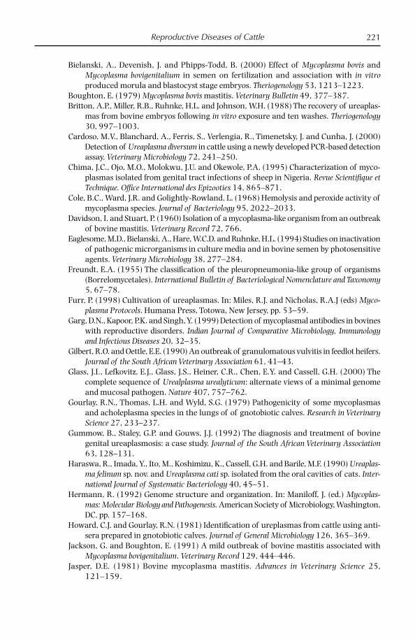
Reproductive Diseases of Cattle 221
Bielanski, A., Devenish, J. and Phipps-Todd, B. (2000) Effect of Mycoplasma bovis and Mycoplasma bovigenitalium in semen on fertilization and association with in vitro produced morula and blastocyst stage embryos. Theriogenology 53, 1213–1223.
Boughton, E. (1979) Mycoplasma bovis mastitis. Veterinary Bulletin 49, 377–387.Britton, A.P., Miller, R.B., Ruhnke, H.L. and Johnson, W.H. (1988) The recovery of ureaplas-
mas from bovine embryos following in vitro exposure and ten washes. Theriogenology 30, 997–1003.
Cardoso, M.V., Blanchard, A., Ferris, S., Verlengia, R., Timenetsky, J. and Cunha, J. (2000) Detection of Ureaplasma diversum in cattle using a newly developed PCR-based detection assay. Veterinary Microbiology 72, 241–250.
Chima, J.C., Ojo, M.O., Molokwu, J.U. and Okewole, P.A. (1995) Characterization of myco-plasmas isolated from genital tract infections of sheep in Nigeria. Revue Scientifi que et Technique. Offi ce International des Epizooties 14, 865–871.
Cole, B.C., Ward, J.R. and Golightly-Rowland, L. (1968) Hemolysis and peroxide activity of mycoplasma species. Journal of Bacteriology 95, 2022–2033.
Davidson, I. and Stuart, P. (1960) Isolation of a mycoplasma-like organism from an outbreak of bovine mastitis. Veterinary Record 72, 766.
Eaglesome, M.D., Bielanski, A., Hare, W.C.D. and Ruhnke, H.L. (1994) Studies on inactivation of pathogenic microrganisms in culture media and in bovine semen by photosensitive agents. Veterinary Microbiology 38, 277–284.
Freundt, E.A. (1955) The classifi cation of the pleuropneumonia-like group of organisms (Borrelomycetales). International Bulletin of Bacteriological Nomenclature and Taxonomy 5, 67–78.
Furr, P. (1998) Cultivation of ureaplasmas. In: Miles, R.J. and Nicholas, R.A.J (eds) Myco-plasma Protocols. Humana Press, Totowa, New Jersey, pp. 53–59.
Garg, D.N., Kapoor, P.K. and Singh, Y. (1999) Detection of mycoplasmal antibodies in bovines with reproductive disorders. Indian Journal of Comparative Microbiology, Immunology and Infectious Diseases 20, 32–35.
Gilbert, R.O. and Oettle, E.E. (1990) An outbreak of granulomatous vulvitis in feedlot heifers. Journal of the South African Veterinary Association 61, 41–43.
Glass, J.I., Lefkovitz, E.J., Glass, J.S., Heiner, C.R., Chen, E.Y. and Cassell, G.H. (2000) The complete sequence of Urealplasma urealyticum: alternate views of a minimal genome and mucosal pathogen. Nature 407, 757–762.
Gourlay, R.N., Thomas, L.H. and Wyld, S.G. (1979) Pathogenicity of some mycoplasmas and acholeplasma species in the lungs of of gnotobiotic calves. Research in Veterinary Science 27, 233–237.
Gummow, B., Staley, G.P. and Gouws, J.J. (1992) The diagnosis and treatment of bovine genital ureaplasmosis: a case study. Journal of the South African Veterinary Association 63, 128–131.
Haraswa, R., Imada, Y., Ito, M., Koshimizu, K., Cassell, G.H. and Barile, M.F. (1990) Ureaplas-ma felinum sp. nov. and Ureaplasma cati sp. isolated from the oral cavities of cats. Inter-national Journal of Systematic Bacteriology 40, 45–51.
Hermann, R. (1992) Genome structure and organization. In: Maniloff, J. (ed.) Mycoplas-mas: Molecular Biology and Pathogenesis. American Society of Microbiology, Washington, DC, pp. 157–168.
Howard, C.J. and Gourlay, R.N. (1981) Identifi cation of ureplasmas from cattle using anti-sera prepared in gnotobiotic calves. Journal of General Microbiology 126, 365–369.
Jackson, G. and Boughton, E. (1991) A mild outbreak of bovine mastitis associated with Mycoplasma bovigenitalium. Veterinary Record 129, 444–446.
Jasper, D.E. (1981) Bovine mycoplasma mastitis. Advances in Veterinary Science 25, 121–159.
Chapter 13222
Kobayashi, H., Hirose, K., Worarach, A., Paugtes, P., Ito, N., Morozumi, T. and Yamamoto, K. (1998) In vitro amplifi cation of the 16S rRNA genes from Mycoplasma bovirhinis, Mycoplasma alkalescens and Mycoplasma bovigenitalium by PCR. Journal of Veterinary Medical Science 60, 1299–1303.
Kreplin, C.M.A. and Maitland, V.F. (1989) Abortion due to Ureaplasma diversum. Canadian Veterinary Journal 30, 435.
Kreusel, S., Bocklisch, H., Pfützner, H., Brys, A., Leirer, R. and Ziegenhals, U. (1989) Ex-perimentelle Infektionen von Bullen mit Mycoplasma bovis und M. bovigenitalium. [Experimental infection of bulls with Mycoplasma bovis or M. bovigenitalium.] Archiv fur Experimentelle Veterinarmedizin 43, 705–712.
Kumar, A., Garg, D.N. and Mahajan, S.K. (1992) Experimental pathogenicity of Mollicutes of bovine udder origin in hamster tracheal ring organ culture. Indian Journal of Experimental Biology 30, 607–610.
Mazzolini, E., Ceccato, C., Agnoletti, F. and Friso, S. (1993) Artriti da micoplasmi in vitel-loni da carne. [Mycoplasmal arthritis in feedlot cattle.] Summa 10, 61–63.
McAuliffe, L., Ellis, R.J., Ayling, R.D. and Nicholas, R.A.J. (2003) Differentiation of Mycoplasma species by 16S rDNA PCR and DGGE fingerprinting. Journal of Clinical Microbiology 41, 4844–4847.
McAuliffe, L., Ellis, R.J., Lawes, J.R., Ayling, R.D. and Nicholas, R.A.J. (2005) 16S rDNA PCR and DGGE, a single generic test for detecting and differentiating Mycoplasma species. Journal of Medical Microbiology 54, 1–9.
Miles, K., McAuliffe, L., Ayling, R.D. and Nicholas R.A.J. (2005) Rapid detection for Myco-plasma dispar and M. bovirhinis using an allele specific PCR. Veterinary Research Communications 241, 103–107.
Miles, R.J. (1992) Cell nutrition and growth. In: Maniloff, J. (ed.) Mycoplasmas: Molecular Biology and Pathogenesis. American Society of Microbiology, Washington, DC, pp. 23–40.
Mulira, G.L. Saunders, J.R. and Barth, A.D. (1992) Isolation of Ureaplasma diversum and mycoplasmas from genital tracts of beef and dairy cattle in Saskatchewan. Canadian Veterinary Journal 33, 46–49.
Nagamoto, H., Shimizu, T., Higashiyama, Y., Yano, Y., Kuroki, H. and Hamana, K. (1996) Antibody response to Mycoplasma bovis of calves introduced in a farm contaminated with the organism. Journal of Veterinary Medical Science 58, 919–920.
Naglic, T., Sankovic, F., Madic, J., Hajsig, D., Seol, B. and Busch, K. (1996) Mycoplasmas associated with bovine conjunctivitis and keratoconjunctivitis. Acta Veterinaria Hungarica 44, 21–24.
Nicholas, R.A.J. (2002a) Mycoplasma bovigenitalium. Animal Health Compendium, CABI, Wallingford, UK. www.cabi.org
Nicholas, R.A.J. (2002b) Ureaplasmas. Animal Health Compendium. CABI, Wallingford, UK. www.cabi.org.uk
Nicholas, R. and Baker, S. (1998) Recovery of mycoplasmas from animals. In: Miles, R. and Nicholas, R. (eds) Methods in Molecular Biology. Mycoplasma Protocols. Humana Press, Totowa, New Jersey, pp. 37–43.
Nicholas, R.A.J., Wessels, M., Orme, P.K., Wood, E. and Sachse, K. (1999) Isolation of Mycoplasma ovine/caprine serogroup 11 from infertile sheep in Britain. Veterinary Record 145, 434–435.
Nicholas, R.A.J., Khan, L., Houshaymi, B., Miles, R.J., Ayling, R.D., Hotzel, H. and Sachse, K. (2002) Close genetic and phenotypic relatedness between Mycoplasma ovine/caprine serogroup 11 and M. bovigenitalium. Systematic and Applied Microbiology 25, 396–402.
Nicholas, R.A.J., Lin, Y.-C., Sachse, K., Hotzel, H., Parham, K., McAuliffe, L., Kelly, D.P. and Wood, A.P. (2008) Proposal that the strains of the Mycoplasma ovine/caprine serogroup

Reproductive Diseases of Cattle 223
11 be reclassifi ed as Mycoplasma bovigenitalium. International Journal of Systematic and Evolutionary Microbiology 58, 308–312.
Pathak, R.C., Singh, P.P. and Kappoor, S.G. (1993) Prevalence and pathogenicity of Myco-plasma bovigenitalium in goats. Études et Syntheses de l’IEMVT 42, 381.
Pfützner, H. (1989) Untersuchungen zur “in-vitro”-Wirkung von Antibiotika auf Myko-plasmen unter den Bedingungen der Aufbereitung von Bullensperma. [In vitro activity of antibiotics against mycoplasmas in bull semen processed for AI.] Archiv fur Experi-mentelle Veterinarmedizin 43, 725–728.
Pilaszek, J. (1988) Wystepowanie drobnoustrojow Mycoplasmatales w narzadzie rozrodc-zym u bydla. I. Izolacja szczepow od buhajow. [Occurrence of Mycoplasmatales in the reproductive tract of cattle. I. Isolation from bulls]. Medycyna Weterynaryjna 44, 369–372.
Pilaszek, J. and Truszczynski, M. (1991) Proby doswiadczalnego zakazenia narzadu roz-rodczego jalowek drobnoustrojami Ureaplasma diversum. [Experimental infections of the reproductive tract of heifers with Ureaplasma diversum]. Medycyna Weterynaryjna 47, 466–468.
Poumarat, F., Longchambon, D. and Martel, J.L. (1992) Application of dot immunobinding on membrane fi ltration (MF dot) to the study of relationships within ‘M. mycoides cluster’ and within ‘glucose and arginine-negative cluster’ of ruminant mycoplasmas. Veterinary Microbiology 32, 375–390.
Poveda, J.B. and Nicholas, R.A.J. (1998) Serological identifi cation by growth and metabo-lism inhibition tests. In: Miles, R. and Nicholas, R. (eds) Methods in Molecular Biology. Mycoplasma Protocols. Humana Press, Totowa, New Jersey, pp.105–112.
Rae, D.O., Chenoweth, P.J., Brown, M.B., Genho, P.C., Moore, S.A. and Jacobsen, K.E. (1993) Reproductive performance of beef heifers: effects of vulvo-vaginitis, Ureaplasma diver-sum and prebreeding antibiotic administration. Theriogenology 40, 497–508.
Reid, S.W., Madhill, D.G. and Vreugdenhil, A.H. (1989) Ureaplasmal vulvovaginitis and infertility in eight southern Ontario dairy herds. Canadian Veterinary Journal 30, 255.
Roberts, D.H. (1968) Immunological response in cows to Mycoplasma bovigenitalium strain 1836. Journal of Hygiene (Cambridge) 66, 585–593.
Ross, R.F. (1993) Mycoplasmas – animal pathogens. In: Kahane, I. and Adoni, A. (eds) Rapid Diagnosis of Mycoplasmas. Plenum Press, New York, pp. 69–110.
Ruhnke, H.L. (1994) Mycoplasmas associated with bovine genital tract infections. In: Whitford, H.W., Rosenbusch, R.F. and Lauerman, L.H. (eds) Mycoplasmosis in Animals. Iowa State University Press, Ames, Iowa, pp. 56–61.
Shepard, M.C. (1954) The recovery of pleuropneumonia-like organisms from negro men with and without nongonococcal urethritis. American Journal of Syphillus 38, 113–124.
Singh, Y., Garg, D.N., Mahajan, S.K. and Kapoor, P.K. (1997) Experimental mastitogenicity of Mollicutes originated from bovines with reproductive disorders in rat mammary gland model. Indian Journal of Animal Sciences 67, 373–375.
Stemke, G.W. and Robertson, J.A. (1996) Ureaplasma gallorale, an isolate from chickens, is most closely related to the human isolate, U. urealyticum. International Journal of Systematic Bacteriology 46, 1183–1184.
ter Laak, E.A., Noordergraaf, J.H. and Dieltjes, R.P.J.W. (1992a) Prevalence of mycoplas-mas in the respiratory tracts of pneumonic calves. Journal of Veterinary Medicine, Series B 39, 553–562.
ter Laak, E.A., Noordergraaf, J.H. and Boomsluiter, E. (1992b) The nasal mycoplasmal fl ora of healthy calves and cows. Journal of Veterinary Medicine, Series B 39, 610–616.
ter Laak, E.A., Noordergraaf, J.H. and Verschure, M.H. (1993a) Susceptibilities of Myco-plasma bovis, Mycoplasma dispar and Ureaplasma diversum strains to antimicrobial agents in vitro. Antimicrobial Agents and Chemotherapy 37, 317–321.

Chapter 13224
ter Laak, E.A., Dijk. J.E. and van Noordergraaf, J.H. (1993b) Comparison of pathological signs of disease in specifi c-pathogen-free calves after inoculation of the respiratory tract with Ureaplasma diversum or Mycoplasma canis. Journal of Comparative Pathology 108, 121–132.
Whitford, H.W., Rosenbusch, R.F. and Lauerman, L.H. (1994) Mycoplasmosis in Animals. Iowa State University Press, Ames, Iowa, pp. 144–145.
Zoldag, L., Stipkovits, L., Thuroczy, J. and Balogh, L. (1993) Mycoplasma izolalas szapor-odasbiologiai zavarokat mutato es egeszseges kutyak nemi szerveibol. [Isolation of mycoplasmas from the genitalia of healthy dogs and those with reproductive disorders.] Magyar Allatorvosok Lapja, 48, 356–359.

© CAB International 2008. Mycoplasma Diseases of Ruminants(R. Nicholas et al.) 225
14 New, Emerging and Unculturable Mycoplasmas in Ruminants
“… it is to be expected that as our understanding of mollicute nutrition increases, improved isolation media will enable growth of hitherto unsuspected species.” Roger Miles, Kings College, London (1992).
Introduction
Increases in the isolations of new or unusual mollicutes from ruminants are seen from time to time; the reason is not always clear but may be due to the introduc-tion of any affected animal from another country or region, leading to a rapid dissemination in the susceptible animal population, such as may have been seen with M. canis in cattle. The sudden appearance of M. canis in the UK in the mid-1990s represented a real increase in isolations because the species grows quickly and would almost certainly have been detected previously by traditional culture methods. The possibility that the mycoplasmas M. bovis or M. bovigenitalium may have been introduced through infected semen cannot be discounted. However, with other more fastidious mollicutes, such as M. dispar, the use of improved media formulations (Ayling et al., 2004) or molecular detection methods such as PCR would account for an increased prevalence. Genetic-based methods have certainly overcome the problems of serological cross-reactions seen with immu-nologically related species but are also prone to misidentifi cation because of the close homology of the 16S rDNA sequence of some mycoplasmas, which is the target of many PCR tests.
Mycoplasma Transmission between Animals and Man
Mycoplasmas are ubiquitous throughout the animal kingdom and virtually every mammal, bird, reptile, amphibian and fi sh that has been examined for
Chapter 14226
mycoplasmas has revealed unique species. Although it is often assumed that Mycoplasma species are extremely host specifi c, it is well known that some species, including M. bovis, M. arginini, M. agalactiae, M. mycoides SC and LC types and Acholeplasma laidlawii have a broader host range and can pass easily between sheep, goats and cattle and that M. gateae is found equally in cats and dogs.
Mycoplasmas are most likely to be acquired by their host as a result of intimate contact and transfer of material between mucosal surfaces, for example from dam to calf or through artifi cial insemination. It is possible that interspecies transfer may also occur on occasion, as between man and farm animals, but whether the foreign mycoplasma will survive for long in its new environment is not known.
Reports of infections in man with animal species of mycoplasma are rare, but infections probably occur more frequently than is generally recognized, the majority of cases remaining undiagnosed because so few laboratories are equip-ped to detect and identify mycoplasmas.
In some cases there is no certain evidence that the infecting organism was acquired from contact with another animal species. This is the case with M. fermentans, a frequent contaminant of laboratory cell lines. The species has been isolated from man on rare occasions but its DNA is very commonly detected by PCR in the synovial fl uid of patients with rheumatoid arthritis (Schaeverbeke et al., 1996) and has been associated with infections in patients with AIDS (Pitcher and Nicholas, 2005) and Gulf War Syndrome (Nicolson and Nicolson, 1996). Despite the apparent frequency with which M. fermentans is detected by PCR, no evidence of the species' natural habitat has been reported and it is far from certain that humans are its only host. Nicholas et al. (1998) reported the fi rst isolation of M. fermentans from genital infections in sheep; since then further isolations have been made from both rams and ewes with genital lesions (Fig. 14.1) in three other sheep fl ocks in England (Nicholas et al., 2000b). No route of trans-mission from sheep to man or vice versa has yet been identifi ed.
Ruminant species may colonize human body sites as secondary invaders without causing disease. Madoff et al. (1979) reported a case where M. bovis, a cause of mastitis, arthritis and respiratory disease in cattle, was isolated from the sputum of a woman with bronchopneumonia and central nervous system abnormalities. The only contact the patient had with cattle was exposure to cow manure while gardening 3 weeks before developing respiratory disease. There was serological evidence of M. pneumoniae but this species was not isolated. Attempts to identify M. bovis antibodies in the patient’s blood were unsuccessful and the disease subsided following tetracycline therapy. The patient’s symptoms were consistent with M. pneumoniae as the primary aetiological agent and the presence of M. bovis could not be explained. In another case of M. bovis infection in a human that responded to tetracycline treatment, there was no evidence of M. pneumoniae (Pitcher and Nicholas, 2005).
Mycoplasma infections are particularly common in immunocompromised patients, and cases of human infection with mycoplasmas normally inhabiting domestic animals are probably due to persistent and intimate contact with these susceptible patients. Armstrong et al. (1971) demonstrated colonization of the throat by M. canis in several family members who were in very close contact with
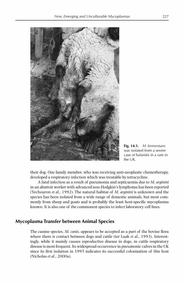
New, Emerging and Unculturable Mycoplasmas 227
their dog. One family member, who was receiving anti-neoplastic chemotherapy, developed a respiratory infection which was treatable by tetracycline.
A fatal infection as a result of pneumonia and septicaemia due to M. arginini in an abattoir worker with advanced non-Hodgkin’s lymphoma has been reported (Yechouron et al., 1992). The natural habitat of M. arginini is unknown and the species has been isolated from a wide range of domestic animals, but most com-monly from sheep and goats and is probably the least host-specifi c mycoplasma known. It is also one of the commonest species to infect laboratory cell lines.
Mycoplasma Transfer between Animal Species
The canine species, M. canis, appears to be accepted as a part of the bovine fl ora where there is contact between dogs and cattle (ter Laak et al., 1993). Interest-ingly, while it mainly causes reproductive disease in dogs, in cattle respiratory disease is most frequent. Its widespread occurrence in pneumonic calves in the UK since its fi rst isolation in 1995 indicates its successful colonization of this host (Nicholas et al., 2000a).
Fig. 14.1. M. fermentans was isolated from a severe case of balanitis in a ram in the UK.

Chapter 14228
The presence of the goat pathogen M. mycoides LC in cattle has been described previously but was thought to be avirulent for this species (Nicholas and Bashiruddin, 1995). However, the spread of LC from goats to calves through infected unpasteurized goat milk in New Zealand led to severe outbreaks of polyarthritis, septicaemia and deaths (Jackson and King, 2002). LC and M. capricolum subsp. capricolum have also been detected serologically in South American camelids (Hung et al., 1991; Nicholas, 1998), but isolates of these mycoplasmas from these animals remain elusive.
M. bovis, a major cause of calf pneumonia and mastitis, was originally classifi ed as a subspecies of the small ruminant pathogen M. agalactiae because of its close immunological and biochemical relationship; however, distinct genetic differences, particularly in the uvrC gene, have been exploited to develop a PCR capable of distinguishing the two species (Subramaniam et al., 1998). This PCR was used to identify M. bovis in goats with mastitis in the UK (Ayling et al., 2004); without this test there would have been real concerns that the UK was experi-encing its fi rst case of contagious agalactia.
Since its fi rst isolation in 1972 in Australia (Carmichael et al., 1972) the unclassifi ed mycoplasma M. ovine/caprine serogroup 11 has been found sporadi-cally in sheep and goats with a variety of clinical conditions, including infertility and vulvovaginitis (Nicholas et al., 2002). The biochemically similar M. bovigeni-talium causes similar conditions in cattle. In a comparison of strains from these two species, Nicholas et al. (2002, 2008) demonstrated very close genetic, biochemical and immunological relationships between the two species and recommended that the M. serogroup 11 should be reclassifi ed as M. bovigenita-lium.
One of the recommended sampling sites for pathogenic mycoplasmas in sheep and goats is the ear canal and it is believed to be due to the presence of ear mites (Psoroptes cuniculi or Railleria caprae), from which mycoplasmas have been isolated (Da Massa and Brooks, 1991). Goat fl eas (order Siphonaptera) too have been implicated in disease transmission; Nayak and Bhowmik (1990) showed that the blood taken from polyarthritic goat kids contained up to 105 organisms/ ml of viable M. mycoides LC organisms, and when fl eas were placed on unaffected kids polyarthritis and septicaemia followed. What is not clear from either study is whether this represents just the passive carriage of mycoplasmas from the blood of acutely affected animals or whether mites and fleas act as reser-voirs in which mycoplasmas replicate. Further study is necessary to clarify these fi ndings.
Unculturable Mycoplasmas
Mycoplasma wenyonii is a wall-less haemotrophic prokaryote previously classifi ed as Eperythrozoon wenyonii in the order Rickettsiales, but recently assigned to the Mycoplasma genus on the basis of 16S rDNA analysis (Neimark et al., 2001). M. wenyonii adheres to erythrocytes but may also be found in plasma unattached to erythrocytes (Neimark et al., 2001). Scanning electron microscopic analysis of M. wenyonii-infected blood shows deformed erythrocytes with invaginations and

New, Emerging and Unculturable Mycoplasmas 229
the presence of either rod- or coccoid-shaped organisms embedded in the membrane of erthrocytes (Neimark et al., 2001; Bretana et al., 2002). Although the mode of transmission is unknown, there is evidence that fl ies, lice and mos-quitoes may serve as mechanical vectors and oral transmission also seems likely (Smith et al., 1990; Prullage et al., 1993; Hofmann-Lehmann et al., 2004). While the precise role of M. wenyonii in disease is still controversial, infections caused by M. wenyonii have been reported to result in parasitaemia and anaemia, with infected cattle rarely dying but on occasions showing acute clinical signs (Smith et al., 1990). Scrotal and hind limb oedema, tachycardia, pyrexia and infertility were reported in a Charolais bull in the USA that was chronically infected with M. wenyonii (Montes et al., 1994). In addition, dairy cows with swollen teats, oedema of the distal portion of the hind limbs, transient fever, prefemoral lymphadeno-pathy, rough coat, dramatically decreased milk production and subsequent infer-tility and weight loss have been reported (Smith et al., 1990). Recently, other as yet unclassifi ed haemotrophic mycoplasmas have been discovered in cattle in Switzerland with severe anaemia that were co-infected with M. wenyonii and Ana-plasma marginal (Hofmann-Lehmann et al., 2004); however, these organisms have not yet been found elsewhere.
Previously, diagnosis of M. wenyonii has been based on blood smears but these are not specific for M. wenyonii and can be difficult to interpret. The PCR and denaturing gradient gel electrophoresis (DGGE), which has previously been described for the detection and differentiation of culturable Mycoplasma species (McAuliffe et al., 2003, 2005), can be used on blood samples as a rapid and specifi c test for M. wenyonii and can also be used as a screening test for other blood-borne pathogens (McAuliffe et al., 2006).
Mycoplasma ovis, formerly Eperythrozoon ovis, is an reythrocytic agent that causes haemolytic anaemia in sheep and goats and it occurs worldwide. M. ovis was previously thought to be a rickettsia, however, phylogenetic analysis revealed that this wall-less bacterium is not a rickettsia but a mycoplasma. This myco-plasma is related closely to several other uncultivated, blood-borne mycoplasmas that comprise a recently identifi ed group, the haemotrophic mycoplasmas (hae-moplasmas). It is likely that M. ovis is present in many fl ocks causing asymptom-atic infection. In some instances the presence of bacterial infection progesses to disease, characterised by the destruction of red blood cells, leading to anaemia and jaundice. Goats can also be infected. Deaths may occur in severely affected young sheep, especially if they are stressed by handling or movement. Losses of up to 30% of the fl ock have occurred in these circumstances. As with other haemotrophic mycoplasmas, it is thought that M. ovis is transmitted mainly by blood-feeding arthropod vectors.
References
Armstrong, D., Yu, B.H., Yagoda, A. and Kagnoff, M.F. (1971) Colonization of humans by Mycoplasma canis. Journal of Infectious Diseases 124, 607–609.
Ayling, R.D., Bashiruddin, S.E. and Nicholas, R.A.J. (2004) Mycoplasmas and related organisms identifi ed from ruminants 1990–2000. Veterinary Record 155, 413–416.
Chapter 14230
Bretana, A., Nanez, B., Contreras-Bretana, M. and Giardina, S. (2002) Multiple infection in bovines from the tropics: observation of blood parasites by scanning and transmission electron microscope. Parasitologia 44, 173–178.
Carmichael, L.E., St George, T.D., Sullivan, N.D. and Horsfall, N. (1972) Isolation, propaga-tion and characterization studies of an ovine mycoplasma responsible for proliferative interstitial pneumonia. Cornell Veterinarian 62, 654–679.
Da Massa, A.J. and Brooks, D.L. (1991) The external ear canal of goats and other animals as a mycoplasma habitat. Small Ruminant Research 4, 85–93.
Hofmann-Lehmann, R., Meli M.L., Dreher U.M., Gonczi E., Deplazes P., Braun U., Engels M., Schupbach J., Jorger K., Thoma R., Griot C., Stark K.D., Willi B., Schmidt J., Kocan K.M. and Lutz, H. (2004) Concurrent infections with vector-borne pathogens associ-ated with fatal hemolytic anemia in a cattle herd in Switzerland. Journal of Clinical Microbiology 42, 3775–3780.
Hung, A.L., Alvarado, A., Lopez, T., Perales, R., Li, O. and Garcia, E. (1991) Detection of antibodies to mycoplasmas in South American camelids. Research in Veterinary Science 51, 250–253.
Jackson, R. and King, C. (2002) Mycoplasma mycoides subsp. mycoides (large colony) infec-tion in goats – a review with special reference to New Zealand. Surveillance 29, 8–12.
Madoff, S., Pixley, B.Q., DelGuidice, R.A. and Moellering, R.C. (1979) Isolation of Myco-plasma bovis from a patient with systemic illness. Journal of Clinical Microbiology 9, 709–711.
McAuliffe, L.M., Ellis, R., Ayling, R.D. and Nicholas, R.A.J. (2003) Detection and differen-tiation of Mycoplasma species by rDNA PCR and DGGE fi ngerprinting. Journal of Clinical Microbiology 41, 4844–4847.
McAuliffe, L., Ellis, R., Lawes, J., Ayling, R.D. and Nicholas, R.A.J. (2005) 16S rDNA and DGGE: a single generic test for detecting and differentiating Mycoplasma species. Journal of Medical Microbiology 54, 1–9.
McAuliffe, L., Lawes, J., Bell, S., Barlow, A., Ayling, R.D. and Nicholas, R.A.J. (2006) The detection of Mycoplasma (formerly Eperythrozoon) wenyonii by 16S rDNA and DGGE. Veterinary Microbiology 117, 292–296.
Montes, A.J., Wolfe, D.F., Welles, E.G., Tyler, J.W. and Tepe, E. (1994) Infertility associated with Eperythrozoon wenyonii infection in a bull. Journal of the American Veterinary Medical Association 204, 261–263.
Nayak, N.C. and Bhowmik, M.K. (1990) Goat fl ea (order Siphonaptera) as a possible vector for the transmission of caprine mycoplasmal polyarthritis with septicaemia. Preventative Veterinary Medicine 9, 259–266.
Neimark, H., Johansson, K.E., Rikihisa, Y. and Tully, J.G.(2001) Proposal to transfer some members of the genera Haemobartonella and Eperythrozoon to the genus Mycoplasma with descriptions of ‘Candidatus Mycoplasma haemofelis’, ‘Candidatus Mycoplasma haemo-muris’, ‘Candidatus Mycoplasma haemosuis’ and ‘Candidatus Mycoplasma wenyonii’. International Journal of Systematic and Evolutionary Microbiology 51, 891–899.
Nicholas, R.A.J. (1998) Surveillance for contagious agalactia in Great Britain. In: Leori, G., Santini, F., Scanziani, E. and Frey, J. (eds) Mycoplasmas of Ruminants: Pathogenicity, Diagnostics, Epidemiology and Molecular Genetics, Vol. 5. European Commission, Brussels, pp. 95–97.
Nicholas, R.A J. and Bashiruddin, J. B. (1995) Mycoplasma mycoides subsp. mycoides SC variant: the agent of contagious bovine pleuropneumonia and member of the Myco-plasma mycoides cluster. Journal of Comparative Pathology 113, 1–27.
Nicholas, R.A.J., Grieg, A., Baker, S., Ayling, R.D., Heltander, M., Johannsson, K.E., Houshaymi, B. and Miles, R.J. (1998) Isolation of Mycoplasma fermentans from a sheep. Veterinary Record 142, 220–221.
New, Emerging and Unculturable Mycoplasmas 231
Nicholas, R.A.J., Baker, S.E., Ayling, R.D. and Stipkovits, L. (2000a) Mycoplasma infections in growing cattle. Cattle Practice 8, 115–118.
Nicholas, R.A.J., Ayling, R.D. and Miles, R.J. (2000b) Studies on two unusual mycoplasmas isolated from the reproductive tract of sheep in Britain. In: Bergonier, D., Berthelot, X. and Frey, J. (eds) Mycoplasmas of Ruminants: Pathogenicity, Diagnostics, Epidemiology and Molecular Genetics, Vol. 4. European Commission, Brussels, pp. 223–226.
Nicholas, R.A.J., Khan, L., Houshaymi, B., Miles, R.J., Ayling, R.D., Hotzel, H. and Sachse, K. (2002) Close genetic and phenotypic relatedness between Mycoplasma ovine/caprine serogroup 11 and M. bovigenitalium. Systematic and Applied Microbiology 25, 396–402.
Nicholas, R.A.J., Lin, Y.-C., Sachse, K., Hotzel, H., Parham, K., McAuliffe, L., Kelly, D.P. and Wood, A.P. (2008) Proposal that the strains of the Mycoplasma ovine/caprine sero-group 11 be reclassifi ed as Mycoplasma bovigenitalium. International Journal of Systematic and Evolutionary Microbiology 58, 308–312.
Nicolson, G.L. and Nicolson, N.L. (1996) Diagnosis and treatment of mycoplasmal infec-tion in Persian Gulf War illness-CFIDS patients. International Journal of Occupational Medicine, Immunology and Toxicology 5, 69–78.
Pitcher, D. and Nicholas, R.A J. (2005) Mycoplasma host specifi city: fact or fi ction? The Veterinary Journal 170, 300–306.
Prullage, J.B., Williams, R.E. and Gaafar, S.M. (1993) On the transmission of Eperythrozoon suis by Stomoxys calcitrans and Aedes aegypti. Veterinary Parasitology 50, 125–135.
Schaeverbeke, T., Gilroy, C.B., Bébéar, C., Dehais, J. and Taylor-Robinson, D. (1996) Mycoplasma fermentans in joints of patients with rheumatoid arthritis and other joint disorders. Lancet 347, 1418.
Smith, J.A., Thrall, M.A., Smith, J.L., Salman, M.D., Ching, S.V. and Collins, J.K. (1990) Eperythrozoon wenyonii infection in dairy cattle. Journal of the American Veterinary Medical Association 196, 1244–1250.
Subramaniam, S., Bergonier, D., Poumerat, F., Capaul, S., Schlatter, Y., Nicolet, J. and Frey, J. (1998) Species identifi cation of Mycoplasma bovis and Mycoplasma agalactiae based on uvrC genes by PCR. Molecular and Cellular Probes 12, 161–169.
ter Laak, E.A., Tully, J.G., Noordergraaf, H.H., Rose, D.L., Carle, P., Bové, J.M. and Smits, M.A. (1993) Recognition of Mycoplasma canis part of the mycoplasma fl ora of the bovine respiratory tract. Veterinary Microbiology 34, 175–181.
Yechouron, A., Lefevre, J., Robson, H.G., Rose, D.L. and Tully, J.G. (1992) Fatal septicae-mia due to Mycoplasma arginini: a new human zoonosis. Clinical Infectious Disease 15, 434–438.

This page intentionally left blank

233
Index
Acholeplasma laidlawii 210Amplifi ed fragment length polymorphism
(AFLP)See typing, molecular and individual
Mycoplasma speciesAntimicrobial sensitivity testing 58–65
ATP-dependent luminometry 63contagious agalactia 190–193CBPP 92breakpoints 60E-tests 63factors affecting 61fl ow cytometry 63indicator systems 62–63M. bovigenitalium 213–214M. bovis 151M. mycoides subsp. capri/M. mycoides
subsp. mycoides LC 190–193M. mycoides subsp. mycoides SC 92mutation prevention
concentration 60–61minimum inhibitory concentrations
(MIC) 59–65, 92, 190–193mycoplasmacidal activity (MMC)
59–60, 92standardisation 63
Arthritiscontagious agalactia 101–103M. bovis 144–145M. mycoides subsp. capri/M. mycoides
subsp. mycoides LC 185–186
Atypical pneumoniaantimicrobial susceptibility testing
190–193biochemistry 176–1177causative organism 175–176clinical signs 172–173control 177–178diagnosis 174, 177epidemiology 171–173
molecular 171–172pathology 173–175PCR 177transmission 171–173treatment 177–178virulence factors 177
See also M. ovipneumoniae
Biofi lms 58, 140
Cattle, respiratory mycoplasmoses 132–168
M. alkalescens 159–160M. bovirhinis 157–159M. bovigenitalium 212M. bovis 133–154
arthritis 144–145brain isolation 147–148characteristics 139–141clinical signs 141–143

Index234
Cattle, respiratory mycoplasmoses continued
M. bovis continuedcomparison with M. mycoides subsp
mycoides SC 134diagnosis 148–149ELISA 149economic losses 135eye disease 145epidemiology 136–138
molecular 138–139host 135–136immunohistochemistry 149–150mastitis 144–146occurrence 135otitis 146–148pathology 141–144PCR 149samples 148survival 140treatment 149–152vaccines 153–154virulence factors 140
M. canis 160–161M. dispar 154–157
characteristics 155diagnosis 156–157pathology 155–156treatment 157
M. mycoides subsp. mycoides SC 69–97
U. diversum 218Chemotherapy
atypical pneumonia 177–178bovine respiratory mycoplasmoses
149–152caprine respiratory mycoplasmoses
189–190CBPP 90–92CCPP 128contagious agalactia 109infectious keratoconjunctivitis 202,
205reproductive diseases 213–214, 219See also antimicrobial sensitivity
testingContagious agalactia 98–113
antimicrobial susceptibility testing 190–193
causative organisms 106–107clinical signs 101–106
control 109diagnosis 107–109
biochemical tests 108DGGE 108differential 108–109ELISA 108immunoblotting 108isolation 107serology 107PCR 107
epidemiology 99molecular typing 100–101
hosts 99occurrence 99pathology 101–106treatment 109vaccines 109–111
Contagious bovine pleuropneumonia (CBPP) 53, 69–97, 133, 134
Africa 69–70, 84–85Asia 70, 85Australia 72, 84–85causative organism 82clinical signs 74–76control 87–90diagnosis 85–87
culture 86immunoblotting 86–87penside tests 86PCR 86, 89–90serology 85–86
economic impact 72–73epidemiology 83–85
molecular 83–85participatory 85
Europe 70–71, 84–85hosts 73immunology 89New Zealand 72origins 71–72pathogenesis 77pathology 77–81Portugal 86–87transmission 74–75treatment 90–92USA 72–73vaccines 85, 87–89
Contagious caprine pleuropneumonia (CCPP) 114–131, 170
biochemistry 126clinical signs 119–124
Index 235
control 128–129diagnosis 126–128epidemiology, molecular 125gazelles 123, 125history 114–115hosts 116immunology 124isolation 126–127occurrence 115–116pathology 119–124penside tests 128PCR 29–30, 127–128samples 126serology 128transmission 124–125treatment 128vaccines 128
Denaturing gradient gel electrophoresis (DGGE) 15–27
detectionavian mycoplasmas 18bovine mycoplasmas 20, 22canine mycoplasmas 20, 23CBPP 89–90clinical samples 17contagious agalactia 108equine mycoplasmas 23–24human mycoplasmas 18–19mixed culture 17porcine mycoplasmas 23, 25sea mammal mycoplasmas 21–22small ruminant mycoplasmas 21, 23
DNA extraction 16
Endometritissee M. bovigenitalium
Eperythrozoon wenyoniisee Mycoplasma wenyonii
Eye diseasesee infectious keratoconjunctivitis
Firmicutes 3
Gene sequencingsee typing, molecular and individual
Mycoplasma species
Goats, respiratory mycoplasmosessee also atypical pneumoniasee also contagious caprine
pleuropneumoniasee also diseases caused by M. mycoides
subsp. capri/M. mycoides subsp. mycoides LC
M. mycoides subsp. capri/M. mycoides subsp. mycoides LC 179–198
abortion 181antimicrobial susceptibility testing
190–193arthritis 185–186, 189biochemistry 184, 187camelids 181characteristics 181–185control 190CCPP 184, 186diagnosis 186–189
ELISA 187PCR/DGGE 188samples 187
economic impact 181epidemiology 180experimental infections 185–186hosts 180–181occurrence 180insects, role of 190MICs 190–193taxonomy 179treatment 189–190
Histophilus somni 132, 137, 208Host specifi city 225–228
Immunoblottingantigen preparation 53–54CBPP 56, 86–87SDS-PAGE 54sensitivity and specifi city 55–56
Infectious keratoconjunctivitiscontagious agalactia 102–104, 203M. agalactiae 102–104, 203M. bovis 134, 202M. bovoculi 199–202M. conjunctivae 199, 202–205
clinical signs 203–204control 205epidemiology 203–204
Index236
Infectious keratoconjunctivitis continuedcontagious agalactia continued
diagnosis 204–205treatment 205
M. mycoides subsp. capri/M. mycoides subsp. mycoides LC 185–186, 203
Insertion sequence analysissee typing, molecular and individual
Mycoplasma speciesInsect transmission 190
M. wenyonii 228–229
Latex agglutination testsCBPP 86CCPP 128
Mannheimia haemolytica 132–133, 150, 169, 171, 173
Mastitiscontagious agalactia 101–102M. agalactiae 101–102M. bovigenitalium 210M. bovine group 7 154M. bovis 144–145M. californicum 210
Minimum inhibitory concentrations (MIC) 59–65
Morexella bovis 202, 210Multilocus sequence techniques (MLST)
see typing, molecular and individual Mycoplasma species
Mycoplasmacidal activity (MMC) 59–60Mycoplasma sppM. agalactiae 7, 9, 11, 21, 117, 139, 179,
203antimicrobial susceptibility testing
190–193biochemical tests 108, 118comparison 182contagious agalactia 98–113ELISA 108immunoblotting 108isolation 108PCR 29–30, 31, 107–108PFGE 100RAPD 100real time PCR 29typing methods 47VNTR 100–101
vaccines 109–111Western (immuno) blotting 53
M. alvi 154M. anatis 20M. alkalescens 17, 18, 20, 133, 140,
159–160M. arginini 18, 21, 117, 118, 171, 173,
178, 182in humans 226
M. arthritidis 21M. bovigenitalium 7, 133, 140, 154,
208–215characteristics 211–212control 214–215diagnosis 213epidemiology 210hosts 211mastitis 212MIC 214occurrence 210pathology 212–213PCR 29, 32–33reproductive disease 208–215treatment 213–214
M. bovine group 7 29, 154MLST 48typing methods 47
M. bovirhinis 10, 17, 18, 20, 133, 140, 157–159
M. bovis 7, 9, 12, 20, 179AFLP 36–37biofi lm 140bovine respiratory disease 133–157characteristics 139–141in humans 226infectious keratoconjunctivitis 202insertion sequence analysis 44–46PCR 29, 31PFGE 40RAPD 37–40real time PCR 29reproductive disease 208, 210resistance to antimicrobials 58survival 140
typing methods 47, 138–139virulence factors 140see also cattle respiratory
mycoplasmosesM. bovoculi 20, 140, 154
characteristics 200clinical signs 200–201

Index 237
control 202diagnosis 201–202epidemiology 200occurrence 200treatment 202
M. buccale 21M. californicum 20, 140, 154, 210M. canadense 20, 140, 154M. canis 7, 20, 26, 133, 140, 154,
160–161in humans 226PCR 29, 32see also cattle respiratory disease
M. capricolum subsp. capricolum 7, 21, 28, 117, 171, 179
characterstics 182contagious agalactia 98–100MLST 48PCR 29–30
M. capricolum subsp. capripneumoniae 7, 9, 12, 21, 179
antimicrobial susceptibility testing 190–193
biochemistry 118, 126CCPP 114–131characteristics 116, 182DGGE 127gene sequencing 46medium 127PCR 29–30, 127–128typing methods 47, 125
M. cloacale 20M. columbinasale 20, 26M. columbinum 20, 26M. columborale 20M. conjunctivae 7, 18, 21, 117, 182, 199,
202–205clinical signs 203–204control 205diagnosis 204–205epidemiology 203–204gene sequencing 46PCR 29, 31, 205real time PCR 29treatment 205typing methods 47
M. cottewii 21, 23M. cynos 20, 26
typing methods 47M. dispar 7, 20, 133, 140, 154–157
DGGE 157
PCR 29, 157see also cattle respiratory
mycoplasmosesM. edwardii 20, 23, 26M. equigenitalium 20, 23, 24M. equirhinis 21, 23, 24M. fastidiosum 21, 23, 24M. faucium 21M. felis 21, 23, 24M. fermentans 21, 226
in sheep 226–227PCR 29
M. fl occulare 21, 23, 25M. gallinaceum 20M. gallinarum 20M. gallisepticum 20
AFLP 36gene sequencing 46typing methods 47
M. gallopavonis 20M. gateae 21, 25M. genitalium 21, 25M. glycophilum 20M. hominis 21
resistance to antimicrobialsM. hyopneumoniae 21, 23, 25, 30
PFGE 40gene sequencing 46typing methods 47
M. hyorhinis 21, 23, 25M. hyosynoviae 21, 23, 25
AFLPM. imitans 20M. iners 20M. iowae 20M. leopharyngis 23, 26M. lipofaciens 20M. lipophilum 21M. maculosum 20, 23, 26M. meleagridis 20M. mycoides cluster 28, 181
PCR 28–29MLST 48
M. mycoides subsp. capri 7, 21, 23, 25, 28, 170
biochemistry 118characteristics 181–185CCPP 115MLST 48PCR 29–30see also M. mycoides subsp. mycoides LC
Index238
see also goat respiratory mycoplasmoses
M. mycoides subsp. mycoides LC 7, 9, 10, 21, 23, 25, 28, 170, 179
biochemistry 118in calves 227–228comparison with M. mycoides subsp.
mycoides SC 183–185contagious agalactia 98–113MLST 48PCR 29–30in sheep 183typing methods 47vaccines 110see also M. mycoides subsp. caprisee also goat respiratory
mycoplasmosesM. mycoides subsp. mycoides SC 7, 9, 10,
11, 17, 20, 28, 134, 140, 171antimicrobial testing 92Asian strains 85African/Australian strains 43–44,
83–84CBPP 69–97characteristics 82comparison with M. bovis 134comparison with M. mycoides subsp.
mycoides LC 183–185European strains 43–44, 83–84epidemiology, molecular 83- 85
AFLP 36MLST 47, 84PFGE 84VNTR 41–44, 84see also contagious bovine
pleuropneumoniaPCR 29–30, 86
real time PCR 29typing methods 47virulence 82–83Western (immuno) blotting 53–57
M. ovipneumoniae 7, 9, 13, 18, 21, 117, 170
antimicrobial susceptibility testing 190–193
biochemistry 118, 176–177CCPP 124, 127characteristics 175–176, 182medium, vegetable 5,177PCR 29PFGE 40–42
typing methods 47virulence factors 177see also atypical pneumonia
M. ovine/caprine serogroup 11 21, 209, 211–212, 228
See also M. bovigenitaliumM. opalescens 20, 23M. orale 21M. phocicerebrale 21, 25M. phocidae 21M. phocirhinis 21M. pneumoniae 21, 25, 226
resistance to antimicrobials 59M. primatum 21M. pullorum 20M. putrefaciens 21, 23, 117, 179, 182
antimicrobial susceptibility testing 190–192
biochemistry 118contagious agalactia 98–101,
107–110PCR 29, 32
M. salivarum 21M. spumans 20, 23M. spermatophilum 21M. subdolum 21, 24M. synoviae 20
gene sequencing 46PFGE 40typing methods 47
M. verecundum 20, 154M. wenyonii 228–229M. yeatsii 21, 22Mycoplasma culture 5–6
collection of samples 6–9transport of samples 9
Mycoplasma growth 3Mycoplasma isolation 7–13
subculturing 10–11Mycoplasma medium 3–5
bacterial inhibitors 6PRM 5–6vegetable proteins 6, 155
Mycoplasma nutrition 3Mycoplasma resistance 58–59
Pasteurella multocida 132, 137Pasteurella trehalosi 169Polymerase chain reaction (PCR) 15–27,
28–34, 35

Index 239
see individual Mycoplasma species16S rDNA geneDGGE15–27DNA extraction 16false positive/negative 31–32limitations 30nested 31primers 16, 17real time 28sensitivity 30–31specifi city 30–31
Pulse fi eld gel electrophoresis (PFGE)see typing, molecular and individual
Mycoplasma species
Random amplifi ed polymorphic DNA anaysis (RAPD)
see typing, molecular and individual Mycoplasma species
Reproductive diseasessee M. bovigenitaliumsee M. fermentanssee Ureaplasmas
Sheep mycoplasma diseasessee atypical pneumoniasee contagious agalactiaesee infectious keratoconjunctivitis
Sodium dodecyl sulphate polyacrylamide electrophoresis (SDS) 54
Typing, molecular 35–52AFLP 36, 125, 138–139gene sequencing 46, 125insertion sequence analysis 44–46MLST 35, 46–48PFGE 35, 40–41, 100, 138–139,
171RAPD 37–40, 100, 138–139, 171
VNTR 41–44, 100
Unculturable mycoplasmas 228–229Ureaplasma diversum 7, 133, 140, 154,
reproductive disease 208, 210, 215–220
characteristics 216–217control 219–220diagnosis 218economic impact 216epidemiology 215–216hosts 216pathology 218PCR/DGGE 219treatment 219
respiratory disease 218Ureaplasmas 215–220
see also Ureaplasma diversum
VaccinesCBPP 85, 87–89CCPP 128contagious agalactia 109–111M. bovis
Variable number tandem repeat analysis (VNTR)
see typing, molecular and individual Mycoplasma species
Viruses 132–133, 150, 170Vulvitis, granular
see M. bovigenitaliumsee U diversum
Vulvovaginitissee M. bovigenitaliumsee U. diversum
Western (immuno) blottingsee immunoblotting
Zoonoses 225–227