myelography 9/30/10 online ed.. central nervous system 2 basic parts- what are they? brain spinal...
TRANSCRIPT

MyelographyMyelography
9/30/10 online ed.

Central nervous systemCentral nervous system
2 basic parts- 2 basic parts- What are they?What are they?
BrainBrain
Spinal CordSpinal Cord
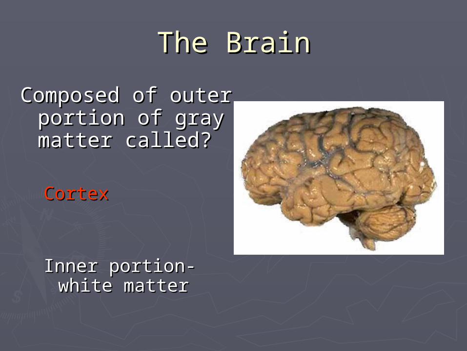
The BrainThe Brain
Composed of outer Composed of outer portion of gray portion of gray matter called?matter called?
CortexCortex
Inner portion- white Inner portion- white mattermatter

The BrainThe Brain
► Forebrain-largest part Forebrain-largest part of brain know as?of brain know as? CerebrumCerebrum Divided into lobes and Divided into lobes and
lobules by sulcilobules by sulci
► MidbrainMidbrain Connects cerebrum toConnects cerebrum to
pons and cerebellumpons and cerebellum

Cerebellum-Cerebellum-largest part of largest part of hind brainhind brain
PonsPons
Medulla Medulla oblongataoblongata
HindbrainHindbrain
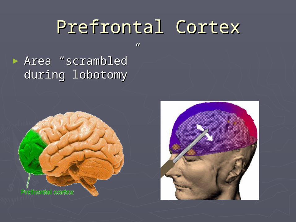
Prefrontal CortexPrefrontal Cortex
► Area “scrambled” Area “scrambled” during lobotomyduring lobotomy

Spinal CordSpinal Cord
► Continuous with Continuous with medulla oblongatamedulla oblongata
► Extends from brain Extends from brain to approximately L2to approximately L2
► Connected to Connected to 31 31 pairspairs of of spinal spinal nervesnerves

The MeningesThe Meninges
► Layered Layered coverings of brain coverings of brain and spinal cordand spinal cord
► Pia mater- inner sheathPia mater- inner sheath Highly vascularHighly vascular
► Arachnoid- central sheathArachnoid- central sheath Separated from pia mater by Separated from pia mater by
subarachnoid spacesubarachnoid space
► Dura mater- outer sheathDura mater- outer sheath Outermost, protective layerOutermost, protective layer

Ventricles-Ventricles- 4 fluid-filled cavities within the brain 4 fluid-filled cavities within the brain
What is that fluid?What is that fluid?
Cerebral spinal fluid (CSF)Cerebral spinal fluid (CSF)
Two upper pair are identical, Two upper pair are identical, known as known as left and right lateral left and right lateral ventricalsventricals
33rdrd Ventrical- Ventrical- just inferior to body just inferior to body of lateral ventricalsof lateral ventricals
44thth Ventrical Ventrical located in hindbrain located in hindbrain
All ventricles communicate with All ventricles communicate with each other through special each other through special connecting channels known as connecting channels known as interventricular foraminainterventricular foramina

R and L Lateral VentriclesR and L Lateral Ventricles
located in located in forebrainforebrain►BodyBody►anterior anterior hornhorn►posteriorposterior horn horn►inferiorinferior horn horn

Ventricles Ventricles cont’dcont’d
► Lateral Lateral ventriclesventricles
► 3rd ventricle3rd ventricle
► 4th ventricle4th ventricle
Superior Aspect

PneumoencephalographyPneumoencephalography
► Introduced in 1919Introduced in 1919
► Performed extensively throughout Performed extensively throughout late 20th centurylate 20th century
► Cerebrospinal fluidCerebrospinal fluid -a small amount -a small amount drained from around drained from around brainbrain and and replaced with replaced with air, oxygen, or air, oxygen, or helium as contrast helium as contrast to allow the to allow the structure of brain to show up more structure of brain to show up more clearly on an X-rayclearly on an X-ray
► Derived from cerebral Derived from cerebral
ventriculography- air is injected ventriculography- air is injected through holes drilled in skullthrough holes drilled in skull
► Pt turned upside down in special Pt turned upside down in special chair that can rotate vertically 360 chair that can rotate vertically 360 degrees to get air to fill ventricalsdegrees to get air to fill ventricals

Pneumoencephalography Pneumoencephalography cont’dcont’d
► Extremely painful, very dangerous Extremely painful, very dangerous
► Test was generally not well tolerated by pts- Test was generally not well tolerated by pts- headachesheadaches and and severe vomitingsevere vomiting common side common side
effectseffects
► Replacement of spinal fluid was by natural Replacement of spinal fluid was by natural generation- took as long as 2-3 monthsgeneration- took as long as 2-3 months
►MRI and CT have largely replaced MRI and CT have largely replaced Pneumoencephalography.Pneumoencephalography.

Myelography- Myelography- Radiographic exam of spinal cordRadiographic exam of spinal cord
X-ray examination performed by radiologist to detect abnormalities of the spine, spinal cord, or surrounding structures
Contrast material is injected into the fluid-filled space around spinal cord
X-rays are taken

Myelogram Myelogram cont’dcont’d
► Air used for early myelogramsAir used for early myelograms Injected via lumbar punctureInjected via lumbar puncture
► In 1922, iodized In 1922, iodized poppy seed oilpoppy seed oil was used was used Accidentally discovered with no apparent side Accidentally discovered with no apparent side
effectseffects
► Nonionic, water-soluble compoundsNonionic, water-soluble compounds now used now used Demonstrates a low neurotoxicityDemonstrates a low neurotoxicity
► CT and MRI CT and MRI now chiefly used to image now chiefly used to image nervous system nervous system

Myelography Myelography
► Injections can be given at:Injections can be given at:
Cistern (below occipital bone -can be hazardous because the needle is inserted close to brain stem)
Cervical spine
Thoracic spine
lumbar region (most common)
► Injection is in subarachnoid space –which is?
(space between arachnoid and pia mater)

Myelography IndicationsMyelography Indications
► Intraspinal abnormalitiesIntraspinal abnormalities
► Nerve root abnormalitiesNerve root abnormalities
► Disk prolapse Disk prolapse (slipped disk),(slipped disk), herniation herniation
► SpondylosisSpondylosis- - degenerative arthritis of spinal vertebra and related tissue degenerative arthritis of spinal vertebra and related tissue
► SpondylolisthesisSpondylolisthesis
► Spinal stenosis Spinal stenosis ((spinal canal narrows and compresses spinal cord and nerves)narrows and compresses spinal cord and nerves)
► TumorsTumors
► MetastasesMetastases

Myelography Myelography ContraindicationsContraindications
►Central aneurysms-Central aneurysms-(balloon-like bulge in an artery (balloon-like bulge in an artery
caused by weakening of artery wall)caused by weakening of artery wall)
►Arterio-venous malformationsArterio-venous malformations
► lumbar puncture within one weeklumbar puncture within one week
►Previous reaction to contrastPrevious reaction to contrast

Preliminary RadiographsPreliminary Radiographs
► PurposePurpose Determine accurate bony Determine accurate bony
anatomyanatomy
To exclude pathologiesTo exclude pathologies that wouldn’t need that wouldn’t need myelographymyelography
Distinguish congenital Distinguish congenital abnormalitiesabnormalities
For correlation with For correlation with myelography, MRI and CT myelography, MRI and CT images when reportingimages when reporting
Preliminary RadiographsPreliminary Radiographs
APAP
LateralLateral
Both anterior oblique views to Both anterior oblique views to demonstrate pars interarticularis demonstrate pars interarticularis (neck of Scotty dog)(neck of Scotty dog)
Lateral L5-S1Lateral L5-S1

Contrast MediaContrast Media
► IohexolIohexol – Nonionic, water-soluble contrast – Nonionic, water-soluble contrast
► IotrolanIotrolan (Isovist)(Isovist) – More recent contrast agent – More recent contrast agent Less toxic than IohexolLess toxic than Iohexol

Procedure SequenceProcedure Sequence
► Prepare room, sterile trays, and contrastPrepare room, sterile trays, and contrast
► Pt placed prone on tablePt placed prone on table
► Area to be punctured is cleaned and Area to be punctured is cleaned and prepped with sterile towelsprepped with sterile towels
► Local anesthetic injected in area of punctureLocal anesthetic injected in area of puncture

Procedure Sequence Procedure Sequence (cont’d)(cont’d)
► Lumbar puncture Lumbar puncture needle inserted needle inserted under fluoroscopic under fluoroscopic guidance until guidance until fluid appearsfluid appears
► CSF may be taken CSF may be taken for analysisfor analysis

Procedure Sequence Procedure Sequence (cont’d)(cont’d)
► Contrast material Contrast material injectedinjected
► Flow monitored Flow monitored fluoroscopicallyfluoroscopically
Pt. tilted into Pt. tilted into trendelenberg and trendelenberg and reverse-trendelenberg reverse-trendelenberg to control flow of to control flow of contrast during spot contrast during spot films Overheadsfilms Overheads
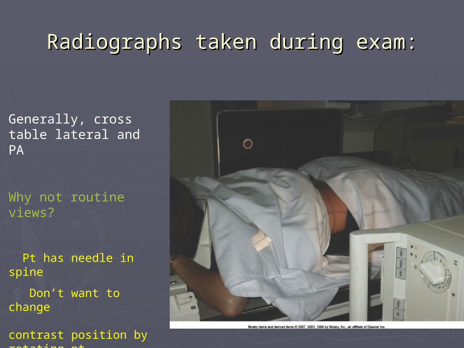
Radiographs taken during exam:Radiographs taken during exam:
Generally, cross table lateral and PA
Why not routine views?
Pt has needle in spine
Don’t want to change contrast position by rotating pt

Lumbar MyelogramLumbar Myelogram
PAPA LateralLateral

Myelography Myelography accuracy rate
When compared with surgical findings:When compared with surgical findings:
MRI – 96% Myelography – 81% CT – 57% CT and Myelography together - 84%

CervicalCervical
MyelograMyelogramm
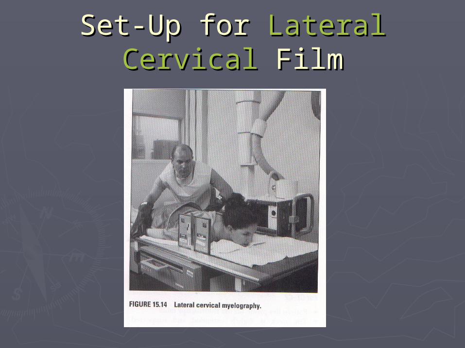
Set-Up for Set-Up for Lateral CervicalLateral Cervical FilmFilm

CervicalCervical Radiographs Radiographs
►OverheadsOverheads PAPA PA Oblique projectionsPA Oblique projections Cross-table lateral filmsCross-table lateral films

Alternative RadiographsAlternative Radiographs►Lateral flexion/extensionLateral flexion/extension
Demonstrates: Demonstrates:
►Stenosis Stenosis
►spinal instablilityspinal instablility
►deg. of movement of disk protrusiondeg. of movement of disk protrusion

Patient Care ConcernsPatient Care Concerns
►Maintain head in acute extension during Maintain head in acute extension during examination!examination!
► Bedrest 8 – 24 hours after procedure with Bedrest 8 – 24 hours after procedure with head elevatedhead elevated
► Encourage hydrationEncourage hydration
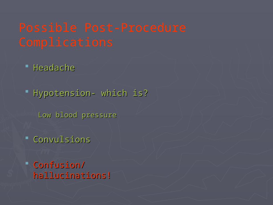
HeadacheHeadache
Hypotension- which is?Hypotension- which is?
Low blood pressureLow blood pressure
ConvulsionsConvulsions
Confusion/hallucinations!Confusion/hallucinations!
Possible Post-Procedure Complications

Advanced Imaging Advanced Imaging TechniquesTechniques

DiskographyDiskography
► Radiographic exam Radiographic exam of intervertebral of intervertebral disksdisks
► Contrast is injected Contrast is injected directly into diskdirectly into disk Determines disk Determines disk
morphologymorphology Reproduces pain Reproduces pain
caused by disk caused by disk disease itselfdisease itself

Diskogram FilmsDiskogram Films

CT Brain ScanCT Brain Scan
► Often Often 1st to evaluate 1st to evaluate head and spine traumahead and spine trauma
► Very accurateVery accurate diagnosis of acute diagnosis of acute intracranial injuriesintracranial injuries ContusionsContusions HemorrhageHemorrhage Fracture evaluationFracture evaluation
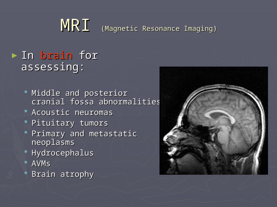
MRI MRI (Magnetic Resonance Imaging)(Magnetic Resonance Imaging)
► In In brain brain for assessing:for assessing:
Middle and posterior Middle and posterior cranial fossa abnormalitiescranial fossa abnormalities
Acoustic neuromasAcoustic neuromas Pituitary tumorsPituitary tumors Primary and metastatic Primary and metastatic
neoplasmsneoplasms HydrocephalusHydrocephalus AVMsAVMs Brain atrophyBrain atrophy

MRIMRI
► In In spine-spine-for assessing: for assessing: Demyelinating disease-Demyelinating disease- (any condition resulting in (any condition resulting in
damage to the protective damage to the protective covering (myelin sheath) that covering (myelin sheath) that surrounds nerves in brain and surrounds nerves in brain and spinal cord)spinal cord)
Spinal cord compressionSpinal cord compression Paraspinal massesParaspinal masses Postradiation therapy Postradiation therapy
changes in spinal cord changes in spinal cord tumorstumors
Metastatic diseaseMetastatic disease Herniated disks Herniated disks Congenital anomalies Congenital anomalies