myocardial diseases - columbia university · myocardial diseases context ¥ cardiac cycle ¥...
TRANSCRIPT

Mario C Deng & Charles C Marboe
Columbia University
New York, USA
Myocardial Diseases
context
• Cardiac cycle
• Valvular heart diseases
• Ischemic heart diseases
• Congenital heart diseases
• Myocardial diseases

objectives
• classify myocardial diseases into three major phenotypes
• describe their clinical presentation during the initial encounter
• delineate the diagnostic process and the role of different tests
• interpret these results in the context of pathophysiology
• employ the stages of heart failure to delineate therapeutic steps
patientteamhistory/exam/tests?
patho(physio)logy/etiology?
ethics/economics?
time
prognosis/therapy?
patient-physican encounter
Doc, can you help me with my
advanced heart failure?
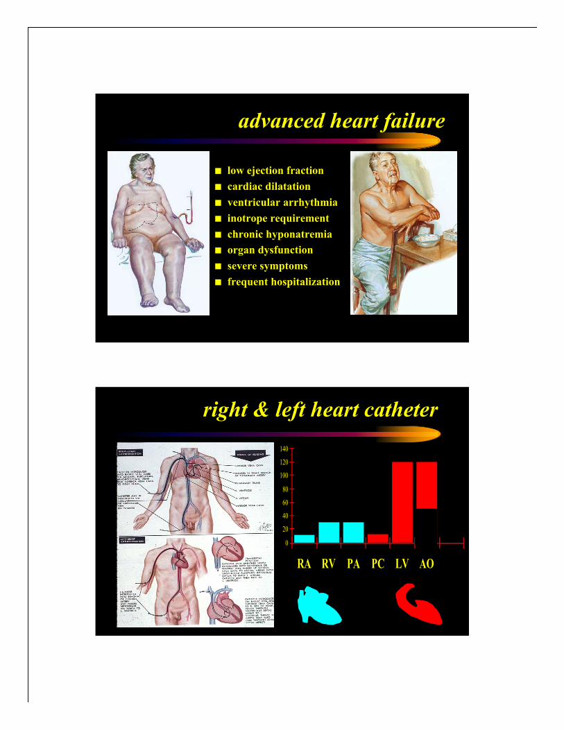
advanced heart failure
! low ejection fraction
! cardiac dilatation
! ventricular arrhythmia
! inotrope requirement
! chronic hyponatremia
! organ dysfunction
! severe symptoms
! frequent hospitalization
right & left heart catheter
0
20
40
60
80
100
120
140
RA RV PA PC LV AO

cardiac cycle - ECG & pressures
cardiac muscle function
Preload
•The length of a cardiac
muscle fiber prior to the
onset of contraction.
•Frank Starling
Muscle Length (mm)
Ten
sio
n (
g)
b
ac
d
Afterload
Muscle Length (mm)
Ten
sio
n (
g)
a c
!Lc
!La
e
•The force against which
a cardiac muscle fiber
must shorten.
•Isotonic Contraction
Contractility
Muscle Length (mm)
Ten
sio
n (
g)
a
g
f
b
e
+norepinephrine
•The force of contraction
independent of preload
and afterload.
•Inotropic State
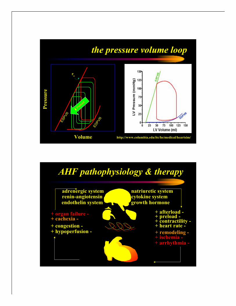
the pressure volume loop
Volume
Pre
ssu
re
ESPVR
esP
ED
PVR
"" Pre
load
Pre
load
http://www.columbia.edu/itc/hs/medical/heartsim/
AHF pathophysiology & therapy
+ hypoperfusion -+ congestion -
adrenergic system
growth hormonecytokine systemrenin-angiotensin
endothelin system
natriuretic system
+ afterload -+ preload -+ contractility -+ heart rate -
+ remodeling -
+ cachexia -
+ ischemia -+ arrhythmia -
+ organ failure -
Columbia
University
Medical
Center
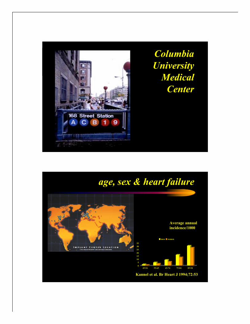
age, sex & heart failure
Kannel et al. Br Heart J 1994;72:53
Average annual
incidence/1000
0
5
10
15
20
25
30
35
45-54 55-65 65-74 75-84 85-94
men women

0
0.2
0.4
0.6
0.8
1
0 6 12 18 24 30 36 42 48 54 60
months after diagnosissu
rvival r
ate
epidemiology
Htx0.001%
advanced0.1%
heart failure1-2%
population
AHF medical
cancer!!
hypertrophic
cardiomyopathy normaldilated
cardiomyopathy
macroscopic pathology

cardiomyopathy phenotypes
! dilated cardiomyopathy
! hypertrophic cardiomyopathy
! restrictive cardiomyopathy
systems biology strategy
• level distinction
• relationships within levels
• relationships between levels
• iterative strategy
clinical clinical
picture 1picture 1
clinical clinical
picture 2picture 2
proteomeproteome
transcriptometranscriptome
genomegenome

NYPH
Hammer
Health
Sciences
Building
cardiomypathy phenotypes
! dilated cardiomyopathy
! hypertrophic cardiomyopathy
! restrictive cardiomyopathy

transgenic animals
Shuldiner AR. NEJM 1996;334:653
Cardiac
Compartment-
specific
Overexpression
of a Modified
Retinoic Acid
Receptor
Produces Dilated
Cardiomyopathy
and Congestive
Heart Failure in
Transgenic Mice
Colbert CM...Robbins J
specific cardiomyopathies
! Ischemic
! Valvular
! Hypertensive
! Inflammatory (Idiopathic, Autoimmune, Infectious)
! Metabolic (Endocrine, Amyloid)
! General system Disease (Connective Tissue Disorders)
! Muscular Dystrophies
! Neuromuscular Disorders
! Sensitivity and Toxic Reactions
! Peripartum

! 55 y male
! married, 2 kids
! large anterolat wall AMI
! 10/31/04 Impella pump
! 11/03/04 HeartMate 1 MCSD
! evaluation for heart transplant
! 2/17/05 heart transplant
!stable post-transplant course
!back to work and normal life
initial presentation follow-up
!benefits of hi-tech medicine
teaching points
team patient
ischemic dilated cardiomyopathy
GE #4734815 *1950 m
! married, 2 kids
! large anterolat wall AMI
! 10/31/04 Impella pump
! 11/03/04 HeartMate 1 MCSD
! evaluation for heart transplant
! 2/17/05 heart transplant
!stable post-transplant course
!back to work and normal life
initial presentation follow-up
!benefits of hi-tech medicine
teaching points
team patient
Xray ischemic cardiomyopathy
GE #4734815 *1950 m

! married, 2 kids
! large anterolat wall AMI
! 10/31/04 Impella pump
! 11/03/04 HeartMate 1 MCSD
! evaluation for heart transplant
! 2/17/05 heart transplant
!stable post-transplant course
!back to work and normal life
initial presentation follow-up
!benefits of hi-tech medicine
teaching points
team patient
ECG ischemic cardiomyopathy
GE #4734815 *1950 m
DCM TTE - parasternal axis

DCM TTE – apical 2/4 chamber view
Calculated CO= 2.1 L/min
Tei index 0.85
DCM TTE – AV/MV velocity

Decel time= 102 msec
DCM TTE – E deceleration time
DCM TTE – early mitral flow

E/prop vel = 2.7
E/Ea = 16
PASP= 56mmHG
DCM TTE – PA pressure
endomyocardial biopsy

NYPH - South West View
hypertrophic
cardiomyopathy normaldilated
cardiomyopathy
macroscopic pathology

Masson
trichrome
stain
extensive
interstitial
fibrosis
(blue) with
myocytes in
red and
epicardial
fat/pericard
ium to the
left
idiopathic dilated cardiomyopathy
Hematoxylin
& eosin
stain:
Myocyte
hypertrophy
(very
enlarged and
irregular
nuclei)
idiopathic dilated cardiomyopathy

myocarditis
inflammatory
infiltrate in
the
myocardium
associated
with myocyte
damage
myocarditis
inflammatory
infiltrate in
the
myocardium
associated
with myocyte
damage

giant cell myocarditis
multinucleated
giant cells
Trypanosom
a cruzi
Amastigotes
chagas disease

Columbia
University
College of
Physicians &
Surgeons
dilated cardiomyopathy
• pathology• enlargement of all four chambers, mild hypertrophy,
interstitial fibrosis
• pathophysiology• Frank-Starling mechanism, neurohormonal activation,
myocardial remodeling
• etiology• genetic, infectious, inflammatory, toxic, metabolic,
neuromuscular
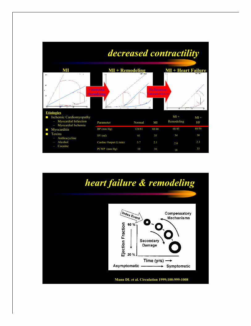
decreased contractility
Etiologies
! Ischemic Cardiomyopathy– Myocardial Infarction
– Myocardial Ischemia
! Myocarditis
! Toxins– Anthracycline
– Alcohol
– Cocaine33
2.3
38
80/50
MI +
HF
MI MI + Remodeling
Ventricular
Remodeling
MI + Heart Failure
Na Retention
Vasoconstriction
18
2.0
34
68/45
MI +
Remodeling
16
2.1
35
68/46
MI
10PCWP (mm Hg)
3.7Cardiac Output (L/min)
61SV (ml)
124/81BP (mm Hg)
NormalParameter
heart failure & remodeling
Mann DL et al. Circulation 1999;100:999-1008
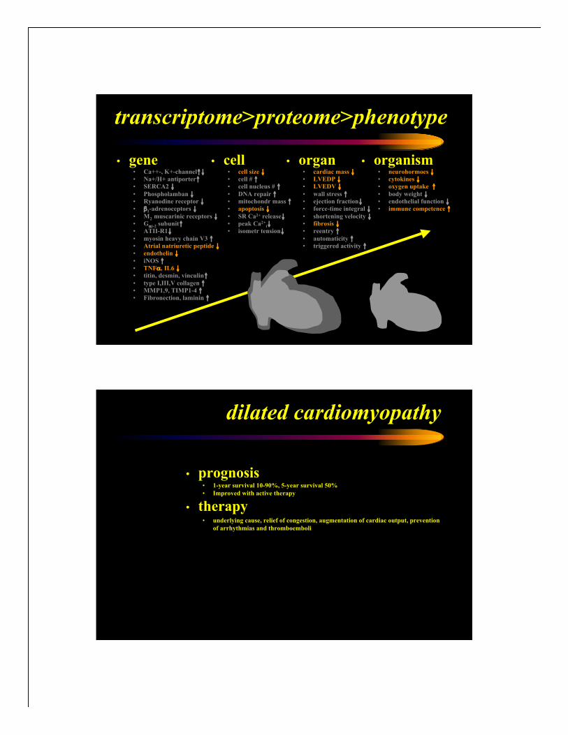
• organism• neurohormoes #• cytokines #• oxygen uptake $
• body weight #• endothelial function #• immune competence $
• gene• Ca++-, K+-channel$#• Na+/H+ antiporter$• SERCA2 #
• Phospholamban #• Ryanodine receptor #• %1-adrenoceptors #
• M2 muscarinic receptors #• G&i-2 subunit$• ATII-R1#
• myosin heavy chain V3 $• Atrial natriuretic peptide #• endothelin #
• iNOS $• TNF&, IL6 #• titin, desmin, vinculin$
• type I,III,V collagen $• MMP1,9, TIMP1-4 $• Fibronection, laminin $
transcriptome>proteome>phenotype
• cell• cell size #• cell # $• cell nucleus # $
• DNA repair $• mitochondr mass $• apoptosis #
• SR Ca2+ release#• peak Ca2+
i#• isometr tension#
• organ• cardiac mass #• LVEDP #• LVEDV #
• wall stress $• ejection fraction#• force-time integral #
• shortening velocity #• fibrosis #• reentry $
• automaticity $• triggered activity $
dilated cardiomyopathy
• prognosis• 1-year survival 10-90%, 5-year survival 50%
• Improved with active therapy
• therapy• underlying cause, relief of congestion, augmentation of cardiac output, prevention
of arrhythmias and thromboemboli

Framingham Study - mortality
Wang TJ et al. Circulation. 2003;108:977
Years
No ALVD (EF >50%), no
HF history
Mild ALVD (EF 40% to
50%)
Mod.-Severe ALVD (EF
<40%)
Systolic HF (EF '50%)
0 2 4 6 8 10 12
0.0
0.2
0.4
0.6
0.8
1.0
Su
rv
iva
l
P<.0001
Stage AHigh riskwith no
symptoms
Stage BStructural
heartdisease,
nosymptoms
Stage C Structural
disease,prior orcurrent
symptoms
Stage DRefractorysymptoms requiring
specialintervention
CHF stages and steps of treatment
Risk factor reduction, patient and family education Treat HTN, DM, CAD, dyslipidemia. ACEI when appropriate
ACE inhibitors, ? ARB’s, beta-blockers when appropriateACE inhibitors and beta-blockers in all patients
Aldosterone antagonists
Sodium restriction, diuretics, and digoxin
Short-term inotrope, nesiritideMitral or CABG surgery
Inotropes, nesiritide
CRT, ICD if applicable
VAD, TX
hospice
Hunt SA et al. J Am Coll Cardiol 2001:38:2101
NYPH
Garden
cardiomyopathy phenotypes
! dilated cardiomyopathy
! hypertrophic cardiomyopathy
! restrictive cardiomyopathy

hypertrophic cardiomyopathy genetics
! autosomal dominant trait
– 2/3 of patients have family
history
– more than 200 mutations in
10 genes encoding
contractile sarcomeric
proteins
– two genes for non-
sarcomeric proteins and
mitochondrial genome
Rosenthal N. NEJM 1994;331:39
HCM mutation frequencies
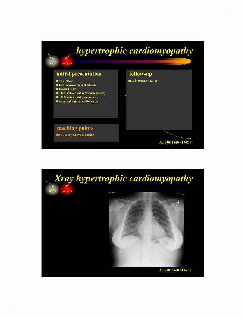
! 44 y female
! heart murmur since childhood
! married ,4 kids
! 3/6/06 mitral valve repair & myectomy
! 3/8/06 mitral valvle replacement
! complicated postoperative course
!good longterm recovery
initial presentation follow-up
!HOCM surgically challenging
teaching points
team patient
hypertrophic cardiomyopathy
AJ #5015860 *1962 f
! heart murmur since childhood
! Married ,4 kids
! 3/6/06 mitral valve repair & mywectomy
! 3/8/06 mitral valvle replacement
!stable post-transplant course
initial presentation follow-up
!HOCM surgically challenging
teaching points
team patient
Xray hypertrophic cardiomyopathy
AJ #5015860 *1962 f
! heart murmur since childhood
! Married ,4 kids
! 3/6/06 mitral valve repair & mywectomy
! 3/8/06 mitral valvle replacement
!stable post-transplant course
initial presentation follow-up
!HOCM surgically challenging
teaching points
team patient
ECG hypertrophic cardiomyopathy
AJ #5015860 *1962 f
hypertrophic cardiomyopathy
• history• sudden death during vigorous exercise 1/500, suncope, angina, dyspnea
• physical exam• S4, systolic murmur (LVOT obstruction – increased by Valsalva, MR)
• diagnostic tests• X-ray
• ECG (LAH, LVH)
• Echocardiogram (asymmetric hypertrophy)
• Catheterization (LVOT gradient)
• Genetic testing
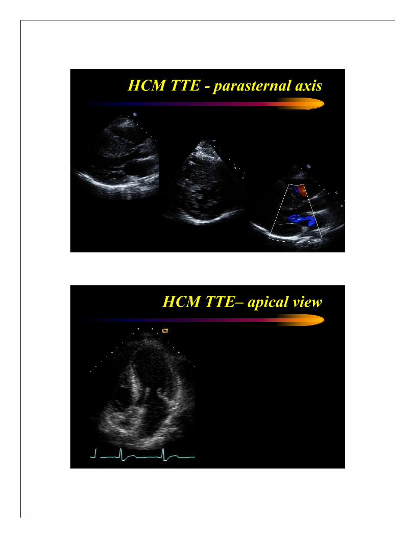
HCM TTE - parasternal axis
HCM TTE– apical view
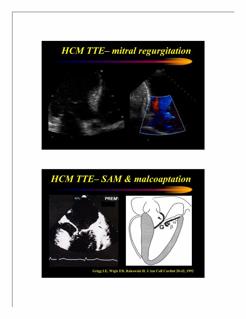
HCM TTE– mitral regurgitation
Grigg LE, Wigle ED, Rakowski H. J Am Coll Cardiol 20:42, 1992
HCM TTE– SAM & malcoaptation

Pollick C, Rakowski H, Wigle ED. Circulation 66:1087, 1982
HCM TTE– SAM & obstruction
Pollick C, Rakowski H, Wigle ED. Circulation 69:43, 1984
HCM TTE– LVOT obstruction

cardiomyopathy phenotypes
! dilated cardiomyopathy
! hypertrophic cardiomyopathy
! restrictive cardiomyopathy
amyloidosis cardiomyopathy
PRIMARY: amyloid light chain (AL)
lambda: kappa = 2:1
SECONDARY: serum amyloid A (AA)
SENILE CARDIAC: (SCA); transthyretin
FAMILIAL: autosomal dominant with mutations in
transthyretin, gelsolin, apolipoprotein A-I, lysozyme,
or fibrinogen genes.

iron storage disorders
! Iron overload – Hemosiderosis – following multiple blood
transfusions.
! Hereditary Hemochromatosis
Autosomal recessive
HFE gene on chromosome 6
Increased intestinal absorption of dietary iron
! 51 y male
! banker,2 kids
! rapidly progressive heart failure
! heart transplant evaluation
! heart transplantation 2003
! autologous stem cell transplantation (CAMP9)
!sucessful post-heart/stemcell transplant course
initial presentation follow-up
!amyloid-related cardiomyopathy DD
challenging
teaching points
team patient
restrictive cardiomyopathy
LD #4379458 *1952 m

! Banker, kids
! Rapidly progressive heart failoure
! Heart transplant evaluation
!sucessful post-heart/stemcell transplant course
initial presentation follow-up
!amyloid-related cardiomyopathy DD
challenging
teaching points
team patient
Xray restrictive cardiomyopathy
LD #4379458 *1952 m
! Banker, kids
! Rapidly progressive heart failoure
! Heart transplant evaluation
!sucessful post-heart/stemcell transplant course
initial presentation follow-up
!amyloid-related cardiomyopathy DD
challenging
teaching points
team patient
ECG restrictive cardiomyopathy
LD #4379458 *1952 m

restrictive cardiomyopathy
• history• Fatigue, exercise tolerance #
• physical exam• rales, neck veins $, ascites, peripheral edema, KUSSMAUL SIGN
• diagnostic tests• Xray: normal sized heart, congestion
• ECG: ST/T-changes, a-fib, AB-block, BBB
• echocardiography
• endomyocardial bipsy
RCM TTE– parasternal view

RCM TTE– apical view
Decel time= 102 msec
RCM TTE– restrictive mitral filling

! Abnormally low E’! (Atrial mechanical failure)! (Low systolic velocity)
RCM TTE– tissue doppler
Impaired relaxation- reduced propagation velocity
RCM TTE– tissue doppler

hypertrophic
cardiomyopathy normaldilated
cardiomyopathy
macroscopic pathology

macroscopic pathology
concentric
hypertrophy
microscopic pathology HCM
myocyte
disarray

Copyright ©2001 BMJ Publishing Group Ltd.
Mudhar, H S et al. J Clin Pathol 2001;54:321-325
Amyloid encircling
a myocyte
(original
magnification,
x1890)
microscopic pathology amyloid
Amyloid: 7-10
nm fibrils
haphazardly
arranged
microscopic pathology amyloid

Congo Red stain of
amyloid deposits in
the heart
microscopic pathology amyloid
Congo Red stain
under polarized
light: Amyloid
deposits are
birefringent.
microscopic pathology amyloid
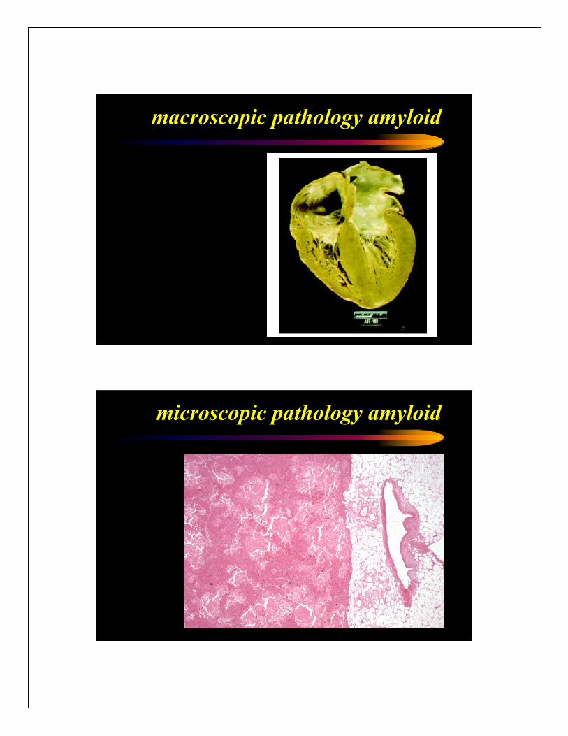
macroscopic pathology amyloid
microscopic pathology amyloid

microscopic pathology amyloid
microscopic pathology amyloid

Endomyocar
dial Biopsy:
Iron storage
disease in the
heart
iron storage disease
Prussian Blue
stain: Iron is
blue
Iron deposits
in myocytes
and interstitial
macrophages
iron storage disease

Columbia Presbyterian 1872
hypertrophic cardiomyopathy
• pathology• asymmetric septal hypertrophy, myocardiac fibers in disarray, compensatory
hypertrophy and fibroblast proliferation
• pathophysiology• compliance and relaxation reduced , dynamic LV outflow tract obstruction,
abnormal motion of the anterior mitral leaflet
• etiology• sarcomere complex mutations (b-myosin heavy chain, cardiac trop T, myosin-
binding protein C (automal dominant mechanism)

restrictive cardiomyopathy
• pathology• abnormally rigid ventricles (not necessarily hypertrophied), endocardial fibrosis or scarring
or myocardial infiltration
• pathophysiology• upward shilt of passive ventricular filling curve > elevated pulmonary and systemic venous
pressures
• reduced cavity size > stroke volume/cardiac output #
• etiology• infitrative: amyloidosis, sarcoidosis
• storage disease: hemochromatosis, glycogen storage diseases
• endocardial fibrosis
• hypereosinophilic syndrome
• metastatic tumors
• radiation therapy
• noninfiltrative: scleroderma, idiopathic
decreased filling
Etiologies
• Mitral Stenosis
• Constriction
• Restrictive Cardiomypoathy
• Cardiac Tamponade
• Hypertrophic
Cardiomyopathy
• Infiltrative Cardiomyopathy
Normal HCM
Ventricular
Remodeling
12
3.4
57
112/74
HCM
10PCWP (mm Hg)
3.7Cardiac Output (L/min)
61SV (ml)
124/81BP (mm Hg)
NormalParameter
27
4.0
66
131/87
HCM +
HF
HCM + HF
Na Retention
Vasoconstriction

LV outflow tract obstruction
ventricular remodeling

hypertrophic cardiomyopathy
• prognosis• dependent on mutation
• Sudden death 4-6% per year (children), 2-4% (adults)
• therapy• AVOID strenuous exercise
• B-blockers (myocardial oxygen demand#, LVOT gradient #)
• CA-channel antagonists
• amiodarone (a-fib)
• antibiotic prophylaxis
• Defibrillator (patient with elevated risk)
• dual chamber PM
• Septal ablation with ethanol
• myomectomy
restrictive cardiomyopathy
• prognosis• Very poor prognosis
• therapy• salt restriction
• diuretics (cautious use)
• Maintainance of SR
• Intravavitary thrombus: anticoagulation

amyloidosis management
Heart-liver transplantation? Heart-autologous BM transplantation?
NYPH
Broadway
Entrance

summary cardiomyopathiesteam patient
systemic approachBB, CA, cave volumesystolic HF guidelinestherapy
r/o infitrativeDD restrctiver/o myocarditisbiopsy
RA/PC$,square rootcompl#, LVOT gradCAD?, RA/PC$, CO#cardiac catheter
LV wall $, LVEF okasymm LVH, SAMchamber dilat, regurgecho
low volt, AV cond#LVHSR$, ST/T, IC abnornECG
PVHLA enlargementLV enlargement, PVHchest Xray
Kussmaul signS4, valsalva+ murmurS3, S4, MRphysical exam
right heart failureSOB, cP, syncopeleft heart failurehistory
restrictivehypertrophicdilatedphenotype
Braunwald E. Heart Disease (4th Ed). Saunders, Philadelpia
top 10 controversies
urgentHtx/
MCSD
chronic neurohorm blockade& evaluation for
comfortcare
hospice
ICU
floor/home
medPCICABG
medresynch/IABPvalve OP/SVER
medPM/ICD/ablantitach OP0
team patientassessment
potential Htx or chronic MCSD
end-of-life situation?
yes no
unsuccessful recompensation?
yes no
ischemia? pump failure? arrhythmias?
classification or staging
risk stratification
choice of BB/ACEI
role of added ARB
risks of aldo-antagonists
role of infusion therapy
indication for ICD
indication for CRT
timing of MCSD
selection for Htx

Columbia University Medical Center
teaching
patient care
research